l orad breast cancer detection - activexray.com · the selenia’s innovative detector technology...
TRANSCRIPT

L O R A D B R E A S T C A N C E R D E T E C T I O N
HOL-1236_Selenia_r6.qxd 6/17/03 12:33 PM Page 3

Clarityfor Digital MammographyIntroduction of the Selenia™ full field digital mammography system signals the
beginning of a new era in the world of digital mammography. This elegant and
innovative system combines the latest advances in technology with sophisticated
information management capabilities to bring you the total solution for your
digital mammography needs.
■ Revolutionary DirectRay® Direct Conversion Detector
preserves image sharpness by completely eliminating light diffusion
■ Largest digital detector in the industry, 24 x 29 cm field of view accommodates
almost all breast sizes
■ Renowned High Transmission Cellular (HTC®) Grid significantly reduces
radiation scatter for higher contrast images
■ Exclusive Smart Paddle System™ allows accurate, easy positioning
A Vision of
Imagine the possibilities...
■ Custom designed softcopy workstation streamlines
workflow and provides flexible user configurability
■ Flexible image management capabilities: DICOM
compatible connectivity solutions for any
clinical setting
■ Remarkably small footprint fits easily in a standard
exam room
HOL-1236_Selenia_r6.qxd 6/17/03 12:33 PM Page 4

Advanced technology and sophisticatedinformation managementcapabilities in a remarkablysmall footprint
HOL-1236_Selenia_r6.qxd 6/17/03 12:33 PM Page 5

The Selenia’s innovative detector technology brings the advantages of direct-to-
digital imaging to mammography for the first time. Powered by our DirectRay
digital image receptor, the Selenia uses amorphous selenium to directly convert
x-rays to electronic signals, without first converting them to light, a step required
in systems using indirect conversion technology.
By completely eliminating the image-degrading effects of light diffusion, our
proprietary direct conversion process preserves image sharpness, giving you and
your patients incredibly sharp images – quickly, efficiently, and consistently.
ImagingAdvantage
Experience the Hologic Direct-to-Digital
In systems using indirect conversion detectors,
an intensifying screen captures and converts
x-rays to visible light, which are then converted
to electronic signals.
In this process, visible light diffuses laterally
across a large distance. Light diffusion results
in reduced image sharpness and loss of
tissue contrast, for less than optimal
imaging performance.
Electron-hole pairs
Amorphous selenium
Pixel array
Line spread function
X-ray
X-ray
Light scintillation
Columnar CsI (TI)
Pixel array
Line spread function
With Selenia’s direct conversion detector,
x-rays are absorbed by the amorphous selenium;
negative and positive electrical charges are
directly generated.
Under the influence of an external electrical field,
the electrical charges are pulled directly towards
a pixel electrode and collected on a pixel
capacitor. Because the electrical charges travel
along electrical field lines, there is no lateral
movement of the charge and no diffusion across
pixels. The result is an exceptionally sharp
digital image.
HOL-1236_Selenia_r6.qxd 6/17/03 12:33 PM Page 6
LORAD BREAST CANCER DETECTION
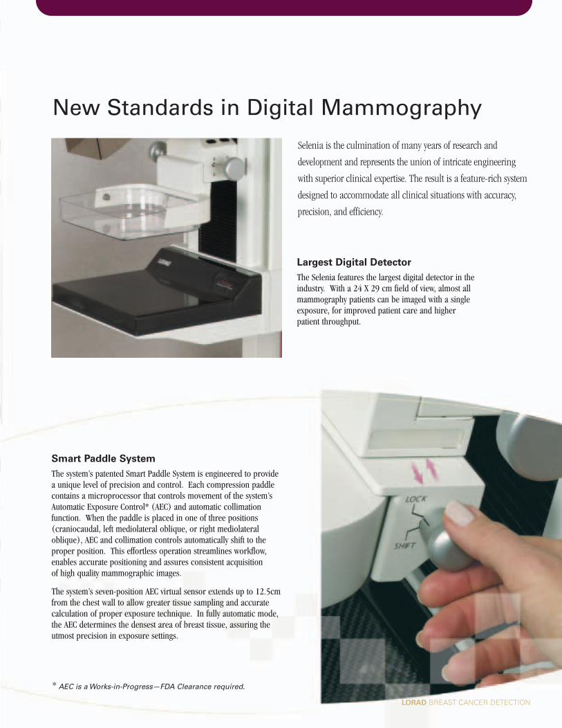
Selenia is the culmination of many years of research and
development and represents the union of intricate engineering
with superior clinical expertise. The result is a feature-rich system
designed to accommodate all clinical situations with accuracy,
precision, and efficiency.
* AEC is a Works-in-Progress—FDA Clearance required.
Smart Paddle System
The system’s patented Smart Paddle System is engineered to provide a unique level of precision and control. Each compression paddlecontains a microprocessor that controls movement of the system’sAutomatic Exposure Control* (AEC) and automatic collimation function. When the paddle is placed in one of three positions (craniocaudal, left mediolateral oblique, or right mediolateraloblique), AEC and collimation controls automatically shift to theproper position. This effortless operation streamlines workflow,enables accurate positioning and assures consistent acquisition of high quality mammographic images.
The system’s seven-position AEC virtual sensor extends up to 12.5cmfrom the chest wall to allow greater tissue sampling and accurate calculation of proper exposure technique. In fully automatic mode,the AEC determines the densest area of breast tissue, assuring theutmost precision in exposure settings.
Largest Digital Detector
The Selenia features the largest digital detector in theindustry. With a 24 X 29 cm field of view, almost all mammography patients can be imaged with a single exposure, for improved patient care and higher patient throughput.
New Standards in Digital Mammography
HOL-1236_Selenia_r6.qxd 6/17/03 12:34 PM Page 7

The High Transmission Cellular (HTC) Grid
Patterned on our industry-renowned M-IV screen-film platform, the Selenia incorporates the gold standard features of the M-IV in a system specifically designed to address image quality, workflow, and efficiency in digital mammography.
Our exclusive HTC Grid, optimized for the Selenia’s digital detector, provides evengreater enhancements in image quality. The HTC Grid is the industry’s leading technology designed to increase both the absorption of radiation scatter and the transmission of primary x-ray, for higher contrast images.
The HTC Grid’s focused cellular pattern reduces radiation scatter in both the X and Ydirections. Its structure is self-supporting, so interspace material is eliminated and primary transmission is increased. Its micro-processor controlled movement isdesigned to eliminate grid artifacts.
The fully integrated HTC Grid automatically retracts when the Selenia’s magnificationplatform is detected. This unique feature allows magnification views to be obtainedwithout removing or handling the Bucky, for safe and streamlined workflow.
FeaturesInnovative Design
Trusted
Fully Automatic
Self-Adjusting Paddle
In addition, our Fully-Automatic Self-Adjusting Tilt (FAST)Paddle has been incorporated as a standard componentof the Selenia. The FAST Paddle automatically adjusts to the natural contour of the breast, providing more uniform compression across the entire breast forimproved imaging.
The FAST Compression Paddle is especially usefulwhere there is a disproportionate thickness at thechest wall compared to the anterior of the breast.By automatically conforming to the natural contourof the breast, imaging of the structures in the sub-areola regions may be improved withoutcompromising the image quality of the breast at the chest wall.
HOL-1236_Selenia_r6.qxd 6/17/03 12:34 PM Page 8

Selenia’s acquisition station features integrated x-ray control capabilities and an
image acquisition console with local storage and archive capabilities. Intuitive
display screens and user interfaces provide enhanced ease of use and higher
patient throughput.
With this flexible system, images can be acquired within seconds, allowing
the technologist to confirm proper positioning. Exam parameters can
be adjusted quickly and efficiently to meet the requirements of the
examination. An average of two weeks of exams can be stored locally on the
acquisition station, to effectively accommodate recalls. The operator has
the option of using the trackball or function keys to activate the generator control.
Built-in quality control measurements and service tools streamline maintenance
and assure optimal performance with minimal effort. A repeat analysis report is
generated automatically upon request, greatly simplifying MQSA reporting.
DICOM Compatible
The acquisition station is DICOM
3.0 compatible, allowing images to
be sent to the Selenia Softcopy
Workstation, DICOM compatible
hardcopy printers, and long-term
archives. Patient demographics
can be input by means of the
barcode reader or by accessing
modality worklist, ensuring
accurate, efficient patient flow.
Intuitive display screens andinterfaces provide enhanced ease ofuse for higher patient throughput.
State-of-the-Art Image Acquisition
HOL-1236_Selenia_r6.qxd 6/17/03 12:34 PM Page 9

CustomizedSoftcopy Image Review
* PCE is a Works-in-Progress—FDA Clearance required.
The Selenia Softcopy Workstation provides the ideal pathway to tomorrow’s filmless
mammography environment. This system incorporates the features needed to
maximize efficiency and accuracy in softcopy image review, including automatic
image processing, versatile image manipulation options, full resolution
viewing capabilities, high-volume throughput, user-friendly controls and full
DICOM connectivity.
The workstation supports high-volume reading of screening and diagnostic
mammography. Dual 5-mega pixel monitors with 1024 true shades of gray allow
full resolution display of images. An image display time of 0.2 seconds per patient
ensures maximum efficiency in case throughput.
Imagine the possibilities...
Selenia’s Softcopy Workstation provides automatic image
processing functions that optimally enhance each digital
mammogram based on breast tissue compositions.
The Softcopy Workstation also utilizes an exclusive image
processing feature, Peripheral Contrast Enhancement
(PCE)*, to enable instantaneous visualization of all
breast tissue with optimal image contrast across the
entire breast, from chest wall to skin line.
HOL-1236_Selenia_r6.qxd 6/17/03 12:34 PM Page 10

An ergonomically designed workflow keypad places all controls at the physician’s fingertips. Views and functions used mostfrequently are grouped and ordered, to streamline workflow and allow the physician’s attention to remain focused oncase review. Each user can easily define their own preferences forhanging protocol and workflow. The system saves user preferences, which are automatically accessed upon login.
The workflow keypad provides single-click image manipulation and allows the physician to enact a variety of image display andmanipulation tools including:
" Digital Magnifying Glass – image detail can be viewed atincreased resolution to better visualize fine detail, in both normal and inverse video mode
" Window/level adjustment – brightness and contrast changescan be interactively applied to images
" Display of prior studies – user can quickly toggle between current and prior studies from multiple years
" Single-click view comparison – user can select flexible comparisonchoices among views
" Roaming – images can be displayed at full resolution for efficientreview of sub-regions
" Marking and annotation – suspicious areas can be marked,annotated, and the results archived in electronic or hardcopyformat for future reference
" Measurement tools – measurement lines can be drawn tomark suspicious areas, with automatic indication of length (in millimeters)
" Image layout and orientation – images can be displayed indifferent layouts at different resolutions, including ability tointeractively mirror and flip
The Selenia Softcopy Workstation is DICOM 3.0 compatible and supports connectivity to the Selenia Acquisition Station, long-termarchives, and DICOM compatible printers. In addition, DICOMimages from other modalities, including ultrasound and MRI, can be displayed (for viewing only) on the workstation.
HOL-1236_Selenia_r6.qxd 6/17/03 12:34 PM Page 11

The Selenia is a remarkably versatile system designed to meet the unique
connectivity and bandwidth requirements of digital mammography. Built with
a flexible architecture and incorporating DICOM open standards, this system is
configured to interface with a wide variety of information management system
components — connecting you to the benefits of digital mammography and
enabling seamless integration of all workflow functions from image acquisition
to long-term archive.
ConnectivitySolutions for any Clinical Setting
Selenia System
Provides mammographic
image acquisition
DVD Archive
Provides permanent,
longterm storage of
studies and rapid
retrieval of prior exams
Approved DICOM
Compatible Printers
Support hardcopy printing
of digital images
Workflow Manager
Manages routing,
archival and
retrieval of studies
Selenia Softcopy
Workstation
Supports image viewing
and interpretation
PACS Broker
Allows integration with HIS/RIS
and enables modality worklist
features (including scheduling)
at the acquisition station
Imagine the possibilities...
HOL-1236_Selenia_r6.qxd 6/17/03 12:34 PM Page 12

The Selenia was designed to accommodate future advances in breast imaging
technologies. Its flexible architecture allows incorporation of system
enhancements to support emerging applications such as tomosynthesis,
digital subtraction angiography, and dual energy imaging. In addition,
the Selenia Softcopy Workstation is fully configured to support Computer
Aided Detection (CAD)* technologies.
We have also developed an effective pathway to allow our M-IV screen-film
systems to be upgraded to Selenia systems – easily and efficiently. This allows
you to protect your original equipment investment and plan your migration to
digital mammography at the pace that’s best for your facility and your practice.
With our Selenia Total Solution you can be connected to the world of digital
mammography — today and tomorrow.
*Works-in-Progress
Because of our unwavering committment to women's health, Hologicconstantly seeks better ways to detect breast cancer earlier. The LoradSelenia mammography system with direct-to-digital imaging is the mostadvanced technology available to ensure the best possible breast healthand breast cancer screening.
Womenis a Way of Life for Us
Caring for
A Platform for the Future
HOL-1236_Selenia_r6.qxd 6/17/03 12:34 PM Page 13

B-LM-SEL US/International (5/03) © Lorad 2003. Printed in USA. Specifications are subject to change without prior notice.
The power of Hologic is the power of clear innovation and a singular
focus . . . to challenge the boundaries of science and technology everyday
to raise the standards of image quality. Our passion has led to discoveries
that contribute to earlier detection, more accurate diagnoses, and better
overall patient care. As we focus on the future, we are bound by our
clarity of vision. A vision created solely to enhance yours.
Corporate Headquarters
35 Crosby Drive Bedford, MA 01730-1401 USATel: 781.999.7300Fax: 781.280.0668www.hologic.com
Asia and Pacific Rim
Room 302, Hung Kei Building5-8 Queen Victoria StreetCentral, Hong KongHong KongTel: 852.3102.9200Fax: 1.452.696.7846
Europe
Horizon ParkLeuvensesteenweg 510, BUS 311930 Zaventem, BelgiumTel: 32.2.711.4680Fax: 32.2.725.2087
Osteoporosis Assessment ■ DirectRay® Digital ImagingLORAD® Breast Cancer Detection ■ FLUOROSCANTM C-arm Imaging
LORAD
36 Apple Ridge RoadDanbury, CT 06810 USATel: 203.207.4500Fax: 203.207.4501
HOL-1236_Selenia_r6.qxd 6/17/03 12:33 PM Page 2