kumar and aidya, int phys med ehabil 21, 1 physical ... · physical medicine rehabilitation kumar...
TRANSCRIPT
International Journal of
Physical Medicine & RehabilitationKumar and Vaidya, Int J Phys Med Rehabil 2014, 3:1
http://dx.doi.org/10.4172/2329-9096.1000253
Research Article Open Access
Volume 3 • Issue 1 • 1000253Int J Phys Med RehabilISSN: 2329-9096 JPMR, an open access journal
Effectiveness of Myofascial Release on Spasticity and Lower Extremity Function in Diplegic Cerebral Palsy: Randomized Controlled TrialChandan Kumar1* and Snehashri N Vaidya2
1Associate Professor, Smt Kashibai Navale College of Physiotherapy, Narhe, Pune, Maharashtra, India2MGM’S School of Physiotherapy, Aurangabad, India
*Corresponding author: Chandan Kumar, Associate Professor, Smt Kashibai Navale College of Physiotherapy, Narhe, Pune, Maharashtra, India, Tel: 8087518006; E-mail: [email protected]
Received October 31, 2014; Accepted December 16, 2014; Published December 20, 2014
Citation: Kumar C, Vaidya SN (2014) Effectiveness of Myofascial Release on Spasticity and Lower Extremity Function in Diplegic Cerebral Palsy: Randomized Controlled Trial. Int J Phys Med Rehabil 3: 253. doi:10.4172/2329-9096.1000253
Copyright: © 2014 Kumar C, et al. This is an open-access article distributed under the terms of the Creative Commons Attribution License, which permits unrestricted use, distribution, and reproduction in any medium, provided the original author and source are credited.
Keywords: Spastic diplegic cerebral palsy; Myofascial release; MAS; MTS; GMFM
Introduction“Cerebral Palsy (CP) described as a group of permanent disorders
of the development of the movement and posture, causing activity limitation that is attributed to non- disturbances that occurs in developing fetal or infant brain. The motor disorders of cerebral palsy are often accompanied by disturbances of sensation, perception, cognition, communication, behavior, by epilepsy and by secondary musculoskeletal problems [1]. This term is commonly used name for a group of conditions characterized motor dysfunction due to non-progressive brain damage early in life. It is stated as static encephalopathy in which even though the primary lesion, anomaly or injury is static, the clinical pattern of presentation may change with time due to growth and developmental plasticity and maturation of Central Nervous System (CNS) [2].
The current all over worldwide prevalence in children ages 3 to 10 years is 2.4 per 1000 children [3] with incidence being 2 to 3 per 1000 live births [4]. The male female ratio is 2.42: 1, with males being more severely disabled [2].
CP can be classified according to the severity of motor deficits as mild, moderate, or severe. Several other classification systems exist based on the pathophysiology, etiology, and distribution of motor deficits [5].
In CP the lesion in the central nervous system frequently results in spasticity of various muscle groups.6 approximately 75% of children with CP have spasticity, a state of increased muscle tone and heightened deep tendon reflexes [6,7].
Spasticity is defined as a velocity-dependent resistance to stretch. Spastic CP is caused by damage to the pyramidal parts of the brain4. Bone and joint changes in cerebral palsy result from muscle spasticity and contracture. The spine and the joints of the lower extremity are most commonly affected. Scoliosis may progress rapidly and may continue after skeletal maturity. Progressive hip flexion and adduction lead to windswept deformity, increased femoral anteversion, apparent coxa valga, subluxation, deformity of the femoral head, hip dislocation, and formation of a pseudoacetabulum. In the knee, flexion contracture,
patella alta, and patellar fragmentation are the most commonly seen abnormalities. Progressive equinovalgus and equinovarus of the foot and ankle are associated with rocker-bottom deformity and subluxation of the talonavicular joint. Early recognition of progressive deformity in subjects with cerebral palsy allows timely treatment and prevention of irreversible change [8].
One of the survey describing problems in adult CP reported that 77% of CP children were having problems with spasticity, 80% had contractures and 18% had pain every day [9].
The increase in muscle tone is responsible for relative failure of muscle growth and may produce functional problems. Spastic deformities of the lower limbs affect ambulation, bed positioning, sitting, chair level activities, transfers, and standing up [6].
There are three potential aims of treating the spasticity - to improve function, to reduce the risk of unnecessary complication and to alleviate pain [10-13].
There are various ways to tackle the spasticity which include the medical, surgical and physiotherapy management. The common physiotherapy approaches to reduce spasticity are stretching, strengthening of the antagonistic muscles, positioning, inhibitive casting and bracing, and weight bearing exercises [13].
Some manual techniques such as massage, myofascial release (MFR), and acupressure are also used along with other physiotherapy techniques [13].
AbstractPurpose: To find out the effectiveness of Myofascial Release in combination with conventional physiotherapy on
spasticity of calf, hamstring and adductors of hip and on lower extremity function in spastic diplegic subjects.
Methodology: 30 spastic diplegic subjects of age group 2-8 years were taken by random sampling method from MGM College and other private clinics in Aurangabad. 15 subjects were assigned in each group. Group A: Myofascial release and conventional PT treatment. Group B: conventional PT treatment. Both the groups received training for 4 weeks. Baseline and Post treatment measures of Modified Ashworth Scale (MAS), Modified Tardieu Scale (MTS) and Gross Motor Function Test (GMFM-88) were evaluated.
Results: Mean difference of MAS and R2 value of MTS in group A was more than group B, for calf, hamstring and adductors, whereas GMFM showed nearly equal improvement in both groups.
Conclusion: Overall, it can be concluded from our study that the MFR along with conventional treatment reduces spasticity in calf, hamstring and adductors of hip in spastic diplegic subjects.
Citation: Kumar C, Vaidya SN (2014) Effectiveness of Myofascial Release on Spasticity and Lower Extremity Function in Diplegic Cerebral Palsy: Randomized Controlled Trial. Int J Phys Med Rehabil 3: 253. doi:10.4172/2329-9096.1000253
Page 2 of 9
Volume 3 • Issue 1 • 1000253Int J Phys Med RehabilISSN: 2329-9096 JPMR, an open access journal
Myofascial therapy can be defined as “the facilitation of mechanical, neural and psycho physiological adaptive potential as interfaced by the myofascial system” [14]. The purpose of deep myofascial release is to release restrictions (barriers) within the deeper layers of fascia. This is accomplished by a stretching of the muscular elastic components of the fascia, along with the crosslinks, and changing the viscosity of the ground substance of fascia [15].
Myofascial Release (MFR) techniques are utilized in a wide range of settings and diagnoses; pain, movement restriction, spasm, spasticity, neurological dysfunction, i.e., cerebral palsy, head and birth injury, Cardiovascular Accidents (CVA), scoliosis [16].
Myofascial release and Static stretching are expected to have an effect on the spastic/tight muscles, efficacies of these methods need to be established in clinical practice. Moreover, there are few studies done on Myofascial release to reduce spasticity which showed immediate & short term effect. there are insufficient published evidences available for effect of MFR technique on spasticity, so present study is focus on to find out the effectiveness of Myofascial Release on calf muscle, hamstring muscle and adductor muscles of hip spasticity in spastic diplegic subjects.
Methodology• Type of Study: Experimental study
• Study Design: Randomized controlled trial.
• Study Setting: Neuroscience department of Physiotherapy OPD, MGM Hospital Aurangabad, other hospitals and private clinics of Aurangabad, Maharashtra, India.
• Sample Size: 30 Subjects
• Group A- 15 (MFR and Conventional Physiotherapy Treatment)
• Group B- 15 (Conventional Physiotherapy Treatment)
• Type of sampling: Simple randomized.
• Duration of intervention: 4 weeks.
• Duration of study: 1 year
Inclusion Criteria• Spastic diplegic type of CP subjects
• Age group: 2-8 years
• Both genders
• Modified Ashworth scale (grade1 - grade 3) for hip adductor, hamstrings & calf muscles.
• Ambulatory subjects with or without ambulatory aids.
Exclusion Criteria• Subjects who has undergone prior orthopedic surgery.
• Subjects who has received Botulinium toxin injection in the past 6 months,
• Subjects who has undergone serial casting in past 6 months
• Subjects who are taking oral or Intrathecal Myorelaxant drugs
• Subjects who has severe limitations in passive range of motion at lower extremities
• Subjects who has any cognitive and perceptual disorders.
• Patient who has contracture.
Outcome Measures Modified ashworth scale (MAS)
The Modified Ashworth Scale is considered the primary clinical measure of muscle spasticity in subjects with neurological conditions. The Modified Ashworth Scale can be applied to muscles of both the upper or lower body. The Modified Ashworth Scale shows conflicting results regarding its reliability and validity [17-19].
Modified tardieu scale (MTS)
Tardieu is a scale for measuring spasticity that takes into account resistance to passive movement at both slow and fast speed. The quality of the muscle reaction at specified velocities and the angle at which the muscle reaction occurs are incorporated into the measurement of spasticity using the Modified Tardieu Scale (Morris, 2002). Modified Tardieu describes R1 and R2; R1 is the angle of muscle reaction, R2 is the full PROM. The angle of full ROM (R2) is taken at a very slow speed (V1). The angle of muscle reaction (R1) is defined as the angle in which a catch or clonus is found during a quick stretch (V3). R1 is then subtracted from R2 and this represents the dynamic tone component of the muscle. The Tardieu Scale differentiates spasticity from contracture, and having had good reliability and validity [20-22].
Gross motor function test (GMFM-88)
The GMFM is a measure designed to assess change in gross motor function for children aged 5 months to 16 years with Cerebral Palsy (CP). The 88-item GMFM is a performance based measure with 5 dimensions: lying and rolling; crawling and kneeling; sitting; standing; and walking, running and jumping. The GMFM-88 had good reliability and validity in assessing the gross motor functions of children with cerebral palsy [23,24].
Procedure
Step 1: All the subjects from specified source of data were assessed and those who fulfilled inclusion criteria were taken up for the study.
Step 2: All thirty subjects were randomly allocated in to two groups, Group A (Conventional PT treatment, MFR) and Group B (Conventional PT Treatment), with 15 subjects in each.
Step 3: The procedure was explained to parents of all the children and written informed consent from the parents was taken.
Step 4: All the subjects were evaluated with MAS and MTS for calf muscle, hamstring muscle and adductor muscles of hip in supine position, at two instances, before intervention and after intervention. Even Gross Motor Function Measure (GMFM) was evaluated before and after treatment.
Step 5:
Group A: (Experimental Group) Intervention Protocol.
MFR for calf muscle- MFR for calf muscle was given with patient in prone position with 120 second hold. For giving the MFR, finger pads were allowed to sink in to the central portion of the calf. It was held for 120 seconds to allow the tissue to soften and then myofascial structures were spread in a lateral direction until feeling of first fascial barrier [25] (Figure 1).
MFR for hamstring- MFR for hamstring was given with patient in prone position with crossed hands or thumb. Then myofascial structures were stretched and it was held for 120 seconds to allow the tissue to soften [14].

Citation: Kumar C, Vaidya SN (2014) Effectiveness of Myofascial Release on Spasticity and Lower Extremity Function in Diplegic Cerebral Palsy: Randomized Controlled Trial. Int J Phys Med Rehabil 3: 253. doi:10.4172/2329-9096.1000253
Page 3 of 9
Volume 3 • Issue 1 • 1000253Int J Phys Med RehabilISSN: 2329-9096 JPMR, an open access journal
MFR for adductors of hip-MFR for adductors was given with patient in supine position with crossed hands or with thumb. Then myofascial structures were stretched and then it was held for 120 seconds to allow the tissue to soften [14] (Figure 2).
MFR for all the muscles were performed in 15 minutes. Following MFR, conventional PT treatment similar to controlled group was given to all the subjects, 6 days a week for four weeks.
Group B: (Controlled Group)
Conventional PT Treatment: Passive stretching of calf, hamstring and hip adductors.
Resistive exercises.
Weight bearing exercises
• Bridging (bilateral & unilateral), kneeling, kneel sit to stand, single kneeling, sit to stand, single leg standing, mini squats, stepping(forward, backward & sideways).
• Reaching exercises in sitting, kneeling and standing.
• Exercises in different functional position.
• Gait training.
• All the exercises were performed in 60 minutes. There was no subdivision of time for each activity. Patients were performed exercises on the bases of their motor control for 60 minutes in a day and 6 days in a week for 4 weeks.
Step 6: At the end of 4th week session effect of intervention was seen by MAS, MTS and GMFM-88. Data for MAS, MTS and GMFM was recorded and analyzed using appropriate statistical test.
Ethical approval and informed consent
Before implementing the study, an approval from University ethical committee was taken. Also the Informed consent was taken from subjects parents, who were willing to participate in study.
Data and statistical analysisMean score, Standard deviation, degree of freedom, confidence
interval, P value and significance were calculated to express the results. SPSS version 20 (Statistical Package for Social Sciences) for Windows and Microsoft Office Excel- 2007 was used to statistically analyze obtained data from the study.
Unpaired t test was performed to compare pre and post interventional assessment values in between two groups and paired t test was used to compare pre and post intervention values within the groups. Statistical level of significance is set at alpha=0.05.
The obtained data from the patient was organized in a master charts and various tables along with graphs derived for statistical analysis for easy interpretation of results.
Results Flow Chart (Selection criteria)
44 subjects were assessed for eligibility. 6 subjects were excluded as they were not meeting the inclusion criteria, total 38 subjects were randomized (19 in MFR group and 19 in conventional Ex) there was 4 drop out form MFR group and 4 from conventional group. This left 30 participants: 15 in MFR Group and 15 in control Group (Chart 1).
A total of 19 males (63.3%) and 11 females (36.7%) were participated in the study. In Group A there were 60% male and 40% female and in group B there were 66.7% male and 33.3% female. The age of the patient in this study ranged from 2-8 years (Table 1).
Above table shows gender wise and age wise distribution of the subjects. No statistically significant difference was found between the groups showing that subjects are matched for baseline characteristics (Table 2).
Figure 1: MFR for hamstring.
Figure 2: MFR for adductors of hip.
Assessed for eligibility
(n=44)
6 Excluded as
not meeting inclusion
Included (n=38)
Randomized allocation
done
Group-A (Experimental)
n=19
Group-B (Control)
n=19
4 Lost to follow up
4 Lost to follow up
Analyzed
n=15
Analyzed
n=15
Chart 1: Flow Chart (Selection criteria).

Citation: Kumar C, Vaidya SN (2014) Effectiveness of Myofascial Release on Spasticity and Lower Extremity Function in Diplegic Cerebral Palsy: Randomized Controlled Trial. Int J Phys Med Rehabil 3: 253. doi:10.4172/2329-9096.1000253
Page 4 of 9
Volume 3 • Issue 1 • 1000253Int J Phys Med RehabilISSN: 2329-9096 JPMR, an open access journal
Above table shows baseline measurement of GMFM score for Group A and Group B, MAS and MTS
Score for Right calf, left calf, right hamstrings, left hamstrings, right hip adductor and left hip Adductor of Group A and Group B. P value shows no significant difference among Groups (Table 3).
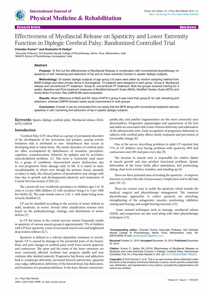
Above table shows pre and post intervention values of MAS and MTS score of calf, hamstring and adductors. P value shows significant difference among them (Graph 1).
Above Graph 1 shows pre and post intervention score comparison in Group A (Table 4).
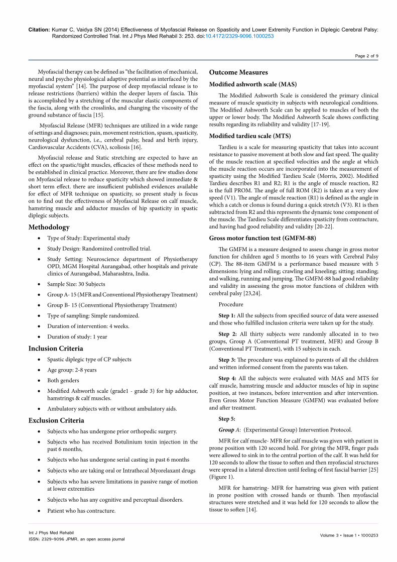
Above Table 4 shows pre and post intervention values of MAS and MTS score of calf, hamstring and adductors of group B. P value shows significant difference among them (Graph 2).
Above Graph 2 shows pre and post intervention score comparison in Group B (Table 5).
Above Table 5 shows Post intervention score of GMFM of Group
Group A (n=15) Group B (n=15)
GenderMale 09 10
Female 06 05Age (Mean ± SD) 5.26 ± 1.70 5.3 ± 1.85
Table 1: Demographic Data.
Group A Group Bp - Value Sign.
Mean ± SD Mean ± SDGMFM 63.38±14.32 62.81 ± 12.60 p=0.909 Not Significant
Calf (Rt)MAS 2.03 ± 0.54 2.16 ± 0.55 p=0.515 Not significant
MTSR1 -1.6 ± 4.89 -1.4 ± 5.16 p=0.914 Not significantR2 16.73 ± 1.53 16.4 ± 2.06 p=0.619 Not significant
Calf (Lt)MAS 2.03 ± 0.54 2.03 ± 0.54 p=1.000 Not significant
MTSR1 -1.26 ± 4.71 -1.53 ± 5.19 p=0.884 Not significantR2 16.93 ± 1.48 16.66 ± 1.98 p=0.681 Not significant
Hams (Rt)MAS 1.86 ± 0.51 2 ± 0.56 p=0.506 Not Significant
MTSR1 85.75 ± 15.25 85.53 ± 13.75 p=0.970 Not SignificantR2 143.4 ± 7.52 143.4 ± 6.76 p=1.000 Not Significant
Hams (Lt)MAS 1.86 ± 0.39 1.93 ± 0.59 p=0.721 Not Significant
MTSR1 86.86 ± 14.00 87.73 ± 12.34 p=0.859 Not SignificantR2 141.73 ± 5.87 143.2 ± 5.72 p=0.494 Not Significant
Add (Rt)MAS 1.93 ± 0.49 1.93 ± 0.59 p=1.000 Not Significant
MTSR1 19.66 ± 4.9 19.06 ± 3.08 p=0.695 Not SignificantR2 37.53 ± 1.99 37.93 ± 2.28 p=0.613 Not Significant
Add (Lt)MAS 1.86 ± 0.39 1.76 ± 0.41 p=0.508 Not Significant
MTSR1 20.06 ± 5.95 20.33 ± 3.03 p=0.878 Not SignificantR2 37.33 ± 2.28 37.6 ± 2.29 p=0.752 Not Significant
Table 2: Baseline comparison of GMFM, MAS and MTS of Right calf, left calf, Right Hamstrings, left Hamstrings, Right hip adductors and left hip adductors.
Group APre Post
p Value Sig.Mean ± SD Mean ± SD
GMFM 63.38 ± 14.32 67.24 ± 14.08 P=0.000 Significant
Calf (Rt)MAS 2.03 ± 0.54 1.16 ± 0.36 P=0.000 Significant
MTSR1 -1.6 ± 4.89 9.46 ± 2.97 P=0.000 SignificantR2 16.73 ± 1.53 18.13 ± 13.5 P=0.000 Significant
Calf (Lt)MAS 2.03 ± 0.54 1.13 ± 0.35 P=0.000 Significant
MTSR1 -1.26 ± 4.71 9.53 ± 3.60 P=0.000 SignificantR2 16.93 ± 1.48 18.26 ± 1.27 P=0.000 Significant
Hams (Rt)MAS 1.86 ± 0.51 1.13 ± 0.22 P=0.000 Significant
MTSR1 85.75 ± 15.25 111.33 ± 12.19 P=0.000 SignificantR2 143.4 ± 7.52 145.93 ± 7.38 P=0.000 Significant
Hams (Lt)MAS 1.86 ± 0.39 1.06 ± 0.75 P=0.000 Significant
MTSR1 86.86 ± 14.00 108.4 ± 13.08 P=0.000 SignificantR2 141.73 ± 5.87 144.06 ± 6.36 P=0.000 Significant
Add (Rt)MAS 1.93 ± 0.49 1.1 ± 0.20 P=0.000 Significant
MTSR1 19.66 ± 4.9 27.73 ± 5.18 P=0.000 SignificantR2 37.53 ± 1.99 38.8 ± 1.56 P=0.000 Significant
Add (Lt)MAS 1.86 ± 0.39 1.06 ± 0.17 P=0.000 Significant
MTSR1 20.06 ± 5.95 27.93 ± 6.28 P=0.000 SignificantR2 37.33 ± 2.28 38.8 ± 1.74 P=0.000 Significant
Table 3: MAS and MTS at Pre and Post Intervention score comparison in Group A. P<0.05* shows a statistically significant result.
Citation: Kumar C, Vaidya SN (2014) Effectiveness of Myofascial Release on Spasticity and Lower Extremity Function in Diplegic Cerebral Palsy: Randomized Controlled Trial. Int J Phys Med Rehabil 3: 253. doi:10.4172/2329-9096.1000253
Page 5 of 9
Volume 3 • Issue 1 • 1000253Int J Phys Med RehabilISSN: 2329-9096 JPMR, an open access journal
A and Group B. P valueis 0.904 which shows no significant difference among Groups.
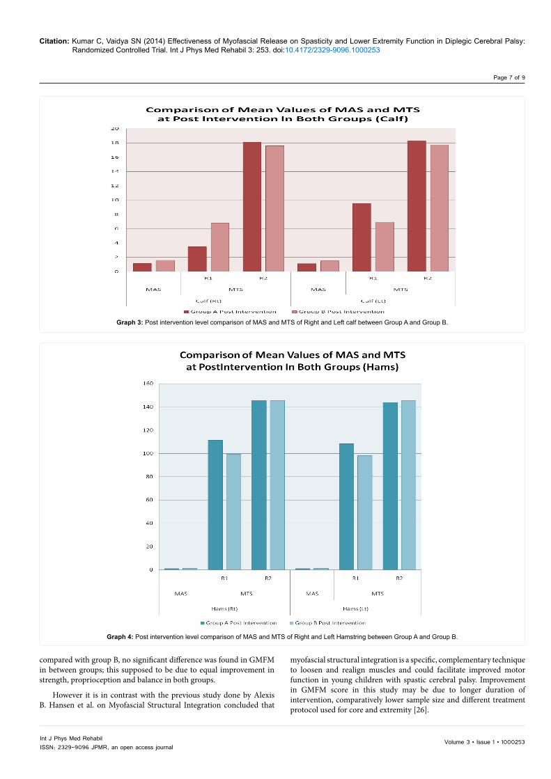
Post intervention measurement of MAS and MTS score for Right and Left calf of Group A and Group B. P value shows significant difference for MAS and R1 whereas no significant difference for R2 of MTS.
Post intervention measurement of MAS and MTS score for Right & Left hamstring of Group A and Group B. P value shows significant difference for MAS and R1 whereas no significant difference for R2.
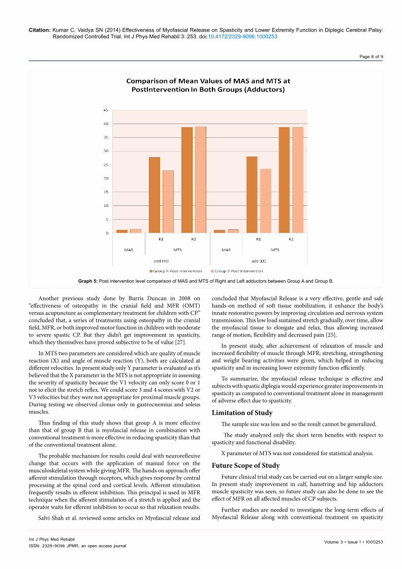
Post intervention measurement of MAS and MTS score for Right & Left adductors of Group A and Group B. P value shows significant difference for MAS and R1 whereas no significant difference for R2 (Graph 3).
Above Graph 3 shows post intervention comparison of MAS and MTS of Right and Left calf for Group A and Group B (Graph 3).
Above Graph 4 shows post intervention comparison of MAS and MTS of Right and Left Hamstring for Group A and Group B (Graph 4).
Above Graph 5 shows post intervention comparison of MAS and MTS of Right and Left adductors for Group A and Group B.
DiscussionThe present study was carried out to find out effectiveness of
myofascial release on spasticity in subjects diagnosed with spastic diplegia. Calf, hamstring and adductors of hip were considered for the study, as spastic diplegic subjects usually have spasticity in these muscle groups.
Graph 1: MAS and MTS at Pre and Post Intervention score comparison in Group A.
Group BPre Post
p Value Sign.Mean ± SD Mean ± SD
GMFM 62.81 ± 12.6 66.64 ± 12.82 P=0.000 Significant
Calf (Rt)MAS 2.16 ± 0.55 1.6 ± 0.43 P=0.000 Significant
MTSR1 -1.4 ± 5.16 6.8 ± 2.39 P=0.000 SignificantR2 16.4 ± 2.06 17.6 ± 1.35 P=0.001 Significant
Calf (Lt)MAS 2.03 ± 0.54 1.5 ± 0.53 P=0.000 Significant
MTSR1 -1.53 ± 5.19 6.93 ± 2.52 P=0.000 SignificantR2 16.66 ± 1.98 17.73 ± 1.66 P=0.000 Significant
Hams (Rt)MAS 2 ± 0.56 1.43 ± 0.41 P=0.000 Significant
MTSR1 85.53 ± 13.75 99.53 ± 11.83 P=0.000 SignificantR2 143.4 ± 6.76 145.53 ± 6.45 p=0.030 Significant
Hams (Lt)MAS 1.93 ± 0.59 1.4 ± 0.43 P=0.000 Significant
MTSR1 87.73 ± 12.34 98.06 ± 13.34 P=0.000 SignificantR2 143.2 ± 5.72 145.46 ± 5.18 P=0.000 Significant
Add (Rt)MAS 1.93 ± 0.59 1.36 ± 0.44 P=0.000 Significant
MTSR1 19.06 ± 3.08 22.93 ± 3.88 P=0.000 SignificantR2 37.93 ± 2.28 39 ± 2 p=0.001 Significant
Add (Lt)MAS 1.76 ± 0.41 1.26 ± 0.31 P=0.000 Significant
MTSR1 20.33 ± 3.03 23.53 ± 3.33 P=0.000 SignificantR2 37.6 ± 2.29 38.66 ± 1.87 p=0.001 Significant
Table 4: MAS and MTS at Pre and Post Intervention score comparison in Group B. P<0.05* shows a statistically significant result.
Citation: Kumar C, Vaidya SN (2014) Effectiveness of Myofascial Release on Spasticity and Lower Extremity Function in Diplegic Cerebral Palsy: Randomized Controlled Trial. Int J Phys Med Rehabil 3: 253. doi:10.4172/2329-9096.1000253
Page 6 of 9
Volume 3 • Issue 1 • 1000253Int J Phys Med RehabilISSN: 2329-9096 JPMR, an open access journal
When analysis was done for demographic information of participants, no statistically significant difference was found showing that subjects are matched for baseline characteristics. There was no significant difference between pre GMFM, pre MAS and pre MTS score in two groups for calf, hamstring and adductor muscle, which shows that two groups are statistically matched at baseline level as shown in Tables 1 and 2.
When comparison was done between pre and post intervention level for both the groups, the values for MAS, MTS and GMFM score were statistically significant for all calf, hamstring and adductor muscle as shown in Tables 3 and 4.
When post intervention comparison was done between group A
and group B it was found that there was significant difference between post MAS, post R1 value of MTS for calf, hamstring and adductor muscle; whereas no significant difference was found in post R2 value of MTS and post GMFM score in between the groups as shown in Table 5.
This is in agreement with previous study done by Salvi Shah in 2012 on immediate effect of Myofascial release on spasticity in spastic cerebral palsy subjects suggesting that the combination of Myofascial Release with stretching alone on calf muscle has better outcomes in treatment of spasticity than stretching alone according to R1 value of MTS, whereas no significant improvement was seen in MAS and R2 value of MTS [25].
Though there is significant reduction of spasticity in group A
Graph 2: MAS and MTS at Pre and Post Intervention score comparison in Group B.
Sr no Group A Group B
p - Value Sign.Mean ± SD Mean ± SD
1. GMFMPost Intervention 67.24 ± 14.08 66.64 ± 12.82 p=0.904 Not Significant
2. Calf (Rt)MAS 1.16 ± 0.36 1.6 ± 0.43 p=0.006 Significant
MTSR1 9.46 ± 2.97 6.8 ± 2.39 p=0.011 SignificantR2 18.13 ± 13.5 17.6 ± 1.35 p=0.290 Not Significant
3. Calf (Lt)MAS 1.13 ± 0.35 1.5 ± 0.53 p=0.035 Significant
MTSR1 9.53 ± 3.60 6.93 ± 2.52 p=0.030 SignificantR2 18.26 ± 1.27 17.73 ± 1.66 p=0.334 Not Significant
4. Hams (Rt)MAS 1.13 ± 0.22 1.43 ± 0.41 p=0.021 Significant
MTSR1 111.33 ± 12.19 99.53 ± 11.83 p=0.012 SignificantR2 145.93 ± 7.38 145.53 ± 6.45 p=0.876 Not Significant
5. Hams (Lt)MAS 1.06 ± 0.75 1.4 ± 0.43 p=0.010 Significant
MTSR1 108.4 ± 13.08 98.06 ± 13.34 p=0.041 SignificantR2 144.06 ± 6.36 145.46 ± 5.18 p=0.514 Not Significant
6. Add (Rt)MAS 1.1 ± 0.20 1.36 ± 0.44 p=0.043 Significant
MTSR1 27.73 ± 5.18 22.93 ± 3.88 p=0.008 SignificantR2 38.8 ± 1.56 39 ± 2 p=0.763 Not Significant
7. Add (Lt)MAS 1.06 ± 0.17 1.26 ± 0.31 p=0.043 Significant
MTSR1 27.93 ± 6.28 23.53 ± 3.33 p=0.024 SignificantR2 38.8 ± 1.74 38.66 ± 1.87 p=0.842 Not Significant
Table 5: Post intervention level comparison of GMFM score, MAS score and MTS score of Right calf, left calf, Right Hamstrings, left Hamstrings, Right hip adductors and left hip adductors between Group A and Group B.
Citation: Kumar C, Vaidya SN (2014) Effectiveness of Myofascial Release on Spasticity and Lower Extremity Function in Diplegic Cerebral Palsy: Randomized Controlled Trial. Int J Phys Med Rehabil 3: 253. doi:10.4172/2329-9096.1000253
Page 7 of 9
Volume 3 • Issue 1 • 1000253Int J Phys Med RehabilISSN: 2329-9096 JPMR, an open access journal
compared with group B, no significant difference was found in GMFM in between groups; this supposed to be due to equal improvement in strength, proprioception and balance in both groups.
However it is in contrast with the previous study done by Alexis B. Hansen et al. on Myofascial Structural Integration concluded that
myofascial structural integration is a specific, complementary technique to loosen and realign muscles and could facilitate improved motor function in young children with spastic cerebral palsy. Improvement in GMFM score in this study may be due to longer duration of intervention, comparatively lower sample size and different treatment protocol used for core and extremity [26].
Graph 3: Post intervention level comparison of MAS and MTS of Right and Left calf between Group A and Group B.
Graph 4: Post intervention level comparison of MAS and MTS of Right and Left Hamstring between Group A and Group B.
Citation: Kumar C, Vaidya SN (2014) Effectiveness of Myofascial Release on Spasticity and Lower Extremity Function in Diplegic Cerebral Palsy: Randomized Controlled Trial. Int J Phys Med Rehabil 3: 253. doi:10.4172/2329-9096.1000253
Page 8 of 9
Volume 3 • Issue 1 • 1000253Int J Phys Med RehabilISSN: 2329-9096 JPMR, an open access journal
Another previous study done by Burris Duncan in 2008 on “effectiveness of osteopathy in the cranial field and MFR (OMT) versus acupuncture as complementary treatment for children with CP” concluded that, a series of treatments using osteopathy in the cranial field, MFR, or both improved motor function in children with moderate to severe spastic CP. But they didn’t get improvement in spasticity, which they themselves have proved subjective to be of value [27].
In MTS two parameters are considered which are quality of muscle reaction (X) and angle of muscle reaction (Y), both are calculated at different velocities. In present study only Y parameter is evaluated as it’s believed that the X parameter in the MTS is not appropriate in assessing the severity of spasticity because the V1 velocity can only score 0 or 1 not to elicit the stretch reflex. We could score 3 and 4 scores with V2 or V3 velocities but they were not appropriate for proximal muscle groups. During testing we observed clonus only in gastrocnemius and soleus muscles.
Thus finding of this study shows that group A is more effective than that of group B that is myofascial release in combination with conventional treatment is more effective in reducing spasticity than that of the conventional treatment alone.
The probable mechanism for results could deal with neuroreflexive change that occurs with the application of manual force on the musculoskeletal system while giving MFR. The hands on approach offer afferent stimulation through receptors, which gives response by central processing at the spinal cord and cortical levels. Afferent stimulation frequently results in efferent inhibition. This principal is used in MFR technique when the afferent stimulation of a stretch is applied and the operator waits for efferent inhibition to occur so that relaxation results.
Salvi Shah et al. reviewed some articles on Myofascial release and
concluded that Myofascial Release is a very effective, gentle and safe hands-on method of soft tissue mobilization, it enhance the body’s innate restorative powers by improving circulation and nervous system transmission. This low load sustained stretch gradually, over time, allow the myofascial tissue to elongate and relax, thus allowing increased range of motion, flexibility and decreased pain [25].
In present study, after achievement of relaxation of muscle and increased flexibility of muscle through MFR; stretching, strengthening and weight bearing activities were given, which helped in reducing spasticity and in increasing lower extremity function efficiently.
To summarize, the myofascial release technique is effective and subjects with spastic diplegia would experience greater improvements in spasticity as compared to conventional treatment alone in management of adverse effect due to spasticity.
Limitation of StudyThe sample size was less and so the result cannot be generalized.
The study analyzed only the short term benefits with respect to spasticity and functional disability.
X parameter of MTS was not considered for statistical analysis.
Future Scope of StudyFuture clinical trial study can be carried out on a larger sample size.
In present study improvement in calf, hamstring and hip adductors muscle spasticity was seen, so future study can also be done to see the effect of MFR on all affected muscles of CP subjects.
Further studies are needed to investigate the long-term effects of Myofascial Release along with conventional treatment on spasticity
Graph 5: Post intervention level comparison of MAS and MTS of Right and Left adductors between Group A and Group B.

Citation: Kumar C, Vaidya SN (2014) Effectiveness of Myofascial Release on Spasticity and Lower Extremity Function in Diplegic Cerebral Palsy: Randomized Controlled Trial. Int J Phys Med Rehabil 3: 253. doi:10.4172/2329-9096.1000253
Page 9 of 9
Volume 3 • Issue 1 • 1000253Int J Phys Med RehabilISSN: 2329-9096 JPMR, an open access journal
when compared with conventional treatment and also on the functional activity of spastic subjects
Clinical ImplicationThe results of this study have important clinical implication for
developing effective intervention along with conventional treatment for subjects with spastic diplegia when compared with conventional treatment for reducing spasticity. They can be easily incorporated in any rehabilitation technique.
ConclusionThe findings of this study suggests that the combination of
myofascial release and conventional treatment was shown to reduce spasticity in for spastic diplegic subjects but functional changes were not seen compared to conventional treatments.
References
1. Rosenbaun P, Peneth N (2007) The definition and classification of cerebral palsy. Dev Med Child Neurol 49: 8-14.
2. Sharma P, Sharma U, Kabra A (1990) Cerebral Palsy-Clinical profile and predisposing factors. Indian Pediatr 36: 1038-1042.
3. Dan B, Cheron G (2004) Reconstructing Cerebral Palsy. J PediatrNeurol 2: 57-64.
4. Green LB, Hurvitz ED (2007) Cerebral palsy, physical medicine rehabilitation clinic. 859-882.
5. Jan MM (2006) Cerebral Palsy: Comprehensive Review and Update. Ann Saudi Med 26: 123-132.
6. Love SC, Valentineb JP, Blairc EM, Priced CJ, Coled JH, et al. (2001) The effect of botulinum toxin type A on the functional ability of the child with spastic hemiplegia a randomized controlled trial, Eur J Neurol 8: 50-58.
7. Whisler SL, Lang DM (2012); Effect of myofascial release and other advanced myofascial therapies on children with cerebral palsy: six case reports. Explore 8: 199-205.
8. Morrell DS, Pearson JM, Sauser DD (2002) Progressive bone and joint abnormalities of the spine and lower extremities in cerebral palsy. Radiographics 22: 257-268.
9. Andersson C, Mattsson E (2001) Adults with cerebral palsy: a survey describing problems, needs, and resources, with special emphasis on locomotion. Dev Med Child neurol 43: 76-82.
10. Barnes MR (1998) Review management of spasticity. Age and Ageing 27: 239-245.
11. Nerita NC Chan (2011) Physiotherapy in Spasticity Management for Children with Cerebral Palsy 16.
12. Manheim CJ (2001) The Myofascial Release Manual. (3rd edn).
13. Barnes JF (1991) Pediatric Myofascial Release, Physical Therapy Forum-MFR Techniques.
14. Barnes JF (1995) Myofascial Release-The Missing Link in Your Treatment.
15. Gregson JM, Leathley M, Moore P, Sharma AK, Smith TL, et al. (1999) Reliability of the Tone Assessment Scale and the Modified Ashworth Scale as clinical tools for assessing poststroke spasticity. Arch Phys Med Rehabil 80: 1013-1016.
16. Bohannon RW, Smith MB (1987) Inter-rater reliability of a Modified Ashworth Scale of muscle spasticity. Phys Ther 2: 206-208.
17. Ansari NN, Naghdi S, Younesian B, Shayeghan M (2008) Inter and intra-rater reliability of the Modified Modified Ashworth Scale in subjects with knee extensor poststroke spasticity. Physiotherapy theory and practice 24: 205-213.
18. Tardieu G, Shentoub S, Delarue R (1954) A la recherché d’une technique de mesure de la spasticite. Rev Neurol 91: 143-144.
19. Tardieu G, Rondont O, Mensch J, Dalloz JC, Monfraix C, et al. (1957) Responses electromyographiques a l’etirement musculaire chezl’homme normal. Rev Neurol 97: 60-61.
20. Patrick E, Alda L (2006) The Tardieu Scale differentiates contracture from spasticity whereas the Ashworth Scales is confounded by it. Clinical Rehabilitation 20: 173-182.
21. Russell D, Rosenbaum P, Gowland C, Hardy S, Lane M, et al. (1993) Gross Motor Function Measure Manual (2nd edn). Hamilton: McMaster University.
22. Finch E, Brooks D, Stratford PW, Mayo NE (2002) Physical rehabilitation outcome measures: A guide to enhanced clinical decision making (2nd edn). Hamilton: B. C. Decker.
23. Salvi Shah, AktaBhalara (2012) Myofascial Release. International Journal of Health Sciences & Research 2.
24. Hansen AB, Price KS, Heidi M (2001) Feldman Myofascial structural integration: A promising complementary therapy for young children with spastic cerebral palsy. Journal of Evidence Based Complementary & Alternative Medicine 1-5.
25. Duncan B, McDonough S, Worden MK, Schnyer R, Andrews J, et al. (2008) Effectiveness of Osteopathy in the Cranial Field and Myofascial Release Versu Acupuncture as Complementary Treatment for Children With Spastic Cerebral Palsy: A Pilot Study. J Am Osteopath Assoc 108.
26. Numanolu A, Gunel MK (2002) Intraobserver Reliability of Modified Asworth Scale And Modified Tardieu Scale in the assessment of spasticity in children with cerebral palsy. Acta Orthop Traumatol Turc 46: 196-200.
27. Greenman (2003) Principles of Manual Medicine. (3rd edn), 157.
Submit your next manuscript and get advantages of OMICS Group submissionsUnique features:
• Userfriendly/feasiblewebsite-translationofyourpaperto50world’sleadinglanguages• AudioVersionofpublishedpaper• Digitalarticlestoshareandexplore
Special features:
• 300OpenAccessJournals• 25,000editorialteam• 21daysrapidreviewprocess• Qualityandquickeditorial,reviewandpublicationprocessing• IndexingatPubMed(partial),Scopus,EBSCO,IndexCopernicusandGoogleScholaretc• SharingOption:SocialNetworkingEnabled• Authors,ReviewersandEditorsrewardedwithonlineScientificCredits• Betterdiscountforyoursubsequentarticles
Submityourmanuscriptat:http://www.omicsonline.org/submission/
Citation: Kumar C, Vaidya SN (2014) Effectiveness of Myofascial Release on Spasticity and Lower Extremity Function in Diplegic Cerebral Palsy: Randomized Controlled Trial. Int J Phys Med Rehabil 3: 253. doi:10.4172/2329-9096.1000253