kois--l_kdfas_rev_3
TRANSCRIPT

8/6/2019 KOIS--L_KDFAS_REV_3
http://slidepdf.com/reader/full/kois-lkdfasrev3 1/8
1 L-KDFAS REV 3
U.S. Patents 6,413,085 & 6,582,931
Foreign Patents Pending
5402
1. Based on Dr. John C. Kois’s research of an averageaxis-incisal distance of 100mm, the Kois Dento-FacialAnalyzer was developed to simplify the procedures oftransferring and mounting study casts for both estheticsand function. Dr. Kois research is substantiated andcorroborated by Bonwill’s Equilateral Triangle, Monson’sSpherical Theory (4”=100.12mm), Weinberg studies in1963 as well as others.
2. Dr. Kois’s studies were done using different Ethnicbackgrounds and genders. This bar chart shows thedistribution of measurements from the hinge axis tothe incisal edge of the maxillary central incisor. As youcan see, approximately 80% are within 5mm of the av-erage 100mm axis-insical distance, which is approxi-mately the same percentages reported in research com-paring arbitrary earbows.
3. Traditionally, dentists are taught to make the incisal-canine line parallel to the eyes. If the eyes are slanted,then the teeth would also be made slanted. The dentalmidline is critical and is always related to the facial mid-line. Therefore, we need to register the facial midlinewhich dictates the dental midline. Then the occlusalplane will be made perpendicular to the dental midline.
4. This system registers the steepness and tilts of the
occlusal plane related in three planes of space. Thehorizontal portion of the Analyzer Bow will register anocclusal-horizontal plane of reference. The Vertical Rodwill register the facial mid-line for the sagittal plane ofreference; and the average axis-incisal distance of100mm relates to the frontal plane of reference.
Kois Dento-Facial Analyzer System Instructions“A Simplified Face-Bow for Esthetics and Function”
RESEARCH:
h 4300
4305Includes:4322 D
M Panadent Corporation
580 S. Rancho Avenue • Colton, California 92324, USA
Tel: (909) 783-1841 • USA & Canada (800) 368-9777
These instructions apply to the following items:

8/6/2019 KOIS--L_KDFAS_REV_3
http://slidepdf.com/reader/full/kois-lkdfasrev3 2/8
2 L-KDFAS REV 3
U.S. Patents 6,413,085 & 6,582,931
Foreign Patents Pending
5402
1. Attach Vertical Indicator Rod to Analyzer bow by slid-ing white attachment disk on the rod into the keyway sloton the Analyzer bow.
2. Attach disposable Index Tray to Analyzer bow by align-ing protruding pins on Index Tray into the holes in the bitefork section of the Analyzer bow. Seat Index Tray all theway flat onto Analyzer bow.
3. It is best to place 4 Bite-Tabs™ impression com-pound onto posterior and bicuspid area of the Index Tray.If using registration material other than Bite-Tabs™, firstapply an adhesive to occlusal surfaces of Index Tray.
4. Place Index Tray into a bowl of hot water to temperBite-Tabs™ impression compound. The compound canbe squeezed into a cone shape if more height is re-
quired.

8/6/2019 KOIS--L_KDFAS_REV_3
http://slidepdf.com/reader/full/kois-lkdfasrev3 3/8
3 L-KDFAS REV 3
U.S. Patents 6,413,085 & 6,582,931
Foreign Patents Pending
5402
1. Having posterior portion of Analyzer Bow down outof occlusion, set incisal edge of maxillary incisors tothe wall or ledge on Index Tray. This registers the in-cisal point of the average 100mm axis-incisal distance
for function.
2. Align Vertical Indicator Rod to patient’s facial midlineto register the dental midline of the teeth to the frontalplane for esthetics. The Vertical Indicator Rod can bepositioned posteriorly in keyway slot of Analyzer Bow tobe close to the patient’s nose.
3. While maintaining incisal contact with the Index Trayand vertical rod alignment to the facial midline, rotateAnalyzer Bow up in the posterior until the lateral wingsare level to the horizon. This should be done whilelooking at the front of the patient. The Bioesthetic LevelGauge™ is not required. However, it can be added toverify that the bow is level in the sagittal plane.
4. These procedures can be more simply done with thepatient in a supine position. By doing this, the head issupported by the dental chair head rest. Align the incisaledge to the wall on the Index Tray. You can look at thevertical rod related to the facial midline better from be-hind the patient. Have the lateral wings hang straight downas you make the registration of the teeth.
5. You have now captured the steepness and tilts of theocclusal plane in the registration material on the horizon-tally aligned Index Tray. Remove Index Tray from AnalyzerBow and send to the lab for mounting of study casts. Thisdisposable Index Tray now becomes a permanent bitefork registration record.
REGISTRATION:
8/6/2019 KOIS--L_KDFAS_REV_3
http://slidepdf.com/reader/full/kois-lkdfasrev3 4/8
4 L-KDFAS REV 3
U.S. Patents 6,413,085 & 6,582,931
Foreign Patents Pending
5402
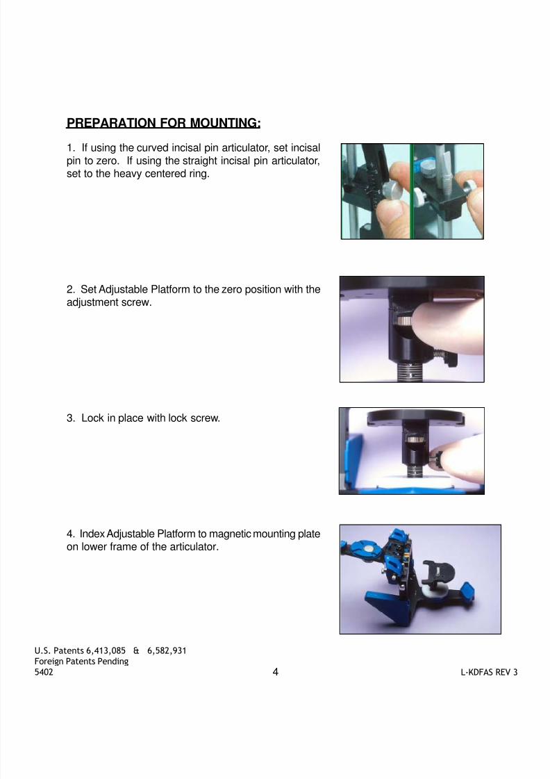
PREPARATION FOR MOUNTING:
1. If using the curved incisal pin articulator, set incisalpin to zero. If using the straight incisal pin articulator,set to the heavy centered ring.
2. Set Adjustable Platform to the zero position with theadjustment screw.
3. Lock in place with lock screw.
4. Index Adjustable Platform to magnetic mounting plate
on lower frame of the articulator.

8/6/2019 KOIS--L_KDFAS_REV_3
http://slidepdf.com/reader/full/kois-lkdfasrev3 5/8
5 L-KDFAS REV 3
U.S. Patents 6,413,085 & 6,582,931
Foreign Patents Pending
5402
MOUNTING:
1. Place Index Tray on Adjustable Platform by align-ing protruding pins of Index Tray to the holes onthe Adjustable Platform. Seat Index Tray all theway down flat to the Adjustable Platform.
2. Index study cast into impressions on the IndexTray. The Adjustable Platform now becomes a built-in bite fork support system.
3. Add plaster to mounting plate and cast to mountmaxillary study cast in usual manner. It has beenengineered that the incisal edges are now 100mmfrom the axis.
4. Mount mandibular study cast in usual manner us-ing interocclusal record and the Mandibular Mount-ing Stand. Note that the front of the articulator canbe adjusted down with the support pin to control theplaster.
5. The steepness and tilts of the occlusal plane,the dental midline, incisal edge position as well asgingival contours can now be diagnosed for sym-metry and balance.

8/6/2019 KOIS--L_KDFAS_REV_3
http://slidepdf.com/reader/full/kois-lkdfasrev3 6/8
6 L-KDFAS REV 3
U.S. Patents 6,413,085 & 6,582,931
Foreign Patents Pending
5402
DIAGNOSTIC OPTIONS:
1. While the Analyzer bow is on the patient, havethe patient smile. Measure and chart the heightof the lip commissures from the Index Tray (ie.right side, mesial of 2cd bicuspid, 3mm; left side,distal of 1st bicuspid, 2mm).
2. Mark the charted height of the lip commissureson the study cast to evaluate lip curvature andsmile lines.
3. This chart shows the different facial landmarksto evaluate lateral facial proportions. This chartshows that the width of the eyes are 60% widerthan the mouth. The mouth is 60% wider thanthe nose, and the nose is 60% wider than thetwo central incisors. Divide that distance by 2 toget the width of one central incisor.
4. A reusable set of 7 Golden Proportion Wax-ing Guides are available ranging from 7-10mmin .5mm increments (7, 7.5, 8, 8.5, 9, 9.5, 10mm)to correspond to the width of one central incisor.Place appropriate Waxing Guide on AdjustablePlatform by indexing protruding pins of WaxingGuide to the holes on the Adjustable Platform.
5. Anterior tooth widths can now be diagnosedfor proper anterior proportions for optimum es-thetics. There are also three 1 mm lines anteri-orly and posteriorly if you want a guide to movethe incisal edges forward or backward for betterlip support.

8/6/2019 KOIS--L_KDFAS_REV_3
http://slidepdf.com/reader/full/kois-lkdfasrev3 7/8
7 L-KDFAS REV 3
U.S. Patents 6,413,085 & 6,582,931
Foreign Patents Pending
5402
6. This chart shows the different facial landmarksto evaluate vertical facial proportions. Knowingthat we can not change the inner canthus of theeyes or the ayla of the nose, we will use thesepoints as references to evaluate incisal edge po-sition vertically in the face for diagnosing toothlengths. Using the nasial-labial angle and the newincisal edge position, we can elvaluate mentonfor diagnosing vertical dimension for optimum fa-cial esthetics.
7. Remove Vertical Indicator Rod from Analyzerbow. Have patient bite their teeth together andplace white attachment disk to the incisal edge ofthe patient’s maxillary central incisor.
8. Adjust slideable O-rings on vertical rod to spe-
cific landmarks on patient’s face (ie. inner can-thus and ayla of the nose, incisal edge and men-ton). The vertical rod can also be placed onpatient’s chart to record the O-Ring points on therod for a permanent reference of the patient’sfacial proportions.
9. Using the inner canthus of the eye and the aylaof the nose as reference points, evaluate incisal
edge position vertically in the face. This pictureshows that approximately 3mm could be added tothe incisal edge length to improve this patient’s mid-face proportions.
DIAGNOSTIC OPTIONS CONTINUED...

8/6/2019 KOIS--L_KDFAS_REV_3
http://slidepdf.com/reader/full/kois-lkdfasrev3 8/8
8 L-KDFAS REV 3
U.S. Patents 6,413,085 & 6,582,931
Foreign Patents Pending
5402
10. Using adjustment screw, adjust platform down 3mmto the incisal length to be fabricated. The study cast isnow suspended 3mm above waxing guide by the incisalpin.
11. The technician can now add wax or porcelain until ittouches the waxing guide for a determined incisal length
to be fabricated. The Golden Proportion Waxing Guidecan also be used at the same time for tooth widths.
12. Knowing the new incisor length that will be restored,measure from the nasial-labial to new incisal edge to befabricated and evaluate menton position for proper ver-tical dimension. This picture shows that the vertical di-mension could be restored approximately 2mm to im-prove this patient’s lower facial proportions.
13. The steepness and tilts of the occlusal plane relatedto the hinge axis, smile and gingival symmetry and bal-ance, lip curvature, tooth and facial proportions can nowbe diagnosed to achieve a superior treatment plan foroptimum esthetics and function.
DIAGNOSTIC OPTIONS CONTINUED...
4322: If used more than one time, patient cross-contamination may occur.
i
D