knt nd eltrrph anl f th rn nd r b rn wln n tvld sh · 2010-03-22 · knt nd eltrrph anl f th rn nd...
TRANSCRIPT
ORIGINAL ARTICLES
Kinematic and Electromyographic Analysis of the Trunkand Lower Limbs During Walking in Negative-Heeled Shoes
Jing Xian Li, PhD*Youlian Hong, PhD -I-
Background: We investigate kinematic adaptation and muscle activities in the trunk andlower extremities of healthy subjects during treadmill walking in negative-heeled sportsshoes versus normal sports shoes.
Methods: Thirteen healthy female university students participated in the study. We ana-lyzed sagittal-movement kinematics and electromyographic findings from the erectorspinae, rectus abdominus, rectus femoris, biceps femoris, tibialis anterior, and lateralgastrocnemius muscles of the dominant side in two shod conditions.
Results: Negative-heeled gait is characterized by faster cadence, shorter stride length, in-creased maximal extension angles in the trunk and hip, increased flexion angle in the knee,larger dorsiflexion in the stance phase, and a larger range of motion of the ankle joint. Neg-ative-heeled gait resulted in a significantly larger integrated electromyographic value, alonger duration of electromyographic activity, and a higher mean amplitude of electromyo-graphic activity in the tibialis anterior, lateral gastrocnemius, and biceps femoris muscles.
Conclusions: Negative-heeled gait compared with normal gait places a higher physio-logic demand on the tibialis anterior, lateral gastrocnemius, and biceps femoris muscleswhen walking on a level surface. Thus, negative-heeled shoes could be of value if usedin an exercise rehabilitation or training program where inclined walking is not availableowing to a flat terrain. (J Am Podiatr Med Assoc 97(6): 447-456, 2007)
The strength of the muscles across the ankle andknee joints is important in maintaining stability and inreducing the likelihood of injury. For patients withankle and knee joint injuries, many approaches havebeen developed to enhance the muscular strengtharound these joints as part of rehabilitation, one ofwhich is graded walking) Negative-heeled shoes, alsoknown as earth shoes, were designed to mimic uphillwalking so as to build up and exercise the muscles inthe trunk and lower limbs. In normal sports shoes,the heel is approximately 1.5 cm higher than the toepart.' In contrast, in negative-heeled shoes the toepart is 1.5 cm higher than the heel, which tilts the footinto approximately 10° of dorsiflexion.
Previous studies 3 in trunk and lower-extremitykinematics have shown that gait adaptation on uphilltreadmill inclination during walking is characterized
*School of Human Kinetics, University of Ottawa, Ottawa,Ontario.
tDepartment of Sports Science and Physical Education,The Chinese University of Hong Kong, Hong Kong, China.
Corresponding author: Jing Xian Li, PhD, School of HumanKinetics, University of Ottawa, 125 University, Ottawa, On-tario KIN 6N5, Canada.
by an increasingly flexed posture of the hip, knee,and ankle at initial foot contact and by a progressiveforward tilt of the pelvis and trunk. An earlier report' , 5
showed a significant decrease in walking speed andcadence when walking up a steep ramp of greaterthan 9°. A reduction in step length was also observedduring descent walking!' Electromyographic stud-ies' , 7 9 regarding muscle activation in uphill treadmillwalking and ramp ground walking have found amodifying level of activation of the relevant flexorand extensor muscles in the lower extremity.
Some studies show the influence of shoe heelheight on locomotor adaptation. It was found thatwalking in high-heeled shoes produced shorter stridelengths and higher stance time percentage than walk-ing in normal sports shoes.m Kinematically, gait whilewearing high-heeled shoes, compared to gait whilewearing low-heeled shoes, was characterized by sig-nificantly increased knee flexion at heel strike andduring the stance phase and significantly lower kneeflexion and hip flexion during the swing phase.
Studies that present the kinematics of the trunkand lower limbs when walking in negative-heeled
Celebrating Ail? vcrivw of continuous publication:1907-2007
Journal of the American Podiatric Medical Association • Vol 97 • No 6 • November/December 2007 447
shoes are limited, and the kinematic changes thatoccur when walking in these shoes are not consis-tent. The earliest study" of negative-heeled shoesstated that there were no changes in the gait patternof subjects walking barefoot, in tennis shoes, or innegative-heeled shoes. However, comparison of theflexion and extension of the back, hip, knee, andankle joints of subjects when standing and walking innegative-heeled shoes, positive-heeled shoes, andbare feet suggested that the greatest compensationfor heel height occurs distally." Another study" re-ported that walking speed was significantly reducedwhile walking in negative-heeled shoes as a conse-quence of a shorter stride length combined with anincreased cadence. Walking patterns differed drasti-cally at the ankle joint, but no significant differencewas found at the knee and hip joints. Kinematic adap-tation in different walking conditions is attributed tothe changes in muscle activity in the trunk and lowerextremities. However, for negative-heeled shoes, nostudy about muscle activity in the trunk and lower ex-tremities has been reported.
The purpose of the present investigation was to es-tablish a basic understanding of the activity levels ofthe muscles of the erector spinae, rectus abdominus,rectus femoris, biceps femoris, tibialis anterior, andlateral gastrocnemius and to evaluate the kinematicadaptation of the trunk and lower extremities whenwalking in negative-heeled shoes. This knowledge willprovide a basis for the recommendation of walking innegative-heeled shoes to strengthen the muscles ofthe trunk and lower extremities through locomotionadaptation, which could be of value if used in an exer-cise rehabilitation or training program where inclinedwalking is not available owing to a flat terrain.
Materials and Methods
Subjects and Shoes
A total of 13 female subjects (mean ± SD age, 23.08 ±3.9 years; body weight, 50.18 ± 5.3 kg; and height, 1.63± 0.05 m) volunteered to participate in the study. All ofthe subjects were in good health and had no experi-ence walking in negative-heeled shoes. They had noprevious history of muscle weakness, neurologic dis-ease, or any drug therapy. All of the subjects werefully informed of the purpose and the procedures in-volved in the study, to which they gave informed con-sent according to the guidelines of the Chinese Uni-versity of Hong Kong's ethics committee. All of thesubjects were provided with two types of shoes: nor-mal sports shoes and negative-heeled shoes. Bothtypes of shoes were similar in construction and mate-
rials except for heel height. Normal sports shoes tiltedthe sole into approximately 10° of plantarflexion,whereas negative-heeled shoes tilted the sole into 10°of dorsiflexion. Because heel heights vary accordingto the sizes of shoes, in this study, only size 37 (Euro-pean) shoes were selected.
Data Collection and Analysis
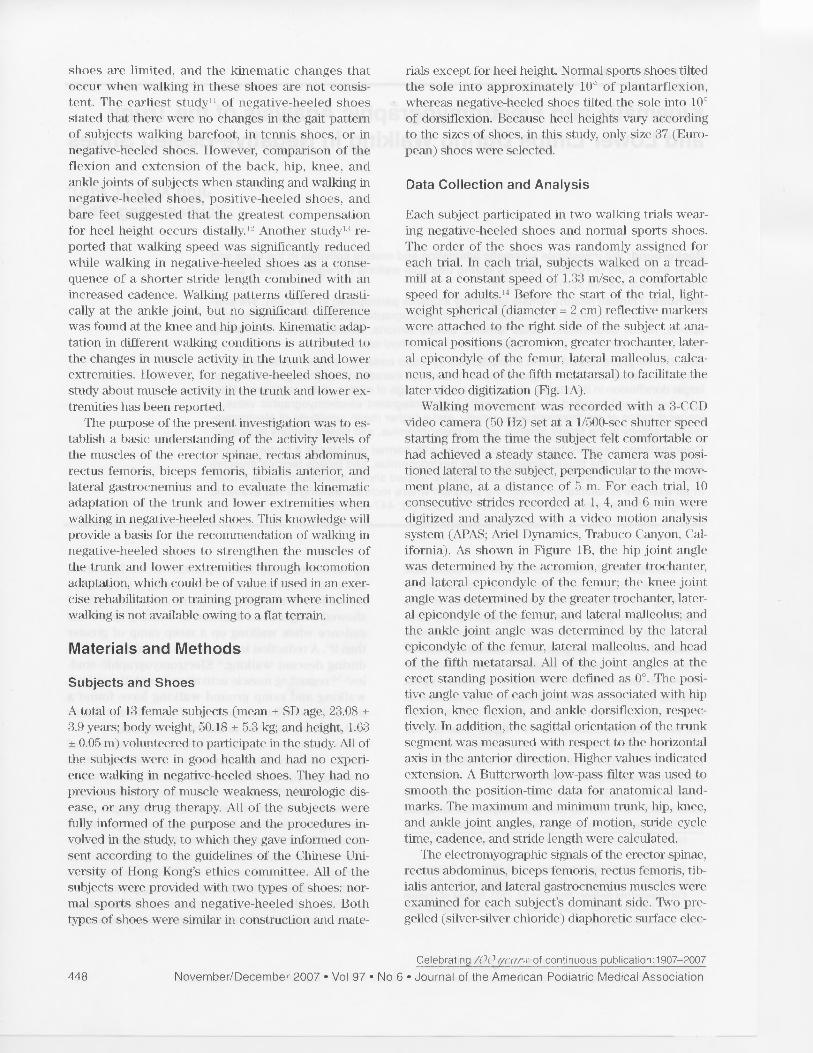
Each subject participated in two walking trials wear-ing negative-heeled shoes and normal sports shoes.The order of the shoes was randomly assigned foreach trial. In each trial, subjects walked on a tread-mill at a constant speed of 1.33 m/sec, a comfortablespeed for adults.' 4 Before the start of the trial, light-weight spherical (diameter = 2 cm) reflective markerswere attached to the right side of the subject at ana-tomical positions (acromion, greater trochanter, later-al epicondyle of the femur, lateral malleolus, calca-neus, and head of the fifth metatarsal) to facilitate thelater video digitization (Fig. 1A).
Walking movement was recorded with a 3-CCDvideo camera (50 Hz) set at a 1/500-sec shutter speedstarting from the time the subject felt comfortable orhad achieved a steady stance. The camera was posi-tioned lateral to the subject, perpendicular to the move-ment plane, at a distance of 5 m. For each trial, 10consecutive strides recorded at 1, 4, and 6 min weredigitized and analyzed with a video motion analysissystem (APAS; Ariel Dynamics, Trabuco Canyon, Cal-ifornia). As shown in Figure 1B, the hip joint anglewas determined by the acromion, greater trochanter,and lateral epicondyle of the femur; the knee jointangle was determined by the greater trochanter, later-al epicondyle of the femur, and lateral malleolus; andthe ankle joint angle was determined by the lateralepicondyle of the femur, lateral malleolus, and headof the fifth metatarsal. All of the joint angles at theerect standing position were defined as 0°. The posi-tive angle value of each joint was associated with hipflexion, knee flexion, and ankle dorsiflexion, respec-tively. In addition, the sagittal orientation of the trunksegment was measured with respect to the horizontalaxis in the anterior direction. Higher values indicatedextension. A Butterworth low-pass filter was used tosmooth the position-time data for anatomical land-marks. The maximum and minimum trunk, hip, knee,and ankle joint angles, range of motion, stride cycletime, cadence, and stride length were calculated.
The electromyographic signals of the erector spinae,rectus abdominus, biceps femoris, rectus femoris, tib-ialis anterior, and lateral gastrocnemius muscles wereexamined for each subject's dominant side. Two pre-gelled (silver-silver chloride) diaphoretic surface elec-
Celebrating /(k ) of continuous publication:1907-2007
448 November/December 2007 • Vol 97 • No 6 • Journal of the American Podiatric Medical Association
A B Trunk
Figure 1. A, Lateral view of the kinematic model. B, Segment and joint angle definitions are illustrated for the sagit-tal plane. For every segment and joint angle, the arrow direction indicates the positive angle from neutral.
trodes were placed, 3 cm apart, over prepared musclesites. A single ground electrode was placed on the an-terior superior iliac crest of the pelvis. The raw elec-tromyographic signals were sampled at 1,000 Hzusing a software program (LabVIEW; National Instru-ments Corp, Austin, Texas) and were stored in a com-puter for postevent analysis. Video filming and elec-tromyographic signal collection were synchronizedwith an external light trigger. The raw electromyo-graphic data were high-pass filtered digitally at 20 Hz,full-wave rectified, and linear enveloped, with a cutofffrequency of 5 Hz. The electromyographic parametersused in this study included the average integratedelectromyographic value in 1 sec, the mean amplitudeof electromyographic activity, and the duration of elec-tromyographic activity in each muscle during a stridecycle. The corresponding time point at which electro-myographic amplitude exceeded or went below 2 SDof the mean amplitude of the electromyographic ac-tivity of the muscle was set as the criterion of theonset and offset of the electromyographic activity, re-spectively.
To reduce small variations in the duration of eachgait cycle between subjects, the time of the gait cyclewas normalized (0% to 100%). For each subject, thekinematic and electromyographic parameters at thesame normalized time point of each stride were aver-aged across the ten consecutive strides and furtheraveraged across the three time points for each shodcondition. The parameters were finally averaged acrossall of the subjects for each shod condition.
Statistical Analysis
Differences in the measured parameters betweennegative-heeled shoes and normal sports shoes wereexamined with a paired-sample t test, with the levelof significance set at P < .05.
Results
Kinematic Parameters
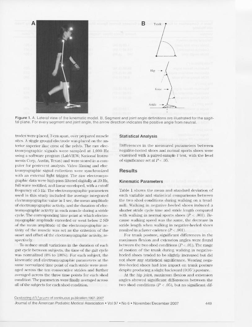
Table 1 shows the mean and standard deviation ofeach variable and statistical comparisons betweenthe two shod conditions during walking on a tread-mill. Walking in negative-heeled shoes induced ashorter stride cycle time and stride length comparedwith walking in normal sports shoes (P < .001). Be-cause walking speed was the same, the decrease instride length when walking in negative-heeled shoesresulted in a faster cadence (P < .001).
For trunk posture, significant differences in themaximum flexion and extension angles were foundbetween the two shod conditions (P < .01). The rangeof motion of the trunk during walking in negative-heeled shoes tended to be slightly increased but didnot show any statistical significance. Wearing nega-tive-heeled shoes had less impact on trunk posturedespite producing a slight backward (0.95°) posture.
At the hip joint, maximum flexion and extensionangles showed significant differences between thetwo shod conditions (P < .05), but no significant dif-
Celebrating /0(?//coitir of continuous publication:1907-2007
Journal of the American Podiatric Medical Association • Vol 97 • No 6 • November/December 2007 449
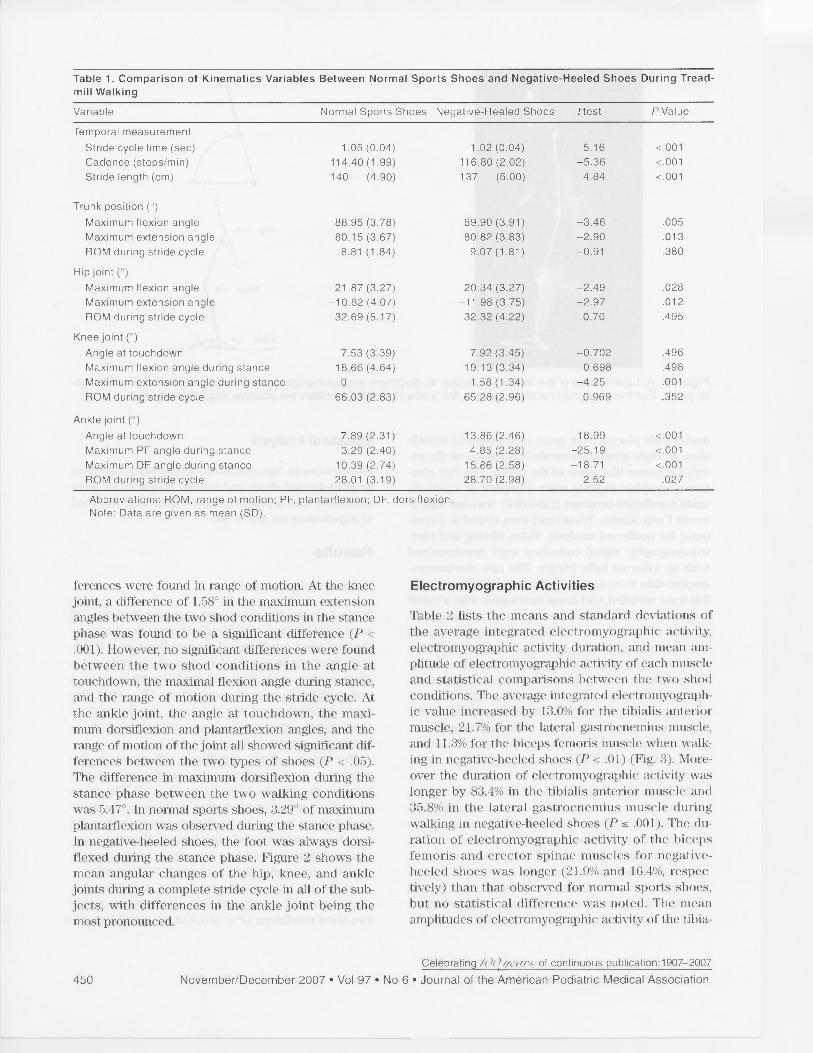
Table 1. Comparison of Kinematics Variables Between Normal Sports Shoes and Negative-Heeled Shoes During Tread-mill Walking
Variable Normal Sports Shoes Negative-Heeled Shoes t test P Value
Temporal measurementStride cycle time (sec) 1.05 (0.04) 1.02 (0.04) 5.16 <.001Cadence (steps/min) 114.40 (1.99) 116.80 (2.02) -5.36 <.001Stride length (cm) 140 (4.90) 137 (5.00) 4.84 <.001
Trunk position (°)Maximum flexion angle 88.95 (3.78) 89.90 (3.91) -3.46 .005Maximum extension angle 80.15 (3.67) 80.82 (3.83) -2.90 .013ROM during stride cycle 8.81 (1.84) 9.07 (1.81) -0.91 .380
Hip joint (°)Maximum flexion angle 21.87 (3.27) 20.34 (3.27) -2.49 .028Maximum extension angle -10.82 (4.07) -11.98 (3.75) -2.97 .012ROM during stride cycle 32.69 (5.17) 32.32 (4.22) 0.70 .495
Knee joint (°)Angle at touchdown 7.53 (3.39) 7.92 (3.45) -0.702 .496Maximum flexion angle during stance 18.66 (4.64) 19.13 (3.34) -0.698 .498Maximum extension angle during stance 0 1.58 (1.34) -4.25 .001ROM during stride cycle 66.03 (2.83) 65.28 (2.96) 0.969 .352
Ankle joint (°)Angle at touchdown 7.89 (2.31) 13.86 (2.46) -18.99 <.001Maximum PF angle during stance -3.29 (2.40) 4.85 (2.28) -25.19 <.001Maximum DF angle during stance 10.39 (2.74) 15.86 (2.58) -18.71 <.001ROM during stride cycle 28.01 (3.19) 28.70 (2.98) -2.52 .027
Abbreviations: ROM, range of motion; PF, plantarflexion; DF, dorsiflexion.Note: Data are given as mean (SD).
ferences were found in range of motion. At the kneejoint, a difference of 1.58° in the maximum extensionangles between the two shod conditions in the stancephase was found to be a significant difference (P <.001). However, no significant differences were foundbetween the two shod conditions in the angle attouchdown, the maximal flexion angle during stance,and the range of motion during the stride cycle. Atthe ankle joint, the angle at touchdown, the maxi-mum dorsiflexion and plantarflexion angles, and therange of motion of the joint all showed significant dif-ferences between the two types of shoes (P < .05).The difference in maximum dorsiflexion during thestance phase between the two walking conditionswas 5.47°. In normal sports shoes, 3.29° of maximumplantarflexion was observed during the stance phase.In negative-heeled shoes, the foot was always dorsi-flexed during the stance phase. Figure 2 shows themean angular changes of the hip, knee, and anklejoints during a complete stride cycle in all of the sub-jects, with differences in the ankle joint being themost pronounced.
Electromyographic Activities
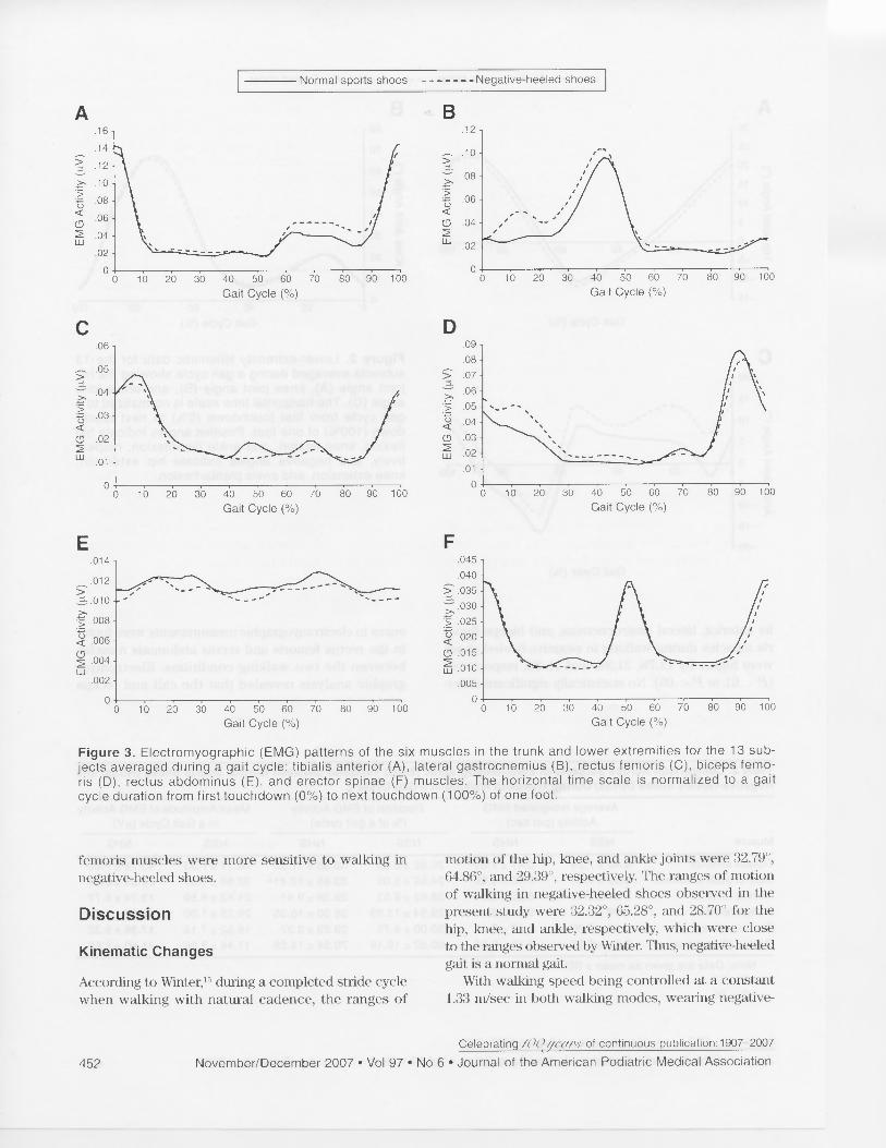
Table 2 lists the means and standard deviations ofthe average integrated electromyographic activity,electromyographic activity duration, and mean am-plitude of electromyographic activity of each muscleand statistical comparisons between the two shodconditions. The average integrated electromyograph-ic value increased by 13.0% for the tibialis anteriormuscle, 21.7% for the lateral gastrocnemius muscle,and 11.3% for the biceps femoris muscle when walk-ing in negative-heeled shoes (P < .01) (Fig. 3). More-over the duration of electromyographic activity waslonger by 83.4% in the tibialis anterior muscle and35.8% in the lateral gastrocnemius muscle duringwalking in negative-heeled shoes (P s .001). The du-ration of electromyographic activity of the bicepsfemoris and erector spinae muscles for negative-heeled shoes was longer (21.9% and 16.4%, respec-tively) than that observed for normal sports shoes,but no statistical difference was noted. The meanamplitudes of electromyographic activity of the tibia-
Celebrating /0( 7 //e0T4' of continuous publication:1907-2007
450 November/December 2007 • Vol 97 • No 6 • Journal of the American Podiatric Medical Association

Normal sports shoes Negative-heeled shoes
A
Gait Cycle (%)
C
Gait Cycle (%)
lis anterior, lateral gastrocnemius, and biceps femo-ris muscles during walking in negative-heeled shoeswere higher by 12.7%, 21.3%, and 11.4%, respectively(P < .01 or P < .05). No statistically significant differ-
B
20 40 60
80 100
Gait Cycle (%)
Figure 2. Lower-extremity kinematic data for the 13subjects averaged during a gait cycle showing the hipjoint angle (A), knee joint angle (B), and ankle jointangle (C). The horizontal time scale is normalized to agait cycle from first touchdown (0%) to next touch-down (100%) of one foot. Positive angles indicate hipflexion, knee flexion, and ankle dorsiflexion, respec-tively, and negative angles indicate hip extension,knee extension, and ankle plantarflexion.
ences in electromyographic measurements were foundin the rectus femoris and rectus abdominis musclesbetween the two walking conditions. Electromyo-graphic analysis revealed that the calf and biceps
Table 2. Comparison Across Subjects of Electromyographic (EMG) Variables Between Normal Sports Shoes (NSSs) andNegative-Heeled Shoes (NHSs) During Treadmill Walking in a Gait Cycle
Muscle
Average Integrated EMGActivity (per sec)
Duration of EMG Activity(% of a gait cycle)
Mean Amplitude of EMG Activityin a Gait Cycle (IN)
NSS NHS NSS NHS NSS NHS
Tibialis anterior 0.40 ± 7.24 0.47 ± 7.61 20.92 ± 6.30 38.38 ± 11.32a 40.17 ± 7.33 45.28 ± 7.86aLateral gastrocnemius 0.33 ± 7.04 0.40 ± 9.62 24.92 ± 5.02 33.85 ± 12.41a 32.68 ± 7.61 39.65 ± 9.84aRectus femoris 0.21 ± 8.40 0.20 ± 5.64 28.62 ± 9.53 28.38 ± 9.91 21.42 ± 8.59 19.74 ± 5.77Biceps femoris 0.29 ± 0.10 0.31 9.95 29.54 ± 13.83 36.00 ± 16.35 29.25 ± 1.00 32.59 ± 1.00bErector spinae 0.18 ± 6.80 0.17 ± 6.18 30.00 ± 9.75 29.23 3.37 18.52 ± 7.18 17.38 ± 6.32Rectus abdominus 0.12 ± 5.29 0.11 ± 5.56 60.62 ± 19.19 70.54 ± 14.28 11.48 ± 5.56 11.50 ± 5.60
Note: Data are given as mean ± SD.
bP<.05.
Celebrating /()(!yaftr,v of continuous publication:1907-2007
Journal of the American Podiatric Medical Association • Vol 97 • No 6 • November/December 2007 451
A
>. .10
▪ .08
.06CD2 .04
.02
010 20 30 40 50 60 70 80 90 100
Gait Cycle (%)
10 20 30 40 50 60 70 80 90 100Gait Cycle (%)
x .010
.5 .008
< .006
2
•
.004Lt..]
.002
.014
.012
.005
0 00 10 20 30 40 50 60 70 80 90 100
Gait Cycle (%)
045
.040
.035
.030
.025
1--5 .020(.D .015▪ .01 0
Normal sports shoes Negative-heeled shoes
10 20 30 40 50 60 70 80 90 100Gait Cycle (%)
C D06 .09
.08
>.05
-f > .07.04 >, .06>....
'5 5 .05▪ .03 0 .04< <CD .02 CD .032 W Li, .02
01.01
010 20 30 40 50 60 70 80 90 100
Gait Cycle (%)
E
F
10 20 30 40 50 60 70 80 90 100Gait Cycle (%)
Figure 3. Electromyographic (EMG) patterns of the six muscles in the trunk and lower extremities for the 13 sub-jects averaged during a gait cycle: tibialis anterior (A), lateral gastrocnemius (B), rectus femoris (C), biceps femo-ris (D), rectus abdominus (E), and erector spinae (F) muscles. The horizontal time scale is normalized to a gaitcycle duration from first touchdown (0%) to next touchdown (100%) of one foot.
femoris muscles were more sensitive to walking innegative-heeled shoes.
Discussion
Kinematic Changes
According to Winter, 15 during a completed stride cyclewhen walking with natural cadence, the ranges of
motion of the hip, knee, and ankle joints were 32.79°,64.86°, and 29.39°, respectively. The ranges of motionof walking in negative-heeled shoes observed in thepresent study were 32.32°, 65.28°, and 28.70° for thehip, knee, and ankle, respectively, which were closeto the ranges observed by Walter. Thus, negative-heeledgait is a normal gait.
With walking speed being controlled at a constant1.33 m/sec in both walking modes, wearing negative-
Celebrating /00, fierrt,r of continuous publication:1907-2007
452 November/December 2007 • Vol 97 • No 6 • Journal of the American Podiatric Medical Association
heeled shoes showed a significantly shorter stridecycle time, associated with faster cadence and short-er stride length. In studying uphill walking, Leroux etal3, 7 found that as the treadmill slope increased, thestride length progressively increased and, consequent-ly, the stride cycle duration increased. These authorsfound that the increase in hip flexion was important,in direct association with the increase in stride length.The present results show a decrease in hip flexion,which may have been due to the increase in dorsiflex-ion of the ankle joint and the difference between thetasks of level and uphill walking, which may haveconsequently resulted in the decrease in stride lengthand a shorter stride-cycle duration. In addition, themaximum extension angle of the knee joint at take-off when walking in negative-heeled shoes was signif-icantly smaller than when walking in normal sportsshoes (Table 1), which possibly contributed to short-ening of the stride length.' 6 To keep a constant speed,subjects had to increase their cadence to compensatefor the decrease in stride length.
While walking in normal sports shoes, initial con-tact was followed by plantarflexion (-3.29°) by plac-ing the forefoot completely on the ground during theearlier stance phase. The ankle joint changed again todorsiflexion (10.39°) from midstance through take-off. While walking in negative-heeled shoes, the anklejoint was held in less dorsiflexion (4.85°) after initialcontact, then changed gradually to the greatest dorsi-flexion (15.86°) at take-off. The range of motion ofthe ankle joint was significantly greater in negative-heeled shoes than in normal sports shoes, probablybecause of the structure of the negative-heeled shoe,with the heel being lower than the toe.
Leroux et al' , and Lange et al' reported that in-creasing the treadmill gradient induced an increasing-ly flexed posture of the hip, knee, and ankle at initialcontact and a progressive forward tilt of the pelvisand trunk. A similar result occurred when walking innegative-heeled shoes: the foot could not retain theplantarflexed position owing to the slope of the shoes.Therefore, the ankle joint maintained dorsiflexionthroughout the stance and swing phases. In this re-gard, it could be said that greater ankle joint dorsi-flexion would lengthen muscles such as the gastrocne-mius and soleus and increase the duration of contact, 3,7
facilitating power generation during the propulsionphase of walking. The findings of the electromyograph-ic analysis also confirmed the longer working dura-tion of muscle in a gait cycle. Yamamoto et all' foundthat walking exercise in negative-heeled shoes at amoderate speed induced an increase in blood flow inthe calf. As the ankle became more dorsiflexed, thelength of the moment arm of the Achilles tendon in-
creased, thus promoting the ability of this tendon toproduce active propulsion. The opposite result hasbeen found with walking in high-heeled shoes.'
After an injury to or surgical operation on the gas-trocnemius and soleus muslces or Achilles tendon,the common rehabilitation exercise protocol involvesdorsiflexion stretching with a towel or strap, standingon an inclined plane, performing an initial toe raiseon a box or step, lifting the body to maximum ankleplantarflexion and then lowering it to ankle dorsiflex-ion, and walking on a treadmill with a slight incline.' 8
The purpose of these exercises is to increase therange of motion of the ankle joint and the strength ofthe calf muscle and the Achilles tendon. Similarly, dur-ing sports the highest stress on the Achilles tendonoccurs during eccentric contraction of the gastrocne-mius soleus complex, for example, when pushing offthe weightbearing foot and simultaneously extendingthe knee in uphill running. Eccentric exercise is rec-ommended for rehabilitation of those who undergothese stresses in their sports. 19 As described previous-ly herein, walking in negative-heeled shoes causeddorsiflexion that could increase the range of motionof the ankle joint and improve the strength of the calfmuscle and Achilles tendon. Therefore, walking innegative-heeled shoes might be a viable alternativemethod of exercising to those previously describedherein during postsurgical rehabilitation of the calfmuscle and the Achilles tendon.
For the knee joint, the only significant differencewas exhibited in the maximum extension angle attake-off. The results for extension angle in the pres-ent study were consistent with the suggestion that ex-tension of the knee contributes to an increase instride length. 16 This was confirmed in the presentstudy where the stride length of walking in normalsports shoes was significantly greater than that ofwalking in negative-heeled shoes (Table 1). The kneejoint would achieve a greater extension angle in nega-tive-heeled shoes than in uphill walking before thefoot makes contact with level ground. However, thedifference between the tasks of level and uphill walk-ing might require different mechanisms of posturecontrol at the knee joint.
Trunk orientation, therefore, plays an importantrole in maintaining balance in walking. The maximumhip flexion angle, extension angle, range of motionand trunk flexion angle implied that the whole trunkmoved posteriorly, causing the center of gravity toshift backward while wearing negative-heeled shoescompared with walking in normal sports shoes. Thisfinding agreed with the results of previous stuclies, 2,2°which reported that the center of gravity was shiftedforward when walking in high-heeled shoes. Leaning
Celebrating /0(?//ears of continuous publication:1907-2007
Journal of the American Podiatric Medical Association • Vol 97 • No 6 • November/December 2007 453

the trunk forward facilitates movement of the centerof gravity outside the area of support and assists thelower limbs in generating more momentum duringtake-off in uphill walking" 3, 7 and level walking. 21, 22
From this point of view, walking in negative-heeledshoes induced the upper body to tilt backward, whichmay be disadvantageous in the propulsion phasecompared with walking in normal sports shoes.
In this kinematic analysis, walking in negative-heeled shoes enabled the ankle to maintain dorsiflex-ion, which benefited active propulsion. However, theincrease in ankle dorsiflexion resulted in the centerof gravity shifting backward, which may cause diffi-culty in generating active propulsion. These adverseaspects may compensate each other so that an adapt-ed walking pattern can be sustained. This posture maybe helpful in keeping the upper body in an upright po-sition, thereby achieving a more graceful posture. Interms of imitating uphill walking, similar ankle dorsi-flexion was found in both walking modes. However,owing to the different tasks associated with the twowalking modes, the postural adaptation in the kneeand hip joints and the trunk orientation showed con-siderable differences. Therefore, a follow-up study isneeded to determine whether postural adaptationsshow similar patterns in walking in negative-heeledshoes for a relatively long period.
Electromyographic Changes
The electromyographic activity of the quadriceps,hamstring, tibialis anterior, and gastrocnemius mus-cles during level walking has previously been pro-filed.23, 24 Studies have demonstrated that during levelwalking, the tibialis anterior muscle shows two peaksof electromyographic activity: first at the swing-stancetransition and second at the stance-swing transition.The gastrocnemius muscle shows a single peak of ac-tivity recorded during push-off, and the hamstringmuscle shows its greatest activity during decelerationin the swing phase. The quadriceps muscles achievepeak activity at heel strike and are relatively inactiveby midstance until the next heel strike. Electromyo-graphic activity patterns observed in these muscleswhile wearing normal sports shoes in this study werethe same as those observed by previous researchers.Other studies" 7, 9, 25 have included the electromyo-graphic activities of the thigh and calf muscles ingraded walking. Brandell 25 examined the effects ofspeed and 5° and 10° upward grades on electromyo-graphic activity of the quadriceps and calf muscula-ture. The author concluded that increases in speedand grade resulted in increased electromyographicactivity of the vasti and calf muscles, with a consis-
tently greater increase in the vasti muscles. Tokuhiroet aP studied electromyographic activities of thelower-limb muscles during slope walking at 3°, 6°, 9°,and 12°. They found that in upslope walking, the du-ration of electromyographic activity in the tibialis an-terior, semitendinosus, and gastrocnemius muscleswas longer and that of the rectus femoris muscle wasshorter than in level walking. Leroux et al 7 providedfurther evidence of increased electromyographic ac-tivity in thigh and calf muscles during uphill walkingat 5°, 10°, and 15° of treadmill grade. The peak ampli-tude of electromyographic activity of the vastus later-alis, medial hamstring, soleus, medial gastrocnemius,and tibialis anterior muscles was progressively in-creased during uphill walking in healthy subjects. Thechanges in peak amplitude of electromyographic ac-tivity in the thigh and calf muscles were consistentwith the results obtained by Brandell. 25
The present electromyographic results show thatduring walking in negative-heeled shoes, electromyo-graphic activity patterns in the thigh and calf musclesare similar to the patterns observed in uphill walking,which showed significantly longer duration of elec-tromyographic activity in calf muscles, observableslightly shorter duration in the rectus femoris muscle,and significantly higher peak amplitude of integratedelectromyographic activity in the calf and biceps fem-oris muscles. Changes in mean amplitude of electro-myographic activity of the rectus femoris musclewhen walking in negative-heeled shoes were consis-tent with the findings reported by Leroux et a1, 7
which not only showed a significant increase but ac-tually recorded a slight decrease. This finding indi-cates weaker muscle activity during walking in nega-tive-heeled shoes. Figure 3 shows that walking innegative-heeled shoes induced higher muscle activityin the biceps femoris muscle, which contributed tomore knee flexion, as observed in the kinematic analy-sis. The hamstring and quadriceps muscles are impor-tant to normal knee function. The hamstring musclesprovide dynamic stability to the knee by resistingmediolateral and anterior translational forces on thetibia. 26 The coactivation of the antagonist musclesabout the knee aid the ligaments in maintaining jointstability, equalizing articular surface pressure distri-bution, and controlling tibial translation. 27,28 The elec-tromyographic activity of the hamstring muscle duringlevel walking has shown that the hamstring musclesdecelerate the leg before heel contact and then actsynergistically with the quadriceps muscles duringthe stance phase to stabilize the knee." The electro-myographic activity patterns in the rectus femorisand biceps femoris muscles during walking in nega-tive-heeled shoes seemingly enhanced the co-contrac-
Celebrating /0(?ficar.i' of continuous publication:1907-2007
454 November/December 2007 • Vol 97 • No 6 • Journal of the American Podiatric Medical Association
tion of these two muscles. Therefore, walking in neg-ative-heeled shoes might be helpful in exercisingthese two muscles.
Significant changes in electromyographic activitywere found in calf muscles when walking in negative-heeled shoes. The tibialis anterior and gastrocnemiusmuscles showed longer duration and higher meanamplitude of electromyographic activity, which indi-cates that walking in negative-heeled shoes had higherdemand on calf muscle activity. Except for the longerduration and higher mean amplitude of electromyo-graphic activity, tibialis anterior muscle electromyo-graphic activity showed relatively higher activity inthe swing phase. This change was attributed to lessplantarflexion of the ankle joint at the swing phase.Gastrocnemius muscle electromyographic activitywhen walking in negative-heeled shoes was charac-terized by the changes in the stance phase, where atwo-peak activity pattern was observed. The first peakoccurred after touchdown, and the second peak ap-peared at push-off. After touchdown, the foot rolledforward and tended to full stance. Because of the struc-ture of negative-heeled shoes, the ankle joint at thestance phase underwent larger dorsiflexion, and thegastrocnemius muscle had to work harder to roll for-ward to reach full stance. Because the forefoot partwas tilted upward by 10°, it needed more effort to liftthe heel and move the body forward, thus increasingthe amplitude and duration of electromyographic ac-tivity during push-off. This finding was not observedin the electromyographic activity pattern when walk-ing in normal sports shoes.
The significant increases in electromyographic ac-tivities of the thigh and calf muscles are encouragingbut do not necessarily support a strengthening effect.The mean amplitude of electromyographic activity in-creased from 11.3% to 21.7%, and electromyographicactivity duration was raised from 35.8% to 83.4%.These increases in electromyographic activity demon-strate greater muscular involvement, and, thus, therecould be a training effect of negative-heeled shoes ifused in exercise rehabilitation. It is possible that neg-ative-heeled shoes might also be a good form of train-ing for persons prone to shin splints that are thoughtto be related to a weak tibialis anterior muscle and ashort gastrocnemius muscle. However, it is not wellunderstood what level of electromyographic activityis required to achieve a strengthening effect, and thislevel may differ depending on the population.
Conclusion
The findings of this study indicate that walking innegative-heeled shoes alters trunk and lower-extremi-
ty kinematics and results in higher muscle activitiesof the tibialis anterior, lateral gastrocnemius, and bi-ceps femoris muscles. These changes represent anadaptation of human gait by which posture and limbstability are maintained, as the ankle joints are forcedinto a dorsiflexion position. To compensate for thechanged ankle joint posture, the trunk was posturedslightly backward, the hip more extended, and theknee joint more flexed. Kinematic changes in thelower limb and trunk were realized through increasedmuscle activities in the ankles with the tibialis anteri-or and lateral gastrocnemius muscles and in the kneeflexor biceps femoris muscle. Negative-heeled gait isa normal gait, but it generates higher muscle activi-ties in the lower limbs. Thus, walking in negative-heeled shoes might be considered good training forthe enhancement of these muscles.
Financial Disclosures: This study was supported bygrants from the Chinese University of Hong Kong.Conflict of Interest: None reported.
References
1. LANGE GW, HINTERMEISTER RA, SCHLEGEL T, ET AL: Elec-tromyographic and kinematic analysis of graded tread-mill walking and the implications for knee rehabilitation.J Orthop Sports Phys Ther 23: 294, 1996.
2. STEFANYSHYN D, NIGG B, FISHER V, ET AL: The influence ofhigh heeled shoes on kinematics, kinetics, and muscleEMG of normal female gait. J Appl Biomech 16: 309, 2000.
3. LEROUX A, FUNG J, BARBEAU H: Postural adaptation towalking on inclined surfaces: I. Normal strategies. GaitPosture 15: 64, 2002.
4. PRENTICE SD, HASLER EN, GROVES JJ, ET AL: Locomotoradaptations for changes in the slope of the walking sur-face. Gait Posture 20: 255, 2004.
5. KAWAMURA K, TOKUHIRO A, TAKECHI H: Gait analysis ofslope walking: a study on step length, stride width, timefactors and deviation in the center of pressure. Acta MedOkayama 45: 179, 1991.
6. SUN J, WALTERS M, SVENSSON N, ET AL: The influence of Sur-face slope on human gait characteristics: a study of urbanpedestrians walking on an inclined surface. Ergonomics39: 677, 1996.
7. LEROUX A, FUNG J, BARBEAU H: Adaptation of the walk-ing pattern to uphill walking in normal and spinal-cordinjured subjects. Exp Brain Res 126: 359, 1999.
S. SIMONSEN EB, DYHRE-POULSEN P, VOIGT M: Excitability ofthe soleus H reflex during graded walking in humans.Acta Physiol Scand 153: 21, 1995.
9. TOKUHIRO A, NAGASHIMA H, TAKECHI H: Electromyographickinesiology of lower extremity muscles during slopewalking. Arch Phys Med Rehabil 66: 610, 1985.
10. OPILA-CORREIA KA: Kinematics of high-heeled gait. ArchPhys Med Rehabil 71: 304, 1990.
11. MANN RA, HAGY JL, SCHWARZMAN A: Biomechanics of theEarth shoe. Orthop Clin North Am 7: 999, 1976.
12. DE LATEUR BJ, GIACONI RM, QUESTAD K, ET AL: Footwearand posture: compensatory strategies for heel height.
Celebrating /0(2 ticar.s: of continuous publication:1907-2007
Journal of the American Podiatric Medical Association • Vol 97 • No 6 • November/December 2007 455
Am J Phys Med Rehabil 70: 246, 1991.13. BENZ D, STACOFF A, BALMER E, ET AL: Walking pattern with
missing-heel shoes. J Biomech 31(suppl): 132, 1998.14. WHITTLE M: Gait Analysis, Reed Educational and Pro-
fessional Publishing Ltd, Chicago, 2002.15. WINTER D: The Biomechanics and Motor Control of
Human Gait: Normal, Elderly and Pathological, Uni-versity of Waterloo Press, Waterloo, Ontario, 1991.
16. VALMASSY R: Clinical Biomechanics of the Lower Extrem-ities, Mosby Inc, St Louis, 1996.
17. YAMAMOTO T, OHICUWA T, ITOH H, ET AL: Walking at mod-erate speed with heel-less shoes increases calf bloodflow. Arch Physiol Biochem 108: 398, 2000.
18. MANDELBAUM B, GRUBER J, ZACHAZEWSKI J: "Achilles Ten-don Repair and Rehabilitation," in Rehabilitation for thePostsurgical Orthopedic Patient, ed by L Maxey, J Mag-nusson, p 323, Mosby Inc, St Louis, 2001.
19. BROTZIVIAN S, BRASEL J: "Foot and Ankle Rehabilitation," inClinical Orthopaedic Rehabilitation, ed by S Brotzman,p 245, Mosby Inc, St Louis, 1996.
20. EBBELING C, HAMILL J, CRUSSEMEYER J: Lower extremitymechanics and energy cost of walking in high-heel shoes.J Orthop Sports Phys Ther 19: 190, 1994.
21. THORSTENSSON A, NILSSON J, CARLSON H: Trunk movement
in human locomotion. Acta Physiol Scand 121: 9, 1984.22. VOGT L, BANZER W: Measurement of lumbar spine kinemat-
ics in incline treadmill walking. Gait Posture 9: 18, 1999.23. DUBO HI, PEAT M, WINTER DA, ET AL: Electromyographic
temporal analysis of gait: normal human locomotion. ArchPhys Med Rehabil 57: 415, 1976.
24. NILSSON J, THORSTENSSON A, HALBERTSMA J: Changes inleg movements and muscle activity with speed of loco-motion and mode of progression in humans. Acta Phys-iol Scand 123: 457, 1985.
25. BRANDELL BR: Functional roles of the calf and vastus mus-cles in locomotion. Am J Phys Med 56: 59, 1977.
26. PALMITIER RA, AN KN, SCOTT SG, ET AL: Kinetic chain ex-ercise in knee rehabilitation. Sports Med 11: 402, 1991.
27. BARATTA R, SOLOMONOW M, ZHOU BH, ET AL: Muscular coac-tivation: the role of the antagonist musculature in main-taining knee stability. Am J Sports Med 16: 113, 1988.
28. DRAGANICH LF, JAEGER RJ, KRALJ AR: Coactivation of thehamstrings and quadriceps during extension of the knee.J Bone Joint Surg Am 71: 1075, 1989.
29. BATTYE CK, JOSEPH J: An investigation by telemetering ofthe activity of some muscles in walking. Med Biol Eng4: 125, 1966.
Celebrating /0(? geor,, , of continuous publication: 1907-2007
456 November/December 2007 • Vol 97 • No 6 • Journal of the American Podiatric Medical Association