kinetics of inactivation of indicator pathogens during thermophilic anaerobic digestion
TRANSCRIPT
wat e r r e s e a r c h 4 4 ( 2 0 1 0 ) 5 9 6 5e5 9 7 2
Avai lab le a t www.sc iencedi rec t .com
journa l homepage : www.e lsev ie r . com/ loca te /wat res
Kinetics of inactivation of indicator pathogens duringthermophilic anaerobic digestion
Sudeep C. Popat a,1, Marylynn V. Yates b, Marc A. Deshusses c,*aDepartment of Chemical & Environmental Engineering, University of California, Riverside, CA 92521, USAbDepartment of Environmental Sciences, University of California, Riverside, CA 92521, USAcDepartment of Civil & Environmental Engineering, Duke University, Durham, NC 27708, USA
a r t i c l e i n f o
Article history:
Received 2 February 2010
Received in revised form
9 July 2010
Accepted 14 July 2010
Available online 23 July 2010
Keywords:
Class A biosolids
Thermophilic anaerobic digestion
Pathogen inactivation
Ascaris suum
Helminth eggs
Poliovirus
Enteric viruses
* Corresponding author. Tel.: þ1 919 660 548E-mail address: [email protected]
1 Present address: Center for Environment0043-1354/$ e see front matter ª 2010 Elsevdoi:10.1016/j.watres.2010.07.045
a b s t r a c t
Thermophilic anaerobic sludge digestion is a promising process to divert waste to beneficial
use, but an important question is the required temperature and holding time to achieve
a given degree of pathogen inactivation. In this study, the kinetics of inactivation of Ascaris
suum andvaccine strain poliovirus type 1 (PVS-1), selected as indicators for helminth ova and
enteric viruses respectively, were determined during anaerobic digestion at temperatures
ranging from 51 to 56 �C. Inactivation of both indicator organismswas fast with greater than
two log reductions achieved within 2 h for A. suum and three log reductions for PVS-1,
suggesting that the current U.S. regulations are largely conservative. The first-order inacti-
vation rate constants k followed Arrhenius relationship with activation energies of 105 and
39 KJmol�1 forA. suum and PVS-1, respectively indicating thatA. suumwasmore sensitive to
temperature. Although inactivation was fast, the presence of compounds in the sludge that
are known to be protective of pathogen inactivation was observed, suggesting that compo-
sition-dependent timeetemperature relationships are necessary.
ª 2010 Elsevier Ltd. All rights reserved.
1. Introduction classes: viable helminth eggs, enteric viruses and Salmonella
Land application of biosolids is of considerable interest to the
wastewater treatment community, because it provides the
opportunity to put sewage sludge, which otherwise needs to be
disposed of, towards beneficial use such as crop growth
(National Research Council Committee on Toxicants and
Pathogens in Biosolids Applied to Land, 2002). The use and
disposal of biosolids in theUnited States is regulated by theU.S.
Environmental Protection Agency (EPA) under 40 CFR Part 503
(U.S. Environmental Protection Agency, 2003). Treated biosolids
need to meet either of two requirements in terms of pathogen
presence: Class A or Class B. Class A requires that treated
biosolids contain no detectable levels of pathogens from three
0; fax: þ1 919 660 5219.u (M.A. Deshusses).al Biotechnology, Biodesiier Ltd. All rights reserved
spp. Land application of Class B biosolids, which require only
reduction in the density of pathogens but not necessarily
complete removal, requires additional management and thus
most treatment facilities want to move towards production of
Class A biosolids.
The CFR Part 503 regulations have identified six alterna-
tives in treating sewage sludge in order to meet Class A
requirements for biosolids (U.S. Environmental Protection
Agency, 2003). One of the alternatives is classified as
“processes to further reduce pathogens” (PFRP) and is
assumed to result in complete inactivation of pathogens. PFRP
include treatment techniques such as composting, heat
drying, heat treatment, thermophilic aerobic digestion, beta
gn Institute, Arizona State University, Tempe, AZ 85287, USA..
Nomenclature
C concentration of indicator pathogen at time t
C0 concentration of indicator pathogen at time zero
k first-order rate constant for inactivation at
temperature T
k0 pre-exponential constant
E activation energy
wat e r r e s e a r c h 4 4 ( 2 0 1 0 ) 5 9 6 5e5 9 7 25966
ray and gamma ray irradiation, and pasteurization. Heat
drying and heat treatment require temperatures of at least
80 �C, while thermophilic aerobic digestion needs to be carried
out in the presence of oxygen. These significantly increase the
treatment cost, and can result in major releases of odors and
thus are not popular. Thermophilic anaerobic digestion is
comparatively less expensive, but it is not classified as a PFRP.
In order to use thermophilic anaerobic digestion, treatment
facilities must use holding at a specified timeetemperature
combination to meet the CFR Part 503 regulations. Thermo-
philic anaerobic digestion is an attractive solution for the
plant operators (Iranpour et al., 2002). However, the current
timeetemperature relationships specified by the EPA may
require excessively long holding times depending on the
temperature. Thus, better information on the inactivation of
pathogens and indicator organisms during thermophilic
anaerobic digestion is warranted.
Aitken et al. (2005) critically evaluated the basis of the
current EPA timeetemperature relationships by determining
the kinetics of inactivation of two indicator pathogens during
thermophilic anaerobic digestion. They used the swine
pathogen Ascaris suum as an indicator for viable helminth
eggs, and the vaccine strain poliovirus type 1 (PVS-1) as an
indicator for human enteric viruses. They found that indi-
cator microorganism inactivation was rapid and the effect of
temperature was prominent, and concluded that the current
EPA guideline was extremely conservative. While most other
researchers have evaluated the kinetics of inactivation of
helminth eggs and enteric viruses in aqueous media, the
study by Aitken et al. (2005) remains one of the very few that
was aimed at quantifying inactivation kinetics in a sewage
sludge matrix as well as studying the effect of temperature.
While the Aitken et al. study suggested that a revision of the
EPA timeetemperature relationships was necessary, addi-
tional studies are needed for several reasons. First, several
kinetic constants in the Aitken et al. study were determined
with only two or three data points and confirmation of their
validity is warranted. Second, the variability of conditions or
of sewage sludge composition, especially the presence of
virucidal or protective agents at different facilities may have
a significant impact on the kinetics of inactivation of
pathogens.
Thus, the objective of our study was to determine the
kinetics of inactivation of A. suum and PVS-1 during thermo-
philic anaerobic digestion of first-stage digested sludge
obtained from amajorwastewater treatment facility, evaluate
the effect of temperature on inactivation kinetics, and
compare the results with those of Aitken et al. (2005) and with
EPA guidelines. An additional objective was to test for the
presence of sodium dodecyl sulfate (SDS) and cysteine in the
sludge as possible agents protecting the indicator organisms
from inactivation.
2. Materials and methods
2.1. Microorganism spikes
A. suum and PVS-1 spikes were obtained from Hoosier Micro-
biological Laboratory (HML, Inc., Muncie, IN), an EPA-approved
commercial laboratory that was also selected to perform the
microbial analyses in samples from all experiments. A. suum
spikes were prepared by the manufacturer by suspending eggs
obtained from feces of naturally infected pigs into a sterile
medium containing deionized water and 0.2% Tween 80. PVS-1
spikes were suspended in a maintenance medium containing
per 100 mL: 45.0 mL Eagles MEM, 45.0 mL Leibovitzs L-15,
0.70 mL NaHCO3, 7.5%, 5.0 mL Fetal Calf Serum, 5.0 mL Sterile
Water, 0.10 mL PenicillineStreptomycin, 0.05 mL Tetracycline,
B 0.02 mL Amphotericin. This medium was designed to result
in minimal viability loss prior to use in spiking. A. suum spikes
for inactivation experiments were 25 mL in volume and con-
tained 106 ova, while those for recovery tests were 10 mL in
volumeand contained 5� 104 ova. PVS-1 spikes for inactivation
experiments were 25 mL in volume and contained 108 PFU,
while those for recovery tests were 10 mL in volume and con-
tained 5� 105 PFU.A. suum spikeswere stored at 4 �C and PVS-1
spikes at �18 �C.
2.2. Microorganism recovery tests
Microorganism recovery tests consisted of spiking known
concentrations ofA. suum and PVS-1 individually into 1 L sterile
polyethylene screw-capped bottles containing either first-stage
digested sludge from a major wastewater treatment facility or
a simple basal salt medium (BSM, 40 g L�1Na2HPO4$7H2O in
water). Both the sludge and the BSMwere at room temperature
(22 �C) for the recovery tests. After shaking for 10min, replicate
samples were taken and analyzed for each organism. Recovery
ofA. suum in sludge averaged 30%,while that of PVS-1was 15%.
Recovery of A. suum in BSM was 64%, while that of PVS-1 was
78%. The lower recovery in sludge indicates significant matrix
effects from the complex composition of sludge. Thus, the
amount of organisms to be spiked into the digesters for the
inactivation experiments had to be significantly higher, espe-
cially since�2-log reduction ofA. suum and�3-log reduction of
PVS-1 needed to be demonstrated.
2.3. Digesters setup
To determine timeetemperature relationships relevant to
practical conditions, six glass digesters (Kimble Chase Life
Science and Research Products LLC, Vineland, NJ), each 22 L in
volume and equipped to run at a constant temperature and
mixing speed, were established (Fig. 1). Each digester was
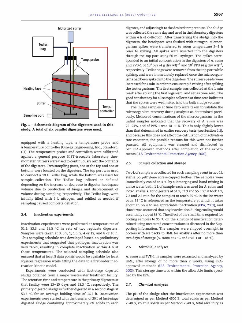
Fig. 1 e Schematic diagram of the digesters used in this
study. A total of six parallel digesters were used.
wat e r r e s e a r c h 4 4 ( 2 0 1 0 ) 5 9 6 5e5 9 7 2 5967
equipped with a heating tape, a temperature probe and
a temperature controller (Omega Engineering, Inc., Stamford,
CT). The temperature probes and controllers were calibrated
against a general purpose NIST-traceable laboratory ther-
mometer. Stirrers were used to continuouslymix the contents
of the digesters. Two sampling ports, one at the top and one at
bottom, were located on the digesters. The top port was used
to connect a 10 L Tedlar bag, while the bottom was used for
sample collection. The Tedlar bag inflated or deflated
depending on the increase or decrease in digester headspace
volume due to production of biogas and displacement of
volume during sampling, respectively. The Tedlar bags were
initially filled with 5 L nitrogen, and refilled as needed if
sampling caused complete deflation.
2.4. Inactivation experiments
Inactivation experiments were performed at temperatures of
51.1, 53.3 and 55.5 �C in sets of two replicate digesters.
Samples were taken at 0, 0.5, 1, 1.5, 2, 4 or 12, and 8 or 16 h.
This sampling schedule was developed based on preliminary
experiments that suggested that pathogen inactivation was
very rapid, resulting in complete inactivation within 4 h at
these temperatures. The selected sampling schedule also
ensured that at least 5 data points would be available for least
squares regression while fitting the data to a first-order inac-
tivation kinetic model.
Experiments were conducted with first-stage digested
sludge obtained from a major wastewater treatment facility.
The retention time and temperature in the primary digesters at
that facility were 13e15 days and 53.3 �C, respectively. Theprimary digested sludge is further digested in a second stage at
53.6 �C for an average holding time of 16.5 h. Inactivation
experiments were started with the transfer of 20 L of first-stage
digested sludge containing approximately 2% solids to each
digester, andadjusting it to thedesired temperature. The sludge
was collected the sameday and used in the laboratory digesters
within 4 h of collection. After transferring the sludge into the
digesters, the headspace was flushed with nitrogen. Microor-
ganism spikes were transferred to room temperature 2e3 h
prior to spiking. All spikes were inserted into the digesters
through the top port using 60 mL syringes. The spikes corre-
sponded to an initial concentration in the digesters of A. suum
and PVS-1 of 104 ova (4 g dry wt)�1 and 106 PFU (4 g dry wt)�1,
respectively. Tedlar bags were removed from the top port while
spiking, and were immediately replaced once the microorgan-
ismshadbeen spiked into thedigesters. The stirrer speedswere
increased for 1min in order to ensure rapidmixing after spiking
the test organisms. The first sample was collected at the 1 min
mark after spiking the first organism, and set as time zero. The
good consistency for all samples collectedat timezero indicates
that the spikes were well mixed into the bulk sludge volume.
The initial samples at time zero were taken to validate the
microorganism recovery during analysis as determined previ-
ously. Measured concentrations of the microorganisms in the
initial samples indicated that the recovery of A. suum was
22e24%, and of PVS-1 was 10e11%. This is only slightly lower
than that determined in earlier recovery tests (see Section 2.2),
and because this does not affect the calculation of inactivation
rate constants, the possible reasons for this were not further
pursued. All equipment was cleaned and disinfected as
per EPA-approved methods after completion of the experi-
ments (U.S. Environmental Protection Agency, 2003).
2.5. Sample collection and storage
TwoLof samplewascollected for eachsamplingevent in two1L
sterile polyethylene screw-capped bottles. The samples were
immediately cooled to 4 �C by submerging and hand mixing in
an ice water bath. 1 L of sample each was used for A. suum and
PVS-1 analysis. For digesters at 51.1, 53.3 and 55.5 �C, it took 1.9,
2.2 and 2.5 min for the samples to reach 35 �C in the ice water
bath. 35 �C is referenced as the temperature at which it takes
about an hour to see appreciable inactivation (EPA, 2003), and
thus itwas assumed that any inactivation during coolingwould
essentially stopat 35 �C.Theeffect of thesmall time required for
cooling samples to 35 �C on the kinetics of inactivation deter-
mined using measured concentrations is discussed in the Sup-
porting Information. The samples were shipped overnight in
coolers with ice packs to HML for analysis after no more than
two days of storage (A. suum at 4 �C and PVS-1 at �18 �C).
2.6. Microbial analyses
A. suum and PVS-1 in samples were extracted and analyzed by
HML, after storage of no more than 2 weeks, using EPA-
approved methods (U.S. Environmental Protection Agency,
2003). This storage time was within the allowable limits speci-
fied by the EPA.
2.7. Chemical analyses
The pH of the sludge after the inactivation experiments was
determined as per Method 4500 B, total solids as per Method
2540 G, volatile solids as per Method 2540 G, total alkalinity as
wat e r r e s e a r c h 4 4 ( 2 0 1 0 ) 5 9 6 5e5 9 7 25968
per Method 2320 B and total volatile fatty acids (VFAs) as per
Method 5560 C (Clesceri et al., 1998). Additionally,
concentrations of sodium dodecyl sulfate (SDS) and cysteine in
first-stage sludge were analyzed by another commercial labo-
ratory, Adamson Analytical Laboratories, Inc. (Corona, CA),
using high-pressure liquid chromatography following extrac-
tion from the sludge using previously describedmethods (Ward
and Ashley, 1978a).
2.8. Data analysis
Concentration vs. time data obtained from the inactivation
experiments was fitted to a first-order kinetic model repre-
sented as follows.
CC0
¼ e�kt (1)
The effect of temperature on the inactivation rate was
modeled using the Arrhenius equation as presented below.
k ¼ k0e�ERT (2)
Linear regression was performed using Microsoft Excel
2007, and first-order rate constants (k) and activation energies
(E ) were determined.
3. Results and discussion
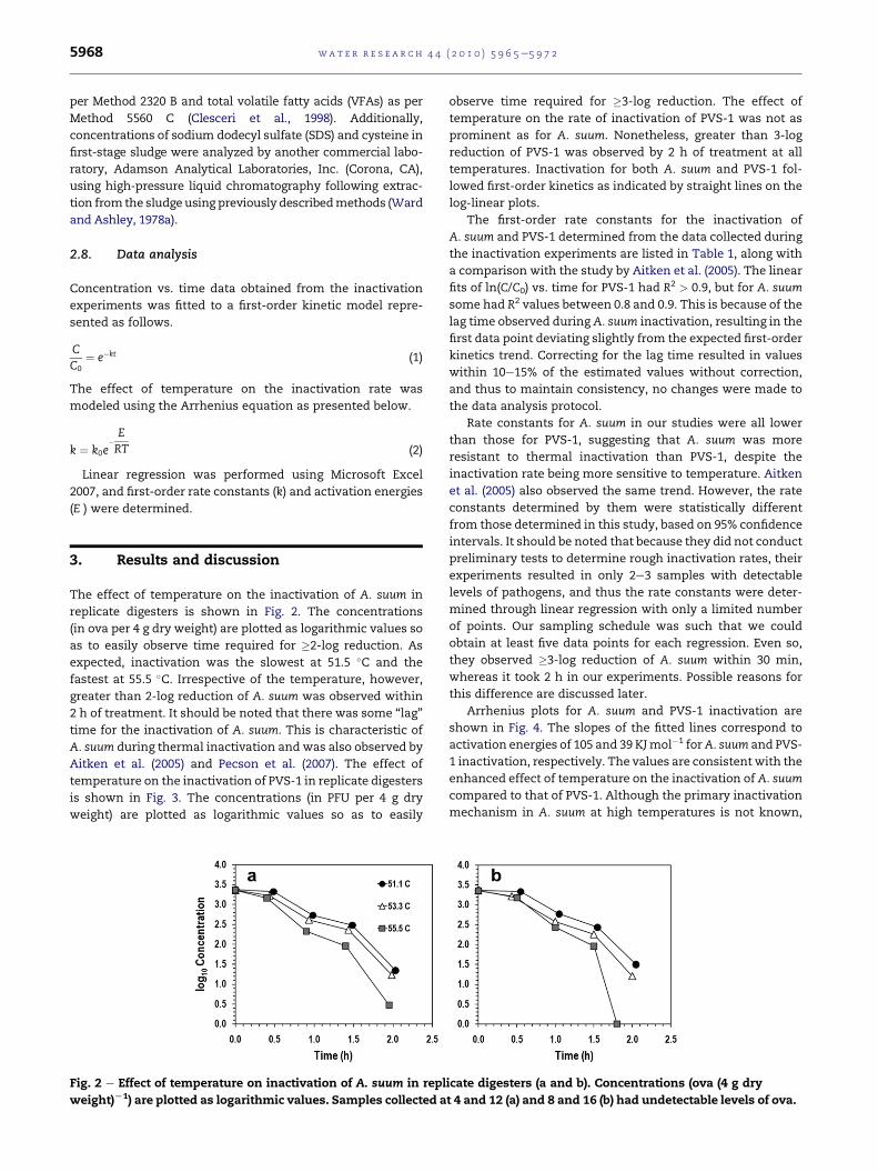
The effect of temperature on the inactivation of A. suum in
replicate digesters is shown in Fig. 2. The concentrations
(in ova per 4 g dry weight) are plotted as logarithmic values so
as to easily observe time required for �2-log reduction. As
expected, inactivation was the slowest at 51.5 �C and the
fastest at 55.5 �C. Irrespective of the temperature, however,
greater than 2-log reduction of A. suum was observed within
2 h of treatment. It should be noted that there was some “lag”
time for the inactivation of A. suum. This is characteristic of
A. suum during thermal inactivation and was also observed by
Aitken et al. (2005) and Pecson et al. (2007). The effect of
temperature on the inactivation of PVS-1 in replicate digesters
is shown in Fig. 3. The concentrations (in PFU per 4 g dry
weight) are plotted as logarithmic values so as to easily
Fig. 2 e Effect of temperature on inactivation of A. suum in repl
weight)L1) are plotted as logarithmic values. Samples collected a
observe time required for �3-log reduction. The effect of
temperature on the rate of inactivation of PVS-1 was not as
prominent as for A. suum. Nonetheless, greater than 3-log
reduction of PVS-1 was observed by 2 h of treatment at all
temperatures. Inactivation for both A. suum and PVS-1 fol-
lowed first-order kinetics as indicated by straight lines on the
log-linear plots.
The first-order rate constants for the inactivation of
A. suum and PVS-1 determined from the data collected during
the inactivation experiments are listed in Table 1, along with
a comparison with the study by Aitken et al. (2005). The linear
fits of ln(C/C0) vs. time for PVS-1 had R2 > 0.9, but for A. suum
some had R2 values between 0.8 and 0.9. This is because of the
lag time observed during A. suum inactivation, resulting in the
first data point deviating slightly from the expected first-order
kinetics trend. Correcting for the lag time resulted in values
within 10e15% of the estimated values without correction,
and thus to maintain consistency, no changes were made to
the data analysis protocol.
Rate constants for A. suum in our studies were all lower
than those for PVS-1, suggesting that A. suum was more
resistant to thermal inactivation than PVS-1, despite the
inactivation rate being more sensitive to temperature. Aitken
et al. (2005) also observed the same trend. However, the rate
constants determined by them were statistically different
from those determined in this study, based on 95% confidence
intervals. It should be noted that because they did not conduct
preliminary tests to determine rough inactivation rates, their
experiments resulted in only 2e3 samples with detectable
levels of pathogens, and thus the rate constants were deter-
mined through linear regression with only a limited number
of points. Our sampling schedule was such that we could
obtain at least five data points for each regression. Even so,
they observed �3-log reduction of A. suum within 30 min,
whereas it took 2 h in our experiments. Possible reasons for
this difference are discussed later.
Arrhenius plots for A. suum and PVS-1 inactivation are
shown in Fig. 4. The slopes of the fitted lines correspond to
activation energies of 105 and 39 KJmol�1 forA. suum and PVS-
1 inactivation, respectively. The values are consistent with the
enhanced effect of temperature on the inactivation of A. suum
compared to that of PVS-1. Although the primary inactivation
mechanism in A. suum at high temperatures is not known,
icate digesters (a and b). Concentrations (ova (4 g dry
t 4 and 12 (a) and 8 and 16 (b) had undetectable levels of ova.
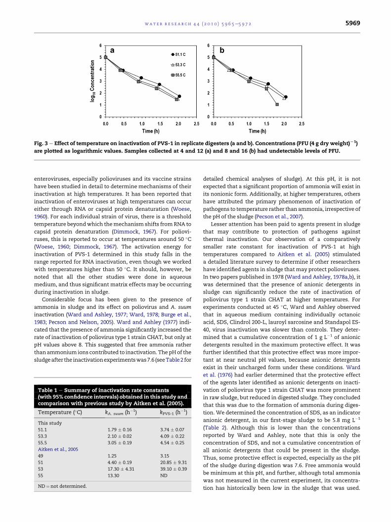
Fig. 3 e Effect of temperature on inactivation of PVS-1 in replicate digesters (a and b). Concentrations (PFU (4 g dry weight)L1)
are plotted as logarithmic values. Samples collected at 4 and 12 (a) and 8 and 16 (b) had undetectable levels of PFU.
wat e r r e s e a r c h 4 4 ( 2 0 1 0 ) 5 9 6 5e5 9 7 2 5969
enteroviruses, especially polioviruses and its vaccine strains
have been studied in detail to determine mechanisms of their
inactivation at high temperatures. It has been reported that
inactivation of enteroviruses at high temperatures can occur
either through RNA or capsid protein denaturation (Woese,
1960). For each individual strain of virus, there is a threshold
temperature beyondwhich themechanism shifts fromRNA to
capsid protein denaturation (Dimmock, 1967). For poliovi-
ruses, this is reported to occur at temperatures around 50 �C(Woese, 1960; Dimmock, 1967). The activation energy for
inactivation of PVS-1 determined in this study falls in the
range reported for RNA inactivation, even though we worked
with temperatures higher than 50 �C. It should, however, be
noted that all the other studies were done in aqueous
medium, and thus significant matrix effects may be occurring
during inactivation in sludge.
Considerable focus has been given to the presence of
ammonia in sludge and its effect on poliovirus and A. suum
inactivation (Ward and Ashley, 1977; Ward, 1978; Burge et al.,
1983; Pecson and Nelson, 2005). Ward and Ashley (1977) indi-
cated that the presence of ammonia significantly increased the
rate of inactivation of poliovirus type 1 strain CHAT, but only at
pH values above 8. This suggested that free ammonia rather
thanammonium ions contributed to inactivation. The pHof the
sludgeafter the inactivationexperimentswas7.6 (seeTable2 for
Table 1 e Summary of inactivation rate constants(with 95% confidence intervals) obtained in this study andcomparison with previous study by Aitken et al. (2005).
Temperature (�C) kA. suum (h�1) kPVS-1 (h�1)
This study
51.1 1.79 � 0.16 3.74 � 0.07
53.3 2.10 � 0.02 4.09 � 0.22
55.5 3.05 � 0.19 4.54 � 0.25
Aitken et al., 2005
49 1.25 3.15
51 4.40 � 0.19 20.85 � 9.31
53 17.30 � 4.31 39.10 � 0.39
55 13.30 ND
ND¼ not determined.
detailed chemical analyses of sludge). At this pH, it is not
expected that a significant proportion of ammonia will exist in
its nonionic form. Additionally, at higher temperatures, others
have attributed the primary phenomenon of inactivation of
pathogens to temperature rather than ammonia, irrespective of
the pH of the sludge (Pecson et al., 2007).
Lesser attention has been paid to agents present in sludge
that may contribute to protection of pathogens against
thermal inactivation. Our observation of a comparatively
smaller rate constant for inactivation of PVS-1 at high
temperatures compared to Aitken et al. (2005) stimulated
a detailed literature survey to determine if other researchers
have identified agents in sludge thatmay protect polioviruses.
In two papers published in 1978 (Ward and Ashley, 1978a,b), it
was determined that the presence of anionic detergents in
sludge can significantly reduce the rate of inactivation of
poliovirus type 1 strain CHAT at higher temperatures. For
experiments conducted at 45 �C, Ward and Ashley observed
that in aqueous medium containing individually octanoic
acid, SDS, Clindrol 200-L, lauroyl sarcosine and Standapol ES-
40, virus inactivation was slower than controls. They deter-
mined that a cumulative concentration of 1 g L�1 of anionic
detergents resulted in the maximum protective effect. It was
further identified that this protective effect was more impor-
tant at near neutral pH values, because anionic detergents
exist in their uncharged form under these conditions. Ward
et al. (1976) had earlier determined that the protective effect
of the agents later identified as anionic detergents on inacti-
vation of poliovirus type 1 strain CHAT was more prominent
in raw sludge, but reduced in digested sludge. They concluded
that this was due to the formation of ammonia during diges-
tion. We determined the concentration of SDS, as an indicator
anionic detergent, in our first-stage sludge to be 5.8 mg L�1
(Table 2). Although this is lower than the concentrations
reported by Ward and Ashley, note that this is only the
concentration of SDS, and not a cumulative concentration of
all anionic detergents that could be present in the sludge.
Thus, some protective effect is expected, especially as the pH
of the sludge during digestion was 7.6. Free ammonia would
be minimum at this pH, and further, although total ammonia
was not measured in the current experiment, its concentra-
tion has historically been low in the sludge that was used.
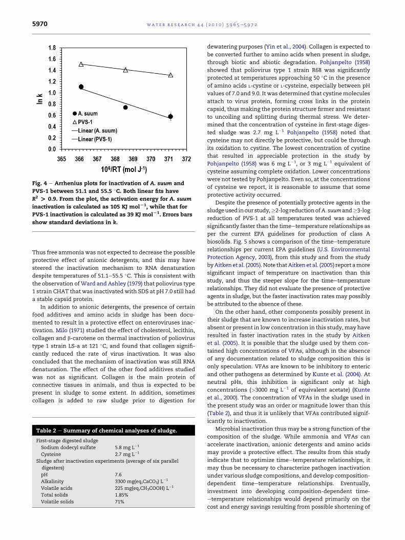
Fig. 4 e Arrhenius plots for inactivation of A. suum and
PVS-1 between 51.1 and 55.5 �C. Both linear fits have
R2 > 0.9. From the plot, the activation energy for A. suum
inactivation is calculated as 105 KJ molL1, while that for
PVS-1 inactivation is calculated as 39 KJ molL1. Errors bars
show standard deviations in k.
wat e r r e s e a r c h 4 4 ( 2 0 1 0 ) 5 9 6 5e5 9 7 25970
Thus free ammonia was not expected to decrease the possible
protective effect of anionic detergents, and this may have
steered the inactivation mechanism to RNA denaturation
despite temperatures of 51.1e55.5 �C. This is consistent with
the observation ofWard and Ashley (1979) that poliovirus type
1 strain CHAT that was inactivated with SDS at pH 7.0 still had
a stable capsid protein.
In addition to anionic detergents, the presence of certain
food additives and amino acids in sludge has been docu-
mented to result in a protective effect on enteroviruses inac-
tivation. Milo (1971) studied the effect of cholesterol, lecithin,
collagen and b-carotene on thermal inactivation of poliovirus
type 1 strain LS-a at 121 �C, and found that collagen signifi-
cantly reduced the rate of virus inactivation. It was also
concluded that the mechanism of inactivation was still RNA
denaturation. The effect of the other food additives studied
was not as significant. Collagen is the main protein of
connective tissues in animals, and thus is expected to be
present in sludge to some extent. In addition, sometimes
collagen is added to raw sludge prior to digestion for
Table 2 e Summary of chemical analyses of sludge.
First-stage digested sludge
Sodium dodecyl sulfate 5.8 mg L�1
Cysteine 2.7 mg L�1
Sludge after inactivation experiments (average of six parallel
digesters)
pH 7.6
Alkalinity 3300 mg(eq,CaCO3) L�1
Volatile acids 225 mg(eq,CH3COOH) L�1
Total solids 1.85%
Volatile solids 71%
dewatering purposes (Yin et al., 2004). Collagen is expected to
be converted further to amino acids when present in sludge,
through biotic and abiotic degradation. Pohjanpelto (1958)
showed that poliovirus type 1 strain R68 was significantly
protected at temperatures approaching 50 �C in the presence
of amino acids L-cystine or L-cysteine, especially between pH
values of 7.0 and 9.0. It was determined that cystinemolecules
attach to virus protein, forming cross links in the protein
capsid, thusmaking the protein structure firmer and resistant
to uncoiling and splitting during thermal stress. We deter-
mined that the concentration of cysteine in first-stage diges-
ted sludge was 2.7 mg L�1 Pohjanpelto (1958) noted that
cysteine may not directly be protective, but could be through
its oxidation to cystine. The lowest concentration of cystine
that resulted in appreciable protection in the study by
Pohjanpelto (1958) was 6 mg L�1, or 3 mg L�1 equivalent of
cysteine assuming complete oxidation. Lower concentrations
were not tested by Pohjanpelto. Even so, at the concentrations
of cysteine we report, it is reasonable to assume that some
protective activity occurred.
Despite the presence of potentially protective agents in the
sludgeused inour study,�2-log reductionofA. suumand�3-log
reduction of PVS-1 at all temperatures tested was achieved
significantly faster than the timeetemperature relationships as
per the current EPA guidelines for production of class A
biosolids. Fig. 5 shows a comparison of the timeetemperature
relationships per current EPA guidelines (U.S. Environmental
Protection Agency, 2003), from this study and from the study
byAitkenetal. (2005).Note thatAitkenet al. (2005) report amore
significant impact of temperature on inactivation than this
study, and thus the steeper slope for the timeetemperature
relationships. They did not evaluate the presence of protective
agents in sludge, but the faster inactivation rates may possibly
be attributed to the absence of these.
On the other hand, other components possibly present in
their sludge that are known to increase inactivation rates, but
absent or present in low concentration in this study,may have
resulted in faster inactivation rates in the study by Aitken
et al. (2005). It is possible that the sludge used by them con-
tained high concentrations of VFAs, although in the absence
of any documentation related to sludge composition this is
only speculation. VFAs are known to be inhibitory to enteric
and other pathogens as determined by Kunte et al. (2004). At
neutral pHs, this inhibition is significant only at high
concentrations (>3000 mg L�1 of equivalent acetate) (Kunte
et al., 2000). The concentration of VFAs in the sludge used in
the present study was an order or magnitude lower than this
(Table 2), and thus it is unlikely that VFAs contributed signif-
icantly to inactivation.
Microbial inactivation thus may be a strong function of the
composition of the sludge. While ammonia and VFAs can
accelerate inactivation, anionic detergents and amino acids
may provide a protective effect. The results from this study
indicate that to optimize timeetemperature relationships, it
may thus be necessary to characterize pathogen inactivation
under various sludge compositions, and develop composition-
dependent timeetemperature relationships. Eventually,
investment into developing composition-dependent time-
etemperature relationships would depend primarily on the
cost and energy savings resulting from possible shortening of
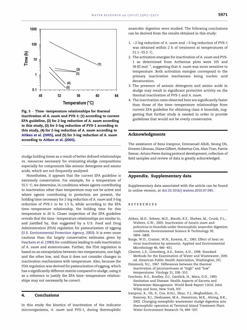
Fig. 5 e Timeetemperature relationships for thermal
inactivation of A. suum and PVS-1: (1) according to current
EPA guideline, (2) for 2-log reduction of A. suum according
to this study, (3) for 3-log reduction of PVS-1 according to
this study, (4) for 2-log reduction of A. suum according to
Aitken et al. (2005), and (5) for 3-log reduction of A. suum
according to Aitken et al. (2005).
wat e r r e s e a r c h 4 4 ( 2 0 1 0 ) 5 9 6 5e5 9 7 2 5971
sludge holding times as a result of better defined relationships
vs. resources necessary for evaluating sludge compositions
especially for components like anionic detergents and amino
acids, which are not frequently analyzed.
Nonetheless, it appears that the current EPA guideline is
extremely conservative. For example, for a temperature of
55.5 �C, we determine, in conditions where agents contributing
to inactivation other than temperature may not be active and
where agents contributing to protection are present, the
holding time necessary for 2-log reduction ofA. suum and 3-log
reduction of PVS-1 to be 1.5 h, while according to the EPA
timeetemperature relationship, the holding time for this
temperature is 20 h. Closer inspection of the EPA guideline
reveals that the timeetemperature relationships are similar to,
and justified by, that suggested by a U.S. Food and Drug
Administration (FDA) regulation for pasteurization of eggnog
(U.S. Environmental Protection Agency, 2003). It is even more
cautious than the largely conservative estimates given by
Feachem et al. (1983) for conditions leading to safe inactivation
of A. suum and enteroviruses. Further, the FDA regulation is
based on an extrapolation between two temperatures, one high
and the other low, and thus it does not consider changes in
inactivation mechanisms with temperature. Also, because the
FDA regulationwas derived for pasteurization of eggnog, which
has a significantly differentmatrix compared to sludge, using it
as a reference to justify the EPA timeetemperature relation-
ships may not necessarily be correct.
4. Conclusions
In this study the kinetics of inactivation of the indicator
microorganisms, A. suum and PVS-1, during thermophilic
anaerobic digestion were studied. The following conclusions
can be derived from the results obtained in this study:
1. >2-log reduction of A. suum and >3-log reduction of PVS-1
was obtained within 2 h of treatment at temperatures of
51.1e55.5 �C.2. The activation energies for inactivation ofA. suum and PVS-
1 as determined from Arrhenius plots were 105 and
39 KJ mol�1, suggesting that A. suum was more sensitive to
temperature. Both activation energies correspond to the
primary inactivation mechanism being nucleic acid
denaturation.
3. The presence of anionic detergents and amino acids in
sludge may result in significant protective activity on the
thermal inactivation of PVS-1 and A. suum.
4. The inactivation rates observed here are significantly faster
than those of the timeetemperature relationships from
current EPA guideline for obtaining class A biosolids, sug-
gesting that further study is needed in order to provide
guidelines that would not be overly conservative.
Acknowledgments
The assistance of Reza Iranpour, Emmanuel Alloh, Seung Oh,
Ernesto Libunao, Diane Gilbert, Hubertus Cox, AlanTran, Parviz
Samar, Arturo Perez during protocol development, collection of
field samples and review of data is greatly acknowledged.
Appendix. Supplementary data
Supplementary data associated with the article can be found
in online version, at doi:10.1016/j.watres.2010.07.045.
r e f e r e n c e s
Aitken, M.D., Sobsey, M.D., Blauth, K.E., Shehee, M., Crunk, P.L.,Walters, G.W., 2005. Inactivation of Ascaris suum andpoliovirus in biosolids under thermophilic anaerobic digestionconditions. Environmental Science & Technology 39,5804e5809.
Burge, W.D., Cramer, W.N., Kawata, K., 1983. Effect of heat onvirus inactivation by ammonia. Applied and EnvironmentalMicrobiology 46, 446e451.
Clesceri, L.S., Greenberg, A.E., Eaton, A.D., 1998. StandardMethods for the Examination of Water and Wastewater, 20thed. American Public Health Association, Washington, DC.
Dimmock, N.J., 1967. Differences between the thermalinactivation of picornaviruses at “high” and “low”temperatures. Virology 31, 338e353.
Feachem, R.G., Bradley, D.J., Garelick, H., Mara, D.D., 1983.Sanitation and Disease: Health Aspects of Excreta andWastewater Management. World Bank Report 11616. JohnWiley and Sons, New York, NY.
Iranpour, R., Oh, S., Cox, H.H.J., Shao, Y.J., Moghaddam, O.,Kearney, R.J., Deshusses, M.A., Stenstrom, M.K., Ahring, B.K.,2002. Changing mesophilic wastewater sludge digestion intothermophilic operation at Terminal Island Treatment Plant.Water Environment Research 74, 494e507.
wat e r r e s e a r c h 4 4 ( 2 0 1 0 ) 5 9 6 5e5 9 7 25972
Kunte, D.P., Yeole, T.Y., Ranade, D.R., 2000. Effect of volatile fattyacids on Shigella dysenteriae during anaerobic digestion ofhuman night soil. World Journal of Microbiology &Biotechnology 16, 519e522.
Kunte, D.P., Yeole, T.Y., Ranade, D.R., 2004. Two-stage anaerobicdigestion process for complete inactivation of enteric bacterialpathogens in human night soil. Water Science & Technology50, 103e108.
Milo Jr., G.E., 1971. Thermal inactivation of poliovirus in thepresence of selective organic molecules (cholesterol, lecithin,collagen, and b-carotene). Applied Microbiology 21, 198e202.
National Research Council Committee on Toxicants andPathogens in Biosolids Applied to Land, 2002. BiosolidsApplied to Land: Advancing Standards and Practices.The National Academies Press, Washington, D. C.
Pecson, B.M., Nelson, K.L., 2005. Inactivation of Ascaris suum eggs byammonia. Environmental Science & Technology 39, 7909e7914.
Pecson, B.M., Barrios, J.A., Jimenez, B.E., Nelson, K.L., 2007.The effects of temperature, pH, and ammonia concentrationon the inactivation of Ascaris eggs in sewage sludge. WaterResearch 41, 2893e2902.
Pohjanpelto, P., 1958. Stabilization of poliovirus by cystine.Virology 6, 472e487.
U.S. Environmental Protection Agency, 2003. Control of Pathogensand Vector Attraction in Sewage Sludge. EPA/625/R-92/013,
Revised 2003. U.S. Environmental Protection Agency,Washington, DC.
Ward, R.L., Ashley, C.S., Moseley, R.H., 1976. Heat inactivation ofpoliovirus in wastewater sludge. Applied and EnvironmentalMicrobiology 32, 339e346.
Ward, R.L., Ashley, C.S., 1977. Identification of the virucidal agentin wastewater sludge. Applied and EnvironmentalMicrobiology 33, 860e864.
Ward, R.L., 1978. Mechanism of poliovirus inactivation byammonia. Journal of Virology 26, 299e305.
Ward, R.L., Ashley, C.S., 1978a. Identification of detergents ascomponents of wastewater sludge that modify the thermalstability of reovirus and enteroviruses. Applied andEnvironmental Microbiology 36, 889e897.
Ward, R.L., Ashley, C.S., 1978b. Heat inactivation of entericviruses in dewatered wastewater sludge. Applied andEnvironmental Microbiology 36, 898e905.
Ward, R.L., Ashley, C.S., 1979. pH modification of the effect ofdetergents on the stability of enteric viruses. Applied andEnvironmental Microbiology 38, 314e322.
Woese, C., 1960. Thermal inactivation of animal viruses. Annalsof the New York Academy of Sciences 83, 741e751.
Yin, X., Han, P., Lu, X., Wang, Y., 2004. A review on thedewaterability of bio-sludge and ultrasound pretreatment.Ultrasonics Sonochemistry 11, 337e348.