kif1bb transports dendritically localized mrnps in neurons ... · pietro pilo boyl • sebastian...
TRANSCRIPT
KIF1Bb transports dendritically localized mRNPs in neuronsand is recruited to synapses in an activity-dependent manner
Despina C. Charalambous • Emanuela Pasciuto • Valentina Mercaldo •
Pietro Pilo Boyl • Sebastian Munck • Claudia Bagni • Niovi Santama
Abstract KIF1Bb is a kinesin-like, microtubule-based
molecular motor protein involved in anterograde axonal
vesicular transport in vertebrate and invertebrate neurons.
Certain KIF1Bb isoforms have been implicated in different
forms of human neurodegenerative disease, with character-
ization of their functional integration and regulation in the
context of synaptic signaling still ongoing. Here, we char-
acterize human KIF1Bb (isoform NM015074), whose
expression we show to be developmentally regulated and
elevated in cortical areas of the CNS (including the motor
cortex), in the hippocampus, and in spinal motor neurons.
KIF1Bb localizes to the cell body, axon, and dendrites,
overlapping with synaptic-vesicle and postsynaptic-density
structures. Correspondingly, in purified cortical
synaptoneurosomes, KIF1Bb is enriched in both pre- and
postsynaptic structures, forming detergent-resistant com-
plexes. Interestingly, KIF1Bb forms RNA–protein
complexes, containing the dendritically localized Arc and
Calmodulin mRNAs, proteins previously shown to be part of
RNA transport granules such as Pura, FMRP and FXR2P, and
motor protein KIF3A, as well as Calmodulin. The interaction
between KIF1Bb and Calmodulin is Ca?2-dependent and
takes place through a domain mapped at the carboxy-terminal
tail of the motor. Live imaging of cortical neurons reveals
active movement by KIF1Bb at dendritic processes, sug-
gesting that it mediates the transport of dendritically localized
mRNAs. Finally, we show that synaptic recruitment of
KIF1Bb is activity-dependent and increased by stimulation of
metabotropic or ionotropic glutamate receptors. The activity-
dependent synaptic recruitment of KIF1Bb, its interaction
with Ca2? sensor Calmodulin, and its new role as a dendritic
motor of ribonucleoprotein complexes provide a novel basis
for understanding the concerted co-ordination of motor pro-
tein mobilization and synaptic signaling pathways.
D. C. Charalambous, E. Pasciuto, C. Bagni, N. Santama contributed
equally to the work.
D. C. Charalambous � N. Santama (&)
Department of Biological Sciences, University of Cyprus,
University Avenue 1, 1678 Nicosia, Cyprus
e-mail: [email protected]
D. C. Charalambous
e-mail: [email protected]
E. Pasciuto � V. Mercaldo � P. Pilo Boyl � S. Munck �C. Bagni (&)
VIB Center for Biology of Disease, Katholieke Universiteit
Leuven, Heresraat 49, 3000 Leuven, Belgium
e-mail: [email protected]
V. Mercaldo
e-mail: [email protected]
P. Pilo Boyl
e-mail: [email protected]
C. Bagni
Department of Biomedicine and Prevention,
Faculty of Medicine, University of Rome Tor Vergata,
Via Montpellier 1, 00100 Rome, Italy
Present Address:V. Mercaldo
Department of Physiology, Faculty of Medicine,
University of Toronto, Toronto, Canada
Present Address:P. Pilo Boyl
Institute of Genetics, University of Bonn,
Karlrobert-Kreiten Str. 13, 53115 Bonn, Germany
Keywords Molecular motors � mRNA transport �Calmodulin � Arc � Synaptic activity � mGluR activation
Abbreviations
Arc Activity-regulated
cytoskeleton-associated protein
CaM Calmodulin
CMT2A Charcot-Marie-Tooth type 2A motor
neuron disease
DHPG S-3,5,-dihydroxyphenylglycine
DMEM Dulbecco’s Modified Eagle’s Medium
FMRP Fragile X mental retardation protein
FXR2P Fragile X-related protein 2
HBSS Hank’s balanced salt solution
KIF1Bb Kinesin family member 1BbMEM Minimal essential medium (Eagle’s)
Pura Purine-rich element binding protein alpha
RT-PCR Reverse-transcription polymerase
chain reaction
Introduction
The members of the KIF1B subfamily (Kinesin-3 as per the
nomenclature of [1]) of motor proteins have attracted
considerable attention because of their important function
in anterograde axonal transport in neurons and their puta-
tive link to neurodegenerative disease. In human, KIF1B is
expressed in two isoform types, KIF1Ba, originally
reported as a mitochondrial transporter [2], and also shown
to interact with postsynaptic density and synaptic scaffold
proteins [3], and at least six, currently known, KIF1Bb-like
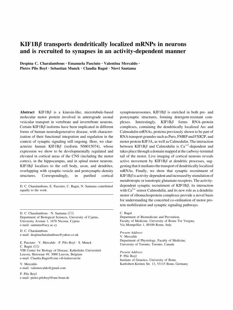
forms (NM015074, AB088210–13, and AX039604). The
AB088210–13 isoforms share a high level of sequence
identity with each other, with small differences over a
region of 40 amino acids (aa), and compared to NM015074
have an extra stretch of six aa and, significantly, an
‘‘alternative C-terminus’’ (Fig. 1). AX039604 shares
characteristics of both, the AB series and NM015074,
being largely identical to AB088210, but with an additional
40-aa stretch and possessing the characteristic C-terminus
of NM015074 (Fig. 1).
The KIF1B family has been linked to several diseases
affecting neural cells of the central nervous system.
KIF1Bb (isoform NM015074) is reported as a transporter
of synaptic vesicle precursors and, in the case of a single
large Japanese family, was implicated in Charcot-Marie-
Tooth type 2A (CMT2A) motor neuron disease [4], a dis-
order commonly associated with mutations in the
mitochondrial GTPase mitofusin-2 [5]. KIF1Bb isoform
AB088210, highly enriched in the motor cortex, is signif-
icantly downregulated in the motor cortex of patients with
sporadic amyotrophic lateral sclerosis, another common
type of motor neuron disease [6]. Mechanistic insights into
the involvement of KIF1B family members in neuropathies
also come from C. elegans, whose KIF1Bb homologue,
UNC-104, is required for the anterograde transport of
synaptic vesicle precursors [7], possibly through interac-
tions with the cargo via a lipid-binding pleckstrin
homology domain in its stalk [8]. In a genome-wide
association study, a single nucleotide polymorphism (SNP)
in intron 5 of the KIF1B gene (shared by KIF1Ba and
KIF1Bb) was found to be associated with susceptibility to
multiple sclerosis (MS) ([9], but see also [10, 11]).
Intriguingly, a tentative explanation for this link is pro-
vided by observations in zebrafish, where mutations or
RNA interference of KIF1Ba/b prevent correct localization
of mRNAs encoding myelin proteins MBP and 36 K to
distal processes in myelinating oligodendrocytes, and cause
reduction of axonal outgrowth and reduced or ectopic
myelination in spinal neurons [12].
Other studies have indicated a wider role for KIF1Bb in
non-neural cell types, such as reports proposing a haplo-
insufficient tumor suppressor function [13–15]. This link of
the KIF1B family to cancer development is further
strengthened by a genome-wide association study that
assigned high statistical significance to an intronic SNP in
the KIF1B gene as a susceptibility locus for hepatocellular
carcinoma in carriers of chronic hepatitis B [16].
1
1
1770
1783Motor FHA PH
C-term
1 1816C-term
C-termalt.
NM015074
AX039604
AB088213
anti-peptide aboligo set 1
oligo set 2
IRSKLSRRCPSQSKY
Fig. 1 Human KIF1Bb isoforms. Schematic comparison of isoforms
NM015074, AX039604 and AB088213 [the latter being representa-
tive of the AB088210–13 subgroup (AB)]. NM015074 (NM) and
AX039604 (AX) share a characteristic C-terminus (red) to which an
anti-peptide antibody was raised (indicated at the top). The AB series
have an alternative C-terminus (denoted by the dark green box) not
recognized by the antibody. AX and AB have an additional 6-aa
stretch (black box) close to the motor domain, and AX also has a
unique additional 40-aa stretch (gray box). The positions of the two
sets of oligonucleotides that are each specific to NM or AX only, and
can thus discriminate between NM and AX by PCR (Fig. 2b, d–f), are
indicated by arrows over the areas to which they hybridize. The
sequences of these oligonucleotide pairs are given in Online Resource
Table S1. The motor (yellow), FHA (light blue), and PH domains
(light green), as well as the two stretches of amino acids (black and
gray boxes), unique in certain isoforms, are highlighted. Numbersdenote aa positions

The existence of multiple isoforms, necessitating iso-
form-specific characterization of gene expression and
function, and use of isoform-specific antibodies, compli-
cate the immediate correlation between the functional
analyses offered by different studies. More importantly,
despite the established involvement of KIF1Bb in vesicular
transport, less is known about the signaling pathways and
mechanisms regulating and modulating the function of
KIF1Bb in neurons. In this work, we identify regulatory
signals, including calcium and receptor signaling, that
influence the recruitment of KIF1Bb isoform NM015074 at
functional synapses, and identified a KIF1Bb-containing
RNA–protein transport complex. The present study there-
fore gives fundamental insights into how mobilization and
interaction of motor proteins are integrated with synaptic
signaling and plasticity.
Materials and methods
Cells
Spinal motor neurons and cortical neurons were obtained
from E18–19 mice. Dissected spinal cords or cortex were
incubated with 4.5 mL HBSS, containing 0.02 % w/v
trypsin and 80 lg/mL DNase I for 15 min at 37 �C,
mechanically dissociated and centrifuged at 1,500g for
5 min. Dissociated cells were plated at a density of
12 9 104 cells/cm2 in MEM (Invitrogen), supplemented
with 10 % v/v horse serum (for spinal cord), or in Neu-
robasal A media (Invitrogen), supplemented with B27.
Primary neuron cultures were maintained 2–3 weeks in
vitro before use.
Mouse NSC-34 neuroblastoma, NIH 3T3 fibroblast cell
lines, human SH-SY5Y neuroblastoma and HeLa cells
were cultured in DMEM (Gibco/BRL), containing 10 %
v/v fetal calf serum (FCS), 2 mM glutamine, 50 U/mL of
penicillin/streptomycin, and maintained at 37 �C in 5 %
CO2. For induction and differentiation, SH-SY5Y cells
were split and maintained for 10–12 days in DMEM with
2 % FCS and 10 lM retinoic acid.
Oligonucleotides and generation of plasmid constructs
All oligonucleotides, used for RT-PCR or plasmid con-
struct generation, were synthesized by MWG Biotech
(Germany). Their sequences and further details are given in
Online Resource Table S1.
Antibodies
A rabbit polyclonal antibody to synthetic peptide IRS-
KLSRRCPSQSKY [aa residues 1,756–1,770 in human
KIF1Bb (NM015074)] was custom-made and affinity-
purified by Peptide Speciality Laboratories, Germany. It
was used at a dilution of 1:500 for immunoblotting and
1:300 for immunofluorescence (IF).
The following primary antibodies were used: mouse anti-
dynein (intermediate chain, 74 kDa) monoclonal antibody
(mab sc-13524) (Santa Cruz, 1:800 in immunoblot), mouse
anti-GST mab (Sigma, 1:4,000 in immunoblot), rat anti-
myelin basic protein (MBP) mab (Millipore, 1:2,500 in
immunoblot), rabbit anti-glial fibrillary acidic protein
(GFAP) polyclonal antibody (Sigma, 1:500 in immunoblot),
mouse anti-non-phosphorylated neurofilament H mab (SMI
32, Eurogentech, 1:1,000 in IF), mouse anti-MAP2 mab
(Abcam, 1:200 in IF), mouse anti-MAP2 mab (Sigma,
1:300 in IF), mouse anti-GAPDH mab (Chemicon, 1:20,000
in immunoblot), mouse anti-PSD-95 mab (BD Biosciences,
1:100 in IF and 1:1,000 in immunoblot), mouse anti-syn-
aptophysin mab (Abcam, 1:100 in IF and 1:1,000 in
immunoblot), mouse anti-SV2 mab (Developmental Stud-
ies Hybridoma Bank, 1:100 in IF), mouse anti-syntaxin 1
mab (Synaptic Systems, 1:5,000), mouse anti-SNAP25 mab
(Synaptic Systems, 1:1,000), mouse anti-CaM mab (Zymed,
1:30 in IF), rabbit anti-CaM (Abcam, 1:1,000 in immuno-
blot), rabbit anti-KIF3A (Abcam, 1:1,000 in immunoblot),
goat anti-MARCKS (Santa Cruz Biotechnology, 1:400 in
immunoblot), rabbit anti-Pura (Millipore, 1:100 for IF,
1:250 in immunoblot), mouse anti-FMRP (IC3, Millipore,
1:100 in IF), rabbit anti-FMRP (rAMII, 1:2,000 in immu-
noblot) [17], mouse anti-FXR2P (1:1,000 in immunoblot),
rabbit anti-FXR2P (Developmental Studies Hybridoma
Bank, 1:50 in IF), rabbit anti-KIF5A (Acris, 1:1,000 in
immunoblot), and rabbit anti-ELAVs (Santa Cruz, 1:1,000
in immunoblot).
The following secondary antibodies (Molecular Probes)
were used for IF: Alexa Fluor (AF) 488 chicken anti-rabbit
IgG (1:700), AF568 goat anti-rabbit IgG (1:2,000), AF488
goat anti-mouse IgG (1:700) and AF568 anti-mouse IgG1
(1:2,000). The following secondary antibodies were used for
immunobloting: HRP donkey anti-rabbit IgG (Santa Cruz,
1:60,000), HRP sheep anti-mouse IgG (Amersham, 1:8,000)
and HRP chicken anti-rat IgG (Santa Cruz, 1:20,000).
Nuclei were stained with Hoechst 33342 (0.5 lg/mL,
Invitrogen). Polymerized filamentous actin (F-actin) was
visualized at postsynaptic densities in mature dendritic
spines of cultured neurons using a phalloidin-AF647 con-
jugate (1:100, Molecular Probes) with 1 h incubation,
followed by three 5-min washes in PBS.
RNA extraction, reverse transcription semi-quantitative
PCR (RT-PCR)
For RT-PCR, poly(A) RNA was extracted from human
cells (HeLa, SH-SY5Y uninduced or induced with RA) or

mouse cells (spinal cord neurons of 20 days in vitro, NSC-
34, NIH 3T3) or mouse tissues (cortex, cerebellum, hip-
pocampus from 3-month-old mice) with the RNeasy
Purification Kit (Qiagen) and 1 lg used for reverse tran-
scription using the Protoscript Reverse Transcription Kit
with a dT23VN primer (New England Biolabs). Human
tissue RNA (from the tissues displayed in Fig. 2a, b) was
purchased from Ambion Inc. and reverse-transcribed to
cDNA in the same manner. RNA from total mouse cortex
was extracted with the Trizol reagent (Invitrogen) and
digested with RNase-free DNAse (RQ1, Promega). Addi-
tionally, mouse RNA from hippocampal neurons in vitro at
stages 2 and 5 (staging as per [18]), embryonic stages E13
and E18, juvenile animals (2.5 weeks) and pure astrocytic
cultures, derived from newborn mice (Fig. 2d), was
obtained as described by [19]. Semi-quantitative PCR
reactions from cells, tissues or cell lines (Fig. 2) were set
up with equivalent cDNA concentrations (starting with the
same amount of RNA) and using cDNA of ribosomal
protein L19 as a calibrator for normalization.
siRNA-mediated silencing
Catalogue and custom-made Stealth siRNA duplexes
(Invitrogen) were used for siRNA-mediated silencing
and scrambled siRNA ‘Low GC’ (a universal control for
siRNAs with low GC content; Invitrogen) was used as
negative control.
adrenal
colonheart
kidneyliver
lungovary
skeletal
spleentesticles
NM015074
-RT
L190.2
kb
0.3
humanA
NM0150740.3
AX039604
cortex
cerebellum
hippocampus
spinal cord neur.
-RTkb
4
L190.2
mouseC
NM015074
L19
0.3
0.2
kb HeLaSHSY5Y
SHSY5Y-RA
NSC-34
NIH 3T3
-RT
cell linesE
F
B
D
AX039604
st.2 st.5 E13E18
juvenileastro
glia
kb
4
NM0150740.3
L190.2
mouse
NM0150740.3
parietal
motortemporal
occipital
AX039604
-RTkb
4
L190.2
human
NIH 3T3
40 80μg
NSC-34
8040kDa
250
130
KIF1Bβ
1530 μg10kDa
170130
907045
KIF1Bβ
GAPDH
cortical neurons
spinal cord
hippoc.
cortex
cerebellum
spinal cord
hippoc.
cortex
cerebellum
anti-KIF1Bβ pre-absorbed
KIF1Bβ
GAPDH
20μg
Fig. 2 a KIF1Bb (NM015074) mRNA expression pattern in human
tissues. Equivalent RT-PCR reactions from various human tissues,
including a mock reverse-transcription reaction as negative control
(-RT). Equivalent reactions for the mRNA of ribosomal protein L19
were used as internal standards (bottom panel). b–d mRNA expres-
sion of KIF1Bb NM015074 versus AX039604 isoforms in the CNS.
Equivalent RT-PCR reactions specific to KIF1Bb isoforms
NM015074 (top panels) and AX039604 (central panels), and
housekeeping gene L19 (bottom panels) for b areas of human cortex;
c mouse neural tissue, including spinal motor neurons; d mouse stage-
2 or stage-5 hippocampal neurons in vitro [18] or the hippocampus of
E13, E18 and juvenile mice, and from proliferating astroglial cultures.
e KIF1Bb (NM015074) mRNA expression in human and mouse cell
lines. Equivalent RT-PCR reactions reveal a pronounced expression
level of KIF1Bb mRNA in the mouse neuroblastoma NSC-34 line,
lower expression levels in mouse NIH 3T3 fibroblasts, and expression
below detection levels for other cell lines. Equivalent reactions for
L19 (bottom panel). f Protein expression of KIF1Bb in different brain
areas by immunoblot (top panels). Total protein extracts from mouse
cultured primary cortical neurons, NSC-34 or NIH 3T3 cells were
analyzed by western blotting, using the anti-peptide KIF1Bbantibody. A signal, consistent with the predicted Mr of
199.01 9 103 for KIF1Bb was detected (e). Also consistent with
mRNA level for these cell lines, protein expression of KIF1Bb in
NSC-34 is higher than in NIH 3T3 cells (bottom panels). Expression
of KIF1Bb protein in equivalent samples from different parts of the
mouse CNS (left); these signals are abolished if the antibody is pre-
absorbed with the antigenic peptide (right). Immunoreactivity for
GAPDH serves as internal loading control

Three pairs of custom-made siRNA duplexes were used
for silencing of KIF1Bb NM015074).50gaguguuucacuuucuggugauaaa30/3
0cu-
cgcaaagugaaagaccacuauuu50 (siRNA1)
50ccuugugauuguuacagcucccuuu30/30ggaacacuaacaaugucg-
agggaaa50 (siRNA2)50ugagguggaugaagcugcaguugau30/3
0acuccaccuacuucgacg-
ucaacua50 (siRNA3)
Transfections of NSC-34 cells with siRNAs were per-
formed using Lipofectamine 2000 (Invitrogen) with the
three sets of siRNAs used as a cocktail (molar ratios 1:1:1)
at a total concentration of 320 pM/transfection. A second
transfection was repeated 72 h after initial treatment. At
120 h, coverslips or cells were harvested for microscopic
imaging or RNA and protein analysis.
Immunoprecipitation and RT-PCR
Mouse brains (p21) were lysed in 150 mM NaCl, 50 mM
Tris–HCl pH 7.4, 1 % v/v Triton X-100, 40 U/mL RNase
OUT (Invitrogen), 1 mM DTT, 5 mM b-glycerol phos-
phate, 0.5 mM Na3VO4, and 10 mg/mL PIC (Sigma).
After 5 min incubation on ice, lysates were centrifuged
for 8 min at 12,000g (4 �C), and 500 lg of protein from
the supernatant were used for the IP. Protein A Sepharose
beads (GE Healthcare) were saturated in 10 % w/v BSA
and heparin (2 mg/mL) in PBS, incubated with 3 lg of
native KIF1Bb antibody or purified rabbit IgGs (2 h at
4 �C), washed three times (150 mM NaCl, 50 mM Tris–
HCl pH 7.40, 1 % v/v Triton X-100), and finally incu-
bated with 500 lg of brain extract plus heparin (1 mg/
mL) for 1 h at 4 �C. Stringent washes with 1 M urea and
low salt (50 mM NaCl) followed the incubation. Proteins
were eluted by boiling of the beads in SDS-PAGE sample
buffer, separated by electrophoresis and immunoblotted to
a PVDF membrane (Millipore). RNA was eluted from the
immunoprecipitates, extracted (phenol/chloroform), and
precipitated in 2.5 volumes ethanol. Prior to RNA
extraction, and as an internal control for equal RNA
extraction from the test and negative control immuno-
precipitations, each immunoprecipitate was spiked with
200 ng of human BC200 RNA. First-strand cDNA syn-
thesis ensued, using p(dN)6 and 100 U of M-MuLV
RTase (Invitrogen), and was followed by PCR with
GoTaq (Promega) to assay for selected cDNAs and the
BC200 cDNA internal control (Fig. 8).
Yeast two hybrid screen and yeast mating assays
To screen for proteins that interact with KIF1Bb, the
GAL4-based Matchmaker Two Hybrid System 3 (Clontech)
was used in conjunction with a human adult brain cDNA
library, constructed in the DNA activation-domain-bearing
pACT2 vector (Clontech). A partial cDNA of KIF1Bb(nucl. 1,980–5,313) was cloned as an in-frame SalI frag-
ment into DNA-binding domain fusion vector pAS2.1 and
used as bait. All procedures were performed according to
the Matchmaker User’s Manual. After five rounds of
selection, putative positive clones were validated by yeast
mating, followed by quadruple auxotrophy selection (-Leu,
-Trp, -His, -Ade) and b-galactosidase filter assays, and
identified by DNA sequencing (MWG Biotech, Germany).
To map the CaM interaction site(s) of KIF1Bb, deletion
constructs of KIF1Bb (M, C1–C5) were generated in
vector pAS2.1 [see Online Resource Table S1; Fig. 7c (see
below)] and tested by yeast mating against full-length CaM
in vector pACT2. Interaction was defined as the ability of
[95 % of resulting clones, containing both constructs, to
grow on quadruple auxotrophy selection and also display
b-galactosidase activity upon filter assay.
Bacterial expression of GST-tagged KIF1Bb constructs
and pull-down assays
For expression and purification of GST-tagged constructs
of KIF1Bb (constructs M, C1–C5 in vector pGEX-4T-1, as
shown in Fig. 7c), competent E. coli of the DL21 (DE3)
Lys-S strain were transformed with the plasmid construct,
and single-colony 1 mL cultures, grown in LB containing
50 lg/mL ampicillin and 30 lg/mL chloramphenicol, were
used (at 1:100) to inoculate 10 mL cultures. These cultures
were induced at exponential growth with 0.05 mM IPTG at
20 �C and sampled 3–18 h later.
For in vitro pull-down assays, the bacterial pellet was
resuspended in 1 mL lysis buffer [PBS containing 1 % v/v
Tween 20 and 1 tablet/50 mL of Complete (a protease
inhibitor cocktail by Roche)], lysed by ultrasonication and
centrifuged at 13,000 rpm for 15 min at 4 �C. Equal parts
of the soluble fraction were each mixed with 20 lL pre-
equilibrated CaM-Sepharose beads (GE Healthcare) in the
absence of Ca2? (5 mM EDTA to bind all free Ca2? ions)
or at increasing concentrations of Ca2? (0.5, 1 and 2 mM
CaCl2) and incubated for 1 h at 4 �C with mild stirring.
Beads were then centrifuged at 2,000 rpm for 2 min at
4 �C, the supernatant kept as the unbound fraction, and the
beads (bound fraction) were then washed three times with
10 bead volumes of PBS, 1 % v/v Tween 20 and Complete.
Beads and the unbound fraction were each mixed with 59
SDS-PAGE sample buffer and analyzed by electrophoresis,
followed by immunoblotting to assess interactions between
the different deletion constructs of KIF1Bb and CaM.
Negative control samples, to exclude non-specific binding
of KIF1Bb constructs to the beads, consisted of samples
processed in an identical manner but using plain Sepharose
beads, rather than CaM-Sepharose beads in the assays.
The same pull-down assay was repeated using native
KIF1Bb. Briefly, a total lysate from three 10 cm-dishes of
80 % confluent NSC-34 cells was prepared by ultrasoni-
cation in 0.5 mL of lysis buffer (150 mM NaCl, 10 mM
Tris pH = 7.4, 0.5 % v/v Tween-20, 10 % v/v glycerol,
10 mM b-mercaptoethanol, and 1 tablet/50 mL of Com-
plete protease inhibitor cocktail). The lysate was
centrifuged at 13,000 rpm for 15 min at 4 �C, and the
soluble fraction was mixed with 20 lL pre-equilibrated
CaM-Sepharose beads and processed in exactly the same
manner as for the assays with recombinant proteins, in the
absence or presence of varying concentrations of Ca2?.
Preparation of purified synaptoneurosomes
Synaptoneurosomes were purified from the cortex of
8-weeks-old mice, based on the original protocol of Nagy
and Delgado-Escueta [20], as used by Pilo-Boyl et al. [21]
and validated by Napoli et al. [22]. Specifically, each
cortex was homogenized in 10 mL homogenization buffer
(0.32 M sucrose, 1 mM EDTA, 1 mg/mL BSA and 5 mM
HEPES pH 7.4), centrifuged at 3,000g for 10 min at 4 �C,
the supernatant re-centrifuged at 14,000 rpm for 12 min at
4 �C and the pellet resuspended in 550 lL Krebs–Ringer
buffer (140 mM NaCl, 5 mM KCl, 5 mM glucose, 1 mM
EDTA and 10 mM HEPES pH 7.4). To this, 450 lL of
Percoll (45 % v/v) were added and mixed, and a synapto-
neurosomes-enriched top layer was collected after
centrifugation at 14,000 rpm for 2 min at 4 �C. The frac-
tion was washed twice in Krebs–Ringer buffer and
resuspended in 400 lL Krebs/HEPES solution (140 mM
NaCl, 3 mM KCl, 1.25 mM NaH2PO4, 25 mM NaHCO3,
2 mM CaCl2 and 20 mM HEPES pH 7.4). The formulation
used provides purified functional synaptoneurosomes
(physiologically defined as synapses containing both pre-
synaptic termini, with synaptic vesicles, and post-synaptic
termini), as previously demonstrated [21, 22]. Where
specified, fractionation of purified synaptoneurosomes into
soluble extrasynaptic matrix, presynaptic matrix, and post-
synaptic densities was carried out as previously described
[23]. Stimulation of purified synaptoneurosomes (100 lg)
was carried out in Hepes–Krebs buffer at 37 �C with a final
concentration of 100 mM DHPG (S-3,5,-dihydroxyphe-
nylglycine) for 5 min, or with glutamatergic stimulators
(100 lM NMDA, or 5 lM AMPA or 100 lM glutamate,
or their combinations, all for 30 min), or with PMA
(phorbol 12-myristate 13-acetate; 200 lM for 30 min), or
with PKC inhibitor Ro320432 (10 lM for 30 min), or with
KCl (55 mM for 15 min). Following these treatments,
synaptoneurosomes were placed on ice for 2 min and
centrifuged at 14,000 rpm for 30 s at 4 �C. Samples of
purified synaptoneurosomes or synaptoneurosome frac-
tions (5–15 lg) were analyzed by SDS-PAGE and
immunoblotting to probe for the enrichment of KIF1Bb,
using appropriate pre- and post-synaptic markers as posi-
tive controls, or non-synaptic, non-neuronal markers as
negative controls (Figs. 5, 6).
Protein electrophoresis, immunobloting
and quantification of protein expression
SDS-PAGE, immunobloting and ECL visualization
(Amersham Pharmacia Biotech) were performed with
standard methods. Protein samples for SDS-PAGE were
prepared either by direct lysis of cells in 29 SDS-PAGE
Laemli sample buffer or by manual homogenization of
brain tissue in lysis buffer (100 mM NaCl, 10 mM MgCl2,
10 mM Tris–HCl, pH 7.5, 1 % v/v Triton X-100, 1 mM
DTT, 5 mM b-glycerol phosphate, 0.5 mM sodium ortho-
vanadate, a cocktail of protease inhibitors, and 1 U/mL
RNAse inhibitor), followed by solubilization with an equal
volume of 29 SDS-PAGE Laemli sample buffer. Digital
images of immunoblots were obtained with a Fuji LAS-
3000 mini camera.
Quantification of immunoblot signals was performed
with the AIDA Image Analyzer software v.4.22. For
quantification of protein expression (Fig. 6a, b, c), intensity
volumes (area 9 height) of protein signals were first nor-
malized to the GAPDH signal. The ratios of the average of
these values from six independent experiments to the
average of the control signal, including the standard devi-
ation (STD), were then calculated and plotted. Statistical
significance values were assessed by two-sample Student’s
t test and assigned as significant with p \ 0.05 (*), or very
significant with p \ 0.01 (**).
Immunolabeling, transient transfections,
and quantification of fluorescence images at dendritic
processes
Immunolabeling of cultured primary neurons was per-
formed as per [24]. NSC-34 cells were fixed/permeabilized
in methanol and processed for immunolabeling as per [25].
NSC-34 cells were transfected with the pEYFP-KIF1Bbexpression vector using Lipofectamine 2000 (Invitrogen)
with a transfection efficiency of 30–40 %. Cells were
retrieved for immunofluorescence 24–48 h post-
transfection.
For the analysis of KIF1Bb in functional synapses on
dendritic processes, we performed triple staining with an
anti-synaptophysin antibody (which marks presynaptic
sites), with phalloidin (which highlights points of F-actin
enrichment in the dendrites and postsynaptic sites), and
with the KIF1Bb antibody (Fig. 6d, e). To assess the
association of KIF1Bb first with the functional synapses,
we further developed an approach described earlier [24]. In

short, a dedicated algorithm was applied using ImageJ and
the JaCoP plugin [26; http://rsb.info.nih.gov/ij/]. With this
algorithm, functional synapses were identified by the co-
appearance of pre- and postsynaptic markers in close
proximity (\0.4 lm). From this co-appearance, an exclu-
sion mask was created for larger aggregations ([650 nm)
and cell bodies, so that only medial and distal dendrites
([35 lm distal from the cell body) were considered for co-
localization analysis. Co-localization of the KIF1Bb signal
with the sites of functional synapses was then analyzed
based on Manders’ coefficients. For the analysis shown on
Fig. 6d, e, values were obtained from 4 different neuronal
culture experiments, analyzing 20 dendrites in total (from
20 cells in all 4 experiments) with about 120 synapses
examined per dendrite. Probability values were obtained
with a Student’s t test with a two-tailed distribution and
two-sample equal variance.
Fluorescence, confocal and time-lapse microscopy
Immunofluorescence preparations were observed with
either a Zeiss Apochromat 963 1.3 NA oil lens on a Zeiss
Axiovert 200 M inverted fluorescence microscope, equip-
ped with a Zeiss AxioCam MRm camera, or with a Nikon
Plan Apo 960 1.4 NA oil lens, fitted on an upright Biorad
confocal scan head, mounted on a Nikon Eclipse E800
upright confocal microscope.
Time-lapse microscopy images were acquired from
mouse cortical primary neurons (div. 10–12) at 24 h after
transfection by calcium-phosphate precipitation [27] with
plasmid constructs pEYFP-KIF1Bb [full-length KIF1Bb,
aa sequence equivalent to construct ‘‘F’’ in Fig. 7c (see
below)] or pEGFP-KIF1Bb-C1 (construct ‘‘C1’’, lacking
the motor domain; Fig. 7c). Neurons were imaged using an
Olympus IX81 inverted microscope, equipped with a
Hamamatsu ImageEM camera and a 1.3 NA UPLAFLN
960 objective lens. Temperature was kept constant (37 �C)
in a humidity chamber. Images were acquired at a rate of
one image per 10 s for at least 10 min. The coordinates of
the granules in the time-lapse videos were analyzed in each
frame for the distance and speed over 180 s using ImageJ
[28], according to [29, 30]. ImageJ was also used to gen-
erate kymographs (Fig. 9, see below).
Animal care
Strict institutional guidelines on animal care were adhered
to for experiments that required use of mouse cells or tis-
sues. Institutional guidelines were in compliance with
international laws and policies governing the use of ani-
mals (European Community Council Directive 86/609, OJa
L 358, 1, December 12, 1987 on the protection of animals
used for experimental and other scientific purposes).
Results
KIF1Bb (isoform NM015074) is a motor protein widely
expressed in the CNS: isoform specificity
and expression within the KIF1B family
To analyze the specific function/s and regulation of
KIF1Bb in neuronal cells, we first studied its gene and
protein expression. To address the need for isoform spec-
ificity within the KIF1Bb subfamily in our experiments, we
raised and employed an anti-peptide antibody to the dis-
tinctive C-terminus of NM015074 (Fig. 1), thus precluding
the detection of the AB088210–13 series of isoforms,
which will accordingly not be further examined in this
work. Additionally, we discriminated between the expres-
sion of NM015074 and AX039604 (which share the
common C-terminus) by utilizing NM- and AX-specific
oligonucleotide pairs (Fig. 1; Online Resource Table 1)
that we showed not to cross-hybridize with those two or the
additional KIF1Bb isoforms (as confirmed in the PCR tests
of Online Resource Fig. S1).
RT and semi-quantitative PCR analysis of NM015074
mRNA showed a widely spread expression profile with
differing abundance across several human tissues (Fig. 2a).
Furthermore, of the four regions of human cerebral cortex
examined, NM015074 was detected in motor and temporal
cortex and AX039604 in the motor cortex (Fig. 2b).
In mouse, both isoforms also appeared in other brain areas
such as the cerebellum and hippocampus as well as in the
spinal cord (Fig. 2c). The developmental expression profile
of both isoforms in the mouse hippocampus was then
examined in more detail in hippocampi of E13 and E18
mouse embryos and juvenile animals, and in hippocampal
primary cultures (immature stage 2 and fully differentiated
stage 5, as per [18]; Fig. 2d). NM015074 mRNA levels
appear to be developmentally regulated with an increased
expression at late embryogenesis (E18) and during early
postnatal development (juvenile). Given that a large pro-
portion of glial cells are present in the in vivo and in vitro
samples, and to distinguish possible differences between
neurons and glia, the analysis was extended to a pure astro-
cytic primary culture. Notably, neither of the two isoforms
was detectable in astrocytes (Fig. 2d), thus indicating that the
detected KIF1Bb expression came exclusively from neurons.
We further investigated KIF1Bb expression in different
human and mouse cell lines (HeLa, neuroblastoma SH-
SY5Y non-induced or induced with retinoic acid, neuro-
blastoma NSC-34 and fibroblast NIH 3T3). KIF1BbNM015074 expression was only detected in the NSC-34
line (high-level expression) and in NIH 3T3 (low-level)
(Fig. 2e). Therefore, the NSC-34 line, in addition to pri-
mary cultured spinal motor neurons, was also used in some
of the subsequent experiments.

The sequence composition of the KIF1B family mem-
bers prevents the design of oligonucleotide primers for
KIF1Bb that can, at the same time, discriminate KIB1BbNM01574 from both, its related beta isoforms (such as the
primers used in Fig. 2) and from the known alpha isoform,
KIF1Ba (NM008441). Therefore, in order to accurately
complete our gene expression analysis, we also designed a
set of KIF1Ba-specific primers and selectively tested per-
tinent tissues (see Online Resource Fig. S2 for validation).
The results revealed KIF1Ba expression in the hippocam-
pus and spinal cord motor neurons, but no expression in the
cortex and NSC-34 line; however, motor neuronal
expression of KIF1Ba does not interfere with our sub-
sequent experiments, because our anti-peptide KIF1Bb-
specific antibody cannot detect the alpha isoform. Indeed,
immunoblotting using this anti-peptide antibody, raised
against the distinctive 15 aa of the KIF1Bb C-terminus,
revealed a specific protein band of the predicted size for
NM015074 (Mr of 199.01 9 103) in primary cortical neu-
rons in culture, as well as in the NSC-34 and NIH 3T3 cell
lines (Fig. 2f). The intensity of the detected protein bands
was at a similar relative ratio between the two lines as their
mRNA levels (shown in Fig. 2e). Moreover, the protein
was detected in the same parts of the nervous system as the
mRNA. Detection of KIF1Bb was effectively abolished by
pre-absorption with the antigenic peptide, as further con-
firmation of its identity (Fig. 2f). For simplicity, KIF1Bbisoform NM015074, which is the subject of our study and
on which subsequent experiments concentrate, will be
referred to as KIF1Bb hereafter.
KIF1Bb is enriched at synaptic sites and co-localizes
with pre- and postsynaptic structures
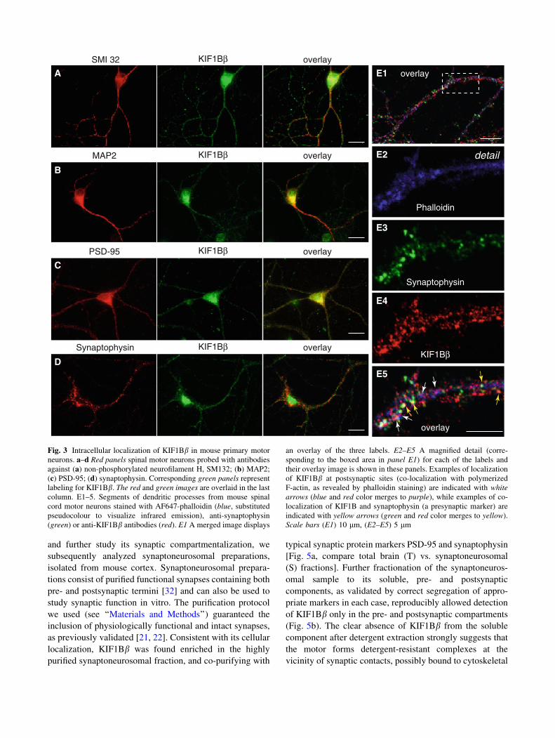
Using the anti-peptide antibody against KIF1Bb, we
mapped the intracellular localization of the protein in pri-
mary mouse spinal motor neurons (Fig. 3), a selective
target for neurodegeneration in CMT2A and a cell type
where expression of KIF1Bb was confirmed by RT-PCR
(Fig. 2c). Double staining of mixed co-cultures of spinal
neurons (motor neurons, interneurons) and glia with SMI
32, a monoclonal antibody specifically labeling motor
neurons, revealed that KIF1Bb is selectively expressed in
motor neurons (Fig. 3a), but not in astroglia (not shown), in
accordance with the RT-PCR results of Fig. 2d showing
absence of signal in pure astroglial cultures.
Initially, we examined the overall topology of KIF1Bblabeling in those motor neurons: KIF1Bb immunostaining
was punctate or granular and detectable both in the cell body,
and in the axon and dendrites (Fig. 3a). With the use of an
antibody to the dendritic microtubule marker MAP2, we
confirmed the expression of KIF1Bb in dendrites (Fig. 3b)
where the motor protein appears co-localized with PSD-95-
positive, postsynaptic structures (Fig. 3c). Furthermore,
KIF1Bb also appears partially co-localized with puncta of
synaptophysin, a known presynaptic vesicle protein
(Fig. 3d). To consolidate these findings, we performed a
triple staining (Fig. 3E1, with E2–E5 showing a detail at
higher magnification) with fluorescently-labeled phalloidin,
which highlights points of filamentous F-actin enrichment
and hence mostly dendritic spines (Fig. 2E2, blue), anti-
synaptophysin antibody as a presynaptic marker (Fig. 3E3,
green), and anti-KIF1Bb (Fig. 3E4, red). Examination of the
labeled patterns consistently revealed colocalization of
KIF1Bb both with synaptophysin puncta, marking presyn-
aptic sites (Fig. 3E5, yellow arrows), and F-actin sites on
dendritic spines, indicating postsynaptic density domains
(Fig. 3E5, white arrows). Thus, KIF1Bb-immunoreactive
structures were present both at pre- and postsynaptic sites.
Furthermore, as independent confirmation, we also
obtained results in clear agreement with this intracellular
localization of KIF1Bb in NSC-34, a spinal cord neuron–
neuroblastoma hybrid cell line, which expresses KIF1Bb(as detected by RT-PCR in Fig. 2e). It is known that NSC-
34 cells maintain many of the morphological and physio-
logical characteristics of spinal motor neurons, including
the ability to form synapses with myotubes [31], and
therefore express synaptic proteins. In NSC-34 cultures, the
anti-KIF1Bb antibody-labeled vesicular structures dis-
playing overlap both with PSD-95 (Fig. 4a) and
presynaptic vesicular protein SV2 (Fig. 4b). Consistently,
no co-localization of KIF1Bb puncta was observed when
NSC-34 cells were labeled with antibodies specific for
other membrane-bound cellular compartments, namely
mitochondria (SOD2), lysosomes (LAMP2), or the Golgi
(58 K Golgin protein) (Online Resource Fig. S3).
The co-localization of KIF1Bb with synaptic structures
in NSC-34 was further confirmed upon transient transfec-
tion of an YFP-tagged full-length KIF1Bb construct. In the
image shown in Fig. 4c, two neighboring cells are imaged,
one untransfected (bottom cell) and one transfected (top
cell); a high degree of co-localization of YFP-KIF1Bbfluorescence and SV2 immunoreactivity can be observed in
the transfected cell.
Finally, the native KIF1Bb-positive structures become
less pronounced upon siRNA-mediated silencing of
KIF1Bb (Fig. 4e). It is notable that, although depletion of
KIF1Bb transcript is very efficient upon prolonged
silencing (Fig. 4d, top panels), the KIF1Bb protein appears
extremely stable even after 120 h of application of the
silencing regime, involving two siRNA transfections
(Fig. 4d, bottom panels). This protein stability likely
accounts for the residual persistence of the, albeit markedly
reduced, vesicular structures of NSC-34 seen in Fig. 4e.
To corroborate biochemically the synaptic localization
of KIF1Bb in neurons, as revealed by immunofluorescence,
and further study its synaptic compartmentalization, we
subsequently analyzed synaptoneurosomal preparations,
isolated from mouse cortex. Synaptoneurosomal prepara-
tions consist of purified functional synapses containing both
pre- and postsynaptic termini [32] and can also be used to
study synaptic function in vitro. The purification protocol
we used (see ‘‘Materials and Methods’’) guaranteed the
inclusion of physiologically functional and intact synapses,
as previously validated [21, 22]. Consistent with its cellular
localization, KIF1Bb was found enriched in the highly
purified synaptoneurosomal fraction, and co-purifying with
typical synaptic protein markers PSD-95 and synaptophysin
[Fig. 5a, compare total brain (T) vs. synaptoneurosomal
(S) fractions]. Further fractionation of the synaptoneuros-
omal sample to its soluble, pre- and postsynaptic
components, as validated by correct segregation of appro-
priate markers in each case, reproducibly allowed detection
of KIF1Bb only in the pre- and postsynaptic compartments
(Fig. 5b). The clear absence of KIF1Bb from the soluble
component after detergent extraction strongly suggests that
the motor forms detergent-resistant complexes at the
vicinity of synaptic contacts, possibly bound to cytoskeletal
SMI 32 KIF1Bβ overlay
KIF1Bβ overlayMAP2
KIF1Bβ overlayPSD-95
KIF1Bβ overlaySynaptophysin
A
B
C
D
overlay
detail
Phalloidin
Synaptophysin
KIF1Bβ
overlay
E1
E2
E3
E4
E5
Fig. 3 Intracellular localization of KIF1Bb in mouse primary motor
neurons. a–d Red panels spinal motor neurons probed with antibodies
against (a) non-phosphorylated neurofilament H, SM132; (b) MAP2;
(c) PSD-95; (d) synaptophysin. Corresponding green panels represent
labeling for KIF1Bb. The red and green images are overlaid in the last
column. E1–5. Segments of dendritic processes from mouse spinal
cord motor neurons stained with AF647-phalloidin (blue, substituted
pseudocolour to visualize infrared emission), anti-synaptophysin
(green) or anti-KIF1Bb antibodies (red). E1 A merged image displays
an overlay of the three labels. E2–E5 A magnified detail (corre-
sponding to the boxed area in panel E1) for each of the labels and
their overlay image is shown in these panels. Examples of localization
of KIF1Bb at postsynaptic sites (co-localization with polymerized
F-actin, as revealed by phalloidin staining) are indicated with whitearrows (blue and red color merges to purple), while examples of co-
localization of KIF1B and synaptophysin (a presynaptic marker) are
indicated with yellow arrows (green and red color merges to yellow).
Scale bars (E1) 10 lm, (E2–E5) 5 lm

or scaffolding structures. The presence of KIF1Bb in both
the pre- and postsynaptic fractions was consistent with the
observed co-localization of KIF1Bb with pre- and
postsynaptic markers by immunofluorescence (Fig. 3c, d,
E1–E5) and indicated that the motor may be bound to the
matrix/structural components on both sides.
KIF1BβPSD-95 / DNA overlay
KIF1BβSV2 / DNA overlay
YFP-KIF1BβSV2 / DNA overlay
KIF1Bβ / DNAEneg. control-silenced KIF1Bβ RNAi
Dkb
0.4
0.4
KIF1Bβ
L19
mockKIF1Bβ
siRNA neg. contr.
siRNA
KIF1Bβ
dynein
KIF1BβsiRNA
neg.siRNA
mock
kDa
72
A
B
C
Fig. 4 Intracellular localization of KIF1Bb in mouse neuroblastoma
NSC-34 cell line. NSC-34 cells stained with antibodies to a PSD-95
(red), b SV2 (red), with concurrent labeling of nuclei with Hoechst
33342 (blue). Green panels represent labeling with anti-KIF1Bb.
Scale bars (a) 20 lm, (b, c) 10 lm. c NSC-34, transiently transfected
with pEYFP-KIF1Bb, green). Anti-SV2 staining is in red, and DNA
in blue. Of the two cells in the field, only the top cell is transfected:
YFP-KIF1Bb localization (green) overlaps extensively with that of
SV2 (red), therefore merging to yellow in the overlay. Scale bar10 lm. d Quantification of silencing efficiency by RT semi-quanti-
tative PCR and immunoblot. Top two panels levels of KIF1Bb mRNA
at 120 h post-transfection in NSC-34 cells silenced with 320 pM of
KIF1Bb-specific siRNA cocktail are compared to control-silenced
cells (negative control) and to mock-treated cells (no siRNA). RT-
PCR detection of L19 mRNA levels serves as internal control. Bottomtwo panels KIF1Bb protein levels are compared in corresponding
samples. Detection of the intermediate chain of dynein serves as
loading control in the three samples. e KIF1Bb immunofluorescence
in silenced cells versus negative control-silenced cells. NSC-34 cells
were immunostained for KIF1Bb (green) and counterstained for DNA
(blue). KIF1Bb-silenced cells display a marked reduction in the signal
of KIF1Bb-positive vesicular structures when imaged at the same
exposure as negative control-silenced cells. Scale bar 10 lm

Synaptic activation causes increased recruitment
of KIF1Bb to the synaptic area
Given the association of KIF1Bb with synaptic structures,
we next asked the question whether synaptic activity might
influence the recruitment of the motor protein to the active
synaptic sites. We therefore quantified KIF1Bb synaptic
recruitment with two independent assays, performed in the
absence or presence of synaptic stimulation. We conducted:
(1) biochemical assays with purified synaptoneurosomes,
and (2) morphological assays using mouse cortical neurons
in culture.
We employed DHPG (S-3,5,-dihydroxyphenylglycine),
a widely used agonist of group I metabotropic glutamate
receptors (mGluR1 and mGluR5) implicated in the induc-
tion of different forms of synaptic plasticity, to stimulate
synaptoneurosomal preparations. We observed, by quanti-
tative immunoblot, a statistically significant (p = 0.023)
increase by 50 % in the concentration of KIF1Bb in the
stimulated synaptoneurosomes, compared with unstimu-
lated controls (Fig. 6a, b), while none of the other synaptic
markers tested (PSD-95, synaptophysin, syntaxin 1 and
SNAP-25) showed significant alteration after stimulation
(Fig. 6c).
For independent confirmation, we quantified the effect
of DHPG stimulation in living cells. To visualize func-
tional synapses on dendrites, we performed triple staining
with phalloidin, which highlights points of polymerized
F-actin enrichment and hence dendritic spines, anti-syn-
aptophysin antibody as a presynaptic marker, and anti-
KIF1Bb (Fig. 6d). For KIF1Bb quantification, we applied a
mask that was created with a dedicated algorithm (modified
from [24]; see further details in ‘‘Materials and Methods’’)
marking an area in the image lying within 0.4 lm of both
synaptophysin puncta and sites of F-actin and thus in close
vicinity to a synaptic contact. In this manner, we restricted
quantification of KIF1Bb signal to that associated with
functional synapses. In confirmation of our biochemical
results, analysis of images from four independent experi-
ments (total of 2400 synapses analyzed) revealed that
DHPG stimulation resulted in increased recruitment of
KIF1Bb to the synaptic area (by 25 %), compared with
non-stimulated cells (statistical significance p = 0.03;
Fig. 6e).
We tested whether a similar activity-dependent increase
of the levels of KIF1Bb at the synapse could occur upon
general stimulation with KCl, metabotropic or ionotropic
glutamate receptor activation (Fig. 6f). Analysis of the
results using KCl or the different agonists, specific for
ionotropic and metabotropic glutamate receptors, and from
the PMA, a phorbol ester that directly activates PKC
(Online Resource Fig. S4), clearly showed that the levels of
KIF1Bb in purified synaptoneurosomes were markedly and
statistically very significantly increased by KCl activation
and glutamate signaling, but preferentially so upon NMDA
receptor activation (p = 0.0014; Fig. 6f).
On the basis of the combined experimental evidence, we
therefore conclude that synaptic concentration of motor
KIF1Bb is increased in an activity-dependent manner.
KIF1Bb interacts directly with Calmodulin (CaM)
in a Ca2?-dependent manner and through
its carboxy-terminal domain
To obtain insight into the functional significance of the
localization and activity-dependent modulation of KIF1Bbrecruitment to the synapse, we employed different
approaches to screen for its interacting partners. We first
sought to identify interacting protein partners for this motor
using a yeast two-hybrid screen. Of the 1.8 9 106 clones of
a human adult CNS cDNA library that were tested, using as
bait a fragment of KIF1Bb containing the non-conserved,
non-motor domain of the protein (construct ‘‘C1’’, nt.
1980–5313), 54 positive clones were identified after five
rounds of selection (details in ‘‘Materials and Methods’’).
A
solublepre post
PSD-95
synaptophysin
KIF1Bβ
B
synaptophysin
PSD-95
KIF1Bβ
MBP
GFAP
T S
Fig. 5 a Detection of KIF1Bb in purified mouse synaptoneurosomes.
KIF1Bb is detected in total lysates from the cortex (T; 5 lg total
protein) and is enriched in a sample of synaptoneurosomes purified
from the cortex (S; 5 lg). The level of detection of synaptophysin, a
presynaptic marker, and PSD-95, a postsynaptic marker, are evidence
of the enrichment of the synaptoneurosomal preparation, which was
essentially devoid of non-neuronal proteins, such as myelin basic
protein (MBP) and glial fibrillary acidic protein (GFAP). b Detection
of KIF1Bb in further fractionated synaptoneurosomes. The synapto-
neurosomal preparation from mouse cortex was further fractionated
into its soluble, pre- and postsynaptic components and probed for
KIF1Bb in parallel with compartment-specific protein markers PSD-
95 and synaptophysin. KIF1Bb was detected in both the pre- and
postsynaptic, but not in the soluble fraction. An amount of 15 lg of
protein was loaded from each fraction

Of these, 25 positive clones contained partial cDNAs
derived from the CALM1 (Calmodulin 1) gene, 21 were
partial cDNAs derived from the CALM2 gene and 8 from
the CALM3 gene. Although these three genes are localized
on distinct human chromosomes (chromosomes 14, 2, and
19, respectively; [33, 34]) and exhibit diverse 5’ and 3’
UTRs, because of the degeneracy of the genetic code, their
ORFs are synonymous and completely identical at the aa
level and thus all encode the same protein, Calmodulin
(CaM).
To ensure that the interaction detected between KIF1Bband CaM was bona fide and to characterize it further, we
carried out several independent sets of experiments. First,
we examined the intracellular localization of KIF1Bb and
A
PSD-95
Synaptophysin
Syntaxin 1
SNAP-25
GAPDH
Control
DHPG
KIF1Bβ
B
Control DHPG
Phalloidin
Synaptophysin
KIF1Bβ
Merged
D
0
0.5
1
1.5
2
1
DH
PG
/ co
ntro
l
control
DHPG
conc.KIF1Bβ
*
0
0.5
1
1.5
1
DHPGcontrol*
conc.KIF1Bβ
Inte
nsity
at s
ynap
ses
(DH
PG
/ co
ntro
l)
E
SNAP-25
DHPGcontrol
C
0
0.5
1
1.5
PSD-95 synaptophysin syntaxin 1DH
PG
/ co
ntro
l
conc.
0.0
0.5
1.0
1.5
2.0
2.5
3.0
KCl DHPG NMDA AMPA NMDA +AMPA
Glutamate PMA
stimulationcontrol
F
* ***
* *
stim
ulat
or/c
ontr
.
conc.KIF1Bβ
Fig. 6 a DHPG stimulation increases the amount of KIF1Bb in
synaptoneurosomes. Immunoblot (representative of 8 independent
experiments) of protein extracts of cortical synaptoneurosomes either
resting (control) or treated with DHPG and probed for KIF1Bb, PSD-
95, synaptophysin, syntaxin1, SNAP-25, and also GAPDH (loading
control). b Quantification of the experiments shows a significant
increase of KIF1Bb protein levels in synaptoneurosomes after DHPG
stimulation (*p \ 0.05; Student’s t test). c Quantification of PSD-95,
synaptophysin, syntaxin1 and SNAP-25 levels in the same experi-
ments as in (b) shows no significant change. d, e DHPG stimulation
recruits KIF1Bb to the synapses. d Cortical neurons resting (leftpanels) or treated with DHPG (right panels), fixed, and stained with
phalloidin, anti-synaptophysin or anti-KIF1Bb antibodies. Merged
images show an overlay of the mask that marks vicinity to a synapse
(green) and the KIF1Bb signal in the control or after DHPG,
respectively (red). Arrows point to strong KIF1Bb signals in the
vicinity of synapses. Scale bar 10 lm. e Quantification of KIF1Bbfluorescence that fell into total synaptic areas, as defined by the mask
described in ‘‘Materials and Methods’’ (n = 20 neurons; *p \ 0.05;
Student’s t test). f Quantification of KIF1Bb signal from 6 indepen-
dent experiments, showing increases of KIF1Bb concentration in
synaptoneurosomes after stimulation of different types of glutamate
receptor (confirmation of PKC stimulation with PMA in Online
Resource Fig. S4). The most pronounced increase corresponds to
stimulation of the NMDA receptor (n = 20 neurons, 6 independent
experiments, **p = 0.0014; Student’s t test)

Input
0.0 0.0 0.0 0.0 0.0 1.0 1.0 1.0 1.0 mM CaCl21
1
1770
660
1218
1549
F
M
C1
C2
C3
C4
C5
GST vector-only neg.contr.
170 C1
95C3
55 C5
55 C4
95 C2
95 M
26 vector
neg.cont. neg.cont.
Motor domain FHA, phosphopeptide binding domain PH, protein-protein interaction domain
anti-GST
A1 A2 A3
KIF1Bβ CaM overlay detail
kDa
1770660
660
17701218
1770
B
C
A
negative control
1 2 3 4 5 6 7 8 9
72
250mM CaCl2
nativeKIF1Bβ
Dynein
0.0 0.0 0.5 0.5 1.0 1.0 2.0 2.0 1.0 1.0 2.0 2.0inpu
t
kDa
1 2 3 4 5 6 7 8 9 10 11 12 13
U B U B U B U B B U B U
U B U B U B U B
Fig. 7 A1–A3 Partial co-localization of KIF1Bb and CaM in spinal
motor neurons. Double immunofluorescence for KIF1Bb (green) and
CaM (red) reveals their co-localization (yellow) in both the cell body
and neuronal processes (detail at higher magnif.). Scale bars 20 lm,
inset 10 lm. b Pull-down assays with cell extracts confirming the
interaction between endogenous KIF1Bb and CaM and demonstrating
its Ca2?-dependence. CaM, immobilized on Sepharose beads, was
incubated with equivalent lysates (10 lg total protein) from NSC-34
cells in the presence of increasing Ca2?concentrations. Bound (B;
lanes 3, 5, 7, 9) and unbound proteins (U; lanes 2, 4, 6, 8) were
probed by immunoblot with anti-KIF1Bb (top panels). Sepharose
beads without CaM were used as negative control (top panels, lanes10–13; B and U). An anti-dynein antibody (bottom panels) was used
as internal control for equal loading. Detection of endogenous
KIF1Bb is obtained only in the presence of CaM (lanes 3, 5, 7, 9) and
is enhanced by increased Ca2? concentration, peaking at about
0.5 mM CaCl2 (lane 5). c Pull-down assays with recombinant
proteins confirming a direct interaction between KIF1Bb and CaM
and mapping the interaction site on KIF1Bb. CaM on Sepharose
beads was incubated with equivalent extracts of E. coli expressing
GST-tagged deletion constructs of KIF1Bb (M, C1–C5; shown in
schematic form), in the absence (lanes 1–5) or presence of 1 mM
Ca2? (lanes 6–9). Bound (B) and unbound (U) proteins were probed
by anti-KIF1Bb the KIF1Bb band is indicated by arrows, a lower
band appearing in some samples is a degradation product). Sepharose-
only beads served as negative control at the same conditions (lanes 4,
5, 8, 9). GST-vector-only was an additional negative control to
exclude interaction of the KIF1Bb constructs and CaM via GST
sequences (vector; bottom panels). F full-length KIF1Bb, M motor-
domain-containing construct, C1–C5 non-motor-domain-containing
constructs. Constructs are also described in Table 1. Numbersaccompanying the construct sketches denote amino-acid residues

CaM in mouse primary spinal motor neurons in culture by
immunofluorescence. We observed extensive co-localiza-
tion, as one would likely expect from interacting proteins
(Fig. 7A1–A3), both, in the cell body and in neuronal
processes (detail at higher magnification in the inset).
Second, we carried out a series of pull-down assays
using CaM-Sepharose beads incubated with extracts from
NSC-34 neuroblastoma cells in the presence of increasing
concentrations of Ca2? (from 0–2 mM CaCl2) (Fig. 7b).
These assays showed that (1) native KIF1Bb was recov-
ered in the pellet fraction only when incubated in the
presence of CaM, consistent with an interaction between
the two proteins (Fig. 7b, compare lanes 3, 5, 7, 9 with
negative controls in lanes 10, 12), and (2) the interaction
was Ca2?-dependent, with recovery of the native KIF1Bbprotein minimal in the absence of Ca2? and peaking in
the presence of 0.5 mM CaCl2 (Fig. 7b, compare lanes 3,
5, 7 and 9 with progressively increasing concentrations of
Ca2?).
Third, we demonstrated that the interaction between
KIF1Bb and CaM is direct, confirmed its dependence on
Ca2? using in vitro pull-down assays with bacterially
expressed, recombinant proteins (Fig. 7c) and mapped the
CaM-binding site on a domain contained within the car-
boxy-terminal part of KIF1Bb (Fig. 7c; Table 1).
Analytically, Fig. 7c illustrates that the E. coli-expressed
GST-tagged C1 fragment of KIF1Bb (construct ‘‘C1’’, the
fragment originally used in the yeast two-hybrid screen)
can bind to CaM immobilized on Sepharose beads, but
only in the presence of Ca2? (Fig. 7c, compare lanes 3
and 7 in panel C1), while the corresponding N-terminus
of KIF1Bb, containing the motor domain (fragment
‘‘M’’), remains unbound in all circumstances (Fig. 7c,
compare lanes 3 and 7 in panel M). By testing progres-
sively smaller GST-tagged subfragments of C1 (constructs
C2–C5), we confined the CaM interaction site(s) of
KIF1Bb to within the last 552 carboxy-terminal aa
(Fig. 7c, observe lanes 3 and 7 in panels C2, C3). If this
carboxy-terminal region is fragmented into two comple-
mentary segments of 331 and 221 aa (constructs C4 and
C5), both segments retain their CaM binding activity
(Fig. 7c, lanes 3 and 7 in panels C4, C5), suggesting that
either (1) there is more than one CaM site, or (2) that the
CaM binding site extends either side of aa 1549, the
position where the subfragments are split. The results of
these in vitro pull-down assays are summarized in Table 1
(left part). It is notable that both the native KIF1Bb(Fig. 7b) and recombinant subfragments C3–C5 (Fig. 3c)
retained some CaM binding activity even in the absence
of Ca2?, either suggesting a negative regulatory function
for the amino-terminal part of KIF1Bb or merely non-
specific interaction with Ca2?-depleted CaM (ApoCaM)
as a result of the protein fragmentation.
Finally, the same fragments of KIF1Bb (M and C1–C5)
were also subcloned as GAL4 DNA-binding-domain
fusions and tested in vivo by yeast mating assays for
interaction with full-length CaM, expressed as a GAL4
DNA activation-domain fusion. The results, also summa-
rized in Table 1 (right part), were identical to those
obtained with the in vitro assays, thus confirming the
position of the CaM binding site(s) within the 552 carboxy-
terminal aa of KIF1Bb.
Table 1 Mapping of the CaM interaction domain of KIF1Bb by in vivo and in vitro assays
In vivo interactions with full-length CaM by mating in
S. cerevisiaeIn vitro pull-down assays of CaM with native KIF1Bb or recombinant protein fragments
expressed in E. coli
Construct Result X gal expos Construct 0.0 mM (5 mM EDTA) 0.5 mM 1.0 mM 2.0 mM (Ca2?)
pGBK/pGAD7 (pos. control) ? 2 h Native KIF1Bba ? ?? ?? ??
M (aa 1–660) – 3 h M (aa 1–660) – NT – NT
C1 (aa 660–1770) ?? 2 h C1 (aa 660–1770) – NT ??? NT
C2 (aa 660–1218) – 3 h C2 (aa 660-1218) – NT – NT
C3 (aa 1218–1770) ?? 3 h C3 (aa 1218–1770) ? NT ?? NT
C4 (aa 1218–1549) ? 3 h C4 (aa 1218–1549) ? NT ? NT
C5 (aa 1549–1770) ? 3 h C5 (aa 1549–1770) ? NT ? NT
pAS2.1 (neg. cont) – 3 h pGEX (neg. cont) – NT – NT
The left part displays mating assays with plasmid pACT2.1-CaM, carrying full-length Calmodulin, and the pAS2.1-KIF1Bb constructs shown
(numbers in parentheses denote amino acid (aa) residues in generated KIF1Bb protein fragments). Interaction was defined as the ability of[95 %
of resulting clones, expressing both constructs, to grow on quadruple selective media (-Leu, -Trp, -His, -Ade) and to also display b-galactosidase
activity upon filter assay. The right part shows assays conducted with native KIF1Bb or GST-tagged recombinant proteins from KIF1Bb deletion
constructs, expressed in E. coli and incubated with CaM-Sepharose beads and in the concentration of Ca2? indicated, with 0.0 mM CaCl2established by chelation with 5 mM EDTA
NT Not testeda The term native KIF1Bb refers to a reaction using total lysate (500 lg) from NCS-34 cells, incubated with CaM-Sepharose beads
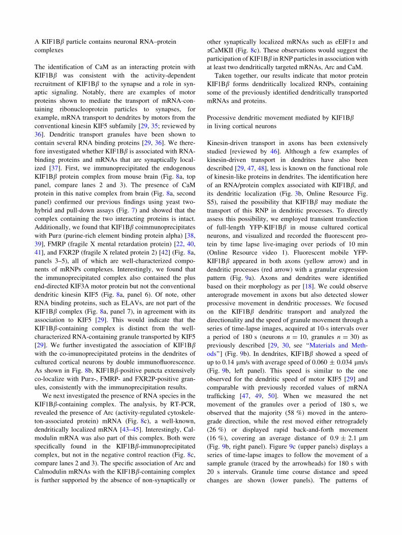
A KIF1Bb particle contains neuronal RNA–protein
complexes
The identification of CaM as an interacting protein with
KIF1Bb was consistent with the activity-dependent
recruitment of KIF1Bb to the synapse and a role in syn-
aptic signaling. Notably, there are examples of motor
proteins shown to mediate the transport of mRNA-con-
taining ribonucleoprotein particles to synapses, for
example, mRNA transport to dendrites by motors from the
conventional kinesin KIF5 subfamily [29, 35; reviewed by
36]. Dendritic transport granules have been shown to
contain several RNA binding proteins [29, 36]. We there-
fore investigated whether KIF1Bb is associated with RNA-
binding proteins and mRNAs that are synaptically local-
ized [37]. First, we immunoprecipitated the endogenous
KIF1Bb protein complex from mouse brain (Fig. 8a, top
panel, compare lanes 2 and 3). The presence of CaM
protein in this native complex from brain (Fig. 8a, second
panel) confirmed our previous findings using yeast two-
hybrid and pull-down assays (Fig. 7) and showed that the
complex containing the two interacting proteins is intact.
Additionally, we found that KIF1Bb coimmunoprecipitates
with Pura (purine-rich element binding protein alpha) [38,
39], FMRP (fragile X mental retardation protein) [22, 40,
41], and FXR2P (fragile X related protein 2) [42] (Fig. 8a,
panels 3–5), all of which are well-characterized compo-
nents of mRNPs complexes. Interestingly, we found that
the immunoprecipitated complex also contained the plus
end-directed KIF3A motor protein but not the conventional
dendritic kinesin KIF5 (Fig. 8a, panel 6). Of note, other
RNA binding proteins, such as ELAVs, are not part of the
KIF1Bb complex (Fig. 8a, panel 7), in agreement with its
association to KIF5 [29]. This would indicate that the
KIF1Bb-containing complex is distinct from the well-
characterized RNA-containing granule transported by KIF5
[29]. We further investigated the association of KIF1Bbwith the co-imunoprecipitated proteins in the dendrites of
cultured cortical neurons by double immunofluorescence.
As shown in Fig. 8b, KIF1Bb-positive puncta extensively
co-localize with Pura-, FMRP- and FXR2P-positive gran-
ules, consistently with the immunoprecipitation results.
We next investigated the presence of RNA species in the
KIF1Bb-containing complex. The analysis, by RT-PCR,
revealed the presence of Arc (activity-regulated cytoskele-
ton-associated protein) mRNA (Fig. 8c), a well-known,
dendritically localized mRNA [43–45]. Interestingly, Cal-
modulin mRNA was also part of this complex. Both were
specifically found in the KIF1Bb-immunoprecipitated
complex, but not in the negative control reaction (Fig. 8c,
compare lanes 2 and 3). The specific association of Arc and
Calmodulin mRNAs with the KIF1Bb-containing complex
is further supported by the absence of non-synaptically or
other synaptically localized mRNAs such as eEIF1a and
aCaMKII (Fig. 8c). These observations would suggest the
participation of KIF1Bb in RNP particles in association with
at least two dendritically targeted mRNAs, Arc and CaM.
Taken together, our results indicate that motor protein
KIF1Bb forms dendritically localized RNPs, containing
some of the previously identified dendritically transported
mRNAs and proteins.
Processive dendritic movement mediated by KIF1Bbin living cortical neurons
Kinesin-driven transport in axons has been extensively
studied [reviewed by 46]. Although a few examples of
kinesin-driven transport in dendrites have also been
described [29, 47, 48], less is known on the functional role
of kinesin-like proteins in dendrites. The identification here
of an RNA/protein complex associated with KIF1Bb, and
its dendritic localization (Fig. 3b, Online Resource Fig.
S5), raised the possibility that KIF1Bb may mediate the
transport of this RNP in dendritic processes. To directly
assess this possibility, we employed transient transfection
of full-length YFP-KIF1Bb in mouse cultured cortical
neurons, and visualized and recorded the fluorescent pro-
tein by time lapse live-imaging over periods of 10 min
(Online Resource video 1). Fluorescent mobile YFP-
KIF1Bb appeared in both axons (yellow arrow) and in
dendritic processes (red arrow) with a granular expression
pattern (Fig. 9a). Axons and dendrites were identified
based on their morphology as per [18]. We could observe
anterograde movement in axons but also detected slower
processive movement in dendritic processes. We focused
on the KIF1Bb dendritic transport and analyzed the
directionality and the speed of granule movement through a
series of time-lapse images, acquired at 10-s intervals over
a period of 180 s (neurons n = 10, granules n = 30) as
previously described [29, 30, see ‘‘Materials and Meth-
ods’’] (Fig. 9b). In dendrites, KIF1Bb showed a speed of
up to 0.14 lm/s with average speed of 0.060 ± 0.034 lm/s
(Fig. 9b, left panel). This speed is similar to the one
observed for the dendritic speed of motor KIF5 [29] and
comparable with previously recorded values of mRNA
trafficking [47, 49, 50]. When we measured the net
movement of the granules over a period of 180 s, we
observed that the majority (58 %) moved in the antero-
grade direction, while the rest moved either retrogradely
(26 %) or displayed rapid back-and-forth movement
(16 %), covering an average distance of 0.9 ± 2.1 lm
(Fig. 9b, right panel). Figure 9c (upper panels) displays a
series of time-lapse images to follow the movement of a
sample granule (traced by the arrowheads) for 180 s with
20 s intervals. Granule time course distance and speed
changes are shown (lower panels). The patterns of

movement (explained as a result of the mixed polarity of
microtubules at proximal dendrites and competition with
retrograde motors, such as dynein) and the measured
speeds are again consistent with those detected for RNPs
transported by KIF5 [29] or other brain derived, dendri-
tically targeted RNPs [30, 51]. Altogether, these results
support our hypothesis that KIF1Bb-containing granules
transport RNA in dendrites.
The complex we identified is not associated to KIF5A
(Fig. 8a), thus suggesting an active role of KIF1Bb in the
RNP transport. To rule out the possibility that the move-
ment we detected was not mediated by another motor
protein (such as KIF3A that we have identified as part of
the complex in Fig. 8a) that carried along KIF1Bb in a
passive, piggy-bag manner, we analyzed the movement of
a truncated KIF1Bb lacking the motor domain (GFP-
InputKIF1Bβ IP
IgG IP
A
KIF1Bβ
CaM
KIF3A
KIF5A
Purα
FMRP
FXR2P
B
C
ELAVs
InputKIF1Bβ IP
IgG IP
CaM
Arc
KIF1Bβ
Trkβ
βactin
eEIF1α
βcatenin
αCaMKII
BC200
1 2 3
1 2 3

KIF1Bb-C1 construct of Fig. 7c). In this case, we did not
observe either specific granule formation or movement,
but, instead, diffuse fluorescence, as previously shown for
KIF5A under the same conditions by Kanai et al. [29]
(Fig. 9d; Online Resource video 2). Similar findings were
obtained using a GFP-empty plasmid (data not shown).
These results indicated that the movement we analyzed by
the use of the full-length construct was bona fide, active
and specific. To better evaluate moving or immobile par-
ticles, we represented in two kymographs the fluorescence
changes along the dendritic segment over 180 s in neurons
transfected with YFP-KIF1Bb or GFP-KIF1Bb-C1,
respectively. As expected, kymograph analysis further
confirmed the absence of moving granules when the trun-
cated KIF1Bb was expressed (Fig. 9e). In conclusion,
therefore, these combined experiments revealed a novel
role for KIF1Bb as a motor protein mediating transport of
RNP particles, which contain RNA-binding proteins and
dendritically targeted Calmodulin and Arc mRNAs.
Discussion
The members of the KIF1A/KIF1B/UNC-104 (kinesin-3)
family of molecular motors have been shown by a number
of studies to transport synaptic vesicle structures [4, 7, 52].
Mechanisms about how signaling pathways may be regu-
lating vesicular transport are gradually emerging.
Association of such vesicles with the stalk domain or the
PH domains of kinesin-3 family motors (see Fig. 1) via
vesicle phosphoinositides is necessary for cargo recogni-
tion and binding [8]. Experimental evidence supports a
model where KIF1A motors exist in an autoinhibited state
in the absence of cargo, and this autoinhibition is relieved
following interaction with cargoes [53]. At least one
adaptor protein, the DENN/MADD, has been identified as
essential in the GTP-dependent axonal transport of Rab3-
containing vesicles [54]. In C. elegans, it has been pro-
posed that, upon cargo delivery at the pre-synaptic site,
kinesin-3 motor KIF1A/UNC-104 is locally degraded at the
synapse, likely via the ubiquitination pathway, and hence is
not recycled back retrogradely [55]. Furthermore, in
C. elegans, UNC-104 motor clustering along axons as well
as their motility is enhanced via multiple interactions with
synaptic scaffolding protein SYD-2 [56]. In non-neuronal
cells, i.e. in non-polarized epithelial cells, the post-Golgi
transport of p75 neurotrophin receptors by kinesin-3 family
motors (KIF1Ba and b) appears to also involve interactions
with the PH domain [57].
While it is now well understood that a multitude of
molecular motors, including KIF1Bb, act in concert to
transport various cellular cargoes to the synapses of neu-
rons, the mechanisms that integrate cargo transport with
synaptic activity, or with synaptic assembly in developing
neurons or with remodeling of synapses in mature neurons
(plasticity), in response to neuronal activity [58] remain
largely uncharacterized.
Here, we provide novel findings that functionally char-
acterize KIF1Bb in the context of synaptic signaling.
KIF1Bb is not neuron-specific but highly expressed in the
CNS and particularly prevalent in the motor cortex and
spinal cord motor neurons (Fig. 2). It is localized both
presynaptically in synaptic boutons and postsynaptically in
postsynaptic density structures of dendritic spines (Figs. 3,
4). KIF1Bb is enriched in isolated synaptoneurosomes, co-
fractionates in both pre- and postsynaptic fractions, and is
exclusively in a bound and detergent-resistant form, with
no detectable soluble KIF1Bb pool (Fig. 5). Stimulation of
metabotrobic or ionotropic glutamate receptors results in
increased KIF1Bb concentration at synaptic sites, revealing
an activity-dependent recruitment of the motor to synapses
(Fig. 6). In this study, Calmodulin was identified as a direct
binding partner of the motor (Fig. 7), via an interaction
domain within the 552 amino acids of the carboxy-terminal
tail of KIF1Bb, partly overlapping with the stalk domain
where interaction with DENN/MADD protein has previ-
ously been localized [54]. The CaM-KIF1Bb interaction is
dependent upon and facilitated by Ca2?, with a possible
weak interaction also with the ApoCaM form (Fig. 7). This
is an important finding because it directly links KIF1Bb to
Ca2? via CaM, a universal Ca2? sensor protein, thus
indicating a possible mechanism of KIF1Bb regulation
Fig. 8 a Immunoprecipitation (IP) for KIF1Bb from mouse brain.
Analysis by western blot, using different antibodies, of the anti-
KIF1Bb-immunoprecipitated proteins revealed that KIF1Bb and its
interacting protein Calmodulin (CaM) are part of the same complex.
Furthermore, the dendritically-localized RNA-binding proteins Pura,
FMRP and FXR2P, as well as the motor protein KIF3A, were also
detected in the same KIF1Bb complex. RNA-binding protein ELAVs
and the motor protein KIF5A were absent. Lane 1 represents 1/20
(25 lg) of the original extract used for immunoprecipitation (input),
lanes 2 and 3 show the immunoprecipitated protein using, respec-
tively, equal amounts of anti-KIF1Bb antibody or rabbit IgGs
(negative control). b Co-localization of KIF1Bb and dendritic
RNA-binding proteins in mouse cultured cortical neurons. Staining
of cortical neurons (12–14 div.) with anti-KIF1Bb (red panels) and
concurrent labeling with antibodies to Pura; FMRP; FXR2P (greenpanels), reveal extensive pairwise co-localization in puncta (arrows)
in a detail of a dendritic process. Scale bar 5 lm. c RT-PCR analysis
of the RNA content of the KIF1Bb IP material. Calmodulin, Arc,
KIF1Bb, TrkB, b-actin, eEIF1a, b-catenin, CamKIIa mRNAs were
amplified, following KIF1Bb immunoprecipitation. Prior to RNA
extraction, each sample was spiked with 100 ng of exogenous human
BC200 mRNA, which was then tested by PCR to show that RNA
extraction, reverse transcription, and amplification efficiency was
equal across samples. Lane 1 represents the RT-PCR reaction of 1/20
of the original extract used for immunoprecipitation (input), lanes 2and 3 represent the RNA immunoprecipitated using equal amounts,
respectively, of anti-KIF1Bb antibody and rabbit IgGs. CaM and ArcmRNAs were detected in the KIF1Bb complex
b

during depolarization-induced Ca2? influx in the presyn-
aptic compartment. Our results would predict that, in
conditions of facilitation of presynaptic Ca2? influx,
triggered by synaptic depolarization, the interaction
between activated CaM and KIF1Bb is enhanced. Increased
strength of the interaction could result in augmented

synaptic delivery of vesicles via KIF1Bb. This mechanism
may thus stimulate synaptic vesicle exocytosis by activating
bound KIF1Bb or redistributing bound KIF1Bb on synaptic
vesicles upon presynaptic depolarization, which in turn
would tie in with the observed activity-dependent increase
of synaptic KIF1Bb recruitment.
Other examples of interaction of kinesin-like motors
with Ca2?-binding proteins have been reported: Ca2?-
dependent binding of CaM to the light chains (KLC) has
been previously documented in the case of heterotetrameric
conventional kinesin [59], and, although the CaM binding
site on KLC has not been determined, binding appears to
inhibit the ATPase activity of the heavy chains. In rat, the
KIF1Bb2 isoform (most similar to the human
AB088210–13 isoform series) interacts with CHP (calci-
neurin B homologous protein) [60], a Ca2?-binding EF-
hand protein with significant similarity to the phosphatase
calcineurin, involved in Ca2?-dependent signal transduc-
tion pathways [61]. In mammalian neurons, kinesin KIF5-
powered mitochondrial mobility is inhibited upon Ca2?
influx in postsynaptic sites, via modulation by Miro1,
another Ca2?-binding EF-hand protein [62, 63]. In plants,
Ca?2-dependent binding of CaM on a kinesin-like motor
(kinesin-like Calmodulin binding protein, KCBP) disrupts
this motor’s microtubule binding [64]. It would appear,
therefore, that regulation of motor protein properties and
activity by Ca2?-sensing proteins, in a Ca2?-dependent
manner, is a more generalized mechanism. This strategy
would be an effective mechanism for integration of motor
protein function and local synaptic activation.
We show here via co-immunoprecipitation experiments
that KIF1Bb forms RNP complexes containing its inter-
acting protein CaM, as well as RNA-binding proteins such
as Pura, FMRP, and FXR2P (with which KIF1Bb co-
localizes in dendritic processes; Fig. 8), and Arc and Cal-
modulin mRNAs (Fig. 8) [43–45]. Both mRNAs have a
bona-fide dendritic localization, and motor protein KIF5
has previously been shown to move anterogradely a large
protein-mRNA complex that includes Arc mRNAs in
dendrites [29]. Although some of the proteins (i.e. Pura and
FMRP) identified here are in common to the large RNP
granule transported by KIF5A [29], we believe that they
are distinct complexes as they have differences both in
their mRNA and protein components. The KIF1Bb com-
plex lacks KIF5A and contains Arc and CaM mRNAs, but
not CaMKII mRNA, while the KIF5A complex lacks
KIF1Bb and CaM proteins and includes Arc and CaMKII
mRNAs.
Our findings raised the possibility that KIF1Bb may be
one additional motor protein that mediates the dendritic
targeting of Arc and CaM mRNA in a distinct RNP com-
plex. Our hypothesis is consistent with all the indirect
experimental findings presented here i.e. (1) the localiza-
tion and enrichment of the motor with synaptic structures,
including the postsynaptic density, (2) its co-immunopre-
cipitation with the two mRNA species, and (3) its activity-
dependent recruitment to the synapse, but we also found
direct evidence of dendritic transport specifically and
actively mediated by KIF1Bb in mouse cultured cortical
neurons (Fig. 9). This is one of the very few examples of a
function of KIF1Bb outside the context of axonal transport,
given the requirement for this motor for the localization of
myelin mRNA to oligodendrocyte processes in zebrafish
[12].
KIF1Bb-mediated dendritic targeting appears of partic-
ular relevance for Arc mRNA, the transcript of an
immediate early gene that, upon synaptic stimulation, is
rapidly transported to active dendritic spines and is locally
translated, thus topically increasing Arc protein concen-
tration at postsynaptic terminals [45, 65, 66]. Arc has a
critical role in multiple forms of protein synthesis-depen-
dent synaptic plasticity [reviewed in 67]: it is involved in
the endocytosis of AMPA receptors, underlying long-term
depression (LTD) [68] and homeostatic plasticity [69, 70],
and in the formation of F-actin, underlying the consolida-
tion of long-term potentiation (LTP) [71]. It has emerged
from different studies that Arc gene expression can be
dynamically regulated by behavioral experience in brain
areas related to learning and memory, thus coupling
changes in neuronal activity patterns to synaptic plasticity
and thereby optimizing information storage in active net-
works [72, 73; reviewed in 74]. Pertinent to our findings,
the translation of Arc is induced within minutes of
Fig. 9 Processive movement of YFP-KIF1Bb granules in the den-
drites of living cortical neurons. a Example of a mouse cultured
cortical neuron transfected with plasmid pEYFP-KIF1Bb (full-length
construct in Fig. 7c), showing fluorescent granules. The dendrite in
this cell is indicated with a red arrowhead and the axon with a yellowarrowhead. Scale bar 10 lm. b Histogram of the maximum speed of
30 granules (average ± SD: 0.060 ± 0.034 lm/s) (left panel); final
distances these 30 granules covered in 180 s (average ± SD:
0.9 ± 2.7 lm) (right panel). c Upper panels a series of still images,
taken at 20-s intervals, from a time-lapse movie of the rectangular
area marked in (a), as an example of the movement of a granule
(arrowheads) for a period of 180 s. Scale bar 2 lm. The full video of
this sequence is given as an Online Resource video1. (The full video
of a sequence derived from a neuron transfected with pEGFP-
KIF1Bb-C1 is given as an Online Resource video2). Bottom panelstime course, during 180 s, of the distances covered by this represen-
tative granule from the start point, as traced by the arrowheads (upperpanels). Corresponding time course of the speeds of the sample
granule during the 180 s recording (right panel). d Equivalent
example of cortical neuron transfected with pEGFP-KIF1Bb-C1
(motor-less construct ‘‘C1’’ in Fig. 7c), showing diffuse fluorescent
labeling. The dendrite in this cell is indicated with a red arrowheadand the axon with a yellow arrowhead. e Kymographs of neurons
transfected with pEYFP-KIF1Bb or pEGFP-KIF1Bb-C1. Rectangularareas marked in (a) and (d) indicate, respectively, the region selected
to generate the kymographs. Red dashed lines indicate the orthogonal
plane represented by the kymographs. Scale bar 1 lm
b

activation of group-I metabotropic glutamate receptors, a
response that is essential to mGluR-dependent LTD [75].
The pathway of Arc translational induction involves the
activation of translational elongation factor eEF2 via Ca2?/CaM [75]. We found DHPG, an agonist of group-I
mGluR, to cause mobilization of KIF1Bb to synaptic sites
and identified Arc mRNA, CaM mRNA and CaM protein
as parts of KIF1Bb ribonucleoprotein complexes. It is
therefore conceivable that, upon metabotropic glutamater-
gic stimulation, synaptic signaling, involving CaM,
activates the transport of Arc (and CaM) mRNAs to den-
dritic spines through the motor function of KIF1Bb. This
could generate a reinforcing loop, since local CaM syn-
thesis may facilitate the recruitment of more KIF1Bb,
consequently further increasing Arc mRNA levels. Given
that Arc mRNA is probably degraded at the first round of
translation, it would need to be continuously supplied over
a long period such as during LTD or LTP [76]. The speeds
of motor-mediated transport of RNPs in axons range from
0.08 lm/s (slow) to 4 lm/s (fast) [51, 77, 78], while RNPs
in dendrites move with lower speeds ranging from 0.01 to
1 lm/s [30, 47–49, 79, 80]. KIF1Bb granules observed
here, under basal (unstimulated) conditions, move within
that range with a speed between 0.01 and 0.14 lm/s,
consistent with our model of KIF1Bb-mediated RNA
transport.
Of note is the detection of plus end-directed motor
protein KIF3A in the ribonucleoprotein complex involving
KIF1Bb (Fig. 8). KIF3A is a motor implicated in many
different important processes, such as intraflagellar trans-
port and ciliogenesis [81], signal transduction [82, 83],
transport of late endosomes and lysosomes [84, 85], the
establishment of neuronal polarity via the targeting of
PAR3 to the distal tip of the axon [86, 87], and even
mitosis [88]. Of particular relevance to this study, KIF3A
in neurons is detected within and probably transports tau-
mRNA-containing RNPs, locally translated in axons [89].
The presence of KIF3A in the KIF1Bb complex therefore
raises the possibility of alternative usage or switching to
distinct motors (i.e. KIF1Bb and KIF3A) bound to a
common cargo (an mRNP complex) in the transport reac-
tion. This is not an unprecedented concept since motor
switching between plus end-directed kinesins and myosin
VA during centrifugal vesicular transport or, in the reverse
direction, the switching between minus end-directed cyto-
plasmic dynein and myosin VI have been demonstrated
[90, 91].
In conclusion, our combined findings about KIF1Bb, in
particular its established role as an axonal transporter and
its current emerging role as a dendritic transporter in
mammalian neurons, are direct pointers to a link of motor
activity with Ca2?-signaling pathways with dendritic tar-
geting of mRNA, translational regulation, and, eventually,
synaptic plasticity, thus providing a firm basis for further
characterization of this functional relationship.
Acknowledgments We warmly thank Carsten W. Lederer for crit-
ically proof reading the manuscript and Teresa Ciotti for her help with
primary neuron cultures. D.C.C. was supported by an A.G. Leventis
Foundation (France) doctoral studentship and a short-term EMBO
Fellowship to visit the laboratory of C.B. for part of this work. Work
in this article was included in part in the doctoral thesis of D.C.C. E.P.
is supported by an FWO fellowship (aspirant). This work was funded
by the University of Cyprus (N.S.), VIB and Telethon
(GGP10150)HEALTH-2009-2.1.2-1 EU-FP7 ‘SynSys’ and FWO
(C.B.).
References
1. Lawrence CJ et al (2004) A standardized kinesin nomenclature.
J Cell Biol 167:19–22
2. Nangaku M et al (1994) KIF1B, a novel microtubule plus end-
directed monomeric motor protein for transport of mitochondria.
Cell 79:1209–1220
3. Mok H, Shin H, Kim S, Lee JR, Yoon J, Kim E (2002) Associ-
ation of the kinesin superfamily motor protein KIF1Balpha with
postsynaptic density-95 (PSD-95), synapse-associated protein-97,
and synaptic scaffolding molecule PSD-95/discs large/zona
occludens-1 proteins. J Neurosci 22:5253–5258
4. Zhao C et al (2001) Charcot-Marie-Tooth disease type 2A caused
by mutation in a microtubule motor KIF1Bbeta. Cell 105:587–597
5. Zuchner S et al (2004) Mutations in the mitochondrial GTPase
mitofusin 2 cause Charcot-Marie-Tooth neuropathy type 2A. Nat
Genet 36:449–451
6. Pantelidou M, Zographos SE, Lederer CW, Kyriakides T, Pfaffl
MW, Santama N (2007) Differential expression of molecular
motors in the motor cortex of sporadic ALS. Neurobiol Dis
26:577–589
7. Hall DH, Hedgecock EM (1991) Kinesin-related gene unc-104 is
required for axonal transport of synaptic vesicles in C. elegans.
Cell 65:837–847
8. Klopfenstein DR, Vale RD (2004) The lipid binding pleckstrin
homology domain in UNC-104 kinesin is necessary for synaptic
vesicle transport in Caenorhabditis elegans. Mol Biol Cell
15:3729–3739
9. Aulchenko YS et al (2008) Genetic variation in the KIF1B locus
influences susceptibility to multiple sclerosis. Nat Genet
40:1402–1403
10. Booth DR et al (2010) Lack of support for association between
the KIF1B rs10492972[C] variant and multiple sclerosis. Nat
Genet 42:469–470
11. Hintzen RQ, AulchenkoYS, Ramagopalan S, Ebers, GC, van
Duijn CM (2010) Reply to ‘‘Lack of support for association
between the KIF1B rs10492972[C] variant and multiple sclero-
sis’’. Nat Genet 40:1402–1403
12. Lyons DA, Naylor SG, Scholze A, Talbot WS (2009) Kif1b is
essential for mRNA localization in oligodendrocytes and devel-
opment of myelinated axons. Nat Genet 41:854–858
13. Schlisio S et al (2008) The kinesin KIF1Bbeta acts downstream
from EglN3 to induce apoptosis and is a potential 1p36 tumor
suppressor. Genes Dev 22:884–893
14. Yeh IT et al (2008) A germline mutation of the KIF1B beta gene
on 1p36 in a family with neural and nonneural tumors. Hum
Genet 124:279–285
15. Munirajan AK et al (2008) KIF1Bbeta functions as a haploin-
sufficient tumor suppressor gene mapped to chromosome 1p36.2
by inducing apoptotic cell death. J Biol Chem 283:24426–24434

16. Zhang H et al (2010) Genome-wide association study identifies
1p36.22 as a new susceptibility locus for hepatocellular carci-
noma in chronic hepatitis B virus carriers. Nat Genet 42:755–758
17. Ferrari F, Mercaldo V, Piccoli G, Sala C, Cannata S, Achsel T,
Bagni C (2007) The fragile X mental retardation protein-RNP
granules show an mGluR1-dependent localization in the post-
synaptic spines. Mol Cell Neurosci 34:343–354
18. Dotti CG, Sullivan CA, Banker GA (1988) The establishment of
polarity by hippocampal neurons in culture. J Neurosci
8:1454–1468
19. Santama N, Krijnse-Locker J, Griffiths G, Noda Y, Hirokawa N,
Dotti CG (1998) KIF2beta, a new kinesin superfamily protein in
non-neuronal cells, is associated with lysosomes and may be
implicated in their centrifugal translocation. EMBO J
17:5855–5867
20. Nagy A, Delgado-Escueta AV (1984) Rapid preparation of syn-
aptosomes from mammalian brain using nontoxic isoosmotic
gradient material (Percoll). J Neurochem 43:1114–1123
21. Pilo BP et al (2007) Profilin2 contributes to synaptic vesicle
exocytosis, neuronal excitability, and novelty-seeking behavior.
EMBO J 26:2991–3002
22. Napoli I et al (2008) The fragile X syndrome protein represses
activity-dependent translation through CYFIP1, a new 4E-BP.
Cell 134:1042–1054
23. Phillips GR et al (2001) The presynaptic particle web: ultra-
structure, composition, dissolution, and reconstitution. Neuron
32:63–77
24. Di Penta. A et al (2009) Dendritic LSm1/CBP80-mRNPs mark
the early steps of transport commitment and translational control.
J Cell Biol 184:423–435
25. Christodoulou A, Lederer CW, Surrey T, Vernos I, Santama N
(2006) Motor protein KIFC5A interacts with Nubp1 and Nubp2,
and is implicated in the regulation of centrosome duplication.
J Cell Sci 119:2035–2047
26. Bolte S, Cordelieres FP (2006) A guided tour into subcellular
colocalization analysis in light microscopy. J Microsc
224:213–232
27. Sans N, Wang PY, Du Q, Petralia RS, Wang YX, Nakka S,
Blumer JB, Macara IG, Wenthold RJ (2005) mPins modulates
PSD-95 and SAP102 trafficking and influences NMDA receptor
surphase expression. Nat Cell Biol 7:1179–1190
28. Rashbad WS (2007-2009) ImageJ. U.S National Institutes of
Health, Bethesda
29. Kanai Y, Dohmae N, Hirokawa N (2004) Kinesin transports
RNA: isolation and characterization of an RNA-transporting
granule. Neuron 43:513–525
30. Elvira G et al (2006) Characterization of an RNA granule from
developing brain. Mol Cell Proteom 5:635–651
31. Cashman NR et al (1992) Neuroblastoma 9 spinal cord (NSC)
hybrid cell lines resemble developing motor neurons. Dev Dyn
194:209–221
32. Titulaer MN, Ghijsen WE (1997) Synaptoneurosomes. A prepa-
ration for studying subhippocampal GABAA receptor activity.
Methods Mol Biol 72:49–59
33. Berchtold MW, Egli R, Rhyner JA, Hameister H, Strehler EE
(1993) Localization of the human bona fide calmodulin genes
CALM1, CALM2, and CALM3 to chromosomes 14q24-q31,
2p21.1-p21.3, and 19q13.2-q13.3. Genomics 16:461–465
34. Friedberg F, Rhoads AR (2001) Sequence homology of the 30-untranslated region of calmodulin III in mammals. Mol Biol Rep
28:27–30
35. Falley K et al (2009) Shank1 mRNA: dendritic transport by
kinesin and translational control by the 50 untranslated region.
Traffic 10:844–857
36. Hirokawa N (2006) mRNA transport in dendrites: RNA granules,
motors, and tracks. J Neurosci 26:7139–7142
37. Martin KC, Zukin RS (2006) RNA trafficking and local protein
synthesis in dendrites: an overview. J Neurosci 26:7131–7134
38. Gallia GL, Darbibian N, Johnson EM, Khalili K (2000) Self-
association of Pura is mediated by RNA. J Cell Biochem
74:334–348
39. Ohashi S, Koike K, Omori A, Ichinose S, Ohara S, Kobayashi S,
Sato TA, Anzai K (2002) Identification of mRNA/protein
(mRNP) complexes containing Puralpha, mStaufen, fragile X
protein, and myosin VA and their association with rough endo-
plasmic reticulum equipped with a kinesin motor. J Biol Chem
277:37804–37810
40. Brown V, Small K, Lakkis L, Feng Y, Gunter C, Wilkinson KD,
Warren ST (1998) Purified recombinant Fmrp exhibits selective
RNA binding as an intrinsic property of the fragile X mental
retardation protein. J Biol Chem 273:15521–15527
41. Antar LN, Dictenberg JB, Plociniak M, Afroz R, Bassell GJ
(2005) Localization of FMRP-associated mRNA granules and
requirement of microtubules for activity-dependent trafficking in
hippocampal neurons. Genes Brain Behav 4:350–359
42. Ceman S, Brown V, Warren ST (1999) Isolation of an FMRP-
associated messenger ribonucleoprotein particle and identifica-
tion of nucleolin and the fragile X-related proteins as components
of the complex. Mol Cell Biol 19:7925–7932
43. Link W et al (1995) Somatodendritic expression of an immediate
early gene is regulated by synaptic activity. Proc Natl Acad Sci
USA 92:5734–5738
44. Lyford GL et al (1995) Arc, a growth factor and activity-regu-
lated gene, encodes a novel cytoskeleton-associated protein that
is enriched in neuronal dendrites. Neuron 14:433–445
45. Steward O, Wallace CS, Lyford GL, Worley PF (1998) Synaptic
activation causes the mRNA for the IEG Arc to localize selec-
tively near activated postsynaptic sites on dendrites. Neuron
21:741–751
46. Vehey KJ, Hammond JW (2009) Traffic control: regulation of
kinesin motors. Nat Rev Mol Cell Biol 10:765–777
47. Ma B, Savas JN, Yu MS, Culver BP, Chao MV, Tanese N (2011)
Huntingtin mediates dendritic transport of b-actin mRNA in rat
neurons. Sci Rep 1:140
48. Guillaud L, Setou M, Hirokawa N (2003) KIF17 dynamics and
regulation of NR2B trafficking in hippocampal neurons. J Neu-
rosci 23:131–140
49. Rook MS, Lu M, Kosik KS (2000) CaMKII 3 untranslated
region-directed mRNA translocation in living neurons: visuali-
zation by GFP linkage. J Neurosci 20:6385–6393
50. Savas JN, Ma B, Deinhardt K, Culver BP, Restituito S, Wu L,
Belasco JG, Chao MV, Tanese N (2010) A role for huntington
disease protein in dendritic RNA granules. J Biol Chem
285:13142–13153
51. Hirokawa N, Niwa S, Tanaka Y (2010) Molecular motors in
neurons: transport mechanisms and roles in brain function,
development, and disease. Neuron 68:610–638
52. Okada Y, Yamazaki H, Sekine-Aizawa Y, Hirokawa N (1995)
The neuron-specific kinesin superfamily protein KIF1A is a
unique monomeric motor for anterograde axonal transport of
synaptic vesicle precursors. Cell 81:769–780
53. Hammond JW, Cai D, Blasius LT, Li Z, Jiang Y, Jih GT, Mey-
hofer E, Verhey KJ (2009) Mammalian kinesin-3 motors are
dimeric in vivo and move by processive motility upon release of
autoinhibition. Plos Biol 7:e72
54. Niwa S, Tanaka Y, Hirokawa N (2008) KIF1Bbeta- and KIF1A-
mediated axonal transport of presynaptic regulator Rab3 occurs in
a GTP-dependent manner through DENN/MADD. Nat Cell Biol
10:1269–1279
55. Kumar J, Choudhary BC, Metpally R, Zheng Q, Nonet ML,
Ramanathan S, Klopferstein DR, Koushika SP (2010) The Ca-
enorhadditis elegans kinesin-3 motor UNC-104/KIF1A is
degraded upon loss of specific binding to cargo. Plos Genetics
6:1–17
56. Wagner OI, Esposito A, Kohler B, Chen C-W, Shen C-P, Wu
G-H, Butkevich E, Mandalapu S, Wenzel D, Wouters FS,
Klopfenstein DR (2009) Synaptic scaffolding protein SYD-2
clusters and activates kinesin-3 UNC-104 in C. elegans. Proc Natl
Acad Sci USA 106:19605–19610
57. Xue X, Jaulin F, Espenel C, Kreitzer G (2010) PH-domain-
dependent selective transport of p75 by kinesin-3 family motors
in non-polarized MDCK cells. J Cell Sci 123:1732–1741
58. Cai Q, Sheng ZH (2009) Molecular motors and synaptic assem-
bly. Neuroscientist 15:78–89
59. Matthies HJ, Miller RJ, Palfrey HC (1993) Calmodulin binding to
and cAMP-dependent phosphorylation of kinesin light chains
modulate kinesin ATPase activity. J Biol Chem 268:11176–
11187
60. Nakamura N, Miyake Y, Matsushita M, Tanaka S, Inoue H,
Kanazawa H (2002) KIF1Bbeta2, capable of interacting with
CHP, is localized to synaptic vesicles. J Biochem 132:483–491
61. Rusnak F, Mertz P (2000) Calcineurin: form and function.
Physiol Rev 80:1483–1521
62. Macaskill AF et al (2009) Miro1 is a calcium sensor for gluta-
mate receptor-dependent localization of mitochondria at
synapses. Neuron 61:541–555
63. Wang X, Schwarz TL (2009) The mechanism of Ca2?-depen-
dent regulation of kinesin-mediated mitochondrial motility. Cell
136:163–174
64. Reddy VS, Reddy AS (2002) The calmodulin-binding domain
from a plant kinesin functions as a modular domain in conferring
Ca2?-calmodulin regulation to animal plus- and minus-end ki-
nesins. J Biol Chem 277:48058–48065
65. Moga DE, Calhoun ME, Chowdhury A, Worley P, Morrison JH,
Shapiro ML (2004) Activity-regulated cytoskeletal-associated
protein is localized to recently activated excitatory synapses.
Neuroscience 125:7–11
66. Rodriguez JJ et al (2005) Long-term potentiation in the rat den-
tate gyrus is associated with enhanced Arc/Arg3.1 protein
expression in spines, dendrites and glia. Eur J Neurosci
21:2384–2396
67. Bramham CR, Worley PF, Moore MJ, Guzowski JF (2008) The
immediate early gene arc/arg3.1: regulation, mechanisms, and
function. J Neurosci 28:11760–11767
68. Chowdhury S et al (2006) Arc/Arg3.1 interacts with the endo-
cytic machinery to regulate AMPA receptor trafficking. Neuron
52:445–459
69. Rial Verde EM, Lee-Osbourne J, Worley PF, Malinow R, Cline
HT (2006) Increased expression of the immediate-early gene arc/
arg3.1 reduces AMPA receptor-mediated synaptic transmission.
Neuron 52:461–474
70. Shepherd JD et al (2006) Arc/Arg3.1 mediates homeostatic
synaptic scaling of AMPA receptors. Neuron 52:475–484
71. Messaoudi E et al (2007) Sustained Arc/Arg3.1 synthesis controls
long-term potentiation consolidation through regulation of local
actin polymerization in the dentate gyrus in vivo. J Neurosci
27:10445–10455
72. Guzowski JF et al (2006) Recent behavioral history modifies
coupling between cell activity and Arc gene transcription in
hippocampal CA1 neurons. Proc Natl Acad Sci USA
103:1077–1082
73. Plath N et al (2006) Arc/Arg3.1 is essential for the consolidation
of synaptic plasticity and memories. Neuron 52:437–444
74. Miyashita T, Kubik S, Lewandowski G, Guzowski JF (2008) A
networks of neurons, networks of genes: an integrated view of
memory consolidation. Neurobiol Learn Mem 89:269–284
75. Park S et al (2008) Elongation factor 2 and fragile X mental
retardation protein control the dynamic translation of Arc/Arg3.1
essential for mGluR-LTD. Neuron 59:70–83
76. Giorgi C, Yeo GW, Stone ME, Katz DB, Burge C, Turrigiano G,
Moore MJ (2007) The EJC factor elF4AIII modulates synaptic
strength and neuronal protein expression. Cell 130:179–191
77. Hirokawa N, Noda Y, Tanaka Y, Niwa S (2009) Kinesin super-
family motor proteins and intracellular transport. Nat Rev Mol
Cell Biol 10:682–696
78. Hirokawa N (2011) From electron microscopy to molecular cell
biology, molecular genetics and structural biology: intracellular
transport and kinesin superfamily proteins, KIFs: genes, structure,
dynamics and functions. J Electr Microsc (Tokyo) 60(Suppl
1):S63–S92
79. Ling SC, Fahner PS, Greenough WT, Gelfand VI (2004) Trans-
port of Drosophila fragile X mental retardation protein-containing
ribonucleoprotein granules by kinesin-1 and cytoplasmic dynein.
Proc Natl Acad Sci USA 101:17428–17433
80. Dynes JL, Steward O (2007) Dynamics of bidirectional transport
of Arc mRNA in neuronal dendrites. J Comp Neurol
500:433–447
81. Nonaka S et al (1998) Randomization of left-right asymmetry due
to loss of nodal cilia generating leftward flow of extraembryonic
fluid in mice lacking KIF3B motor protein. Cell 95:829–837
82. Jimbo T et al (2002) Identification of a link between the tumour
suppressor APC and the kinesin superfamily. Nat Cell Biol
4:323–327
83. Huangfu D, Liu A, Rakeman AS, Murcia NS, Niswander L,
Anderson KV (2003) Hedgehog signalling in the mouse requires
intraflagellar transport proteins. Nature 426:83–87
84. Bananis E et al (2004) Microtubule-dependent movement of late
endocytic vesicles in vitro: requirements for Dynein and Kinesin.
Mol Biol Cell 15:3688–3697
85. Brown CL et al (2005) Kinesin-2 is a motor for late endosomes
and lysosomes. Traffic 6:1114–1124
86. Nishimura T, Kato K, Yamaguchi T, Fukata Y, Ohno S, Kaibuchi
K (2004) Role of the PAR-3-KIF3 complex in the establishment
of neuronal polarity. Nat Cell Biol 6:328–334
87. Shi SH, Cheng T, Jan LY, Jan YN (2004) APC and GSK-3beta
are involved in mPar3 targeting to the nascent axon and estab-
lishment of neuronal polarity. Curr Biol 14:2025–2032
88. Haraguchi K, Hayashi T, Jimbo T, Yamamoto T, Akiyama T
(2006) Role of the kinesin-2 family protein, KIF3, during mitosis.
J Biol Chem 281:4094–4099
89. Aronov S, Aranda G, Behar L, Ginzburg I (2002) Visualization of
translated tau protein in the axons of neuronal P19 cells and
characterization of tau RNP granules. J Cell Sci 115:3817–3827
90. Kural C, Serpinskaya AS, Chou YH, Goldman RD, Gelfand VI,
Selvin PR (2007) Tracking melanosomes inside a cell to study
molecular motors and their interaction. Proc Natl Acad Sci USA
104:5378–5382
91. Watanabe TM, Higuchi H (2007) Stepwise movements in vesicle
transport of HER2 by motor proteins in living cells. Biophys J
92:4109–4120