key characters uniting hemichordates and chordates: homologies or
TRANSCRIPT

REVIEW / SYNTHÈSE
Key characters uniting hemichordates andchordates: homologies or homoplasies?1
Edward E. Ruppert
Abstract: Four chordate characters — dorsal hollow nerve cord, notochord, gill slits, and endostyle — are comparedmorphologically, molecularly, and functionally with similar structures in hemichordates to assess their putativehomologies. The dorsal hollow nerve cord and enteropneust neurocord are probably homoplasies. The neurocord(= collar cord) may be an autapomorphy of Enteropneusta that innervates a unique pair of muscles, the perihemalcoelomic muscles. Despite the apparent lack of organ-level homology, chordates and enteropneusts share a commonpattern of neurulation that preserves a “contact innervation” between neuro- and myo-epithelia, which may be the prim-itive deuterostome pattern of neuromuscular innervation. The chordate notochord and hemichordate stomochord areprobably homoplasies. Other potential notochord antecedents in hemichordates are examined, but no clear homolog isidentified. The comparative morphology of notochords suggests that the “stack-of-coins” developmental stage, retainedinto adulthood only by cephalochordates, is the plesiomorphic notochord form. Hemichordate and chordate gill slits areprobably homologs, but only at the level of simple ciliated circular or oval pores, lacking a skeleton, as occur in adultsof Cephalodiscus spp., developmentally in some enteropneusts, and in many urochordates. Functional morphology,I125-binding experiments, and genetic data suggest that endostylar function may reside in the entire pharyngeal liningof Enteropneusta and is not restricted to a specialized midline structure as in chordates. A cladistic analysis ofDeuterostomia, based partly on homologs discussed in this paper, indicates a sister-taxon relationship betweenUrochordata and Vertebrata, with Cephalochordata as the plesiomorphic clade.
Résumé : Nous comparons quatre caractères des chordés — la corde nerveuse dorsale creuse, la notochorde, les fentesbranchiales, l’endostyle — des points de vue morphologique, moléculaire et fonctionnel aux structures similaires chezles hémichordés afin d’évaluer les homologies putatives. La corde nerveuse dorsale creuse et la neurocorde des enté-ropneustes sont probablement des homoplasies. La neurocorde (= corde du collier) peut être une autapomorphie des en-téropneustes qui innerve une paire spéciale de muscles, les muscles du coelome périhémal. Malgré l’absence apparented’homologies entre les organes, les chordés et les entéropneustes possèdent en commun un même système de neurula-tion qui conserve une « innervation par contact » entre les épithéliums nerveux et musculaires, ce qui peut être unarrangement primitif de l’innervation neuromusculaire chez les deutérostomiens. La notochorde des chordés et la sto-mochorde des hémichordés sont probablement des homoplasies. Nous avons examiné d’autres antécédents potentiels dela notochorde chez les hémichordés, sans toutefois trouver d’homologie claire. La morphologie comparée des notochor-des laisse croire que le stade de développement « en empilement de pièces de monnaie », retenu à l’état adulte seule-ment chez les céphalochordés, est l’état plésiomorphe de la notochorde. Les fentes branchiales des hémichordés et deschordés sont probablement homologues, mais seulement au stade de simples pores ciliés de forme ronde ou ovale etdépourvus de squelette que l’on retrouve chez les adultes de Cepahalodiscus spp. et au cours du développement chezquelques entéropneustes et plusieurs urochordés. La morphologie fonctionnelle, les expériences de liaison d’I125 et lesdonnées génétiques laissent croire que la fonction d’endostyle peut être dévolue à tout le tissu qui tapisse le pharynxchez les entéropneustes, plutôt qu’être restreinte à une structure spécialisée de la ligne médiane comme chez les chor-dés. Une analyse cladistique des deutérostomiens, basée en partie sur les homologies discutées dans notre travail, in-dique que les urochordés et les vertébrés sont des taxons-soeurs et que les céphalochordés sont le clade plésiomorphecorrespondant.
[Traduit par la Rédaction] Ruppert 23
Can. J. Zool. 83: 8–23 (2005) doi: 10.1139/Z04-158 © 2005 NRC Canada
8
Received 19 April 2004. Accepted 19 November 2004. Published on the NRC Research Press Web site at http://cjz.nrc.ca on15 April 2005.
E.E. Ruppert.2 Department of Biological Sciences, Clemson University, Clemson, SC 29634-0326, USA.
1This review is one of a series dealing with aspects of the biology of the Protochordata. This series is one of several virtualsymposia on the biology of neglected groups that will be published in the Journal from time to time.
2Present address: 50 Robin Hood Road, Brevard, NC 28712, USA (e-mail: [email protected]).

Introduction
Historically, morphological homologies were used to sup-port a phylogenetic relationship of Hemichordata (Ptero-branchia and Enteropneusta) with Echinodermata andChordata. The hemichordate–echinoderm relationship wassupported chiefly by homologs detected during develop-ment, such as the trimeric arrangement of enterocoels, thecircumoral larval ciliary bands, and the similarly asymmetriclarval heart–kidney (Hyman 1959; Ruppert and Balser 1986;Nielsen 2001; Ruppert et al. 2004). The adult morphology ofenteropneusts, on the other hand, provided evidence of ahemichordate–chordate relationship. In hemichordates, puta-tive homologs of gill slits, endostyle, dorsal hollow nervecord, notochord, and post-anal tail suggested a clear chor-date alliance. Acting on similar data, Bateson (1886) in-cluded Hemichordata, as a taxon of equivalent rank withUrochordata, Cephalochordata, and Vertebrata, in Chordata.Few, if any, have accepted Bateson’s proposal, but the sister-taxon relationship of hemichordates and the phylogeny ofdeuterostomes are only now beginning to be clarified.
Key among the recent advances is the molecular system-atic analysis of Turbeville et al. (1994), which corroboratedthe sister-taxon relationship of Hemichordata + Echinodermata.This paper and others also propose a phylogeny of deutero-stomes ((Hemichordata + Echinodermata) Chordata) that hasreceived nearly universal support. Controversy and unan-swered questions remain, however, regarding the phylogenyof taxa within phyla and the nature of ancestral states. IsPterobranchia (Hyman 1959) or Enteropneusta (Cameron etal. 2000) the primitive clade of hemichordates? Is Urochor-data (Jefferies 1997) or Cephalochordata (Schaeffer 1987;Cameron et al. 2000; Peterson and Eernisse 2001) the sister-taxon of Vertebrata? Was the ancestral chordate more like anamphioxus or a urochordate tadpole (Lacalli 2005)? Did theancestral deuterostome resemble a pterobranch (Jefferies1997), an enteropneust (Cameron et al. 2000; Peterson andEernisse 2001), an echinoderm (Schaeffer 1987), or perhapseven a chordate (Dewel 2000)? The answers to this and sim-ilar questions rely on the discovery and distribution ofhomologs, whether morphological or molecular, among thedeuterostome taxa.
This paper examines, using contemporary data, the puta-tive homology of four chordate characters with their corre-sponding structures in hemichordates. These are the dorsalhollow nerve cord, notochord, gill slits, and endostyle. Fol-lowing the in-depth homology analysis, a phylogenetic anal-ysis of chordates and deuterostomes is presented based onthese and other data.
Method
Homology can be defined as similarity attributable tocommon ancestry. A structure that is homologous in two ormore species occurred in the common ancestor of thosespecies. In contrast to homology, homoplasy is similarity at-tributable to a cause other than shared ancestry, such as con-vergence on a common function. The wings of birds, bats,and pterosaurs are homoplasies because each evolved inde-pendently to achieve flight. As forelimbs, however, they arehomologs because a forelimb composed of humerus, radius,
ulna, and “hand” was present in the amphibian ancestor ofthese taxa.
Homology reveals itself via correspondences in a four-dimensional positional hierarchy (Remane 1956; Rieger andTyler 1979; Ruppert 1982). Thus, the test of homologyincludes (i) a correspondence in anatomical position (the po-sition of the putative homologs in relation to surroundingstructures), (ii) a correspondence in composition (the posi-tional relationships among components of the putativehomolog), and (iii) correspondences in anatomical positionand composition over time, both developmental time (corre-spondences in ontogeny, including the expression of genesand gene products) and phylogenetic time (correspondencesin extinct fossilized species and in additional extant species).
Hypotheses of homology are readily accepted if all of theabove criteria are satisfied. For example, the undisputedhomology of the chordate notochord (the putative homolog)results from its identical anatomical (axial middorsal) posi-tion, including its topological relationship to nerve cord,aorta, locomotory musculature, and gut; its composition, suchas its cellular construction and notochordal sheath; and com-mon features in development and phylogeny, such as its“stack-of-coins” stage (Geldrollenstadium) origin from chor-damesoderm, patterning by Brachyury, induction of neuru-lation, and universal occurrence in all chordates, includingfossil intermediates such as Yunnanozoon (Chen et al. 1995),Haikouella (Chen et al. 1999), and Pikaia (Shu et al. 1996).On the other hand, hypotheses of homology that are eithersupported by only one homology-recognition criterion, or bynone, may be regarded as dubious homologs or as homo-plasies, respectively.
The enteropneust neurocord (collar cord)and chordate dorsal hollow nerve cord
Homologs or homoplasies?The enteropneust nervous system consists of an epidermal
nerve net and two longitudinal intraepidermal nerve cords,one middorsal and the other midventral, in the collar andtrunk (Knight-Jones 1952; Figs. 1, 2). The larger diameterventral nerve cord and most of the nerve net are associatedwith the extensive ventrolateral trunk musculature, which isa monolayered myoepithelium composed chiefly of longitu-dinal muscle. The myoepithelium also doubles as the so-matic coelomic lining (Fig. 1). The smaller diameter dorsalnerve cord and the dorsal nerve net are associated with theless extensive dorsolateral trunk musculature, which ishistologically similar to the ventrolateral muscle mass. Abrain and sense organs are absent. Other than a slow retro-grade peristalsis confined primarily to the proboscis, theother major bodily movement is a more or less rapid short-ening of the trunk that withdraws the animal into its burrowor retreat for protection.
The conduction path of the escape response probably in-cludes the epidermal sensory cells and nerve net and thenthe two nerve cords. The two cords may rapidly conduct im-pulses to nerve-net motorneurons associated with the longi-tudinal muscles.
The epidermis and dorsal nerve cord in the collar(mesosomal) region are infolded and rolled into a tube toform the so-called neurocord, or collar cord (Fig. 3). Be-
© 2005 NRC Canada
Ruppert 9

cause the neurocord is dorsal and hollow and because itsmorphogenesis (Morgan 1891) resembles chordate neuru-lation, it has been tentatively homologized with the chordatebrain and neural tube. Unlike the chordate neural tube, how-ever, the neurocord is restricted to the very short collarregion of the body, and apparently is not involved in infor-mation processing, but solely in conduction, as evidenced byultrastructural (Dilly et al. 1970) and physiological data(Cameron and Mackie 1996). Although comparative data arescanty, it seems likely that the neurocord is a unique attrib-ute of Enteropneusta and is unlikely to be homologous withthe chordate neural tube. A neural-tube homoplasy occursnot only in Enteropneusta, but also in Echinodermata, spe-cifically in the taxon Cryptosyringida, which includesOphiuroidea, Echinoidea, and Holothuroidea (Ruppert et al.2004). In these taxa, the ectoneural component of each ra-dial nerve occurs in a hollow epineural canal, which is aninvaginated neuron-containing epithelium (neuroepithelium).
Epithelial folding: the primitive deuterostome path toanatomical complexity?
Deuterostome taxa, such as hemichordates, echinoderms,and cephalochordates, develop anatomical complexity andnovelties by folding, including evagination and invagination,of epithelial sheets, whereas urochordates and vertebratesuse mesenchyme extensively in addition to epithelial fold-ing. Thus, the process of epithelial folding during chordateneurulation and enteropneust neurocord morphogenesis may
indicate homology, but at the level of Deuterostomia, andthus cannot be a synapomorphy of Enteropneusta andChordata.
If the infolded neurocord is unique to Enteropneusta, itshould have a novel function, i.e., some function other than“through-conduction”. That particular function, however, isas yet undiscovered, but a hypothesis can be advanced basedon the constant association of the neurocord with twomuscles unique to enteropneusts. The two muscles are the“perihemal coeloms”, so-called because they originate asdiverticula from the coelomic lining of the trunk that enterthe collar (sandwiched between the halves of the dorsal mes-entery), flank the dorsal blood vessel (Fig. 3), and insert intothe base of the proboscis (van der Horst 1939). Contractionof the longitudinal muscle fibers lining these cavities helpsto retract the proboscis into the collar, thereby closing themouth, like inserting a cork in a bottle, and may also help topump blood into the heart. Because these specialized mus-cles are not in contact with the epidermis of the bodysurface, but rather are submerged below it, the epidermis andits nerve net have moved inward together to form the neuro-cord, which may innervate these muscles. The neurocord isimmediately dorsal to, and in contact with, the muscles ofthe perihemal coeloms. Only a basement membrane inter-venes between the axon tracts of the neurocord and theperihemal musculature (Fig. 3). Thus, the untested hypothe-sis is that the enteropneust neurocord is a unique adaptationthat provides motor innervation to a unique pair of muscles.
© 2005 NRC Canada
10 Can. J. Zool. Vol. 83, 2005
Figs. 1 and 2. Anatomy of the branchial body region of Enteropneusta and Cephalochordata in transverse section at the same scale.Fig. 1. The enteropneust Balanoglossus aurantiacus in conventional dorsoventral orientation. Fig. 2. The cephalochordateBranchiostoma virginiae Hubbs, 1922 in inverted (ventral side up) orientation. Scale bar = 1 mm.

Neuroepithelium–myoepithelium contact: the primitivepattern of muscle innervation in deuterostomes?
It may seem odd that a deep pair of muscles (perihemalcoelomic muscles) is innervated by an invaginated section ofneuroepithelium (neurocord) rather than by axonal processesof epidermal motorneurons that extend to the muscles. Prim-itively, deuterostome motor innervation, however, may berepresented by just such an arrangement, as in echinodermtube feet (Florey and Cahill 1977), probably in hemichor-date, especially pterobranch (Fig. 4), muscles, and in cephalo-chordate myomeres (Fig. 5; Ruppert 1997a). In these,motorneurons confined to one epithelium (the neuro-epithelium) presumably innervate the epitheliomuscle cellsof an opposing epithelium (the myoepithelium) by diffusionof neurotransmitter across their shared basement membrane,an extracellular matrix (Figs. 4, 5). Innervation depends oncontact between these two opposed epithelia and not onthe outgrowth of motorneuronal axons that extend to, andform synapses with, the musculature. If this pattern ofneuroepithelium–myoepithelium-contact innervation can beconfirmed for the somatic musculature of hemichordates,then the submergence of the neurocord to contact the peri-hemal musculature, because of historical constraint, may bethe only morphogenetic means of providing motor inner-vation to these muscles. (In enteropneusts, however, thereare reports of epidermal neurons with processes that extendacross the basement membrane and presumably provide mo-tor innervation to the somatic musculature (Nørrevang 1965;
Dilly et al. 1970)). It may be worthwhile to note that thecephalochordate notochord and myomeres are both mono-layered myoepithelia that receive motor innervation by di-rect contact with the neural tube (Fig. 5). Perhaps thecephalochordate pattern of neuromuscular innervation, andthe developmental induction of the neural tube by the noto-chord in chordates, are both reflections of an ancient func-tional integration between opposed monolayered epithelia(Figs. 6–8).
Patterns of myomeral motor innervation and chordatephylogeny
Three general patterns of myomeral innervation occur inchordates. In cephalochordates, the basal end of each myo-meral epitheliomuscle cell is drawn out into a long, slender,neurite-like tail that rests on the extracellular matrix of theneural tube (Fig. 5). Motorneurons confined to the neural
© 2005 NRC Canada
Ruppert 11
Fig. 3. Enteropneust neurocord and perihemal coelomic muscula-ture. Diagrammatic transverse section, based chiefly on Saccogl-ossus kowalevskii, of the collar (mesosomal) region of the body.Note that the epidermal nerve net (neurites, Nt), which innervatesthe longitudinal muscles (Lm) of the trunk, is carried inwardduring neurulation, presumably to innervate the musculature ofthe perihemal coelomic cavities. Bm, basement membrane; Bv,blood vessel; Ep, epidermis; Mc, mesocoel (collar coelom); Nc,neurocord (collar cord); Pc, perihemal coelom; Ph, pharynx lumen.
Figs. 4 and 5. Microanatomical evidence for “contact” motorinnervation in hemichordates and chordates. Motor innervation isthought to occur across the basement membrane (ecm) shared bythe apposed neuroepithelium and myoepithelium. Fig. 4. Trans-verse section of a tentacle of the pterobranch Cephalodiscusgracilis. The neurites (nrv) are situated in the base of the epider-mis (neuroepithelium) and the epitheliomuscle cells (emc) consti-tute the lining (myoepithelium) of the mesocoel (collar coelom).Fig. 5. Transverse section of part of the nerve cord and myomereof a 3-gill-slit-stage larva of B. virginiae. As in Fig. 4, eachepitheliomuscle cell is in contact with the basement membraneshared with the neuroepithelium. Scale bars = 2 µm. ecm,extracellular matrix (basement membrane); mut, muscle “tails”(non-contractile basal ends of epitheliomuscle cells).

tube and separated from the muscle tails by the extracellularmatrix provide contact motor innervation to each musclecell. In urochordates (ascidians), the myomeral muscle cellsare non-epithelial myocytes. As seen in a cross section ofthe tadpole tail, these myocytes are stacked in dorsoventralcolumns, one column on each side of the body. Of these,only the dorsalmost myocyte, or dorsalmost first twomyocytes, in each column receives motor innervation, in amanner similar to that in cephalochordates, by virtue of itscontact with the outer wall of the neural tube (Burighel andCloney 1997; Meinertzhagen and Okamura 2001). Motor ac-tivation spreads electrically, via gap junctions, to the re-maining non-innervated myocytes. Thus, most of themyocytes on each side of the larval tail constitute a func-tional syncytium. Among vertebrates, developing myocytesfuse to form multinucleate myotubes (syncytia), several ofwhich together constitute each muscle (Gilbert 2003).Within a muscle, each myotube is innervated by a moto-rneuron that grows out from the neural tube and forms a spe-cialized synapse, the motor endplate, with the myotube.
A cladistic analysis of the pattern of motor innervation ofchordate myomeres indicates the following phylogeny:(Cephalochordata (Urochordata + Vertebrata)). Based on anoutgroup comparison with echinoderms and pterobranch
hemichordates, an epitheliomusculature, as in cephalo-chordate myomeres, innervated by direct contact with anopposing nerve layer, may be the primitive pattern ofmyomeral motor innervation in Chordata. If so, then asynapomorphy of urochordates and vertebrates would be thereplacement of the epitheliomusculature by non-epithelialmyocytes derived from mesenchyme. An autapomorphy ofcephalochordates is probably the muscle tails, which be-cause they are thin and neurite-like, allow the manymyofilament-swollen muscle cells to contact and share theminute innervation areas on the surface of the neural tube(Lacalli and Kelly 1999). An autapomorphy of urochordatesis possibly the gap-junction coupling of myocytes. Verte-brate autapomorphies are the outgrowth of motorneuronsfrom the neural tube to innervate the myomeres and the fu-sion of embryonic myoblasts into myotubes.
The hemichordate stomochord andchordate notochord
Homologs or homoplasies?The hemichordate stomochord is a middorsal diverticulum
from the wall of the mesosomal buccal cavity that extendsanteriorly into the protosome, where it ends blindly. Within
© 2005 NRC Canada
12 Can. J. Zool. Vol. 83, 2005
Figs. 6–8. Possible evolution of the locomotory neuromuscular system in hemichordates and chordates. In all designs motor innervationoccurs by diffusion of transmitter from the neuroepithelium (epidermis (Ep) + neurons (Nv)) to the myoepithelium (Lm) across theirshared basement membrane (Bm). Fig. 6. Hypothetical ancestor. Fig. 7. Enteropneusta (based on Ptychoderidae). Fig. 8. Cephalochordata.Note that in cephalochordates, the notochord (Nt) is composed of muscle cells, which, like those of the myomeres, are innervated bydirect contact with the wall of the nerve cord. Co, coelom; Gt, gut; Lm, locomotory musculature (shaded), each outlined profile repre-sents a single cell; Nt, notochord.

the protosome it forms the floor of the heart–kidney. Theblood-filled heart lumen lies between the stomochord belowand the contractile pericardium above (Welsch and Storch1970; Welsch et al. 1987; Balser and Ruppert 1990). Rhyth-mic contractions of the pericardium against the more or lessrigid stomochord compress the heart lumen and pump bloodinto the glomerulus and blood vessels supplying the probos-cis (Enteropneusta; Wilke 1972) or oral shield (Ptero-branchia).
Compositionally, the stomochord is a gut-like tube, thewall of which is a monolayered epithelium that encloses anarrow lumen. The tube is surrounded by a well-developedextracellular sheath, the epithelial basement membrane. Theepithelial cells contain stress-bearing microfilaments andlarge intracellular vacuoles (Benito and Pardos 1997). Thestomochord develops as a localized middorsal diverticulumfrom the anteriormost part of the gut.
In contrast to the chordate notochord (described in“Method” above), the hemichordate stomochord is region-ally restricted, lacks an association with the locomotorymusculature and nerve cord, lies below rather than above thedorsal aorta, and has a lumen that is confluent with thebuccal cavity. The extracellular lumen of the stomochordmay correspond to extracellular spaces and cavities associ-ated with the notochords of cephalo- and uro-chordates, buttubulation in urochordates (Burighel and Cloney 1997; un-studied in lancelets) occurs by fenestration of the chordalcells and not, as in hemichordates, by simple outfoldingfrom the gut. Developmentally, the stomochord originatesfrom dorsal endoderm, but does not pass through a lumen-less stack-of-coins stage, is not likely to induce neurulation(of the neurocord in enteropneusts), and does not expressBrachyury transcripts (Peterson et al. 1999).
The evidence weighs heavily against the hypothesis ofhomology of stomochord and notochord. Rather, these twostructures are probably homoplasies that share only a gen-eral skeletal function — their specific skeletal roles differ.The stomochord seems to be an essential functional compo-nent of the hemichordate heart–kidney and may, amongother possible functions, also contribute to the secretion ofthe enteropneust proboscis skeleton, which anchors the pro-boscis to the collar. The notochord, on the other hand, is alongitudinally incompressible axial skeleton that, in conjunc-tion with the longitudinal musculature, produces the lateralundulatory swimming movements typical of chordates. It isdifficult to imagine any intermediate design that might indi-cate homology of stomochord and notochord and, indeed,none have been discovered.
Epithelial folding: the primitive mode of chordogenesis?If the stomochord and notochord are homoplasies, then
what was the evolutionary antecedent of the chordate noto-chord? The notochord may have evolved from a longitudinalfold of the gut wall that pinched free and became a separatestructure, as evidenced by chordogenesis in some vertebrates(Novoselov 1995), and especially in cephalochordates(Conklin 1932; Stach 1999).
Other than the stomochord, two other gut-derived struc-tures in enteropneusts develop as epithelial folds in the lon-gitudinal axis of the body. In Ptychoderidae, the pharynx is
incompletely divided by a constriction into two parallel lon-gitudinal tubes (Fig. 1). One of these, the branchial channel,bears gill slits and is specialized for gas exchange and sus-pension feeding, whereas the other, the food channel, trans-ports ingested food and sediment into the intestine. Of thesetwo pharyngeal divisions, the non-feeding, non-branchial foodchannel is the more likely candidate to be a notochord
© 2005 NRC Canada
Ruppert 13
Figs. 9–13. Diagrammatic longitudinal sections of chordatenotochords at the common “stack-of-coins” developmental stage(Figs. 9, 10, 12) and later differentiation in Urochordata(Fig. 11) and Vertebrata (Fig. 13). Fig. 9. The cephalochordatenotochord perists in the stack of coins throughout development.Small extracellular pockets (Xp), lined by microvilli, occur be-tween cells, and large intracellular vacuoles (Va) occur withincells. Myofilaments (Mf) are a unique attribute of thecephalochordate notochord. Fig. 10. The ascidian larvalnotochord develops a series of extracellular pockets (Xp), linedby microvilli in at least one species, that extend axially and fuseto create an extracellular canal (Fig. 11, Xc) within thenotochord. Fig. 12. The stack-of-coins stage in at least one spe-cies of vertebrate (lamprey ammocoete). A rudimentary cilium(Rc) projects into a small extracellular pocket (Xp). Fig. 13.Later stage vertebrate notochord of vacuolated cells enclosed byan epithelium. Aj, intercellular junction (zonula adhaerens); Sh,notochordal sheath; Tj, tight junctions; Va, intracellular vacuole.
homolog, but its ventral position would require adorsoventral-axis inversion in the evolution of chordates(Figs. 1, 2). On the other hand, at least one species ofGlandiceps (Spengel, 1893) (Spengelidae) has a dorsal ac-cessory gut in the hepatic region of the intestine (van derHorst 1939). The accessory gut is an intestinal shunt thatparallels the main part of the gut, like the esophageal siphonof sea urchins or the intestinal siphon of echiurans (Ruppertet al. 2004). Interestingly, Glandiceps spp. have been re-ported to swim in the water column while swarming, but ap-parently the undulatory movements are dorsoventral and notlateral as in chordates (van der Horst 1939).
Although the pharyngeal food channel of ptychoderidsand the accessory gut of the genus Glandiceps might benotochord homologs, their restricted anatomical position andthe lack of data bearing on the other homology-recognitioncriteria suggest that such a conclusion is premature, if notincorrect. The chordate notochord is invariably correlatedwith neurulation and a specialized swimming (or burrowing)musculature. Such a tight developmental and functional inte-gration of the notochord, neural tube, and myomeres sug-gests that they may have evolved as a unit and not as apiecemeal association of components.
Notochord development and design and chordatephylogeny
The notochord is undoubtedly homologous among thechordates (Ruppert 1997b; see also“Method” above), but dif-fers in functional design in the three subtaxa (Figs. 9–17).Chordate notochords originate developmentally from surfaceblastomeres, designated chordamesoderm, associated withthe dorsal lip of the blastopore. During gastrulation, thechordamesoderm moves inward and constitutes the mid-dorsal wall of the archenteron. The paraxial component ofthe chordamesoderm then separates from the notochord rudi-ment to form the bilateral somites in cephalochordates andvertebrates. The coelomic cavities (myocoels) of the anteriorsomites of cephalochordates (Hirakow and Kajita 1994;Stach 2000) and the ammocoete larva (anterior first three so-mites) of cyclostomes (Jefferies 1986) arise as pockets(enterocoelic folds) from the archenteron wall; they arise byschizocoely of mesenchymal masses in the posterior somitesin these taxa and both anterior and posterior somites of othervertebrates (Gilbert 2003). Somites and myocoels are absentfrom urochordates, but the columns of tail muscle cells arisedirectly from mesenchyme (Cavey 1983; Satoh 1994). Thenotochord component of the chordamesoderm folds (cepha-
© 2005 NRC Canada
14 Can. J. Zool. Vol. 83, 2005
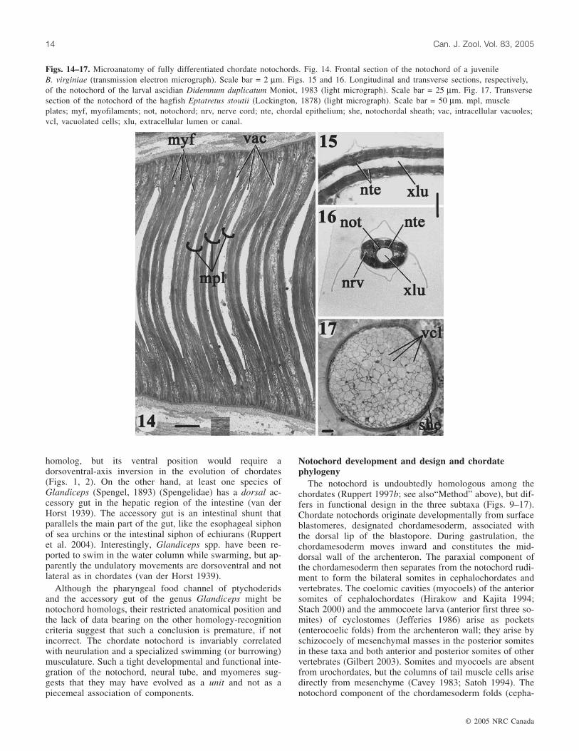
Figs. 14–17. Microanatomy of fully differentiated chordate notochords. Fig. 14. Frontal section of the notochord of a juvenileB. virginiae (transmission electron micrograph). Scale bar = 2 µm. Figs. 15 and 16. Longitudinal and transverse sections, respectively,of the notochord of the larval ascidian Didemnum duplicatum Moniot, 1983 (light micrograph). Scale bar = 25 µm. Fig. 17. Transversesection of the notochord of the hagfish Eptatretus stoutii (Lockington, 1878) (light micrograph). Scale bar = 50 µm. mpl, muscleplates; myf, myofilaments; not, notochord; nrv, nerve cord; nte, chordal epithelium; she, notochordal sheath; vac, intracellular vacuoles;vcl, vacuolated cells; xlu, extracellular lumen or canal.

lochordates, some vertebrates) or establishes itself bymesenchymal movement (some vertebrates) from the wall ofthe archenteron to form the notochord rudiment. Duringurochordate gastrulation, the presumptive chordal cells mi-grate inward and constitute the notochord rudiment directlywithout first forming the roof of the archenteron, which isabsent, except for an endodermal rudiment, from the tail ofthe larva. Although the early morphogenetic mechanismsvary among chordates, sooner or later the chordal cells be-come axially shortened and discoid and arrange themselvesmore or less in single-file, like a stack of coins, to form acylindrical rod. This stack-of-coins stage is the phylotypicstage of the chordate notochord (Figs. 9–13; Boeke 1908a,1908b; Conklin 1932; Munro and Odell 2002).
Of the three chordate subtaxa, only Cephalochordata re-tains the stack-of-coins arrangement of its notochordthroughout life (Figs. 9, 14). The notochords of vertebratesand urochordates depart from the stack-of-coins stage anddevelop taxon-specific configurations (Figs. 10–13), al-though both of these are foreshadowed in the notochordalstructure of cephalochordates.
The adult notochord of cephalochordates consists of dis-coid muscle cells, the muscle plates, stacked like coins alongthe anteroposterior axis and surrounded by a sheath of extra-cellular fibers (Figs. 9, 14). A series of small non-contractileMueller’s cells, of unknown function, form dorsal and ven-tral strips along the notochord (Ruppert 1997a). Myofibrilsarranged in one to several sarcomeres laterally traverse eachof the muscle plates and flank a large central vacuole. Ashort tail (or horn) protrudes dorsally from each muscleplate (and Mueller’s cell) to contact the extracellular matrixof the nerve cord (Flood 1970; Fig. 8). Intercellular junc-tions (zonulae adhaerentes) link the chordal cells, but onlyperipherally, near the notochordal sheath; elsewhere junc-tions are apparently absent (Fig. 9). The muscle plates aresecurely attached to the notochordal sheath by numeroushemidesmosomes. Fluid-filled extracellular spaces and chan-nels have been reported in the notochord, especially in rela-tion to the strips of Mueller’s cells (Flood 1975; Ruppert1997a), but most of these probably are fixation artifacts andshould be reinvestigated. At best, extracellular spaces mayexist as small pockets between the muscle plates (Figs. 9,22). Stiffness is achieved chiefly by contraction of thechordal muscle cells acting against the fluid-filled intra-cellular vacuoles and the non-elastic fibers in the noto-chordal sheath (Guthrie and Banks 1970).
Urochordate and vertebrate notochords transiently expressa stack-of-coins stage, but later depart from this design asthey adopt new functional organizations (Figs. 10–13, 15–17). Urochordates capitalize on the stiffness potential of anosmotically pressurized extracellular canal — the notochordlacks intracellular vacuoles. The chordal cells of the post-stack-of-coins stage of most ascidians and all appendi-cularians enclose an extracellular canal, blind at both ends,that extends the length of the notochord. The canal origi-nates as a series of extracellular pockets, one between eachpair of “coins” in the developing notochord. These pocketselongate axially and eventually fuse to form a continuous ca-nal through the stack of coins (Figs. 10, 11; Cloney 1964,1990; Burighel and Cloney 1997). Tight junctions interjoin
the chordal cells (Figs. 10, 11). All other junctions are ab-sent, including hemidesmosomes between the chordal cellsand the notochordal sheath. Osmotically pressurized extra-cellular fluid, contained by the fenestrated chordal cells andfibers in the notochordal sheath, is probably responsible fornotochordal stiffness.
Boeke (1908a, 1908b), who studied the stack-of-coinsstage in several species of fishes, described a pair of centri-oles in the center of each chordal cell (Fig. 18). In ammo-coete larva of Lampetra richardsoni Vladykov and Follett,1965, at least one of these centrioles is the basal body of arudimentary cilium that projects into a minute extracellularpocket between adjacent chordal cells (Figs. 19, 20). Follow-ing the stack-of-coins stage, the vertebrate notochord be-comes several cells thick. Later, the chord consists ofnumerous vacuolated central cells enclosed by a more or lessnon-vacuolated epithelium (Figs. 12, 13). The chordal epi-thelial cells are interjoined by tight junctions (Fig. 13) andare attached to the sheath by hemidesmosomes. Spotdesmosomes (maculae adhaerentes) interjoin the vacuolatedcells and link them to the epithelial cells. The stiffness of thevertebrate notochord results from osmotically pressurizedfluid in the intracellular vacuoles contained by a network ofintra- and extra-cellular (sheath) fibers.
Cladistic analysis of the chordate notochord indicates thephylogeny (Cephalochordata (Urochordata + Vertebrata)).The sister-taxon relationship of urochordates and vertebratesis supported by two synapomorphies. First, their notochordsdifferentiate beyond the stack-of-coins stage to adopt a novelmorphology: by cell rearrangement and fenestration inurochordates and by the development of a bounding epithe-lium around a core of stratified vacuolated cells in verte-brates. Second, the notochords of both sister-taxa rely onosmotic pressurization and are enclosed in a sheath of cellsjoined together by tight junctions. A vertebrate autapo-morphy is the turgid intracellular vacuoles (extracellularspaces are absent, except developmentally), whereas theextracellular hydrostatic skeleton (intracellular vacuoles areabsent, except developmentally) is a urochordate autapo-morphy. Cephalochordates retain the plesiomorphic adultform (stack of coins), intercellular junctions (zonulaeadhaerentes), and the presence of both extracellular spaces(probably) and intracellular vacuoles. (The presence ofextracellular pockets between chordal cells at the stack-of-coins stage (Figs. 9, 10, 12) is likely to be a chordateautapomorphy.) The pre-cranial extension of the lanceletnotochord into the rostrum and the presence of muscle platesare probably both cephalochordate autapomorphies relatedto their equal head-first and tail-first burrowing performance(Ruppert et al. 2004). The possibility remains, however, thatthe muscle plates are a chordate autapomorphy, and thus acephalochordate plesiomorphy.
Hemichordate and chordate gill slits
Hemichordate gill slitsHemichordate gill slits are ciliated perforations of the
pharyngeal wall that occur chiefly in two forms (van derHorst 1939; Benito and Pardos 1997). The first is a circularpore, lacking specialized skeletal support, which opens di-
© 2005 NRC Canada
Ruppert 15

rectly from the pharynx to the exterior. Such circular poresare found in Cephalodiscus spp., developmentally in entero-pneusts, and as “esophageal pores” in some adult entero-pneusts. The second form is a dorsoventrally elongatedprimary slit that is divided into two secondary slits by auvula-like downgrowth of tissue, called a tongue bar (or sec-ondary gill bar), from the middorsal wall of the primary slit(Fig. 23). The tongue bars and the tissue between the pri-mary slits, called gill bars (or primary gill bars, or septa), areeach supported internally by a collagenous skeletal rod.Each pair of secondary gill slits is further supported by a se-ries of horizontal braces, called synapticles, that span the ad-jacent gill and tongue bars. Collectively, the skeletal rods ofthe gill and tongue bars and synapticles constitute thebranchial skeleton (Fig. 23). This complex second form ofgill slits occurs exclusively in Enteropneusta.
The simple circular pores of Cephalodiscus spp. andEnteropneusta discharge water directly from the pharynx, orvia a short ciliated duct to the exterior. In enteropneusts withcomplex gills, water discharged from the gill slits first entersan atrial (or branchial) sac before being released fromthe sac to the exterior at a so-called gill pore (Fig. 24).Typically, each pair of secondary slits is accompanied by itsown atrial sac and gill pore. Thus, enteropneust branchio-mery is reflected equally in the numbers of pharyngeal gillslits, atrial sacs, and gill pores. But in some enteropneusts,such as Stereobalanus canadensis Spengel, 1893, the sepa-rate atrial sacs on each side of the body have fused to form acommon atrium that discharges through a single exhaust ap-erture (van der Horst 1939).
Gill-slit form in hemichordates is correlated with bodysize. The smallest individuals, the 1 mm long zooids of
© 2005 NRC Canada
16 Can. J. Zool. Vol. 83, 2005
Figs. 18–22. Extracellular spaces between cells of the notochord and notochord-like structures at approximately the stack-of-coins stageof development. Figs. 19–21 are transmission electron micrographs. Fig. 18. Longitudinal section of the notochord of the dogfishSqualus acanthias L., 1758, showing axial centrioles (ctl) in each discoidal cell (from Boeke 1908a). Scale bar = 10 µm. Fig. 19.Transverse section of notochord of young (unstaged) ammocoete of Lampetra richardsoni. Note the axial centriole (ctl). Scale bar =25 µm. Fig. 20. The section adjacent to Fig. 19 at higher magnification. The axial centriole in Fig. 19 is a basal body associated witha rudimentary cilium (cil) that projects into a minute extracellular pocket (Xp). The numerals indicate adjacent cells and the paired ar-rows their adjacent membranes. Scale bar = 0.5 µm. Figs. 21 and 22. Branchiostoma virginiae. The numerals indicate adjacent cells.Scale bars = 5 µm. Fig. 21. Median longitudinal (axial) section through a stack of five discoidal cells of the notochord-like skeletal rodof an oral cirrus. A cilium and microvilli project into the extracellular pockets between adjacent cells. Fig. 22. Longitudinal section ofa notochord showing extracellular pockets into which project microvilli. cil, cilium; ctl centriole/basal body; mvi, microvilli; Xp,extracellular pocket.

Rhabdopleura, lack gill slits; the 3 mm long zooids ofCephalodiscus spp. have a single pair of circular pores; andthe enteropneusts, which range in size from centimetres tometres, include species with gill slits with rudimentarytongue bars at the small end of the range (Protoglossus) tothose with gill slits with full-blown tongue bars among thelarge-bodied species (van der Horst 1939). Among largerspecies, the dorsoventral elongation of each primary slit, itsdivision into two secondary slits by the tongue bar, and thehorizontal partition of each secondary slit by severalsynapticles vastly increase the number of perforations in thepharyngeal wall and the area of tissue surrounding them forgas exchange and, in some species, for suspension feeding(Burdon-Jones 1962; Cameron 2002).
Hemichordate and cephalochordate gill slits: homologsor homoplasies?
The striking resemblance of enteropneust and cephalo-chordate gill slits (Figs. 23–27) has long been thought toindicate homology (Bateson 1886; Hyman 1959). Thesesimilarities include the following: slits develop from circularor oval perforations in the wall of the endodermal pharynx;later, each of the dorsoventrally elongating primary slits is
divided into two secondary slits by the downgrowth of atongue bar; each gill and tongue bar is an axial fold of thepharynx lining, thus the endodermal epithelium of each baris doubled and the basement membrane is sandwiched be-tween the two cell layers; gill and tongue bars are supportedinternally by hairpin-like collagenous skeletal rods (in thebasement membrane); synapticles bridge and support adja-cent gill and tongue bars; cilia are functionally specialized asfrontal particle-conducting and lateral water-pumping tracts;blood vessels occur in the basement membrane (stromal sep-tum) of the gill and tongue bars; blood flow through the gillis from ventral to dorsal; podocytes on the lining of thetrunk coelom are associated with the tongue bars; and nervesoccur in the gill and tongue bars.
Although the resemblances between enteropneust andlancelet gill slits seem to provide overwhelming support forhomology, the differences between them call homology intoquestion. First, enteropneust gill and tongue bars are coveredby multiciliated cells (ca. 30 cilia/cell; Benito and Pardos1997), whereas each lancelet cell bears only one cilium(Ruppert 1997a). In general, multiciliated cells arise devel-opmentally and phylogenetically from monociliated cellsand not vice versa (Rieger 1976; Ax 1995). Second, a tubu-
© 2005 NRC Canada
Ruppert 17
Figs. 23–27. Comparative microanatomy of enteropneust and cephalochordate gills. The collagenous skeleton is lightly shaded;coelomic cavities are darkly shaded; hemal (blood) vessels are black; mucus-secreting cells have mottled shading. Figs. 23 and 24.Enteropneusta. Fig. 23. En face view of a primary gill slit (Sl) divided lengthwise by a tongue bar (Tb) and horizontally by asynapticle. Fig. 24. Horizontal section through the pharyngeal and body wall (transverse section of gill and tongue bars). The arrow in-dicates the direction of water flow. Figs. 25–27. Cephalochordata. Fig. 25. En face view of primary gill slit, as in Fig. 23. Fig. 26.Horizontal section through the pharyngeal wall, atrium (At), and atrial epithelium. The arrow indicates the direction of water flow.Fig. 27. Dorsal ends of the gill bars (Gb) and tongue bars (Tb) and associated structures. At, atrium or atrial sac; Bv, hemal or bloodvessel; Co, coelom; Gb, primary or gill bar; Gp, gill pore; Mc, mucus-secreting cells; Nd, nephridioduct (leads into the atrium); Nv,neurites; Pd, podocytes and cyrtopodocytes; Sk, collagenous skeleton; Sl, gill slit; Sn, synapticle; Tb, secondary or tongue bar.

© 2005 NRC Canada
18 Can. J. Zool. Vol. 83, 2005
lar extension of the trunk coelom extends into and along thelength of each gill bar in lancelets (Fig. 26) but each tonguebar in enteropneusts (Fig. 24). This discrepancy suggests thatthe gill and tongue bars are independently derived in the twotaxa. Third, enteropneust synapticles occur on the atrial sideof the gill and tongue bars and lack blood vessels (Fig. 24),whereas lancelet synapticles are on the side of the pharynx lu-men and contain blood vessels (Fig. 26). This suggests the in-dependent evolution of synapticles in the two taxa. Fourth,conspicuous mucus-secreting cells occur on the frontal sur-face of enteropneust gill and tongue bars (Fig. 24) but at theatrial (abfrontal) surface of lancelet bars (Fig. 26).
Despite the impressive similarity of enteropneust andcephalochordate gill slits, it is difficult to bridge the subtle butreal differences between them. Most of these differences dis-appear, however, at the level of the circular primary gill slit,expressed developmentally in enteropneusts and ascidians(Garstang 1929) and in the adults of Cephalodiscus spp.,some enteropneusts (as esophageal pores), and appendi-cularians. The gill slits of Cephalodiscus spp. lack tonguebars, synapticles, skeletal rods, and blood vessels, but aremonociliated like the gill slits (primary and secondary) ofcephalochordates. Perhaps hemichordate and chordate gill slitsare homologous at the level of such a simple monociliated gillslit, but not homologous in the specialized forms that evolvedin each of their major taxa, including Urochordata (Garstang1929) and Vertebrata. This would mean, for example, that thecomplex and superficially similar secondary gill slits ofenteropneusts and lancelets are homoplasies, like bird and batwings, but the primary gill slits are homologous, like bird andbat forelimbs.
Genetic evidence for the homology of hemichordate andchordate gill slits
The common branchiomeric expression of genes in thepharynx of enteropneusts and chordates provides further sup-port for homology of their gill slits. An ortholog of the verte-brate Pax-1/Pax-9 subfamily of genes is expressedbranchiomerically in the pharynges of two enteropneust spe-cies (Ogasawara et al. 1999; Okai et al. 2000; Lowe et al.2003), two species of Urochordata (Ogasawara et al. 1999),and one species of Cephalochordata (as AmphiPax1; Hollandand Holland 1995). Expression occurs developmentally in theendoderm around each of the two primary gill slits ofSaccoglossus kowalevskii (Agassiz, 1873) (Lowe et al. 2003),in the luminal endoderm of the gill and tongue bars of adultPtychodera flava Eschscholtz, 1825 (Ogasawara et al. 1999;Okai et al. 2000), in the luminal endoderm of the pharyngealvessels in juveniles and adults of two ascidian species(Ogasawara et al. 1999), in the luminal endoderm of embry-onic Branchiostoma floridae Hubbs, 1922 (Holland and Hol-land 1995), and in the pharyngeal-pouch endoderm during thedevelopment of several vertebrates (Müller et al. 1996; Peterset al. 1998). Pax-1/Pax-9 expression is more or less restrictedto endodermally derived pharyngeal structures in hemichor-dates and protochordates, but expression of the duplicatedPax-1 and Pax-9 transcripts in mouse is not only branchio-meric in the pharynx, but also metameric in the developingsomites, suggesting a link in the genetic control of branchio-meric and metameric patterning.
Another set of genes that is specific to the pharyngeal epi-thelium has been localized in hemichordates and urochordatesand suggests homology of hemichordate and chordate gills. InP. flava these six genes are designated PfG1–PfG6 (Okai etal. 2000). PfG1, which encodes a secreted extracellular pro-tein, perhaps a component of pharyngeal mucus, is expressedin the epibranchial ridge and branchiomerically in the tonguebars. PfG2–PfG6 encode C-type lectins typically found inextracellular fluids. These genes are expressed exclusively inthe epibranchial ridge. In the ascidian Halocynthia roretzi(von Drasche, 1884), similar genes (HrPhG1 and HrPhG2)are expressed in the pharyngeal endoderm (Tanaka et al.1996).
Protochordate endostyle
Functional anatomyThe endostyle of cephalo- and uro-chordates is a glandular
ciliated groove in the ventral midline of the pharynx. At itsanterior extremity, the endostyle joins two ciliated peri-pharyngeal bands that extend dorsally and typically join in thedorsal midline to form a longitudinal ciliated tract that takesdifferent names, such as dorsal lamina or epipharyngealgroove, in different taxa (Ruppert et al. 2004).
During filter feeding, the endostyle continuously secretestwo net-like sheets of mucus. One sheet is cast onto the liningof the left side of the pharynx, the other onto the lining of theright side. As each sheet is transported dorsally by frontalcilia, suspended particles are trapped on the net as waterpasses through. When the net and its embedded food reachthe dorsal midline of the pharynx, they are rolled into a cordand conveyed into the esophagus by the dorsal ciliated tract.The peripharyngeal bands also trap particles and transportthem directly to the dorsal ciliated tract.
When viewed in cross section, the endostyle is U-shaped(Fig. 28) and histologically complex. The cells at the bottomof the “U” bear very long cilia that typically extend upwardabove and beyond the arms of the “U”. The two arms of the“U” are composed of distinct bilateral zones of specializedciliated and secretory cells. This complex design is apparentlynecessary to fabricate and transport the mucous feeding nets(Holley 1986), which microscopically consist of a highly reg-ular pattern of meshes, each approximately 1 µm in diameter(Burighel and Cloney 1997).
One or two endostylar zones, typically near the extremitiesof the “U”, are responsible for the binding, concentration, andincorporation of iodine into a secreted protein component ofthe endostylar net (Fig. 29). The enzyme effecting iodinationis a peroxidase. Historically, the experimental binding of I125,as visualized by autoradiography, and peroxidase activity, asevidenced by the periodic acid – Schiff’s reaction, were keytests for the homology of protochordate endostyles and espe-cially of the endostyle and its vertebrate derivative, the thy-roid gland (Barrington 1958; Thorpe et al. 1972). In thevertebrate thyroid gland, the thyroglobulin protein is iodinatedby thyroid peroxidase (TPO) coded for by the TPO gene.TPO is regulated by thyroid transcription factor 1 (TTF-1)protein, which is coded for by the homeobox gene TTF-1(Nkx-2.1) (Mazet 2002; Ogasawara et al. 2001).

Genetic evidence for homology of protochordateendostyle and vertebrate thyroid gland
Gene-expression data, especially for TTF-1 and TPO, con-firm the homology of the protochordate endostyle and verte-brate thyroid gland (Ogasawara 2000; Ogasawara et al.2001; Mazet 2002). In all chordates, TTF-1 expression is ini-tiated in ventral cells of the anterior pharynx at an equivalentdevelopmental stage and remains up-regulated in the differ-entiated endostyle and thyroid. In amphioxus, TTF-1 tran-scripts are expressed in all six zones of the endostyle,whereas in the ascidian and ammocoete endostyles, expres-sion is limited to specific zones. The restricted pattern ofTTF-1 expression in urochordates and vertebrates may be asynapomorphy of these taxa, suggesting the relationship(Cephalochordata (Urochordata + Vertebrata)). TPO tran-scripts are also expressed in all protochordate endostyles andvertebrate thyroids. In the enteropneust P. flava, TTF-1 ex-pression is not restricted to any specific pharyngeal struc-ture, but occurs throughout its endodermal lining (Takacs etal. 2002).
An enteropneust homolog of the chordate endostyle?Although enteropneusts are generally regarded as deposit
feeders, some species, such as Balanoglossus gigas Fr. Müller(Spengel, 1893) (Burdon-Jones 1962), Harrimania plankto-philus (Cameron 2002), and Schizocardium brasilienseSpengel, 1893 (E.E. Ruppert, unpublished data) are at leastfacultative pharyngeal suspension feeders. Particles sus-pended in the inhalant water stream are trapped in mucusand transported ventrally across the pharyngeal lining bycilia. On reaching the ventral midline, the mucus-entrappedparticles are gathered into a string (S. brasiliense, H. plankto-
philus) on the hypobranchial ridge or they enter the foodchannel of the pharynx (B. gigas). Both the hypobranchialridge and the food channel transport particles posteriorlyfrom the pharynx into the esophagus.
The general pattern of particle transport in the entero-pneust pharynx is similar to that in protochordates, but in-verted (Figs. 1, 2). In enteropneusts, particle movement isfrom dorsal to ventral and then posteriorly along the ventralmidline. In protochordates, particles move ventral to dorsaland then posteriorly along the dorsal midline. So if theenteropneust ventral midline (hypobranchial ridge, foodchannel) corresponds functionally to the protochordate dor-sal midline (dorsal lamina, epipharyngeal groove, etc.), thenthe protochordate ventral midline structure (endostyle)should correspond to the enteropneust dorsal midline struc-ture (epibranchial ridge) if the two suspension-feedingpharynges are homologs and not convergently similarhomoplasies. Although an elevated ridge and not a groove,the ciliated epibranchial ridge, like the endostyle, consists ofbilateral zones of specialized secretory cells (Ruppert et al.1999), but does it bind iodine and does it secrete mucousnets?
The results of our I125-binding experiments, using theB. floridae endostyle as a positive control, are shown inFigs. 28–33. The epibranchial ridges of S. kowalevskii (notshown) and Balanoglossus aurantiacus (Girard, 1853)(Figs. 30, 31) both bind iodine, but binding is not restrictedto the epibranchial ridge. Instead, iodine binding occursthroughout the lining of the enteropneust pharynx (Figs. 32,33), unlike in B. floridae, in which it is limited to a zone ofthe endostyle (Fig. 29). These results neither support norreject the homology of the epibranchial ridge and the
© 2005 NRC Canada
Ruppert 19
Figs. 28–31. Iodine binding by the cephalochordate endostyle and enteropneust epibranchial ridge (transverse sections). Figs. 28 and29. Experimental sections of Branchiostoma floridae were incubated for 4.5 h in 0.001 mCi/mL (1 Ci = 37 Gbq) I125 and then exposedto autoradiographic emulsion for 70 days before development, showing control and experimental results, respectively, for the endostyle.The restricted binding shown here is typical for amphioxus and chordate endostyles. Scale bar = 50 µm. Figs. 30 and 31. Sections ofB. aurantiacus incubated 20 h in 0.01 mCi/mL I125 and then exposed to emulsion for 70 days, showing control and experimental re-sults, respectively, for the epibranchial ridge (dorsoventrally inverted). Scale bar = 50 µm.

endostyle. It is possible, for example, that the diffuse bind-ing of iodine in the common ancestor of Enteropneusta andChordata became restricted to the endostyle in chordates.
Observations of dissected living specimens ofS. brasiliense (E.E. Ruppert, unpublished data) show thatparticles are not trapped in mucous nets secreted by theepibranchial ridge. Rather, mucus secretion for particle cap-ture occurs across the entire lining of the pharynx, as indi-cated by the well-developed mucous-gland cells on theupstream luminal surfaces of the gill and tongue bars(Fig. 24) as well as the epibranchial ridge (Ruppert et al.1999). Mucus release, moreover, does not appear to beglobal or continuous, but local and discontinuous, the resultof impingement of particles on a specific area of the pharyn-geal lining. Like the iodine-binding data, these observationsdo not bear critically on the question of homology versushomoplasy. The ancestral deuterostome may have had asuspension-feeding pharynx, as Cameron (2002) has sug-gested, but mucus secretion for particle trapping was gener-alized, only later becoming concentrated and organized bythe specialized chordate endostyle.
The results described in this and the preceding sectionsuggest a generalized distribution of endostylar traitsthroughout the enteropneust pharynx. Although TTF-1 ex-pression is endostyle/thyroid-limited in chordates, it occurspharynx-wide in enteropneusts; iodine binding and mucusproduction for filter feeding are restricted to the chordate
endostyle, but occur throughout the enteropneust pharynx.One interpretation of these data is that chordate endostylarfunctions are the responsibility of the entire pharyngeal lin-ing of enteropneusts. If so, then the endostyle may haveevolved by restriction of these functions to a specializedmidline structure. Nevertheless, an endostyle, per se, is ab-sent from Enteropneusta.
An alternative hypothesis for chordateevolution
The foregoing homology analysis suggests a phylogeny ofChordata in which Urochordata and Vertebrata are sister-taxa and Cephalochordata is the plesiomorphic clade, inagreement with the hypothesis of Jefferies (1997). A phylog-eny of Deuterostomia that supports the Urochordata +Vertebrata hypothesis is shown in Fig. 34. On the otherhand, most cladistic analyses of Chordata, whether morpho-logical, molecular, or both, support a Cephalochordata +Vertebrata sister-taxon relationship (Schaeffer 1987; Wadaand Satoh 1994; Cameron et al. 2000; Ax 2001; Swalla2001).
The Urochordata + Vertebrata hypothesis rests on a signif-icant assumption and is supported by several synapomor-phies. The assumption is that metamerism is a chordate traitthat has been apomorphically lost (along with many otherancestral chordate traits; Fig. 34) in urochordates. If this iscorrect, then Urochordata and its subtaxa exhibit a derivedsimplification of the chordate body design. This can betested by comparing the number of synapomorphies unitingUrochordata and Vertebrata with the number unitingCephalochordata and Vertebrata. Synapomorphies in supportof the Urochordata + Vertebrata hypothesis are listed inFig. 34. Compared with the 13 synapomorphies uniting Uro-chordata and Vertebrata, how many, other than a sharedmetamerism, can be marshaled for Cephalochordata +Vertebrata? Such additional Cephalochordata + Vertebratasynapomorphies might include the occurrence of a pre-oralhood in Cephalochordata and Vertebrata, assuming that theammocoete pre-oral hood is actually homologous with thatof amphioxus, or the presence of a hepatic portal system inCephalochordata and Vertebrata (lost in the highly derivedurochordate hemal system?).
The answer to the question of whether urochordates areprimitive (Cephalochordata + Vertebrata hypothesis) or de-rived (Urochordata + Vertebrata hypothesis) chordates maynot be found in urochordates themselves because of theirsimplified body design and genome (Holland and Gibson-Brown 2003), but rather in a comparison of enteropneustswith Cephalochordata and Vertebrata. For example, thebranchiomeric podocyte clusters of enteropneusts (Benito andPardos 1997), the branchiomeric nephridia of amphioxus(Ruppert 1997a), and, developmentally, the metamericnephrons of vertebrates are likely to be homologous struc-tures. This hypothesis for homology begs a comparison ofthe genetic control network or networks responsible forbranchiomeric and metameric patterning. If indeed thesecontrols are related, then it is likely that the absence inurochordates of metamery and also any regular branchio-mery (as well as filtration nephridia) is a derived loss andnot a primitive chordate character.
© 2005 NRC Canada
20 Can. J. Zool. Vol. 83, 2005
Figs. 32 and 33. Iodine binding by the enteropneust(B. aurantiacus) pharynx. The experimental protocol was asdescribed for Figs. 28–31. Fig. 32. Unstained control section.Fig. 33. Experimental section, corresponding to Fig. 32, showinggeneral binding of iodine by pharyngeal tissues. Scale bar = 50 µm.

© 2005 NRC Canada
Ruppert 21
Fig. 34. A phylogeny of Deuterostomia based chiefly on morphology. N.N. is nomen nominandum (“new name”). Boxed numbers areapomorphies. 1, Deuterostomia: the blastopore becomes the anus; epithelial folding (plesiomorphy?) including enterocoely; branchiomery:undivided (primary) gill slits (as circular or oval pores) lack a skeleton, branchiomeric podocytes; pre-oral heart–kidney with bilateralnephridioducts; hepatic cecum (or ceca?) from anterior intestine. (Plesiomorphies include monolayered, monociliated epithelial tissues;intercellular junctions are zonula adhaerens and septate junctions; connective-tissue cells restricted to fibroblasts, sclerocytes, and non-circulating hemocytes; principal hemal vessels dorsal and ventral, flow anterior in dorsal vessel, posterior in ventral vessel; trimery; mus-culature composed of epitheliomuscle cells in monolayered myoepithelium; motorneurons confined to epidermal nerve net; motorinnervation by diffusion of transmitters across epidermal (neuroepithelium) basement membrane to somatic coelomic lining (myoepithe-lium); metanephridial system.) 2, N. N.: valved mesocoel ducts (collar ducts Enteropneusta; stone canal Echinodermata). 3, Hemichordata:stomochord; post-anal appendage (tail); dorsal anus (developmentally in Enteropneusta). 4, Echinodermata: water-vascular system; calciticstereom ossicles in connective-tissue dermis; mutable connective tissue; (gill slits/branchiomery lost in extant taxa but present in extinctCothurnocystis). 5, Chordata: segmented (metameric) mesoderm (primitively in register with branchiomeres?); notochord arising develop-mentally from two rows of chordal cells that intercalate to form a stack of coins; chordal cells with intracellular vacuoles and intercellularpockets; dorsal hollow nerve cord, including anterior brain with photoreceptive and static sensory structures; non-migratory neural crest;myomeral motorneurons confined to dorsal cord; adenohypophysis consisting of ectoderm + mesoderm; pharyngeal mucous net filter feed-ers with endostyle; dorsoventral axis inversion or reversal of blood-flow direction; pre-oral heart lost, new post-oral ventral heart; newventral anus?; fins; swimming by means of lateral undulations of trunk tail; Pax-1 expression confined to pharynx (endoderm). 6,Cephalochordata: notochord to rostral extremity; chervron-shaped myosepta; left–right myomere pairs out of register; bilateral asymmetryin most tissues and organs; primary gill slits divided by tongue bars; branchiomeric cyrtopodocytes and nephridoducts; muscular stack-of-coins notochord with myoglobin. 7, N.N.: loss of pre-oral kidney; septate junctions replaced by tight junctions; swimming musculaturepartly a functional syncytium; brain with coronet cells (saccus vasculosus); neuromast cells; mesodermal mesenchyme forms novel struc-tures; migratory neural crest (Jeffery et al. 2003); non-epithelial musculature; notochord differentiates beyond stack-of-coins stage; hemalsystem with functionally distinct circulating corpuscles; multiciliated epithelial cells; adenohypophysis consisting of ectoderm + endoderm;Pax-1/9 (Urochordata) or Pax-1 Pax-9 (Vertebrata) expression in developing pharynx and musculature (somites). 8, Urochordata: loss ofmetamerism, coelom (including enterocoelic somites), filtration nephridia (metanephridial system), gut/anus in locomotory “tail”, manyancestral chordate genes (most adults also lose trunk/tail, including notochord, nerve cord, brain, locomotory musculature); non-moltedexoskeletal tunic (apomorphically molted in doliolarians and appendicularians); budding, involving mesenchymal mesoderm, producescolonies (deuterostome plesiomorphy?); periodic heartbeat reversal; post-stack-of-coins notochord with enclosed extracellular canal,intracellular vacuoles absent; 9, Vertebrata: stratified epithelia; endothelium-lined hemal vessels; cephalized brain with paired sense or-gans; processes of motorneurons extend from nerve cord, via ventral roots, to innervate myomeral musculature; metameric nephrons(metanephridial system) join common urinary duct; post-stack-of-coins notochord composed of stratified vacuolated cells enclosed in abounding epithelium; cartilaginous gill skeleton (arches) derived from neural crest; many duplicated ancestral chordate genes.

© 2005 NRC Canada
22 Can. J. Zool. Vol. 83, 2005
Acknowledgments
Thanks to Prof. Jennifer Frick-Ruppert, who performedthe I125-binding experiments on enteropneusts and amphi-oxus, Prof. John P. Wourms, who provided useful literatureand discussion, as well as an anonymous reviewer whosecritique improved the paper.
References
Ax, P. 1995. Multicellular animals: a new approach to the phylo-genetic order in nature. Vol. 1. Springer-Verlag, Berlin and othercities.
Ax, P. 2001. Das System der Metazoa III : ein Lehrbuch derphylogenetischen Systematik. Spektrum Akademischer Verlag,Heidelberg, Germany.
Balser, E.J., and Ruppert, E.E. 1990. Structure, ultrastructure, andfunction of the pre-oral heart–kidney in Saccoglossus kowal-evskii (Hemichordata, Enteropneusta) including new data on thestomochord. Acta Zool. (Stockh.), 71: 235–249.
Barrington, E.J.W. 1958. The localization of organically bound io-dine in the endostyle of amphioxus. J. Mar. Biol. Assoc. U.K.37: 117–126.
Bateson, W. 1886. The ancestry of the Chordata. Q. J. Microsc.Sci. 26: 535–571.
Benito, J., and Pardos, F. 1997. Hemichordata. In Microscopicanatomy of invertebrates. Vol. 15. Hemichordata, Chaetognathaand the invertebrate chordates. Edited by F.W. Harrison and E.E.Ruppert. Wiley–Liss, New York. pp. 15–101.
Boeke, J. 1908a. Das “Geldrollenstadium” der Vertebraten-Chordaund des Skelettes der Mundcirren von Branchiostoma lanceo-latum und seine cytomechanische Bedeutung. Anat. Anz. 33:541–556.
Boeke, J. 1908b. Das “Geldrollenstadium” der Vertebraten-Chordaund des Skelettes der Mundcirren von Branchiostoma lanceo-latum und seine cytomechanische Bedeutung. Anat. Anz. 33:574–580.
Burdon-Jones, C. 1962. The feeding mechanism of Balanoglossusgigas. Bol. Fac. Filos. Cienc. Let. Univ. Sao Paulo Ser. Zool.No. 261. Vol. 24. pp. 255–280.
Burighel, P., and Cloney, R.A. 1997. Urochordata: Ascidiacea. InMicroscopic anatomy of invertebrates. Vol. 15. Hemichordata,Chaetognatha and the invertebrate chordates. Edited by F.W.Harrison and E.E. Ruppert. Wiley–Liss, New York. pp. 221–347.
Cameron, C.B. 2002. Particle retention and flow in the pharynx ofthe enteropneust worm Harrimania planktophilus: the filter-feeding pharynx may have evolved before the chordates. Biol.Bull. (Woods Hole), 202: 192–200.
Cameron, C.B., and Mackie, G.O. 1996. Conduction pathways inthe nervous system of Saccoglossus sp. (Enteropneusta). Can. J.Zool. 74: 15–19.
Cameron, C.B., Garey, J.R., and Swalla, B.J. 2000. Evolution ofthe chordate body plan: new insights from phylogenetic analysesof deuterostome phyla. Proc. Natl. Acad. Sci. U.S.A. 97: 4469–4474.
Cavey, M.J. 1983. Ultrastructure and differentiation of ascidianmuscle. II. Differentiation of the caudal muscle cells in the larvaof Diplosoma macdonaldi. Cell Tissue Res. 230: 77–94.
Chen, J.Y., Dzik, J., Edgecombe, G.D., Ramsköld, L., and Zhu,G.Q. 1995. A possible early Cambrian chordate. Nature (Lond.),377: 72–722.
Chen, J.Y., Huang, D.Y., and Li, C.W. 1999. An early Cambriancraniate-like chordate. Nature (Lond.), 402: 518–522.
Cloney, R.A. 1964. Development of the ascidian notochord. ActaEmbryol. Morphol. Exp. 7: 111–130.
Cloney, R.A. 1990. Urochordata: Ascidiacea. In Reproduction biol-ogy of invertebrates. Vol. IVB. Fertilization, development, andparental care. Edited by K.G. Adiyodi and R.G. Adiyodi. Ox-ford, New Delhi. pp. 391–451.
Conklin, E.G. 1932. The embryology of amphioxus. J. Morphol.54: 69–151.
Dewel, R.A. 2000. Colonial origin for Eumetazoa: major morpho-logical transitions and the origin of bilaterian complexity. J.Morphol. 243: 35–74.
Dilly, P.N., Welsch, U., and Storch, V. 1970. The structure of thenerve fibre layer and neurocord in the enteropneusts. Z.Zellforsch. Mikrosk. Anat. 103: 129–148.
Flood, P.R. 1970. The connection between spinal cord andnotochord in amphioxus (Branchiostoma lanceolatum). Z.Zellforsch. Mikrosk. Anat. 103: 115–128.
Flood, P.R. 1975. Fine structure of the notochord of amphioxus.Symp. Zool. Soc. Lond. No. 36. pp. 81–104.
Florey, E., and Cahill, M.A. 1977. Ultrastructure of sea urchin tubefeet: evidence for connective tissue involvement in motor con-trol. Cell Tissue Res. 177: 195–214.
Garstang, W. 1929. The morphology of Tunicata, and its bearingson the phylogeny of the Chordata. Q. J. Microsc. Sci. 72: 51–187.
Gilbert, S.F. 2003. Developmental biology. 7th ed. Sinauer Associ-ates, Inc., Sunderland, Mass.
Guthrie, D.M., and Banks, J.R. 1970. Observations on the functionand physiological properties of a fast paramyosin muscle — thenotochord of amphioxus (Branchiostoma lanceolatum). J. Exp.Biol. 52: 125–138.
Hirakow, R., and Kajita, N. 1994. Electron microscopic study ofthe development of amphioxus, Branchiostoma belcheri tsingt-auense: the neurula and larva. Acta Anat. Nippon. 69: 1–12.
Holland, L.Z., and Gibson-Brown, J.J. 2003. The Ciona intestinalisgenome: when the constraints are off. Bioessays, 25: 529–532.
Holland, N.D., and Holland, L.Z. 1995. An amphioxus Pax gene,AmphiPax-1, expressed in embryonic endoderm, but not inmesoderm: implications for the evolution of class I paired boxgenes. Mol. Mar. Biol. Biotech. 4: 206–214.
Holley, M.C. 1986. Cell shape, spatial patterns of cilia, and mucus-net construction in the ascidian endostyle. Tissue Cell, 18: 667–685.
Horst, C.J. van der. 1939. Hemichordata. Bronn’s Klass. Ordn.Tierreichs, 4: 1–737.
Hyman, L.H. 1959. The invertebrates: smaller coelomate groups.McGraw–Hill Book Co., Inc., New York, London, and Toronto.
Jefferies, R.P.S. 1986. The ancestry of the vertebrates. British Mu-seum (Natural History), London.
Jefferies, R.P.S. 1997. A defence of the calcichordates. Lethaia, 30:1–10.
Jeffery, W.R., Strickler, A.G., and Yamamoto, Y. 2004. Migratorycrest-like cells form body pigmentation in a urochordate em-bryo. Nature (Lond.), 431: 696–699.
Knight-Jones, E.W. 1952. On the nervous system of Saccoglossuscambrensis (Enteropneusta). Philos. Trans. R. Soc. Lond. B.Biol. Sci. 236: 315–354.
Lacalli, T.C. 2005. Protochordate body plan and the evolutionaryrole of larvae: old controversies resolved? Can. J. Zool. 83(1):216–224.
Lacalli, T.C., and Kelly, S.J. 1999. Somatic motoneurones inamphioxus larvae: cell types, cell position, and innervation pat-terns. Acta Zool. (Stockh.), 80: 113–124.
Lowe, C.J., Wu, M., Salic, A., Evans, L., Lander, E., Stange-

© 2005 NRC Canada
Ruppert 23
Thomann, N., et al. 2003. Anteroposterior patterning inhemichordates and the origins of the chordate nervous system.Cell, 113: 853–865.
Mazet, F. 2002. The Fox and the thyroid: the amphioxus perspec-tive. Bioessays, 24: 696–699.
Meinertzhagen, I.A., and Okamura, Y. 2001. The larval ascidiannervous system: the chordate brain from its small beginnings.Trends Neurosci. 24: 401–410.
Morgan, T.H. 1891. The growth and metamorphosis of tornaria. J.Morphol. 5: 407–458.
Müller, T.S., Ebensperger, C., Neubüser, A., Koseki, H., Balling,R., Christ, B., and Wilting, J. 1996. Expression of avian Pax1and Pax9 is intrinsically regulated in the pharyngeal endoderm,but depends on environmental influences in the paraxial meso-derm. Dev. Biol. 178: 403–417.
Munro, E.M., and Odell, G. 2002. Morphogenetic pattern forma-tion during ascidian notochord formation is regulative andhighly robust. Development (Camb.), 129: 1–12.
Nielsen, C. 2001. Animal evolution. 2nd ed. Oxford UniversityPress, Oxford, New York, and other cities.
Novoselov, V.V. 1995. Notochord formation in amphibians: two di-rections and two ways. J. Exp. Zool. 271: 296–306.
Nørrevang, A. 1965. Fine structure of nervous layer, basementmembrane, and muscles of the proboscis in Harrimania kupfferi(Enteropneusta). Vidensk. Medd. Dan. Naturhist. Foren. 128:325–337 + plates.
Ogasawara, M. 2000. Overlapping expression of amphioxus homo-logs of the thyroid transcription factor-1 gene and thyroidperoxidase gene in the endostyle: insight into evolution of thethyroid gland. Dev. Genes Evol. 210: 231–242.
Ogasawara, M., Wada, H., Peters, H., and Satoh, N. 1999. Devel-opmental expression of Pax 1/9 genes in urochordate andhemichordate gills: insight into function and evolution of thepharyngeal epithelium. Development (Camb.), 126: 2539–2550.
Ogasawara, M., Shigetani, Y., Suzuki, S., Kuratani, S., and Satoh,N. 2001. Expression of thyroid transcription factor (TTF-1)gene in the ventral forebrain and endostyle of the agnathan ver-tebrate, Lampetra japonica. Genesis, 30: 51–58.
Okai, N., Tagawa, K., Humphreys, T., Satoh, N., and Ogasawara,M. 2000. Characterization of gill-specific genes of the acornworm Ptychodera flava. Dev. Dyn. 217: 309–319.
Peters, H., Neubüser, A., Kratochwil, K., and Balling, R. 1998.Pax9-deficient mice lack pharyngeal pouch derivatives and teethand exhibit craniofacial and limb abnormalities. Genes Dev. 12:2735–2747.
Peterson, K.J., and Eernisse, D.J. 2001. Animal phylogeny and theancestry of bilaterians: inferences from morphology and 18SrRNA gene sequences. Evol. Dev. 3: 170–205.
Peterson, K.J., Cameron, R.A., Tagawa, K., Satoh, N., andDavidson, E.H. 1999. A comparative molecular approach tomesodermal patterning in basal deuterostomes: the expressionpattern of Brachyury in the enteropneust hemichordatePtychodera flava. Development (Camb.), 126: 85–95.
Remane, A. 1956. Die Grundlagen des natürlichen Systems, dervergleichenden Anatomie und der Phylogenetik. AkademischeVerlagsgesellschaft Geest & Portig K.G., Leipzig, Germany.
Rieger, R.M. 1976. Monociliated epidermal cells in Gastrotricha:significance for concepts of early metazoan evolution. Z. Zool.Syst. Evol. Forsch. 14: 198–226.
Rieger, R.M., and Tyler, S. 1979. The homology theorem in ultra-structural research. Am. Zool. 19: 655–664.
Ruppert, E.E. 1982. Homology recognition as a basis for compara-tive biology. In Proceedings of the Third International Sympo-sium on the Tardigrada, Johnson City, Tennessee, 3–6 August
1980. Edited by R.P. Higgins and D.R. Nelson. East TennesseeUniversity Press, Johnson City. pp. 31–54.
Ruppert, E.E. 1997a. Cephalochordata (Acrania). In Microscopicanatomy of invertebrates. Vol. 15. Hemichordata, Chaetognathaand the invertebrate chordates. Edited by F.W. Harrison and E.E.Ruppert. Wiley–Liss, New York. pp. 349–504.
Ruppert, E.E. 1997b. Introduction: microscopic anatomy of thenotochord, heterochrony, and chordate evolution. In Microscopicanatomy of invertebrates. Vol. 15. Hemichordata, Chaetognathaand the invertebrate chordates. Edited by F.W. Harrison and E.E.Ruppert. Wiley–Liss, New York. pp. 1–13.
Ruppert, E.E., and Balser, E.J. 1986. Nephridia in the larvae ofhemichordates and echinoderms. Biol. Bull. (Woods Hole), 171:188–196.
Ruppert, E.E., Cameron, C.B., and Frick, J.E. 1999. Endostyle-likefeatures of the dorsal epibranchial ridge of an enteropneust andthe hypothesis of dorsal-ventral axis inversion in chordates.Invertebr. Biol. 118: 202–212.
Ruppert, E.E., Fox, R.S., and Barnes, R.D. 2004. Invertebrate zool-ogy: a functional evolutionary approach. 7th ed. Brooks/Cole,Thomson Learning Inc., Belmont, Calif.
Satoh, N. 1994. Developmental biology of ascidians. CambrigeUniversity Press, New York and other cities.
Schaeffer, B. 1987. Deuterostome monophyly and phylogeny. Evol.Biol. 21: 179–235.
Shu, D.G., Conway Morris, S., and Zhang, X.L. 1996. A Pikaia-like chordate from the Lower Cambrian of China. Nature(Lond.), 384: 157–158.
Stach, T. 1999. The ontogeny of the notochord of Branchiostomalanceolatum. Acta Zool. (Stockh.), 80: 25–33.
Stach, T. 2000. Microscopic anatomy of developmental stages ofBranchiostoma lanceolatum (Cephalochordata, Chordata). Bonn.Zool. Monogr. No. 47.
Swalla, B.J. 2001. Phylogeny of the urochordates: implications forchordate evolution. In The biology of ascidians. Edited byH. Sawada, H. Yokosawa, and C.C. Lambert. Springer-Verlag,Tokyo. pp. 219–224.
Takacs, C.M., Moy, V.N., and Peterson, K.J. 2002. Testing putativehomologues of the chordate dorsal nervous system andendostyle: expression of NK2.1 (TTF-1) in the acorn wormPtychodera flava (Hemichordata, Ptychoderidae). Evol. Dev. 4:405–417.
Tanaka, K.J., Ogasawara, M., Makabe, K.W., and Satoh, N. 1996.Expression of pharyngeal gill-specific genes in the ascidianHalocynthia roretzi. Dev. Genes Evol. 206: 218–226.
Thorpe, A., Thorndyke, M.C., and Barrington, E.J.W. 1972. Ultra-structural and histochemical features of the endostyle of theascidian Ciona intestinalis with special reference to the distribu-tion of bound iodine. Gen. Comp. Endocrinol. 19: 559–571.
Turbeville, J.M., Schulz, J.R., and Raff, R.A. 1994. Deuterostomephylogeny and the sister group of the chordates: evidence frommolecules and morphology. Mol. Biol. Evol. 11: 648–655.
Wada, H., and Satoh, N. 1994. Details of the evolutionary historyfrom invertebrates to vertebrates, as deduced from sequences of18S rDNA. Proc. Natl. Acad. Sci. U.S.A. 91: 1801–1804.
Welsch, U., and Storch, V. 1970. Fine structure of the stomochordof the Enteropneusta, Harrimania kupfferi and Ptychodera flava.Z. Zellforsch. 107: 234–239.
Welsch, U., Dilly, P.N., and Rehkämper, G. 1987. Fine structure ofthe stomochord in Cephalodiscus gracilis. Zool. Anz. 218: 209–218.
Wilke, U. 1972. Der Eicheldarm der Enteropneusten als Stutzorganfür Glomerulus und Perikardialvesikel. Verh. Dtsch. Zool. Ges.66: 93–96.