judiono-2012-b015_published ijfnph.pdf
TRANSCRIPT
International Journal of Food, Nutrition & Public HealthVol. 5 No. 1/2, 2012
The current issue and full text archive of this journal is available at http://www.worldsustainable.org
IJFNPH5,1/2
7
Copyright © 2012 WASD
1Senior Lecturer in Nutrition Department, The Health Polytechnic of Bandung, Min-istry of Health Republic Indonesia, Jl. Babakan Loa, Kelurahan Pasirkaliki, Municipal-ity of Cimahi – West Java, Indonesia 40514 E-mail: [email protected] of University of Diponegoro Semarang - Indonesia, Doctoral Programme Medical Faculty.
Judiono1
The Health Polytechnic of Bandung, Indonesia
Djokomoeljanto2 and Hadisaputro. S2
University of Diponegoro Semarang, Indonesia
Abstract: Purpose and methodology: This study investigated the effect of plain ke-
fir on glycemic, antioxidants, immune response and pancreatic β cell regenera-
tion of hyperglycemia Wistar Rats induced by Streptozotocin.
Findings: Kefir supplementation 3.6 cc / day affect significantly on blood glu-
cose, antioxidants, lipid peroxidation, and pancreatic β-cells. Statistical analysis
showed reduce of glucose (p<0.001), MDA (p<0.001), level of proinflamatory
cytokines (IL1, IL
6,) (p<0.001), except of controls. Antioxidant showed increase
of catalase, GPx (p<0.001) and SOD (p <0.05). Similarly, there was increased
of IL10
(p<0.05) and the normal cells pancreatic (p <0.001), except of control.
TNFα reduced no significant (p> 0.05), except of control. Ancova test showed
MDA and IL10
were the most contributed to the pancreatic β cells regeneration
by 91.0% and 9% determined by TNF-α, antioxidants, blood glucose, body
weight.
Value: Kefir is significantly reduced of glucose, lipid peroxide, level of cytokines
( IL1, IL6) and enhanced IL10, antioxidants capacity and normal pancreatic β
cell expression. Insulin and kefir descriptively reduced TNF α level.
Keywords: Probiotic; Plain Kefir; Hyperglicemia; β Cells Regeneration; Proinflama-
tory Cytokines
BIOMOLECULAR ASPECTS OF PLAIN KEFIR ANTIDIABETIC
POTENTIALS

Biomolecular Aspects of Plain
Kefir Antidiabetic Potentials
8
INTRODUCTION
Diabetes mellitus type 2 (DMT2) is a metabolic disorder char-acterized by syndrome hyperglycemia. This disease is pandem-ic and WHO predicts of people from 8.4 million in 2000 to 21.3 million in 2030 in Indonesia. The prevalence of type 2 diabetes increased yearly in this country. Consequently, it will be affecting on of diabetes complications, morbidity and mor-tality and a lower quality of human resources in the long term. (Hadisaputro, S, et al. 2009)
Excessive free-radicals in diabetes is due to hyperglycemia. It increases through various channels, such as: (1) non-enzymat-ic glycation (AGEs), (2) glucose auto-oxidation, and (3) polyol pathway and (4) protein kinase activation (PKC). (Betteridge, 2000 Ceriello, 2000 Pfaffly, 2001 Djokomoeljanto, 2007 Soeyono, 2007). Those mechanisms enhance the formation of free radicals and cause oxidative stress. Oxidative stress dam-age to proteins, enzymes, membrane lipids and DNA, reduce in antioxidants and immune response, increase lipid peroxida-tion and proinflammatory cytokines as well as pancreatic β cells damage. (Moussa, 2008) Brownlee (2000) found that hypergly-cemia leads to damage the function and structure of pancreatic β cells. (Brownlee, 2000)
The impairment of immune response due to the chron-ic hyperglycemia triggers inflammation through activation of Toll Like Recepor (TLRs) 2 and 4, leading to an increase of proinflammatory cytokines secreation, namely; interleukin-1 (IL1), interleukin-6 (IL
6), tumor necrosis factor alpha (TNFα),
interferon γ (IFNγ), whereas interleukin-10 (IL10
) is decreased. (Mahardhika, et al. 2004 Rytter, et al. 2009) TNFα reduces the glucose transport (GLUT) 1 and 4 in cell and effect de-creases the glucose uptake. Proinflammatory cytokines inhib-it insulin signal by activating receptor kinase inhibitor, NFkB

IJFNPH5,1/2
9
to finnaly induces pancreatic β cell damage to pancreatic β cells apoptosis. (Lee and Simin Liu, 2008)
Diabetes therapy has been demontrated to unabale in achieving maximally blood glucose controlled by the avail-ability of current therapy approaches, such as: (1) changes in behavior therapy, (2) diet, (3) exercise, (4) oral hypoglycemic medication and insulin. (PERKENI,2007.a) The use of insulin and medication are the most effective options, but it is diffi-cult to implement on an ongoing basis, as they relate to socio-economic, level of knowledge and understanding of residual effects after taking the medicine. Attempts on the induction of pancreatic β cell regeneration (Meier, 2008) by using pro-biotics are not reported elsewhere, but the results have been conflicting in relation to the application of therapy. Further studies are therefore important to ensure the consistency of results. (Hadisaputro, et al. 2009)
Plain kefir supplementation identified potentially to reduce hyperglycemia. The mechanism underlying is proba-bly via its bioactive components such as; exopolyssacharide, peptide, antioxidant and immunomodulatory properties. (Brown,2004 Sybesma,2004 Khazrai,2004 Virtanen,2004) Exsopolyssacharide (EPS) activates the hormone glucagon like peptide 1 (GLP 1), gastric inhibitory peptide (GIP) (Khan, 2001) and the enzyme adenylate cyclase through the cyclic ad-enosine monoposfat (cAMP), sensitization of Ca2 ions and activation of protein kinase A, thus it released insulin from the pancreatic β cells. Consequently, the blood glucose can be utilized by the body tissues and cells. Research EPS from non-kefir sources in vivo, showed hypoglycemic via several mecha-nisms such as; stimulating the immune system, regulation of glucose and insulin signaling. (Maeda, H. 2004 Inggrid-Surono, 2007) Moreover, antioxidants capacity will diminish of lip-id peroxidation process by malondealdehid (MDA) reduction

Biomolecular Aspects of Plain
Kefir Antidiabetic Potentials
10
and suppresses the level of proinflammatory cytokines (IL1,
IL6 and TNFα), so it expects the enhancement in pancreatic
β cells through cell regeneration and improving pancreatic β cell organ.
Kefir is also be able to activate regulator T cells (Treg) whose functions are maintain homeostasis of Th1-Th2, with mecha-nisms suppress to inflammation cytokines and increase pro-duction of interleukin-10 (IL
10) in pancreatic β cells. (Susetia-
Totoprajogo, 2010) IL10
suppress proinflammatory response and apoptosis. (Dronavalli, et all. 2008)
Probiotics are able to induce both the innate and adap-tive immune responses, due to their specific molecules on the cell wall, known as pathogen-associated molecular patterns (PAMPs), through immunomodulatory mechanisms. PAMPs recognized by specific receptor- pattern recognition recep-tors (PRRs). One of PAMPs on probiotics is lipoteichoic acid (LTA). LTA is biologically active molecules that are character-istic of gram-positive bacteria and the same with lipopolysa-charide (LPS). (Hughes, et al. 2004) Previous research suggests the reduction of inflammatory therapy on β cells in pancreas contributed the synthesis of proinsulin to insulin by increased cell mass and insulin sensitivity. (Donath, et al. 2009) Synergy of kefir’s bioactive such peptides and immunomodulatory stimulate to the cell regeneration and restoration of cell mass physiology of pancreatic β cell. Regeneration of cell effect of restoration of pancreatic β cell mass, leading to restoration of physiology and insulin secretion. The prevention of hypergly-cemia in order to reduce the occurrence of lipotoxicity and glucotoxicity. Kefir bening is a low-fat milk fermented by ke-fir grains. Kefir grains are symbiotic bacteria and yeast colo-nies. This fermentor containing is more than 35 beneficial to health probiotic bacterias. It is also a simple production tech-nology and easy to implement in the household.

IJFNPH5,1/2
11
Based on the description as mentioned above, the formu-lation of the problem defined as follows: “Is there a difference in improvement of glycemic status, antioxidant, immune re-sponse and cell regeneration of pancreatic β-induced hypergly-cemia rats streptozotocin (STZ) between groups of plain kefir with insulin treatment and group control?.”
MATERIALS AND METhODS
The randomized pretest - posttest control group study design was conducted in male hyperglycemia Wistar rats. (Campbell, et al. 1963 Gross Portney, et al. 1993) The study was conducted in two phase: (1) The first phase was measured the parameters of glycemic status, antioxidants, lipid peroxidation and histo-chemistry of pancreatic β cells, and (2) the second phase was measured the parameters of the immune response. The ani-mals were intra peritoneally injected by 40 mg/kg bw strepto-zotocin (STZ) dissolved in 0,1 M buffer citrate pH 4,5. Rats were randomized into four groups, namely: (1) STZ-induced animals group and recieved insulin treatment 0.76 UI/200 mg bw, (2) STZ-induced animals group and were given orally plain kefir 3.6 cc/200 g bw/day for 30 days, (3) STZ-induced animals group as a positive control (ad libitum), (4) normal ani-mals group as a negative control (ad libitum).
Kefir was made from the 24 hours fermented skim milk by kefir grains commercial inoculum that obtained from the House of Kefir Bening Semarang. Animals were feed by AIN 93 standard diet. (Reeves, Philip G. 1997) Blood glucose were measured with a Super Glucocard II by enzymatic meth-ods. Lipid peroxide was measured by substance Tiobartituric Acid (TBARs) method. Antioxidants status (SOD, GPX) were measured by ELISA. Catalase was measured by Spectrofotometry. Lipid peroxide was measured MDA-TBAR

Biomolecular Aspects of Plain
Kefir Antidiabetic Potentials
12
by Spectrofometry. Immune response were measured to cy-tokines IL
1, IL
6, TNFα, IL
10) by ELISA. Pancreatic histology
was measured by immunohistochemistry. Characterization kefir probiotic microorganisms were measured by Total Plate Count (TPC). Statistical analysis of univariate data pre-sented (mean, SD), bivariate (Wilcoxon, Pairs t-test), bivari-ate (Kruskal Wallis, ANOVA / Post Hoc Duncan’s Multiple Range Test, multivariate Anacova with significance level 0.05. Rats were obtained from the Integrated Research and Development Institute of Unit IV (LPPT ) University of Gajah Mada Yogyakarta. This study was approved by The Research Ethics Committee for Health Research, Faculty of Medicine, Diponegoro University, Semarang and Dr. Kariadi General Hospital.
RESULTS
Table 1 showed that the delta animal weight varried among the groups, except for the positive groups they gained with very small achievement bout 4,01 + 16,82 g. Other groups were gained more than 13,80 + 16,10 g. Statistical analysis were respectively found no difference among groups of ani-mals (p> 0.05). The delta of blood glucose showed that there
Table 1:Summary Data of Changes in the Value of Various Variables Between Groups of Experimental Animals

IJFNPH5,1/2
13
were repectively reduced in the insulin group about -162,29 + 76,75 mg/dL and plain kefir group about -111,00 + 44.23 mg/dL, contrary the control group showed to increase. Insulin was the better reduction of blood glucose compare to kefir, but kefir has challenged to be blood glucose reduction.
Antioxidant status showed an increase of SOD the insulin treatment group about 8,86 + 1,22 mU/mL, plain kefir group about 13,86 + 1,28 mU/mL. GPx increased insulin treatment group about -16,94 + 6,66, and plain kefir about -11,8952+8,1907 mU/mL. Catalase showed an increase the insulin treatment group about 3,10 + 0,34 mU/mL, plain kefir group about 3,33 + 0.13 mU/mL. Delta test () was significantly difference increase SOD (p<0,05), Catalase and GPx (p <0.001).
The delta immune response showed that there were de-creased in the insulin group for IL1 level about -20.93 + 34,59 pg/mL and for IL6 level about -2.70 + 6.20 pg/mL, the plain kefir group for IL1 level about -18.62 + 23.59 pg/mL and for IL6 level also decreased about -3.21 + 7.57 pg/mL, while it was increased in the positive and negative control group. Delta test were significantly difference decrease in the insu-lin and kefir group (p <0.001). Moreover, IL10 level increased only in the plain kefir group about 15.11 + 2.16 pg/mL and the delta test was showed significantly only in the kefir group (p<0.05). TNFα level descriptively no significant decreased in the insulin group about 0.06 + 4.98 pg/mL and kefir with about 1,65 + 4,62 pg/mL, but control groups tend to increase among groups. Taking note kefir was respectively capable to control the TNF α level compare to control groups in descrip-tive manner. Finally, histology tests were detected to increase in pancreatic β cells among treatment groups, except the con-trol positive group. Delta test obtained significantly difference to increase of the normal pancreatic β cells (p <0.001). This study has been able correctly to prove the hypothesis.
Biomolecular Aspects of Plain
Kefir Antidiabetic Potentials
14
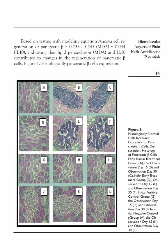
Based on testing with modeling equation Ancova cell re-generation of pancreatic β = 0.733 - 5.545 (MDA) + 0.044 (IL10), indicating that lipid peroxidation (MDA) and IL10 contributed to changes in the regeneration of pancreatic β cells. Figure 1. Histologically pancreatic β cells expression.
Figure 1:Histologically Normal Cells Increased Expression of Pan-creatic β Cells. De-scription: Histology of Pancreatic β Cells Early Insulin Treatment Group (A), the Obser-vation Day 15 (B) and Observation Day 30 (C). Kefir Early Treat-ment Group (D), Ob-servation Day 15 (E) and Observation Day 30 (F). Initial Positive Control Group (G), the Observation Day 15 (H) and Observa-tion Day 30 (I). Ini-tial Negative Control gGroup (A), the Ob-servation Day 15 (K) and Observation Day 30 (L).
IJFNPH5,1/2
15
DISCUSSION
This study has demonstrated the truth of hypotheses and build a new theory research, that the supplementation of plain kefir 3,6 cc/200 g BB / day for 30 days, significantly affect on blood glucose, antioxidants (SOD, Catalase, GPx), peroxidation lipids (MDA), immune response (cytokines IL
1, IL
6, IL
10) and pancre-
atic β-cell function. The research revealed that the mechanism of plain kefir played initially lowering blood glucose and pro inflamatory cytokine, also reduced subsequent effect of free radicals, lipid peroxidation. Reduction of peroxide molecules effects positively secretion pro-inflammation cytokines (IL
1, IL
6,
IL10
), so the cell structure damage and function of pancreatic β inevitable. Kefir prevents against glucotoxiticity and lipotoxitic-ity and reduces the occurrence of hyperglycaemia. This ability is associated with bioactive found in kefir themselves.
In this study, plain kefir have been proven to reduce hy-perglycemia and lipid peroxidation, and increased IL10. Regeneration will be decreased by 5.545 when cells are ex-posed to lipid peroxidation (MDA) after the controlled by vari-able IL10. The R2 (R square) of 0.910 means that the MDA and IL10 variables can explain the occurrence of regeneration of pancreatic β cells by 91.0%, while the rest is determined by other factors, such as: TNF-α exposure, antioxidant status, blood glucose, body weight of animals.
Exopolysaccharide (EPS) is a biopolymer lowering blood glucose, the mechanism is through intestinal microvilli coating process, so that inhibit glucose uptake and glucose does not increase in the body.(Maeda, 2004) Studies in vitro supported this rsearch that kefir lowers of blood glucose. Another mech-anism EPS activates glucagon like peptide 1 (GLP 1), gastric inhibitory peptide (GIP) (Khan, 2001) and the enzyme adenyl-ate cyclase through the cyclic adenosine monoposfat (cAMP)

Biomolecular Aspects of Plain
Kefir Antidiabetic Potentials
16
through sensitization of Ca2 ions and activation of protein ki-nase A, thus increasing insulin release from cells pancreatic β, the occurrence of blood glucose homeostasis and suppression glucotoxicity. (Pickup and Gareth, 1998, Djokomoeljanto, 1999)
Kefir’s peptide improves biological value and digestibility of protein, so it affects the restoration of the pancreatic β cell mass, restoration of its physiology and insulin secretion; be-sides it strengthen the immune system through normallized the pro-inflammation cytokine. It also supported by Meier and Almatsier that the best efforts β cell regeneration is through a systematic effort from inside his own body, the provision of high quality nutrition intake, especially high biological value protein is very importand, because protein will maintain and regenerated the body cells. (Meier,2008, Almatsier, 2001)
Kefir’s antioxidants inhibit oxidation, reducing hydrox-yl radical, superoxide and lipid peroxidation. The antioxi-dant activity occurs in a way delivers on its hydrogen atoms NADP, which will further reduce the existing free radicals. Antioxidant effects to lower through the reduction process malondealdehid peroxidation (MDA) and suppress pro-in-flammatory activity of IL
1 and IL
6, resulting in improvement
of pancreatic β cells through cell regeneration and improve-ment in organ pancreatic β cells. (Susetia-Totoprajogo, 2010)
Intestinal immune system that works well modulate the innate and adaptive immune in the body. At the molecular level, the innate immune system is centered on the activation of NF-kB, induces transcription of several proinflammatory cytokines, response to stimulation by microbial or agent of AGEs. In its role to help bridge the innate immunity system to TLR adaptive system, able to induce a good immune response towards Th1 or Treg. Macrophage cells exposed to probiot-ics maintain immune cells in a state of homeostasis, through

IJFNPH5,1/2
17
immunosuppression and immunomodulating with decreased production of cytokines (IL1, IL6) and increased production of IL10. The role of IL10 inhibit Th1 cells. Increased cytokine IL10, which is proven to maintain homeostatic proliferation of Th1-Th2, and proinflammatory cytokine production can be controlled and inflammation in pancreatic β cells unavoid-able. (Susetia-Totoprajogo, 2010)
Probiotic bacteria and gut mucosal acts synergistic in form of immunomodulation. At the level of intestinal epithelium, probiotic bacteria provide beneficial effects through coloniza-tion and the release of bioactive mixture. Then it reinforce barrier function through modulation of intestinal epithelial cells including the release of cytokines and chemokines. State of good intestinal immune system will affect the whole body immune. (Listiani,2005, Inggrid Surono, 2007, Corthesy,2007).
Increased IL10 related to the period and pancreatic β cell phyisiological, this is supported IL10 suppress proinflamma-tory response and apoptosis in pancreatic β cells. (Dronavalli, et all. 2008) found an association between IL10 and insulin sensitivity. (Bukhari, 2009), Decreased of inflammation in pancreatic β cells is closely related to the improvement of the synthesis of proinsulin to insulin and increase insulin sensitiv-ity and pancreatic β cell mass. (Donath, et al. 2009)
CONCLUSIONS AND RECOMMENDATIONS
Supplementation of the plain kefir with dose about 3,6 cc/200 g bw / day for 30 days in vivo study of Wistar rats STZ induced hyperglycemia, was significantly decreased blood glu-cose, proinflammatory cytokines IL
1, IL
6 and lipid peroxida-
tion (MDA) and increased antioxidants (SOD, catalase, GPX), anti-proliferation cytokine IL
10 and improving of the normal
of pancreatic β cells expression. Insulin and kefir descriptively

Biomolecular Aspects of Plain
Kefir Antidiabetic Potentials
18
reduced TNF β level and not significant. It is very challeng-ing to study on characterization of probiotic properties of via-ble bacteria in kefir to find out the biomolecular mechanisms and apply it in clinically diabetes mellitus therapy.
BIOgRAPhy
DR. Judiono, MPS. He is senior lecture at Nutrition Department. He is also Head of the Research and Development at the Health Polytechnic of Bandung, MOH Republic of Indonesia (R.I). This research is funded by Danone Institute Competitive Doctorate Research Grand for 2010 to 2012, Doctoral Research Awards from the Directorate of Higher Education, MEC R.I. Prof. DR. dr. RRJ. Sri Djokomoeljanto, SpPD, KEMD is Professor at the University of Diponegoro Semarang. He is the Head of Board of Supervisors Doctoral Study Programme of Medical Science. He is neuroendocrinologyst. He was also The Eijkman Award for outstanding contribution in tropi-cal studies from Holland 1998 and Diponegoro Award for Research: “Endemic Goiter and Cretinism Central Java”. Prof. DR. dr. Suharyo Hadisaputro, Sp.PD-KPTI is Professor at the University of Diponegoro Semarang. He is also the Head of Doctoral Study Programme of Medical Science, Post Graduate School. He is Assesor of National Accreditation Board for Higher Education.
ACKNOwLEDgEMENT
This study was financially granted by Indonesian Danone Institute Foundation. The views expressed herein are those of the individual authors, and do not necessarily reflect those of Indonesian Danone Institute Foundation. We express our grat-itude to the late Professor Endang Purwaningsih (†)who has giv-en her support and ideas on the implementation of this study

IJFNPH5,1/2
19
REFERENCES
Almatsier, Sunita. (2003). Prinsip Dasar Ilmu Gizi, Gramedia Pustaka Utama. Jakarta, 2003
American Diabetes Association (ADA). (2008). Diagnosis dan Clasification of Diabetes Melitus. Jurnal Diabetes Care, 2008. (31), 1, January 2008.
Betteridge, D.J. (2000). What is oxidative stress?. Metabolism Clinical and Experimental, (49), 2, Supplemen 1, 2000: p. 3-6
Brown, Amy C., Ana Valiere. (2004). Probiotics and Medical Nutrition Therapy, Nutr Clin Care. 2004; 7(2): 56–68.
Brownlee, Michael. (2004). Banting Lecture 2004. The Pathobiology of Diabetic Complications. A Unifying Mechanism. Diabetes, Vol. 54, June 2005. Page 1615- 1624
Bukhari, A. Obesity induced insulin resistance: Up date on the molecular mechanism. International Symphosium Scientific Paper Presentation On Nutrition and 6th Asia Pacific Clinical Nutrition Society. Makassar, October 10-13, (2009)
Campbell, Donald T., Julian C. Stanley. (1963). Experimental and Quasi-experimental designs for re-search. Rand Mc Nally College Publishing Company, Chicago. 1963. Page 145 – 170.
Ceriello, A. (2000). Oxidative Stress and Glycemic Regulation. Metabolism (49),2 Supplement 2000: p. 27 – 29.
Biomolecular Aspects of Plain
Kefir Antidiabetic Potentials
20
Corthesy, Blaise; Gaskins, H Rex; Mercenier, Annick. (2007). Cross-Talk Between Probiotic Bacteria and the Host Immune System. Journal of Nutrition, 2007. 137: 781S–790S
Djokomoeljanto, R. Insulin: Berperan sentral dalam Diabetes Melitus. Insulin Perannya pada pengelolaan Diabetes Melitus. Editor R. Djokomoeljanto, Darmono, Tony Suhartono. (1999). Badan Penerbit Universitas Diponegoro Semarang, 1999. Hal 1-16.
Djokomoeljanto. RRJ. (2007). Neuropati Diabetik dalam Naskah Lengkap Diabetes Melitus ditinjau dari berb-agai Aspek penyakit dalam editor Darmono, dkk. Badan Penerbit Universitas Diponegoro, 2007. Hal. 1-14.
Donath, Marc Y., Marianne Böni-Schnetzler,Helga Ellingsgaard, andJan A. Ehses. Islet Inflammation Impairs the Pancreatic beta-Cell in Type 2 Diabetes. Physiology 24: 325–331, (2009)
Gross Portney, Leslie., Mary P.W. Foundations of Clinical Research Application to Practice. Connectcut: Apletion & Lange (1993). p.148 – 152
Hadisaputro, S., Riwanto,Ig., Subagio, H.W, Judiono, Laksono,B., Watuguly, T. (2008).Pendekatan Pengobatan Tradisional Untuk Tatalaksana Penyakit Infeksi HIV/AIDS dan Penyakit Degeneratif (Diabetes Mellitus dan Kanker Paru). Laporan Akhir Hibah Pasca Sarjana Universitas Diponegoro, 2008.
Hadisaputro, S., Setyawan, H. (2007). Faktor-Faktor yang berpengaruh terhadap kejadian Diabetes mellitus tipe 2, Naskah Lengkap Diabetes Melitus Ditinjau dari Berbagai Aspek Penyakit Dalam. Semarang: Badan Penerbit

IJFNPH5,1/2
21
Universitas Diponegoro. 2007: p. 133-154.
Hughes, David A., L. Gail Darlington, Adrianne Bendich. (2004).Diet and Human Immune Function. New jersey: Humana Press 2004
Hulley, Stephen B., Cumming, Steven R., Grady, Deborah. (2007) Designing a Randomized Blinded Trial, Chapter 10. Designing Clinical Research, 3rd Edition. Philadelphia, USA: Lippincott Williams & Wilkins. Page: 147 – 161.
Inggrid-Surono. Probiotik Susu fermentasi dan Kesehatan.YAPMMI. Hal. 1-70.
Khan, Steven E. (2001).The importance of β cell failure in the development and progression of type 2 diabe-tes. J. Clin Endocrinology Metabolism, 2001: 86 (9): 4047-4058.
Khazrai, Y.M., S Manfrini and P. Pozzilli. (2004).Diet and di-abetes: prevention and control. Functional foods, cardio-vasCular disease and`diabetes. England: Woodhead
Lee, Cathy C., Simin Liu. (2008) Role of Inflammatory Cytokines in Type 2 Diabetes. Review of Endocrinology, February 2008. Page 19 -21
Listiani, Lanny. (2005). Probiotics in Health system. Makalah Seminar Peran Probiotik dalam menjamin per-tumbuhan & perkembangan anak. Jakarta: 7 Juni, 2005
Maeda, Hiroaki., Xia Zhu, Kazunobu Omura, Shiho Suzuki, Shinichi Kitamura. (2004).Effects of an exopolysaccha-ride (kefiran) on lipids, blood pressure, blood glucose,

Biomolecular Aspects of Plain
Kefir Antidiabetic Potentials
22
and constipation. BioFactors Vol. 22 Issue 1-4 ( 2004). Page. 197-200
Maeda, Hiroaki., Xia Zhu, Kazunobu Omura, Shiho Suzuki, Shinichi Kitamura. Effects of an exopolysaccharide (kefi-ran) on lipids, blood pressure, blood glucose, and consti-pation. BioFactors Vol. 22 Issue 1-4 ( 2004). Page. 197-200 (Abstract)
Mahardhika, E. Dharmana, R. Djokomoeljanto. (2004). Status Zinc dan Imunitas Selular pada Pasien DM Tipe 2 Regulasi Glukosa darah Baik dan Buruk: Fokus pada Jumlah Limfosit dan Fungsi Fagositosis. M.Med Indonesiana, Vol. 39. No. 2 thaun 2004. Hal 80-85
Maritim, A.C., R.A. Sanders., J.B. Watkins III. (2003) Diabetes, Oxidative Stress, and Antioxidants: A Review. J. Biochem Molecular Toxicology, Vol. 17, No.1.. Page: 24-38
Meier, JJ. (2008).Beta cell mass in diabetes: a realistic thera-peutic target?. Diabetologia (2008) 51: 703-713
Miriam Cnop, Nils Welsh, Jean-Christophe Jonas, Anne Jo, Sigurd Lenzen, and Decio L. Eizirik. (2005) Mechanisms of Pancreatic-Cell Death in Type 1 and Type 2 Diabetes. Many Differences, Few Similarities. Diabetes, Vol. 54, Supplement 2, December 2005. Page: S97-S-107
Moussa, S.A. Oxidative Stress in Diabetes Mellitus. Romanian J. Biophys, (2008). Vol. 18 No. 3. Page: 225-236
Ortis, Fernanda., Alessandra K. Cardozo, Daisy Crispim, Joachim Sto¨ rling, Thomas Mandrup-Poulsen,and De´

IJFNPH5,1/2
23
cio L. (2006). Eizirik. Cytokine-Induced Proapoptotic Gene Expression in Insulin-Producing Cells Is Related to Rapid, Sustained, and Nonoscillatory Nuclear Factor-kappa_B Activation. Molecular Endocrinology 2006(8):1867–1879
Perkumpulan Endokrinologi Indonesia (PERKENI). (2007).Konsensus Pengelolaan Diabetes Melitus di Indonesia. Jakarta 2007a.
Pfaffy, (2001). Diabetic Complications, Hyperglicemia & Free Radicals. Biosciences Departement The University of Iowa. Iowa City, 2001. 52242.
Pickup, John C., Gareth Williams. (1998). The biosynthe-sis and secreation of insulin in Textbook of Diabetes, second edition, Volume 1, UK: Blackwell Science, 1998. Page 8.1 – 8.14
Reeves, Philip G. (1997) Components of the AIN-93 Diets as Improvements in the AIN-76A Diet. Journal of Nutrition Vol 127, No.5, May 1, 1997.
Rytter, Elisabet., Bengt Vessby, Rikard A, Clara Johansson, Anders Sjo¨din, Lilianne Abramsson-Zetterberg4, Lennart Mo¨ller and Samar Basu. (2009). Glycaemic sta-tus in relation to oxidative stress and inflammation in wellcon-trolled type 2 diabetes subjects. British Journal of Nutrition (2009), 101, 1423–1426