jpet #178384
TRANSCRIPT
JPET #178384
1
Title Page
Amperometric Measurement of Glutamate Release Modulation by Gabapentin and
Pregabalin in Rat Neocortical Slices: Role of Voltage-Sensitive Ca2+ α2δ-1 Subunit
Authors: Jorge E. Quintero, David J. Dooley, François Pomerleau, Peter Huettl, and
Greg A. Gerhardt
Addresses: Department of Anatomy and Neurobiology, Morris K. Udall Parkinson’s
Disease Research Center of Excellence, Center for Microelectrode Technology,
University of Kentucky, Lexington, KY 40536, USA (JEQ, FP, PH, GAG); Department. of
CNS Pharmacology, Pfizer Global Research & Development, Ann Arbor, MI 48105, USA
(DJD).
JPET Fast Forward. Published on April 4, 2011 as DOI:10.1124/jpet.110.178384
Copyright 2011 by the American Society for Pharmacology and Experimental Therapeutics.
This article has not been copyedited and formatted. The final version may differ from this version.JPET Fast Forward. Published on April 4, 2011 as DOI: 10.1124/jpet.110.178384
at ASPE
T Journals on February 17, 2022
jpet.aspetjournals.orgD
ownloaded from
JPET #178384
2
Running Title: α2δ- ligand Modulation of Glutamate Release Corresponding Author: Jorge E. Quintero, Ph.D.; Dept. of Anatomy & Neurobiology;
University of Kentucky, 306 Whitney Hendrickson Building, Lexington, KY 40536;
[email protected]; Tel: (859) 323-4531; Fax: (859) 257-5310
Number of Text Pages: 23
Number of Tables: 0
Number of Figures: 5
Number of References: 37
Number of Words in Abstract: 241
Number of Words in Introduction: 482
Number of Words in Discussion: 914
Abbreviations: aCSF, artificial cerebrospinal fluid; GBP, gabapentin; PGB, pregabalin;
R-IBG, R-(-)-3-isobutylgaba; VSCC, voltage-sensitive calcium channel; MEAs,
microelectrode arrays; K+, potassium
Section assignment: Neuropharmacology
This article has not been copyedited and formatted. The final version may differ from this version.JPET Fast Forward. Published on April 4, 2011 as DOI: 10.1124/jpet.110.178384
at ASPE
T Journals on February 17, 2022
jpet.aspetjournals.orgD
ownloaded from
JPET #178384
3
ABSTRACT
Gabapentin (GBP; Neurontin®) and pregabalin (PGB; Lyrica®, S-(+)-3-
isobutylgaba) are used clinically to treat several disorders associated with excessive or
inappropriate excitability, including epilepsy; pain from diabetic neuropathy, postherpetic
neuralgia and fibromyalgia; and generalized anxiety disorder. The molecular basis for
these drugs’ therapeutic effects are believed to involve the interaction with the auxiliary
α2δ subunit of voltage-sensitive Ca2+ channels (VSCC) translating into a modulation of
pathological neurotransmitter release. Glutamate as the primary excitatory
neurotransmitter in the mammalian central nervous system contributes, under conditions
of excessive glutamate release, to neurological and psychiatric disorders. This study
used enzyme-based microelectrode arrays to directly measure extracellular glutamate
release in rat neocortical slices and determine the modulation of this release by GBP
and PGB. Both drugs attenuated K+-evoked glutamate release without affecting basal
glutamate levels. PGB (0.1-100 µM) exhibited a concentration-dependent inhibition of
K+-evoked glutamate release with an IC50 of 5.3 µM. R-(-)-3-Isobutylgaba, the
enantiomer of PGB, did not significantly reduce K+-evoked glutamate release. The
decrease of K+-evoked glutamate release by PGB was blocked by the L-amino acid L-
isoleucine, a potential endogenous ligand of the α2δ subunit. In neocortical slices from
transgenic mice having a point mutation (i.e., R217A) of the α2δ -1 (subtype) subunit of
VSCC, PGB did not affect K+-evoked glutamate release yet inhibited this release in wild-
type mice. The results show that GBP and PGB attenuated stimulus-evoked glutamate
release in rodent neocortical slices and that the α2δ -1 subunit of VSCC appears to
mediate this effect.
This article has not been copyedited and formatted. The final version may differ from this version.JPET Fast Forward. Published on April 4, 2011 as DOI: 10.1124/jpet.110.178384
at ASPE
T Journals on February 17, 2022
jpet.aspetjournals.orgD
ownloaded from
JPET #178384
4
Introduction
Several neurological and psychiatric disorders characterized by excessive or
dysfunctional neurotransmitter release are routinely treated with gabapentin [GBP;
Neurontin®, 1-(aminomethyl)cyclohexaneacetic acid] and pregabalin [PGB; Lyrica®, S-
(+)-3-isobutylgaba, S-(+)-4-amino-3-(2-methylpropyl)butanoic acid] (Dooley et al.,
2007). Although multiple mechanisms of action have historically been proposed to
account for the preclinical and clinical profiles of these drugs, there is increasing
evidence for a significant role of the auxiliary α2δ subunit of voltage-sensitive Ca2+
channels (VSCC) (Taylor et al., 1998; Taylor et al., 2007).
The binding of these ligands to the α2δ subunit is believed to be the source of
their efficacy in treating epilepsy; pain from diabetic neuropathy, post-herpetic neuralgia,
and fibromyalgia; and generalized anxiety disorder. With the recent availability of
transgenic mice with point mutations of the α2δ subunit (i.e., α2δ -1 and α2δ -2 subtypes)
(Bian et al., 2006; Bian et al., 2008), preclinical experiments can be designed to test for
altered neurochemical and behavioral effects of GBP and PGB (Field et al., 2006).
In the present study, we used enzyme-based microelectrode arrays (MEA) that
have micrometer-size platinum recording sites with sampling rates of > 1 Hz. These
MEAs were developed in response to the limitations of other techniques or devices used
to measure neurotransmitters, e.g. microdialysis/perfusate sampling coupled to high-
performance liquid chromatography (Barnes et al., 1988; Shinohara et al., 1998;
Burmeister et al., 2000). A drawback of microdialysis or perfusate sampling techniques
is that they sample from a large area (Borland et al., 2005) and at relatively slow
(minutes) sampling rates. Given the rapid nature of neurotransmission of chemical
messengers such as glutamate, faster sampling rates and smaller sampling areas
should be beneficial. Enzyme-coated MEAs have been extensively characterized in the
brain of anesthetized and behaving animals to measure glutamate (Burmeister et al.,
This article has not been copyedited and formatted. The final version may differ from this version.JPET Fast Forward. Published on April 4, 2011 as DOI: 10.1124/jpet.110.178384
at ASPE
T Journals on February 17, 2022
jpet.aspetjournals.orgD
ownloaded from
JPET #178384
5
2002; Binns et al., 2005; Day et al., 2006; Nickell et al., 2006; Rutherford et al., 2007;
Parikh et al., 2010) but not in brain slices.
We used these MEAs to directly measure K+-evoked (extracellular) glutamate
release in rat neocortical slices and to determine the modulation of this release by GBP
and PGB. As the primary excitatory neurotransmitter in the mammalian CNS, glutamate
has often been associated with a variety of pathological conditions (Meldrum, 2000),
several of which are responsive to α2δ-ligands like PGB. A reduction of excessive
glutamate release by α2δ -ligands conceivably translates into clinically relevant
therapeutic effects, especially considering the experimental evidence supporting a
relationship between α2δ subunit binding and the modulation of processes subserving
neurotransmitter release (Dooley et al., 2007).
An additional aspect of this study was to assess the effects of PGB on K+-evoked
(extracellular) glutamate release in neocortical slices from wild-type and α2δ -1 mutant
mice. The α2δ -1 transgenic mice have a point mutation (viz., R217A) that markedly
reduces [3H]-GBP and [3H]-PGB binding in CNS regions (e.g., neocortex) known to
preferentially express the α2δ -1 protein (Bian et al., 2006; Field et al., 2006).
Methods
Animals. Male rats [Sprague-Dawley, 2-8 weeks old; Harlan Laboratories,
Indianapolis, IN] and male mice [wild type and mutant α2δ -1 R217A (Bian et al., 2006;
Field et al., 2006), 2-5 months old; Charles River Laboratories, Wilmington, MA] were
housed in an AAALAC-accredited facility according to the standards outlined in the
Guide for the Use and Care of Laboratory Animals.
Animals were under a 12-hr light-dark cycle, had ad libitum access to food and
water, and were maintained for a minimum of 5 days before euthanasia by decapitation.
The brains were removed by blunt dissection and placed in ice-cold buffer until slice
This article has not been copyedited and formatted. The final version may differ from this version.JPET Fast Forward. Published on April 4, 2011 as DOI: 10.1124/jpet.110.178384
at ASPE
T Journals on February 17, 2022
jpet.aspetjournals.orgD
ownloaded from
JPET #178384
6
preparation. The glutamate recordings occurred during the light phase of the light-dark
cycle. All experimental protocols were approved by The Animal Care and Use
Committee of the University of Kentucky.
Glutamate Release Measurements. Neocortical slices from rats and mice
were prepared using standard protocols (Hascup et al., 2007). Briefly, coronal slices
(0.35-0.4 mm thick), including the frontal and parietal areas exhibiting relatively high [3H]-
GBP and [3H]-PGB binding (Bian et al., 2006), were maintained for at least 1 h at room
temperature in artificial cerebrospinal fluid (aCSF; composition (mM): NaCl (124), KCl
(5), CaCl2 (2.5), MgCl2 (1.5), NaHCO3 (26), NaH2PO4 (1.4), D-glucose (10); saturated
with 95% O2/5% CO2; pH = 7.2–7.4] before the start of an experiment. The slices were
transferred to immersion-style chambers (i.e., one slice/chamber), and superfused at a
rate of 1.5-2.0 ml/min with aCSF (31-33°C). Each chamber was fitted with a Ag/AgCl
reference electrode.
Ceramic-based MEAs (4 platinum sites in a row, 50 x 150 µm each) were
assembled, coated with Nafion®, and subsequently coated with three layers of a 1%
glutamate oxidase (Associates of Cape Cod, East Falmouth, MA)/1% bovine serum
albumin/0.125% glutaraldehyde enzyme solution. Coated MEAs were allowed to cure a
minimum of two days before use. Enzyme-based MEAs measure glutamate through
the enzymatic breakdown of glutamate to yield a reporter molecule of hydrogen peroxide
that is subsequently oxidized on the platinum recording surface to yield an oxidation
current. The MEAs were then calibrated (in vitro) with glutamate in a phosphate-
buffered solution (pH = 7.4) at 31-34°C to (a) generate a standard response curve
(sensitivity > 2 pA/µM); (b) determine the limit of detection (≥ 3 times the signal-to-noise
ratio; < 2.0 µM); and (c) assess the selectivity for glutamate relative to an endogenous
electroactive compound, ascorbic acid (> 30:1). A MEA or MEA/micropipette assembly
This article has not been copyedited and formatted. The final version may differ from this version.JPET Fast Forward. Published on April 4, 2011 as DOI: 10.1124/jpet.110.178384
at ASPE
T Journals on February 17, 2022
jpet.aspetjournals.orgD
ownloaded from
JPET #178384
7
was lowered into the neocortical slice, and extracellular glutamate levels were measured
once basal glutamate levels stabilized for at least 10 min.
Test substances were delivered through the superfusion system for a minimum
of 15 min. before slice stimulation unless stated otherwise. The slices were stimulated
twice (S1, S2) with high K+ by one of two methods to evoke glutamate release: 1) direct,
local application of 70 mM K+ solution (composition (mM): KCl (70), NaCl (79), CaCl2
(2.5); pH = 7.0-7.4) to depolarize the local glutamatergic network via pressure ejection;
or 2) superfusion of 70 mM K+ (i.e., increase of KCl in aCSF with corresponding
decrease of NaCl (59 mM) to maintain iso-osmolarity) to depolarize the whole slice.
Slices were allowed to recover a minimum of 20 minutes between stimulations, and drug
solutions were delivered for a minimum of 15 minutes before stimulation. For local
application, glass micropipettes (inside tip diameter of 10-15 µm) were formed from
stock (1 mm o.d., 0.58 mm i.d.; A-M Systems, Everett, WA), attached, and the tip
centered over the MEA recording site at a tip-to-tip distance of 70-110 µm. The 70 mM
K+ solution was applied at 1-min intervals until at least two to five reproducible glutamate
responses were recorded. Delivery of solution volumes (i.e., 12.5-400 nL over 0.1-3.0
sec) was controlled by a pressure-ejection system (2-12 p.s.i.; Picospritzer II, Parker
Hannifin Corp., Cleveland, OH), and monitored using a stereomicroscope fitted with a
reticule (Gerhardt and Palmer, 1987). Extracellular glutamate levels were measured at 1
Hz using constant potential amperometry (+0.7 V vs Ag/AgCl reference) controlled by a
FAST16 electrochemical recording system (Quanteon, LLC, Nicholasville, KY) and
analyzed offline by customized Excel®- based software.
Calculations and statistics. Glutamate release amplitudes were calculated
from the difference between maximum K+-evoked glutamate release values and basal
values. Values given are X ± S.E. (n ≥ 6). In one set of experiments with PGB, a
This article has not been copyedited and formatted. The final version may differ from this version.JPET Fast Forward. Published on April 4, 2011 as DOI: 10.1124/jpet.110.178384
at ASPE
T Journals on February 17, 2022
jpet.aspetjournals.orgD
ownloaded from
JPET #178384
8
concentration-effect curve with corresponding IC50 value was calculated by nonlinear
regression (Prism 4.0, GraphPad Software Inc., San Diego, CA). If appropriate, the
results were analyzed using the t-statistic for group means, or one- or two-way analysis
of variance followed by post-hoc comparisons using Dunnett’s or Bonferroni multiple
comparison statistic (InStat 3.0, GraphPad Software Inc., San Diego, CA). The minimal
level of significance was p ≤ 0.05 (two-tail criterion).
Materials. Substances were either commercially available (Sigma-Aldrich) or
donated (i.e., GBP, PGB, and R-(-)-3-isobutylgaba (R-IBG) (Pfizer)). Test compounds
were dissolved directly in aCSF.
Results
The effects of GBP and PGB on resting glutamate levels were evaluated in initial
experiments. Neither drug (0.1-100 µM) altered basal glutamate levels in neocortical
slices (data not shown).
In the brains of anesthetized animals, delivery of high K+ to stimulate glutamate
release has been performed with local, pressure-ejected administration (Burmeister et
al., 2002; Day et al., 2006; Quintero et al., 2007; Stephens et al., 2009). Meanwhile,
studies in brain slices permit the use of two methods of delivering high K+ solutions to
activate neural networks: local delivery and superfusion. Local stimulation, using
pressure delivery, produces a comparable stimulus to those used in previous studies
with anesthetized animals. An additional benefit of brain slice recordings is the flexibility
to also employ superfusion of high K+ to evoke release. This sustained depolarization
resembles prolonged or excessive excitability – a condition that characterizes some
neurological disorders such as anxiety. We used both stimulation methods here to
characterize the effects of α2δ subunit ligands and to compare glutamate measurements
with MEAs to previous studies.
This article has not been copyedited and formatted. The final version may differ from this version.JPET Fast Forward. Published on April 4, 2011 as DOI: 10.1124/jpet.110.178384
at ASPE
T Journals on February 17, 2022
jpet.aspetjournals.orgD
ownloaded from
JPET #178384
9
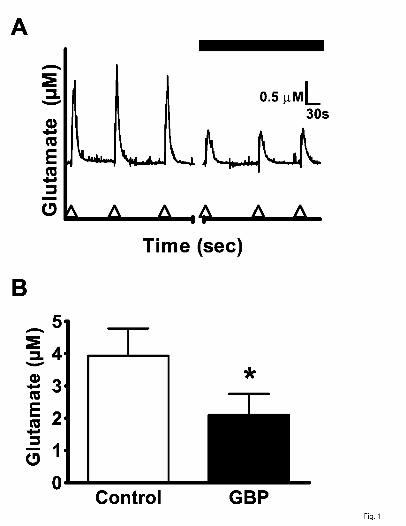
The repeated pressure-ejection delivery of 70 mM K+ solution yielded similar size
glutamate signals with a mean amplitude of 3.9 ± 0.8 µM (Fig. 1A, B). In the presence of
GBP (100 µM), the mean amplitude was significantly decreased by 46% to 2.1 ± 0.7 µM
(Fig. 1A, B).
Because GBP was confirmed to modulate K+-evoked glutamate release, the
more recently developed α2δ ligand, PGB, was chosen for testing in additional
experiments. PGB (100 µM) attenuated pressure-ejection delivery of 70mM K+ solution
(5.7 ± 1.5 µM glutamate, pre-PGB vs. 1.7 ± 1.2 µM glutamate, post-PGB; t(4) = 3.59, p =
0.023). We then transitioned to a paradigm of using repeated stimulation with
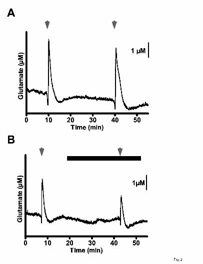
superfused 70 mM K+ (S1, S2). The S2/S1 ratio of control glutamate signals in rat
neocortical slices was 0.97 (Figs. 2A, 3A); this ratio was markedly reduced by 78% to
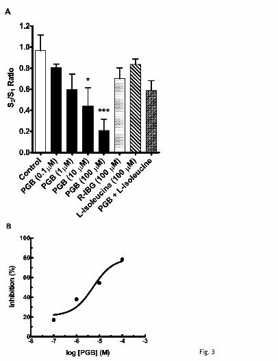
0.21 by PGB (100 µM) (Figs. 2B, 3A). Other S2/S1 ratios for PGB include 0.80 (non-
significant 14% inhibition) at 0.1 µM, 0.60 (non-significant 38% inhibition) at 1 µM, and
0.44 (54% inhibition) at 10 µM (Fig. 3A); an IC50 value of 5.3 µM was determined from
the concentration-effect relationship (Fig. 3B). The enantiomer of PGB, R-IBG (100 µM),
gave an S2/S1 ratio of 0.70 (non-significant 28% inhibition), contrasting sharply with the
effect (78% inhibition) of an equimolar concentration of PGB (Fig. 3A).
PGB and the endogenous amino acid, L-isoleucine, are substrates for the system
L-amino acid transporter and both have similar nanomolar affinity for the α2δ ligand
binding site on the α2δ subunit. The S2/S1 ratio associated with the K+-evoked glutamate
signals in the presence of L-isoleucine (100 µM) was 0.84 (non-significant 13%
inhibition), yet this compound reduced the effect (78% inhibition) of PGB (100 µM) as
indicated by the S2/S1 ratio of 0.59 (non-significant 39% inhibition) (Fig. 3A).
An action of GBP and PGB at the α2δ -1 subtype, rather than the α2δ -2 subtype,
has been proposed to account for the therapeutic effects of these drugs (Bian et al.
2006, 2008; Field et al. 2006). Using neocortical slices from the wild-type and α2δ -1
This article has not been copyedited and formatted. The final version may differ from this version.JPET Fast Forward. Published on April 4, 2011 as DOI: 10.1124/jpet.110.178384
at ASPE
T Journals on February 17, 2022
jpet.aspetjournals.orgD
ownloaded from
JPET #178384
10
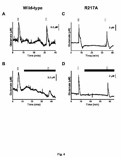
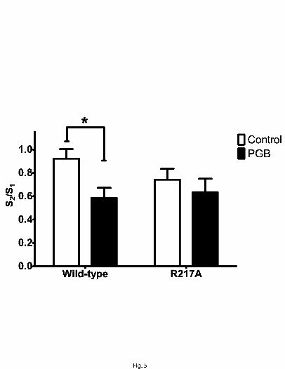
transgenic mice (Bian et al. 2006; Field et al. 2006), PGB (100 µM) significantly
decreased K+-evoked glutamate signals (Fig. 4A and B) in wild-type by 36% [S2/S1 =
0.92 (control) vs 0.59 (PGB); n= 13 and 14, respectively] (Fig.5); in the α2δ -1 transgenic
mice, the glutamate signals were unchanged by this drug [S2/S1 = 0.74 (control) vs 0.63
(PGB); n=9 each] (Fig. 4C and D).
Discussion
Aberrant glutamate neurotransmission is linked to a variety of neurological and
psychological disorders. Thus, identifying mechanisms that could modulate abnormal
glutamate release may provide an avenue for developing new therapeutics for
modulating glutamate signaling. Here we used enzyme-based MEAs to directly
measure extracellular glutamate and observed that both GBP and PGB attenuated the
K+-evoked glutamate release.
GBP has been used as an antiepileptic but its precise mechanism of action is
unknown (Taylor et al., 2007). To help address this, we stimulated the neural network
and measured synaptic spillover of glutamate employing a technique we have previously
used in anesthetized animals of locally delivering high potassium solution to evoke
depolarization and produce a release of glutamate (Burmeister et al., 2002; Day et al.,
2006). We observed in these brain slices an attenuation of glutamate release by GBP
after locally delivering high K+; an effect on neurotransmitter release that is repeatedly
observed after stimulus delivery (Dooley et al., 2000a; Dooley et al., 2000b; Dooley et
al., 2002).
PGB is structurally related to GBP but with greater reported efficacy in clinical
studies (Taylor et al., 2007). Using these MEAs, we used a whole slice superfusion of
high K+ to stimulate neurotransmitter release similar to Dooley et al. (Dooley et al.,
2000b). We have observed that this type of depolarization evokes glutamate release
This article has not been copyedited and formatted. The final version may differ from this version.JPET Fast Forward. Published on April 4, 2011 as DOI: 10.1124/jpet.110.178384
at ASPE
T Journals on February 17, 2022
jpet.aspetjournals.orgD
ownloaded from
JPET #178384
11
that is calcium-dependent (J.E. Quintero and G.A. Gerhardt, unpublished observations).
The dose dependent inhibition of K+-evoked glutamate release by PGB resulted in an
IC50 = 5.3µM compared to an IC50 = 11.8 µM of K+-evoked [3H]Norepinephrine release
(Dooley et al., 2002).
The magnitude of the attenuation of evoked glutamate release was larger than
the reports in some studies that have examined GBP or PGB on stimulus evoked
neurotransmission (Dooley et al., 2000a; Dooley et al., 2000b; Dooley et al., 2002;
Brown and Randall, 2005). One apparent factor that may influence the effectiveness of
these compounds is the type of stimulus; such that GBP and PGB may exert an effect
on the prolonged, depolarization- induced neurotransmitter release that more closely
resembles hyperexcitability as found in pathological states rather than the normal
physiological neurotransmission (Dooley et al., 2000a; Maneuf et al., 2001; Dooley et al.,
2007). Additionally, some of the GBP or PGB effects on stimulus-evoked
neurotransmitter release may have been diluted in studies on slices or synaptic endings
where the whole chamber perfusate is sampled (Dooley et al., 2000a; Fink et al., 2000;
Fink et al., 2002) versus the limited focal area sampled by the 50 x 150 µm size of these
MEAs in slices. For example, with the high resolution technique of whole-cell patch
clamp in cortical slices, GBP and PGB show an effect as high as ~80% on parameters
related to glutamate neurotransmission (Cunningham et al., 2004). Nonetheless, the
effect of GBP and PGB on neurotransmitter release remains controversial given reports
that GBP and PGB have no effect on K+ evoked glutamate release from human
synaptosomes (Brawek et al., 2009) or that the GBP and PGB effect may be linked to
the trafficking of calcium channels to the cell surface (Hendrich et al., 2008; Mich and
Horne, 2008; Bauer et al., 2009; Thorpe and Offord, 2010). The full effect of these
ligands may result both from altering calcium channel trafficking and also from the more
rapid modulation of synaptic function (Taylor, 2009).
This article has not been copyedited and formatted. The final version may differ from this version.JPET Fast Forward. Published on April 4, 2011 as DOI: 10.1124/jpet.110.178384
at ASPE
T Journals on February 17, 2022
jpet.aspetjournals.orgD
ownloaded from
JPET #178384
12
Meanwhile, the pharmacological effects of PGB are stereoselective and this was
borne out by the non-significant changes to the S2/S1 with the enantimore, R-IBG. The α
amino acids, L-isoleucine and L-leucine, have been proposed as potential endogenous
ligands for the α2δ subunit (Thurlow et al., 1993). While L-isoleucine did not produce a
significant change to K+-evoked glutamate release, L-isoleucine did inhibit the PGB
attenuation of K+-evoked glutamate release similar to what had been previously
described with L-isoleucine and GBP in cortical brain slices (Cunningham et al., 2004).
A more complex role in neurotransmission and GBP and PGB effectiveness may be the
case for these α amino acids where these endogenous ligands may act as “positive
modulators required for full functionality of the α2δ subunit” (Hendrich et al., 2008).
Previously, Wang and Offord (1999) showed that the arginine at position 217 in
the α region of the α2δ subunit is critical for GBP binding. Subsequently, in a R217A
knockin mouse that was developed, [3H]GBP and [3H]PGB binding to neocortical
membranes was greatly reduced in R217A mice compared to wild-type mice (Bian et al.,
2006; Field et al., 2006). Accordingly, in slices from R217A mice we concluded that a
functional α2δ subunit is necessary for PGB to attenuate K+ -evoked glutamate release.
While the mean S2/S1 ratio (0.74) in slices from the R217A mice was lower than the
S2/S1 ratio of slices from wild-type mice (0.92), the means were not significantly different.
However, we cannot rule out the possibility of a change in excitability properties of the
neurons and synapses in these animals given that the α2δ subunit is critical for normal
synapse formation or function (Eroglu et al., 2009).
In summary, we showed that GBP and PGB can modulate stimulus-evoked
glutamate release in rat neocortical brain slices and that the α2δ subunit of the VSCC is
involved in the inhibitory effects of these ligands. This ability to modulate excitatory
neurotransmitter release may, in part, explain the efficacy of these molecules in the
clinic. The application of slice recording methodology coupled to the MEA recording
This article has not been copyedited and formatted. The final version may differ from this version.JPET Fast Forward. Published on April 4, 2011 as DOI: 10.1124/jpet.110.178384
at ASPE
T Journals on February 17, 2022
jpet.aspetjournals.orgD
ownloaded from
JPET #178384
13
technology establishes a new means to better assess drugs mechanisms of action by
the direct measurement of neurotransmitter release.
This article has not been copyedited and formatted. The final version may differ from this version.JPET Fast Forward. Published on April 4, 2011 as DOI: 10.1124/jpet.110.178384
at ASPE
T Journals on February 17, 2022
jpet.aspetjournals.orgD
ownloaded from
JPET #178384
14
Authorship contributions:
Participated in research design: Quintero, Dooley, Pomerleau, Huettl, Gerhardt.
Conducted experiments: Quintero.
Contributed new reagents or analytic tools: Gerhardt.
Performed data analysis: Quintero, Dooley, Pomerleau, Gerhardt.
Wrote or contributed to the writing of the manuscript: Quintero, Dooley, Pomerleau,
Huettl, Gerhardt
Other: Directed research efforts; Gerhardt.
This article has not been copyedited and formatted. The final version may differ from this version.JPET Fast Forward. Published on April 4, 2011 as DOI: 10.1124/jpet.110.178384
at ASPE
T Journals on February 17, 2022
jpet.aspetjournals.orgD
ownloaded from
JPET #178384
15
References
Barnes S, Leighton GE and Davies JA (1988) A novel superfusion chamber for the
measurement of endogenous glutamate release from cerebellar slices. Journal of
Neuroscience Methods 23:57-61.
Bauer CS, Nieto-Rostro M, Rahman W, Tran-Van-Minh A, Ferron L, Douglas L, Kadurin
I, Sri Ranjan Y, Fernandez-Alacid L, Millar NS, Dickenson AH, Lujan R and Dolphin AC
(2009) The Increased Trafficking of the Calcium Channel Subunit {alpha}2{delta}-1 to
Presynaptic Terminals in Neuropathic Pain Is Inhibited by the {alpha}2{delta} Ligand
Pregabalin. J. Neurosci. 29:4076-4088.
Bian F, Hannah D and Campbell B (2008) Calcium channel alpha2-delta type 2 subunit
is the major binding protein for pregabalin in cerebellum and septum: an ex vivo
autoradiographic study in wild-type and genetically modified mice, in 2008 Society for
Neuroscience p Program No. 845.813, Washington, D.C.
Bian F, Li Z, Offord J, Davis MD, McCormick J, Taylor CP and Walker LC (2006)
Calcium channel alpha2-delta type 1 subunit is the major binding protein for pregabalin
in neocortex, hippocampus, amygdala, and spinal cord: an ex vivo autoradiographic
study in alpha2-delta type 1 genetically modified mice. Brain Res 1075:68-80.
Binns BC, Huang Y, Goettl VM, Hackshaw KV and Stephens RL, Jr. (2005) Glutamate
uptake is attenuated in spinal deep dorsal and ventral horn in the rat spinal nerve ligation
model. Brain Res 1041:38-47.
This article has not been copyedited and formatted. The final version may differ from this version.JPET Fast Forward. Published on April 4, 2011 as DOI: 10.1124/jpet.110.178384
at ASPE
T Journals on February 17, 2022
jpet.aspetjournals.orgD
ownloaded from
JPET #178384
16
Borland LM, Shi G, Yang H and Michael AC (2005) Voltammetric study of extracellular
dopamine near microdialysis probes acutely implanted in the striatum of the
anesthetized rat. J Neurosci Methods. 146:149-158.
Brawek B, Löffler M, Weyerbrock A and Feuerstein T (2009) Effects of gabapentin and
pregabalin on K+-evoked 3H-GABA and 3H-glutamate release from human neocortical
synaptosomes. Naunyn-Schmiedeberg's Archives of Pharmacology 379:361-369.
Brown JT and Randall A (2005) Gabapentin fails to alter P/Q-type Ca2+ channel-
mediated synaptic transmission in the hippocampus in vitro. Synapse 55:262-269.
Burmeister J, Pomerleau F, Palmer M, Day B, Huettl P and Gerhardt G (2002) Improved
ceramic-based multisite microelectrode for rapid measurements of L-glutamate in the
CNS. J.Neurosci.Methods 119:163-171.
Burmeister JJ, Moxon K and Gerhardt GA (2000) Ceramic-based multisite
microelectrodes for electrochemical recordings. Anal.Chem. 72:187-192.
Cunningham MO, Woodhall GL, Thompson SE, Dooley DJ and Jones RS (2004) Dual
effects of gabapentin and pregabalin on glutamate release at rat entorhinal synapses in
vitro. Eur.J Neurosci 20:1566-1576.
Day BK, Pomerleau F, Burmeister JJ, Huettl P and Gerhardt GA (2006) Microelectrode
array studies of basal and potassium-evoked release of L-glutamate in the anesthetized
rat brain. J Neurochem 96:1626-1635.
This article has not been copyedited and formatted. The final version may differ from this version.JPET Fast Forward. Published on April 4, 2011 as DOI: 10.1124/jpet.110.178384
at ASPE
T Journals on February 17, 2022
jpet.aspetjournals.orgD
ownloaded from
JPET #178384
17
Dooley DJ, Donovan CM, Meder WP and Whetzel SZ (2002) Preferential action of
gabapentin and pregabalin at P/Q-type voltage-sensitive calcium channels: inhibition of
K+-evoked [3H]-norepinephrine release from rat neocortical slices. Synapse 45:171-190.
Dooley DJ, Donovan CM and Pugsley TA (2000a) Stimulus-dependent modulation of
[(3)H]norepinephrine release from rat neocortical slices by gabapentin and pregabalin. J
Pharmacol.Exp.Ther. 295:1086-1093.
Dooley DJ, Mieske CA and Borosky SA (2000b) Inhibition of K(+)-evoked glutamate
release from rat neocortical and hippocampal slices by gabapentin. Neurosci Lett.
280:107-110.
Dooley DJ, Taylor CP, Donevan S and Feltner D (2007) Ca2+ channel alpha2delta
ligands: novel modulators of neurotransmission. Trends Pharmacol Sci 28:75-82.
Eroglu Ç, Allen NJ, Susman MW, O'Rourke NA, Park CY, Özkan E, Chakraborty C,
Mulinyawe SB, Annis DS, Huberman AD, Green EM, Lawler J, Dolmetsch R, Garcia KC,
Smith SJ, Luo ZD, Rosenthal A, Mosher DF and Barres BA (2009) Gabapentin Receptor
[alpha]2[delta]-1 Is a Neuronal Thrombospondin Receptor Responsible for Excitatory
CNS Synaptogenesis. Cell 139:380-392.
Field MJ, Cox PJ, Stott E, Melrose H, Offord J, Su TZ, Bramwell S, Corradini L, England
S, Winks J, Kinloch RA, Hendrich J, Dolphin AC, Webb T and Williams D (2006)
Identification of the alpha2-delta-1 subunit of voltage-dependent calcium channels as a
molecular target for pain mediating the analgesic actions of pregabalin. Proc Natl Acad
Sci U S A 103:17537-17542.
This article has not been copyedited and formatted. The final version may differ from this version.JPET Fast Forward. Published on April 4, 2011 as DOI: 10.1124/jpet.110.178384
at ASPE
T Journals on February 17, 2022
jpet.aspetjournals.orgD
ownloaded from
JPET #178384
18
Fink K, Dooley DJ, Meder WP, Suman-Chauhan N, Duffy S, Clusmann H and Gothert M
(2002) Inhibition of neuronal Ca(2+) influx by gabapentin and pregabalin in the human
neocortex. Neuropharmacology 42:229-236.
Fink K, Meder W, Dooley DJ and Gothert M (2000) Inhibition of neuronal Ca(2+) influx
by gabapentin and subsequent reduction of neurotransmitter release from rat neocortical
slices. Br.J Pharmacol. 130:900-906.
Hascup KN, Rutherford EC, Quintero JE, Day BK, Nickell JR, Pomerleau F, Huettl P,
Burmeister J, Gerhardt GA, Michael AC and Borland LM (2007) Second-by-Second
Measures of L-Glutamate and Other Neurotransmitters Using Enzyme-Based
Microelectrode Arrays, in Electrochemical methods for neuroscience (Michael AC and
Borland LM eds) pp 407-450, CRC Press, Boca Raton, FL.
Hendrich J, Van Minh AT, Heblich F, Nieto-Rostro M, Watschinger K, Striessnig J,
Wratten J, Davies A and Dolphin AC (2008) Pharmacological disruption of calcium
channel trafficking by the α2δ ligand gabapentin. Proceedings of the National Academy
of Sciences 105:3628-3633.
Maneuf YP, Hughes J and McKnight AT (2001) Gabapentin inhibits the substance P-
facilitated K(+)-evoked release of [(3)H]glutamate from rat caudial trigeminal nucleus
slices. Pain 93:191-196.
Meldrum BS (2000) Glutamate as a neurotransmitter in the brain: review of physiology
and pathology. J Nutr 130:1007S-1015S.
This article has not been copyedited and formatted. The final version may differ from this version.JPET Fast Forward. Published on April 4, 2011 as DOI: 10.1124/jpet.110.178384
at ASPE
T Journals on February 17, 2022
jpet.aspetjournals.orgD
ownloaded from
JPET #178384
19
Mich PM and Horne WA (2008) Alternative Splicing of the Ca2+ Channel β4 Subunit
Confers Specificity for Gabapentin Inhibition of Cav2.1 Trafficking. Molecular
Pharmacology 74:904-912.
Nickell J, Salvatore MF, Pomerleau F, Apparsundaram S and Gerhardt GA (2006)
Reduced plasma membrane surface expression of GLAST mediates decreased
glutamate regulation in the aged striatum. Neurobiol Aging.
Parikh V, Ji J, Decker MW and Sarter M (2010) Prefrontal beta2 subunit-containing and
alpha7 nicotinic acetylcholine receptors differentially control glutamatergic and
cholinergic signaling. J Neurosci 30:3518-3530.
Quintero JE, Day BK, Zhang Z, Grondin R, Stephens ML, Huettl P, Pomerleau F, Gash
DM and Gerhardt GA (2007) Amperometric measures of age-related changes in
glutamate regulation in the cortex of rhesus monkeys. Exp Neurol 208:238-246.
Rutherford EC, Pomerleau F, Huettl P, Stromberg I and Gerhardt GA (2007) Chronic
second-by-second measures of L-glutamate in the central nervous system of freely
moving rats. J Neurochem 102:712-722.
Shinohara K, Honma S, Katsuno Y, Abe H and Honma K (1998) Circadian release of
amino acids in the suprachiasmatic nucleus in vitro. Neuroreport 9:137-140.
Stephens ML, Quintero JE, Pomerleau F, Huettl P and Gerhardt GA (2009) Age-related
changes in glutamate release in the CA3 and dentate gyrus of the rat hippocampus.
Neurobiol Aging.
This article has not been copyedited and formatted. The final version may differ from this version.JPET Fast Forward. Published on April 4, 2011 as DOI: 10.1124/jpet.110.178384
at ASPE
T Journals on February 17, 2022
jpet.aspetjournals.orgD
ownloaded from
JPET #178384
20
Taylor CP (2009) Mechanisms of analgesia by gabapentin and pregabalin - Calcium
channel [alpha]2-[delta] [Cav[alpha]2-[delta]] ligands. Pain 142:13-16.
Taylor CP, Angelotti T and Fauman E (2007) Pharmacology and mechanism of action of
pregabalin: The calcium channel [alpha]2-[delta] (alpha2-delta) subunit as a target for
antiepileptic drug discovery. Epilepsy Research 73:137-150.
Taylor CP, Gee NS, Su TZ, Kocsis JD, Welty DF, Brown JP, Dooley DJ, Boden P and
Singh L (1998) A summary of mechanistic hypotheses of gabapentin pharmacology.
Epilepsy Res. 29:233-249.
Thorpe AJ and Offord J (2010) The alpha2-delta protein: an auxiliary subunit of voltage-
dependent calcium channels as a recognized drug target. Curr Opin Investig Drugs
11:761-770.
Thurlow RJ, Brown JP, Gee NS, Hill DR and Woodruff GN (1993) [3H]Gabapentin may
label a system-L-like neutral amino acid carrier in brain. European Journal of
Pharmacology: Molecular Pharmacology 247:341-345.
Wang M, Offord J, Oxender DL and Su TZ (1999) Structural requirement of the calcium-
channel subunit alpha2delta for gabapentin binding. Biochem J 342 ( Pt 2):313-320.
This article has not been copyedited and formatted. The final version may differ from this version.JPET Fast Forward. Published on April 4, 2011 as DOI: 10.1124/jpet.110.178384
at ASPE
T Journals on February 17, 2022
jpet.aspetjournals.orgD
ownloaded from
JPET #178384
21
Footnotes:
This work was supported by Pfizer Inc. and the National Institutes of Health National
Institute on Aging [Grant 00242].
Reprint requests should be directed to: Greg A. Gerhardt, Ph.D.; Dept. of Anatomy &
Neurobiology; University of Kentucky, 306 Whitney Hendrickson Building, Lexington, KY
40536; [email protected].
This article has not been copyedited and formatted. The final version may differ from this version.JPET Fast Forward. Published on April 4, 2011 as DOI: 10.1124/jpet.110.178384
at ASPE
T Journals on February 17, 2022
jpet.aspetjournals.orgD
ownloaded from
JPET #178384
22
Figure Legends
Fig. 1. Effect of GBP(100 µM) on K+-evoked glutamate release from rat neocortical
slices. In (A), glutamate release evoked by repeated pressure ejection delivery of 70
mM K+ solution (arrowheads on abscissa) in the absence (first three traces) and
presence of GBP (last three traces). In (B), the amplitude of K+-evoked glutamate
release [as derived from (A)] was decreased by GBP. Values given are X ± S.E. (n = 6).
The paired t-statistic gave t(5) = 2.930 (p =0.0326). A significant difference from the
control value is indicated by an asterisk (*p ≤ 0.05).
Fig. 2. Effect of PGB (100 µM) on K+-evoked glutamate release in rat neocortical slices.
In (A), detection of glutamate release by MEAs after repeat superfusion with 70 mM K+
(arrowheads; S1, S2) for 50 sec. In (B), PGB (closed bar), present 15 min before S2,
attenuated glutamate release.
Fig. 3. Effects of PGB, (0.1-100 µM), R-(-)-3-isobutylgaba (100 µM), and L-isoleucine
(100 µM) to inhibit K+-evoked glutamate release in rat neocortical slices. In (A),
concentration-effect relationship of PGB and inactivity of R-(-)-3-isobutylgaba, L-
isoleucine, and PGB (100 µM) and L -isoleucine (100 µM) combination after repeat
superfusion with 70 mM K+ (S1, S2) for 50 sec. Substances were present 15 min before
S2. Values given are X ± S.E. (n = 7). Analysis of variance of S2/S1 values for control
and PGB concentrations gave F(4,30) = 5.17 (p = 0.003). A significant difference from
the control value is indicated by an asterisk (*p ≤ 0.05, ***p ≤ 0.001). The S2/S1 values
obtained for the other substances, including the PGB and L -isoleucine combination,
were not significantly different from the control value. The transformed data (B) from (A)
This article has not been copyedited and formatted. The final version may differ from this version.JPET Fast Forward. Published on April 4, 2011 as DOI: 10.1124/jpet.110.178384
at ASPE
T Journals on February 17, 2022
jpet.aspetjournals.orgD
ownloaded from
JPET #178384
23
depict inhibition (%) by PGB relative to the mean control S2/S1 value of 0.97 normalized
to 1.0; the corresponding IC50 value was 5.3 µM.
Fig. 4. Effects of PGB (100 µM) on K+-evoked glutamate release in neocortical slices
from wild-type mice and transgenic mice having a point mutation (i.e., R217A) of the
voltage-sensitive Ca2+ channel α2δ-1 subunit. Glutamate release was assessed with
MEAs after repeated superfusion with 70 mM K+ (S1, S2) for 50 sec. Slices from wild-
type mice were (A) treated as control and (B) exposed to PGB (15 min before S2).
Meanwhile, slices from transgenic mice (R217A) were (C) treated as control and (D)
exposed to PGB (15 min before S2). Open bars: 70 mK K+; closed bars: PGB.
Fig. 5. The point mutation, R217A, prevents PGB from attenuating K+-evoked glutamate
release in slices. The S2/S1 ratio derived from repeated K+ superfusion of slices was
decreased by PGB. A main effect of PGB treatment was identified with a two-way
ANOVA [F(1,41) = 5.57 (p = 0.023)], and a post-hoc Bonferroni test revealed a
significant effect (p < 0.05) of PGB only in slices from wild-type animals. A significant
difference from the control value is indicated by the asterisk (*: p ≤ 0.05). Values given
are X ± S.E.
This article has not been copyedited and formatted. The final version may differ from this version.JPET Fast Forward. Published on April 4, 2011 as DOI: 10.1124/jpet.110.178384
at ASPE
T Journals on February 17, 2022
jpet.aspetjournals.orgD
ownloaded from
This article has not been copyedited and formatted. The final version may differ from this version.JPET Fast Forward. Published on April 4, 2011 as DOI: 10.1124/jpet.110.178384
at ASPE
T Journals on February 17, 2022
jpet.aspetjournals.orgD
ownloaded from
This article has not been copyedited and formatted. The final version may differ from this version.JPET Fast Forward. Published on April 4, 2011 as DOI: 10.1124/jpet.110.178384
at ASPE
T Journals on February 17, 2022
jpet.aspetjournals.orgD
ownloaded from
This article has not been copyedited and formatted. The final version may differ from this version.JPET Fast Forward. Published on April 4, 2011 as DOI: 10.1124/jpet.110.178384
at ASPE
T Journals on February 17, 2022
jpet.aspetjournals.orgD
ownloaded from
This article has not been copyedited and formatted. The final version may differ from this version.JPET Fast Forward. Published on April 4, 2011 as DOI: 10.1124/jpet.110.178384
at ASPE
T Journals on February 17, 2022
jpet.aspetjournals.orgD
ownloaded from
This article has not been copyedited and formatted. The final version may differ from this version.JPET Fast Forward. Published on April 4, 2011 as DOI: 10.1124/jpet.110.178384
at ASPE
T Journals on February 17, 2022
jpet.aspetjournals.orgD
ownloaded from