journal of neuroinflammation - university of kentucky et al 2012 j... · journal of...
TRANSCRIPT

This Provisional PDF corresponds to the article as it appeared upon acceptance. Fully formattedPDF and full text (HTML) versions will be made available soon.
Lipopolysaccharide impairs amyloid beta efflux from brain: altered vascularsequestration, cerebrospinal fluid reabsorption, peripheral clearance and
transporter function at the blood-brain barrier
Journal of Neuroinflammation 2012, 9:150 doi:10.1186/1742-2094-9-150
Michelle A Erickson ([email protected])Pehr E Hartvigson ([email protected])
Yoichi Morofuji ([email protected])Joshua B Owen ([email protected])
D Allan Butterfield ([email protected])William A Banks ([email protected])
ISSN 1742-2094
Article type Research
Submission date 27 February 2012
Acceptance date 6 May 2012
Publication date 29 June 2012
Article URL http://www.jneuroinflammation.com/content/9/1/150
This peer-reviewed article was published immediately upon acceptance. It can be downloaded,printed and distributed freely for any purposes (see copyright notice below).
Articles in JNI are listed in PubMed and archived at PubMed Central.
For information about publishing your research in JNI or any BioMed Central journal, go to
http://www.jneuroinflammation.com/authors/instructions/
For information about other BioMed Central publications go to
http://www.biomedcentral.com/
Journal of Neuroinflammation
© 2012 Erickson et al. ; licensee BioMed Central Ltd.This is an open access article distributed under the terms of the Creative Commons Attribution License (http://creativecommons.org/licenses/by/2.0),
which permits unrestricted use, distribution, and reproduction in any medium, provided the original work is properly cited.

Lipopolysaccharide impairs amyloid beta efflux
from brain: altered vascular sequestration,
cerebrospinal fluid reabsorption, peripheral
clearance and transporter function at the blood–
brain barrier
Michelle A Erickson1,2
Email: [email protected]
Pehr E Hartvigson2
Email: [email protected]
Yoichi Morofuji2,3
Email: [email protected]
Joshua B Owen2,3
Email: [email protected]
D Allan Butterfield4
Email: [email protected]
William A Banks2,3,*
Email: [email protected]
1 Department of Pharmacology and Physiology, Saint Louis University, 1402
South Grand Blvd, St. Louis, MO 63104, USA
2 GRECC, Veterans Affairs Puget Sound Health Care System, 1600 S. Columbian
Way, Seattle, WA 98108, USA
3 Division of Gerontology and Geriatric Medicine, Department of Internal
Medicine, University of Washington School of Medicine, Bldg. 1, Rm. 810A,
1660 Columbian Way, Seattle, WA 98108, USA
4 Department of Chemistry, Center of Membrane Sciences and Sander-Brown
Center on Aging, University of Kentucky, 249 Chemistry-Physics Building,
Lexington, KY 40506, USA
* Corresponding author. Division of Gerontology and Geriatric Medicine,
Department of Internal Medicine, University of Washington School of Medicine,
Bldg. 1, Rm. 810A, 1660 Columbian Way, Seattle, WA 98108, USA
Abstract
Background
Defects in the low density lipoprotein receptor-related protein-1 (LRP-1) and p-glycoprotein
(Pgp) clearance of amyloid beta (Aβ) from brain are thought to contribute to Alzheimer’s
disease (AD). We have recently shown that induction of systemic inflammation by
lipopolysaccharide (LPS) results in impaired efflux of Aβ from the brain. The same treatment
also impairs Pgp function. Here, our aim is to determine which physiological routes of Aβ
clearance are affected following systemic inflammation, including those relying on LRP-1
and Pgp function at the blood–brain barrier.
Methods
CD-1 mice aged between 6 and 8 weeks were treated with 3 intraperitoneal injections of 3
mg/kg LPS at 0, 6, and 24 hours and studied at 28 hours. 125
I-Aβ1-42 or 125
I-alpha-2-
macroglobulin injected into the lateral ventricle of the brain (intracerebroventricular (ICV))
or into the jugular vein (intravenous (IV)) was used to quantify LRP-1-dependent partitioning
between the brain vasculature and parenchyma and peripheral clearance, respectively.
Disappearance of ICV-injected 14 C-inulin from brain was measured to quantify bulk flow of
cerebrospinal fluid (CSF). Brain microvascular protein expression of LRP-1 and Pgp was
measured by immunoblotting. Endothelial cell localization of LRP-1 was measured by
immunofluorescence microscopy. Oxidative modifications to LRP-1 at the brain
microvasculature were measured by immunoprecipitation of LRP-1 followed by
immunoblotting for 4-hydroxynonenal and 3-nitrotyrosine.
Results
We found that LPS: caused an LRP-1-dependent redistribution of ICV-injected Aβ from
brain parenchyma to brain vasculature and decreased entry into blood; impaired peripheral
clearance of IV-injected Aβ; inhibited reabsorption of CSF; did not significantly alter brain
microvascular protein levels of LRP-1 or Pgp, or oxidative modifications to LRP-1; and
downregulated LRP-1 protein levels and caused LRP-1 mislocalization in cultured brain
endothelial cells.
Conclusions
These results suggest that LRP-1 undergoes complex functional regulation following
systemic inflammation which may depend on cell type, subcellular location, and post-
translational modifications. Our findings that systemic inflammation causes deficits in both
Aβ transport and bulk flow like those observed in AD indicate that inflammation could
induce and promote the disease.
Keywords
Alzheimer’s disease, amyloid beta, blood–brain barrier, inflammation, lipopolysaccharide,
LRP1, Pgp, ABCB1, MDR1, cerebrospinal fluid
Background
Alzheimer’s disease (AD) is the most common form of senile dementia [1] and according to
the amyloid cascade hypothesis results from the accumulation of amyloid beta (Aβ) in the
brain [2]. The neurovascular hypothesis of Zlokovic states that a critical pathological event
driving Aβ accumulation in brain is the reduced clearance of Aβ from brain across the blood–
brain barrier (BBB) [3]. A large body of work from multiple groups has shown that the low-
density lipoprotein receptor-related protein-1 (LRP-1) transports Aβ across the BBB in the
brain-to-blood direction, and becomes deficient in AD [4-8]. Evidence for LRP-1 deficiency
includes the correlation of LRP-1 levels with impairment of Aβ efflux in rodent models of
AD [4], LRP-1 downregulation in brain microvasculature of patients with AD [8], and
oxidative modification of LRP-1 in AD hippocampus [9]. Growing evidence also supports a
role of a second efflux transporter, p-glycoprotein (Pgp), in Aβ efflux across the BBB [10].
Similar to LRP-1, results suggest that Pgp dysfunction also occurs in AD [11,12]. Despite
data supporting a deficiency in Aβ transport in AD, little is known about the mechanisms that
could initiate or sustain these transport deficiencies in disease progression.
Two other processes which are thought to contribute to the level of Aβ in brain are clearance
through bulk flow of cerebrospinal fluid (CSF), and clearance of Aβ from the periphery. Bulk
flow denotes the reabsorption of CSF into peripheral compartments, including the systemic
circulation [13]. Inhibition of CSF turnover occurs in AD, and is thought to contribute to
buildup of potential toxins, including Aβ, in the AD brain [14]. Furthermore, reduction of Aβ
in brain restores bulk flow in an AD mouse model [15]. Therefore, deficiency of CSF
turnover reflects an important pathophysiological consideration in AD. Multiple groups have
shown that peripheral clearance of Aβ is important in regulating Aβ levels in brain and may
decrease in AD. This clearance occurs primarily through the liver and kidney [16], and LRP-
1 has been identified as a primary transporter for uptake of Aβ by liver [17]. Two potential
mechanisms could explain how decreased clearance of Aβ from blood contributes to
accumulation in brain. First, the receptor for advanced glycation endproducts (RAGE) has
been identified as an influx transporter for Aβ [18]. Therefore, decreases in peripheral
clearance of Aβ would promote entry into the brain. Second, decreased Aβ clearance from the
periphery is associated with impaired efflux [19]. The mechanism governing this
phenomenon is presently unclear, but could be attributed to concentration gradients or
endothelial dysfunction due to RAGE activation [20]. Understanding the mechanisms that
contribute to impairment of BBB efflux, CSF bulk flow, and reduced peripheral clearance of
Aβ in AD may provide clues for the important early stages of AD pathogenesis.
Inflammation and oxidative stress in the brain are concurrent with AD and roles for each in
the pathogenesis of AD have been proposed. Aβ causes inflammation in the brain through
Toll-like receptor and complement activation [21-23]. Elevated levels of proinflammatory
cytokines and acute phase proteins are localized around Aβ plaques in AD [21], suggesting
that the AD brain is in a chronic proinflammatory state. Oligomeric Aβ1-42 can also cause
oxidative stress by integrating into membranes and catalytically generating the lipid
peroxidation product, 4-hydroxynonenal (HNE) [24], and through activation of the ROS-
generating enzyme NADPH oxidase in microglia [25]. In addition to causal roles of Aβ
initiating neuroinflammation and oxidative stress, inflammation and/or oxidative stress can
themselves cause Aβ accumulation in the brain. The amyloid precursor protein, from which
Aβ is cleaved, is transcriptionally regulated similarly to heat shock proteins and is responsive
to the proinflammatory cytokine IL-1 [26]. Others have shown that lipopolysaccharide (LPS)-
induced inflammation increases Aβ accumulation and deposition in brain [27,28]. Oxidative

stress upregulates proteins involved in Aβ production, such as presenilin 1 [29]. Because Aβ
is present in brain under physiological conditions and upregulated by stressors, some have
postulated that Aβ plays important roles in the stress response. Modest, transient upregulation
of Aβ in the brain may serve as an antioxidant defense [30] and promote clearance of
damaged cells in the brain by microglia [31]. Under severe or chronic conditions of cellular
stress, it is therefore feasible that Aβ accumulation could transition to pathological levels,
resulting in formation of toxic oligomers that drive the AD process. This warrants further
investigation into mechanisms by which inflammation and oxidative stress contribute to BBB
efflux of Aβ.
Evidence supports that induction of systemic inflammation by the proinflammatory molecule
LPS alters both LRP-1 and Pgp at the BBB. Our group has previously reported that peripheral
administration of LPS inhibits Aβ efflux transport out of the brain [32]. LPS also is known to
increase LRP-1 proteolytic processing in macrophages and neurons [33,34]. Because
increased oxidative stress occurs in brains with systemic inflammation [35] and increased
oxidative modifications on LRP-1 are found in AD [9], it is also possible that oxidative
modifications on LRP-1 contribute to its dysfunction following LPS. Many groups have
observed decreased functional Pgp in inflammatory models [36-38], including the regimen
shown to impair Aβ efflux [7]. Because LRP-1 and Pgp are located at the abluminal and
luminal membranes of the brain endothelial cell respectively, it has been proposed that LRP-1
facilitates the initial uptake of Aβ from the brain interstitial fluid, followed by Pgp pumping
Aβ out of the endothelial cell into the blood [11]. Pgp may also regulate Aβ levels in brain by
restricting the entry of circulating Aβ [39].
In this study our goal was to investigate mechanisms by which LPS alters Aβ transport out of
the brain. To do this, we first measured LPS-induced changes in partitioning between the
neurovasculature and parenchyma of 125
I-labeled murine Aβ1-42 or the LRP-1 ligand alpha-2-
macroglobulin (a2M) injected in the lateral ventricle of the brain. We then measured effects
of LPS on peripheral clearance of Aβ, effects on CSF bulk flow, and changes in
microvascular LRP-1 and Pgp. Our findings highlight that multiple routes of Aβ clearance are
impaired by LPS, and therefore may have a synergistic effect on Aβ accumulation in brain.
Methods
Animal use and treatment regimens
All animal protocols were performed in an Association for Assessment and Accreditation of
Laboratory Animal Care accredited facility and approved by the animal committee of the VA
and St Louis University Medical Centers. Male CD-1 mice were purchased from Charles
River and kept on a 12/12 hour light/dark cycle with food and water freely available. Mice at
6–8 weeks of age were treated with 3 intraperitoneal (IP) injections of 3 mg/kg LPS from
Salmonella typhimurium (Sigma, St. Louis, MO, USA) dissolved in sterile normal saline over
a 24-hour period as previously described [32]. Briefly, the first injection was given in the
morning, and the second and third injections were given at 6 and 24 hours following the first
injection, respectively. All mice were studied at 28 hours following the first injection. Mice
given this injection regimen displayed overt sickness behavior and weight loss. No mice died
as a result of this treatment regimen. A total of 225 mice were used in this study: 90 were
used for detection of oxidative modifications to LRP-1 and Pgp measurement, 30 for LRP-1
measurement, 44 for measurement of Aβ and a2M vascular sequestration, 20 for CSF bulk
flow measurement, 21 for measurement of peripheral Aβ clearance, and 20 for primary
endothelial cell culture.
Iodination of Aβ, a2M, and albumin
Murine Aβ1-42 was purchased from Bachem (Torrance, CA, USA) and bovine serum albumin
(BSA) and human a2M from Sigma (St. Louis, MO, USA). Lyophilized Aβ was resuspended
at a concentration of 1 mg/ml in 0.1 M ammonium hydroxide to prevent aggregation,
aliquoted, and stored frozen at −80 °C for up to 3 months. Lyophilized a2M was resuspended
in water at a 1 mg/ml concentration and stored at −20 °C. Activation of a2M was done by
incubating in a final concentration of 0.2 M methylamine overnight at room temperature as
described previously [40]. Using the chloramine-T method [41], 5 μg of Aβ, albumin, or a2M
was labeled with 0.5 mCi 125
I or 131
I (Perkin Elmer, Waltham, MA, USA), and separated from
free 125
I on a Sephadex G-10 column (Sigma, St. Louis, MO, USA) to yield radioactively
labeled Aβ (I-Aβ), albumin (I-albumin), or a2M (I-a2M). To assess stability of I-Aβ and I-
albumin, an aliquot of the labeled peptide fraction was precipitated in 15% trichloroacetic
acid. All iodinated proteins consistently showed greater than 95% activity in the precipitate,
and I-Aβ and I-a2M was always used within 24 hours of radioactive labeling. We have found
that this method of Aβ labeling shows specificity for LRP-1-dependent BBB efflux from
brain [7].
Measurement of inulin efflux
Inulin is not transported across the BBB and lacks binding sites in brain tissue [4]. Therefore,
any efflux of inulin from brain would represent a bulk flow route. To measure inulin efflux, 14 C-inulin (Perkin Elmer, Waltham, MA, USA) was diluted to a concentration of 1 × 10
6
CPM/μl in BSA/lactated Ringer’s solution; saline or LPS-treated mice were anesthetized with
40% urethane, and 1 μl 14 C-inulin was injected into the lateral ventricle of the brain
(intracerebroventricular (ICV)) by reflecting the scalp and drilling a hole 1 mm lateral and
0.5 mm posterior to the bregma, followed by injection at a depth of 2.5 mm using a 26 g
Hamilton syringe. Venous blood and brains were collected 10 minutes post-injection (t10).
To account for central nervous system (CNS).distribution of 14 C-inulin, an identical
treatment group was overdosed with urethane, and 14 C-inulin was injected ICV 10 minutes
post-mortem (t0). Only brains were collected for this group. For quenching normalization of
brains, 1 μl of injectate (injection check) was added in triplicate to a matrix of solubilized
brain in liquid scintillation cocktail. Injection checks for serum were in liquid scintillation
counter (LSC) cocktail. Radioactivity in solubilized brain and 50 μl of serum was measured
using a Packard Tri-carb LSC. Brain efflux was calculated by first determining the percent of
injected material remaining in brain in t10 and t0 groups:
% / 100 /Inj brain CPM in brain CPM injection check
Delta values were calculated by subtracting individual values of %Inj/brain for each t10
mouse from the average %Inj/brain of each t0 group:
% / % / 0 % / 10Delta Inj brain Average Inj brain t Inj brain t
Appearance of ICV-injected material in serum was calculated by determining the percent of
injected material per microliter:

% / 100 / / 50Inj l CPM in serum CPM injection check l
Measurement of vascular sequestration of Aβ and a2M
Vascular sequestration of ICV-injected I-Aβ or I-a2M was measured using a modified
version of the capillary depletion method [42]. I-Aβ or I-a2M was diluted in BSA/lactated
Ringer’s solution to a concentration of 2 × 10⁵ CPM/μl. As described under ‘Measurement of
inulin efflux’, 1 μl I-Aβ or I-a2M was injected in the lateral ventricle of the brain. Blood from
the jugular vein and brains were collected at 10 minutes post-injection, and brains were
immediately put in ice-cold vascular depletion buffer (10 mM HEPES, 141 mM NaCl, 4 mM
KCl, 2.8 mM CaCl, 1 mM MgSO4, 1 mM NaH2PO4, 10 mM D-glucose), and stored on ice
until processing. Blood was allowed to clot at room temperature, and then centrifuged at
5,000 g to separate the serum from blood cells. Brains were homogenized with 6–8 passes of
a Teflon pestle, and homogenates diluted in an equal volume of 40% ice-cold dextran (Sigma,
St. Louis, MO, USA). Homogenates were centrifuged for 20 minutes at 3,500 g at 4 °C, and
the parenchymal layer and dextran interface were removed and transferred to a separate tube.
The remaining vascular pellet, as well as the dextran/parenchymal layers, were then counted
separately in a gamma counter. Data for activity present in the vascular or parenchymal
fractions were expressed as % total CPM:
%
/ 100
Total CPM CPM in parenchymal or vascular fraction
CPM in vascular fraction CPM in parenchymal fraction
Measurement of Aβ uptake by liver and kidney, and clearance in serum
In BSA/lactated Ringer’s solution 3 × 10⁵ CPM of
131I-Aβ and
125I-albumin were prepared
and injected together into the jugular vein of mice treated with LPS or saline. The liver, the
left kidney, and blood from the carotid artery were collected at 1, 2, 5, 10, and 20 minutes.
Blood was allowed to clot, spun at 5,000 g to separate serum from blood cells, and 50 μl
serum was counted along with harvested tissues from liver and kidney in a gamma counter.
The rate of I-Aβ tissue uptake was determined using multiple-time regression analysis [43].
For this analysis, experimental clock time was re-expressed as exposure time to correct for
clearance of I-Aβ from the blood. Exposure time was calculated from the formula:
0/
t
Exposuretime Cpt t dt Cpt
where t equals experimental clock time, Cp represents the level of radioactivity in the serum
over time and Cpt is the level of radioactivity in the serum at time t. Tissue/serum ratios were
then calculated from the following formula:
/ /Tissue serum ratio serum volume tissue CPM tissue weight serum CPM
To correct for alterations in vascular space and/or vascular permeability which occur with
LPS administration, tissue/serum ratios for I-albumin were subtracted from those for I-Aβ.
The corrected tissue/serum ratios were plotted against exposure time calculated for Aβ, and
the unidirectional influx constant determined from the slope of the linear portion of the curve.
Serum clearance was calculated by plotting the log serum CPM/50 μl versus experimental
clock time. The slope of this line is proportional to half-life by the equation:
log 2 /Half life in serum slope
Microvessel isolation
Isolation of brain microvessels from mice treated with saline or LPS was performed
according to a modified protocol [44]. Briefly, three or ten brains (for western blotting and
immunoprecipitation, respectively) were pooled per treatment group and homogenized in ice-
cold DMEM + 0.5% PMSF with 6–8 passes of a Teflon pestle, followed by filtration once
through a 300 μm nylon mesh, and twice through two 100 μm nylon mesh filters. Filtrates
were then mixed with an equal volume of cold 40% dextran dissolved in DMEM, and
centrifuged at 3,500 g for 30 minutes at 4 °C. The upper parenchymal layer was removed and
washed once with ice-cold PBS + protease inhibitor cocktail (Sigma, St. Louis, MO, USA),
and stored at −80 °C prior to protein extraction. The dextran gradient was discarded, and the
microvessel pellet resuspended in DMEM. Suspended vessels were then poured onto a 25 μm
mesh and washed extensively with DMEM to remove cellular debris. Washed vessels were
then removed from the mesh, checked for purity by light microscopy, and washed once with
PBS + protease inhibitor cocktail. The washed microvascular pellet was then stored at −80 °C
prior to protein extraction. The typical microvascular protein yield for this procedure is
approximately 10 μg per mouse.
Culture of primary human, mouse, and immortalized rat brain microvascular
endothelial cells and treatment with LPS
Primary human brain microvascular endothelial cells (HBECs) were purchased from Cell
Systems (Kirkland, WA, USA) and cultured according to the company’s instructions. Cells at
passage 6 were used in this study. Immortalized rat brain endothelial cells (RBE4), a gift
from Dr Pierre Couraud, were seeded on rat tail collagen in a type 1 coated tissue culture
plate (TPP, Trasadingen, Switzerland and maintained in Ham's F10/α medium 1:1 (Gibco,
Invitrogen, St. Louis, MO, USA), 10% fetal bovine serum, 1 ng/ml basic fibroblast growth
factor (Sigma, St. Louis, MO, USA), 300 μg/ml geneticin (Gibco, St. Louis, MO, USA), and
50 μg/ml gentamicin (Sigma, St. Louis, MO, USA). All cells were maintained at 37 °C in a
humidified atmosphere of 5% CO2 and 95% air. Culture medium was changed twice a week,
and endothelial cells at passages 7 were used in this study. Primary cultures of mouse brain
capillary endothelial cells (MBECs) were isolated from 8-week-old CD1 mice according to
published protocols [45] with modifications. MBECs were seeded on dishes (flasks, plates)
coated with collagen type IV and fibronectin (both 0.1 mg/ml). MBEC cultures were
maintained in DMEM/F12 supplemented with 10% plasma-derived serum (PDS, Animal
Technologies, Inc., Tyler, TX, USA), 1% GlutaMAX supplement (Gibco, St. Louis, MO,
USA), basic fibroblast growth factor (bFGF, Roche Applied Sciences, Indianapolis, IN, USA,
1 ng/ml), heparin (100 μg/ml), insulin (5 μg/ml), transferrin (5 μg/ml), sodium selenite (5
ng/ml) (insulin-transferrin-sodium selenite media supplement), and gentamicin (50 μg/ml) at
37 °C with a humidified atmosphere of 5% CO2/95% air; pericytes were eliminated from the
culture by including puromycin (4 μg/ml) [46] in this medium (MBEC medium I). Red blood
cells, cell debris, and nonadherent cells were removed 24 hours after plating by washing with
medium. On the third day, the cells received a new medium which contained all components
of MBEC medium I except puromycin (MBEC medium II). When the cultures reached 80%
confluency (fifth day in vitro), the purified endothelial cells were passaged by brief treatment
with 0.25% Trypsin-EDTA (Gibco, St. Louis, MO, USA) solution, and used to construct in
vitro BBB models on transwell inserts (Corning Inc., Corning, NY, USA). All cells were
treated with 0.1 mg/ml LPS dissolved in culture medium for 4 hours (HBEC) or 24 hours
(MBEC and RBE4). Fresh culture medium was used as a control. Cells were then extracted
for protein or fixed for immunostaining.
Protein extraction and immunoprecipitation of LRP-1
Protein from washed cells, isolated brain microvessels, and vascular-depleted brain
parenchyma were extracted in ice-cold lysis buffer (PBS plus 1% NP-40, 1 mM PMSF, and
protease inhibitor cocktail) by scraping (cells) or homogenization (tissues) followed by
shaking vigorously for 30 minutes at 4 °C. Extracts were then centrifuged at 20,000 g for 10
minutes at 4 °C, and supernatants were used for protein analysis. Protein was quantified in all
extracts by bicinchoninic acid assay (Thermo Scientific, Rockford, IL, USA).
Immunoprecipitation of LRP-1 from brain microvessel extracts was performed using a
modified protocol which has been described previously [9]. Briefly, 75 μg of microvessel
extract was diluted in 500 μl IP buffer (0.05% NP-40 plus protease inhibitor cocktail in PBS),
and precleared by incubating with 50 μl washed protein A/G sepharose beads (Calbiochem,
Billerica, MA, USA) for 90 minutes at 4 °C. The precleared supernatant was then incubated
overnight at 4 °C with 10 μg anti-LRP-1 rabbit monoclonal primary antibody (Epitomics,
Burlingame, CA, USA), and the antigen-antibody complexes immunoprecipitated by
incubating with 50 μl washed protein A/G beads for 1 hour at 4 °C. The beads were then
washed 5 times in IP buffer, and the antigen-antibody complex eluted by adding 25 μl buffer
for SDS-PAGE (1 × LDS, 1 × dTT, Invitrogen, Grand Island, NY, USA) and heating at 70 °C
for 10 minutes.
Immunoblot analysis
For analysis of 3-nitrotyrosine (3-NT) and HNE modified LRP-1, 5 μl of immunoprecipitated
microvascular or parenchymal LRP-1 from saline or LPS-treated mice was resolved in
duplicate on a 4–12% Bis-Tris gel (Invitrogen, Grand Island, NY, USA). Protein was then
transferred to nitrocellulose membranes using an iBlot transfer device (Invitrogen, Grand
Island, NY, USA), washed 5 minutes in PBS-T, and blocked for 1 hour in 5% milk dissolved
in PBS-T. All antibody incubations were done for 1 hour at room temperature, except for Pgp
which was done at 4 °C overnight. One membrane was probed with anti-HNE mouse
monoclonal antibody (R and D systems, Minneapolis, MN, USA; 2 μg/ml) and the other
membrane probed with anti-3-NT mouse monoclonal antibody (Millipore, St. Charles, MO,
USA; 2 μg/ml). Both membranes were then washed, and probed with anti-mouse secondary
antibody conjugated to horseradish peroxidase (Santa Cruz Biotechnology, Inc., Santa Cruz,
CA, USA; 1:5,000). Following band visualization, both blots were stripped and confirmed for
absence of signal. Blots were then re-probed with an anti-LRP-1 antibody which recognizes
the small subunit (Epitomics Burlingame, CA, USA; 0.2 μg/ml), washed, and probed with
anti-rabbit secondary (Santa Cruz Biotechnology, Inc., Santa Cruz, CA, USA; 1:10,000).
Band intensities for HNE and 3-NT were then normalized for LRP-1 signal. For analysis of
LRP-1 in isolated microvessels, 15 μg were loaded on a 3–8% Tris-acetate gel (Invitrogen,
Grand Island, NY, USA), and transferred onto a nitrocellulose membrane. Prior to probing
for LRP-1, the blot was stained with SYPRO ruby (Invitrogen, Grand Island, NY, USA), and
protein bands were quantified under UV exposure for normalization, as described previously.
The blot was then blocked, and probed with anti-LRP-1 antibody which recognizes the large
subunit (2 μg/ml 1 hour at room temperature), followed by probing with secondary anti-rabbit
(1:5,000). The same blot was then re-probed for the small subunit of LRP-1 using the
antibody from Epitomics (0.1 μg/ml), followed by anti-rabbit secondary. Immunoblotting of
HBEC lysates was done using the same method for isolated brain microvessels, with the
following modifications: 8 μg was used for loading, blots were only probed for the small
subunit of LRP-1, and bands were normalized to gamma-tubulin (Santa Cruz Biotechnology,
Inc., Santa Cruz, CA, USA; 1 μg/ml). Immunodetection of Pgp was done in microvascular
extracts by loading 3 μg of protein on a 4–12% Bis-Tris gel under nonreducing conditions.
The lower half of the blot was stained with SYPRO ruby, and the upper half probed for Pgp
(primary: C219, Covance, Princeton, NJ, USA; 1 μg/ml in 2% milk, secondary: Santa Cruz;
1:5,000). As a significant hook effect has been reported for Pgp [47], it was confirmed that
our protein loading conditions fell within linear range of antibody signal for Pgp. All
immunoreactive bands were visualized using West Pico chemiluminescent substrate (Thermo
Scientific, Rockford, IL, USA) and all images were captured using an ImageQuant LAS4000
CCD imaging system (GE Life Sciences, Piscataway, NJ, USA) except for HBEC blots,
which were captured on film. Densitometric analysis was done using IQTL software (GE Life
Sciences, Piscataway, NJ, USA).
Dot blot analysis
RBE4 lysates were diluted to a final concentration of 4 μg/ml in PBS, and 1 μg of protein was
loaded onto a nitrocellulose membrane in duplicate using a Bio-dot apparatus (BioRad,
Hercules, CA, USA). Membranes were then probed for the small subunit of LRP-1 and
analyzed by densitometry as described above. The antibody used for detection is specific for
LRP-1, and shows no signal in the PEA-13 knockout cell line [47].
Immunocytochemistry
MBECs grown on Transwell inserts were washed in PBS and fixed with 4% PFA for 10
minutes at 4 °C. Cells were permeabilized with 0.1% TRITON-X100, blocked with 5% BSA
and then incubated with anti-LRP1 rabbit monoclonal antibody (Epitomics, Burlingame, CA,
USA) and anti-ZO-1 rat monoclonal antibody (Millipore, St. Charles, MO, USA) followed by
incubation with corresponding Alexa Flour-488 or Alexa Flour-568 conjugated secondary
antibody (Invitrogen, Grand Island, NY, USA ). Inserts were mounted in antifade media
containing DAPI (nuclear) counterstain and photographed with a Nikon ECLIPSE E800
fluorescence microscope.
Statistical analysis
All statistical analysis was done using Prism 5 software (GraphPad Inc, San Diego, CA,
USA). Data from liver and kidney uptake as well as serum clearance were analyzed by linear
regression, and the remaining data were analyzed by two-tailed Student’s t-tests. Data are
shown as mean +/− SEM. In all figures, * P <0.05, ** P <0.01, and ***P < 0.001 compared to
saline.
Results
Effects of LPS on vascular sequestration of Aβ and a2M
To characterize the defect in Aβ transport by brain vasculature, the method of capillary
depletion was applied which is routinely used to determine whether circulating compounds
are sequestered by brain endothelial cells [42]. In this case, however, it was used to determine
whether ICV-injected I-Aβ showed significant changes in partitioning between the brain
capillary and parenchymal compartments. We reasoned that decreased vascular partitioning
of Aβ would indicate decreased binding/internalization at the abluminal surface, whereas
increased vascular partitioning would indicate a post-internalization defect, that is, in the
intracellular transport and luminal efflux phases. To establish whether our results reflected an
LRP-1-dependent process, the same study was repeated with the LRP-1 ligand a2M. Figure 1
shows that LPS treatment significantly shifted brain distribution of I-Aβ and I-a2M from the
parenchymal compartment to the vascular compartment (Figure 1a,b; Figure 1d,e), consistent
with an LRP-1-dependent post-binding inhibition of Aβ efflux. To confirm that this was
associated with impaired Aβ efflux, venous blood was also collected from these mice just
prior to decapitation. I-Aβ in serum was significantly decreased with LPS and I-a2M showed
a decreased trend (p = 0.0538; Figure 1c and f), supporting our previous findings that Aβ
efflux from brain is inhibited by systemic inflammation [32].
Figure 1 Distribution of ICV-injected murine 125
I-Aβ1-42 (a–c) or activated 125
I-a2M (d,–
f) in the vasculature (a,d), parenchyma (b,e), and serum (c,f) after treatment with LPS.
Data analyzed by two-tailed t-test, n = 10–11 per group, *P < 0.05; ***P < 0.001
LPS effects on clearance of Aβ in the periphery
Previous studies have shown that Aβ in the circulation is cleared primarily by the liver and
less so by the kidneys [16]. Because alterations in peripheral clearance would affect the
serum levels of I-Aβ shown in Figure 1, we determined the effect of LPS on Aβ clearance
from serum by liver and kidney. Figure 2a shows that clearance of I-Aβ from serum is
reduced with LPS treatment (half-life increased from 4 minutes for saline to 8.4 minutes for
LPS), and this is coupled with a significantly decreased unidirectional influx rate of Aβ into
liver (Figure 2b, Ki = 34.67 ± 3.444 and 8.356 ± 6.433 μl/g-min saline and LPS, respectively)
and kidney (Figure 2c, Ki = 41.26 ± 10.46 and 0.5608 ± 3.859 μl/g-min saline and LPS,
respectively). Therefore, serum Aβ levels shown in Figure 1c underestimate the magnitude of
efflux impairment by LPS.
Figure 2 Peripheral clearance of murine 125
I-Aβ1-42 from blood (a), and uptake by liver
(b) or kidney (c) following LPS. Clearance rate (%Inj/ml-min) of Aβ from serum and
unidirectional influx rates for liver and kidney (μl/g-min) are shown in (d). Data analyzed by
linear regression analysis, n = 7–10 per group, *P < 0.05, **P < 0.01
Effects of LPS on CSF bulk flow
In addition to saturable efflux across the BBB, Aβ clearance from brain through bulk flow of
CSF partially contributes to our measures of total Aβ efflux. To test the effects of LPS on
CSF bulk flow, we measured brain efflux of the bulk flow marker inulin [4]. Figure 3a shows
disappearance of 14 C-inulin from brain after 10 minutes, corrected for its CNS distribution at

time zero. Therefore, the 42% decrease in the delta value indicates that LPS treatment
significantly decreases CSF bulk flow. Figure 3b shows that serum levels of 14 C-inulin also
significantly decrease with LPS, further demonstrating that CSF bulk flow is impaired in this
model.
Figure 3 Brain efflux of ICV-injected 14 C inulin (a) and corresponding appearance in
serum (b) after treatment with LPS. Lower values indicate slower efflux. Data analyzed by
two-tailed t-test, n = 10 per group, *P < 0.05, **P < 0.01
Brain microvascular expression of LRP-1 and pgp, and oxidative modification
of LRP-1
To determine effects of LPS on efflux transporter expression, we first measured levels of
LRP-1 and Pgp in brain microvessels isolated from mice treated with LPS or saline. Figure 4
shows that neither LRP-1 (Figure 4a–c) nor Pgp (Figure 4d,e) levels are altered significantly
with LPS in isolated brain microvessels. Because it was found that oxidative modification of
LRP-1 significantly increases in the AD hippocampus, and inflammation is associated with
increased oxidative stress in the brain, we determined whether similar patterns in oxidative
modification were present in isolated brain microvessels in our model. Figure 5 shows that no
significant alterations in oxidative modification of LRP-1 occur with LPS. Interestingly,
oxidative modifications to LRP-1 were not detectable in capillary-depleted brain homogenate
from either group tested (data not shown).
Figure 4 Protein expression of LRP-1 and Pgp in pooled brain microvessels isolated
from mice treated with saline or LPS. Representative immunoblot images shown for LRP-
1 (a) and Pgp (d). Results from densitometric analysis of the small (b) and large (c) subunits
of LRP-1, and Pgp (e) are shown as bar graphs. Data analyzed by two-tailed t-test, n = 4–5
microvessel pools (3 brains/pool) per group
Figure 5 Oxidative modifications to brain microvascular LRP-1 following LPS
treatment. Representative immunoblots for 3-nitrotyrosine (3-NT) and 4-hydroxynonenal
(HNE)-modified LRP-1 and immunoprecipitated LRP-1 are shown in (a). Results from
densitometric analysis of 3-NT (b) and HNE (c)-modified LRP-1 normalized to total LRP-1
levels are shown as bar graphs. Data analyzed by two-tailed t-test, n = 4–5 microvessel pools
(10 brains/pool) per group
LPS effects on LRP-1 expression in vitro
Although no changes were found for LRP-1 expression in isolated brain microvessels
following LPS treatment in vivo, we have recently found that cultured brain microvascular
pericytes upregulate LRP-1 when treated with LPS in vitro [48]. Mechanical preparations of
isolated brain microvessels include pericytes, due to their juxtaposition to endothelial cells
[49]. This raises the possibility that pericyte upregulation of LRP-1 masks downregulation of
LRP-1 at the endothelial cell. To test this, we treated primary cultures of HBECs, the rat
brain endothelial cell line RBE4, and primary cultures of mouse brain endothelial cells
(MBECs) with LPS and measured LRP-1 protein expression or localization following
treatment. Figure 6 shows that LRP-1 is significantly downregulated following LPS treatment
in both HBECs (Figure 6a,b) and the RBE4 cell line (Figure 6c). We were unable to detect
Pgp in primary pericytes using the same antibody (data not shown). In addition to

downregulation, Figure 7 shows that LRP-1 mislocalization occurs following LPS treatment
in MBECs. This change is associated with mislocalization of the tight junction protein ZO-1.
Figure 6 Decreased protein expression of LRP-1 in cultured HBECs and RBE4 cells
following LPS treatment. Immunoblots of LRP-1 and γ-tubulin from HBECs are shown in
(a), and densitometric analysis of LRP-1 small subunit expression shown in (b). Data from
dot blot analysis of LRP-1 in RBE4 cells is shown in (c). Data analyzed by two-tailed t-test,
n = 3 per group (HBEC) or 6 per group (RBE4), *P < 0.05
Figure 7 Mislocalization of LRP-1 and ZO-1 in cultured MBECs following LPS
treatment. MBECs grown on Transwell inserts were treated with LPS and stained for LRP-1
(red), ZO-1 (green), and DAPI (blue). Images captured at 400× magnification
Discussion and conclusions
In this study we have shown that peripheral administration of LPS inhibits CSF bulk flow,
central and peripheral clearance of Aβ, and increases vascular sequestration of Aβ. All four of
these positive results demonstrate that systemic inflammation alters the distribution of Aβ in
ways that would favor its accumulation in brain. Other groups have shown LRP-1-dependent
efflux of human forms of Aβ across the BBB [4,50], however, we have observed that murine
Aβ1-42 also undergoes LRP-1-dependent BBB transport and is cleared at a faster rate across
the murine BBB than human Aβ1-42 [7,51]. Although we did not observe direct changes to
LRP-1 or Pgp in vivo, our findings of increased vascular partitioning of Aβ and decreased
LRP-1 expression in cultured brain endothelial cells provides important clues of how BBB
transport dysfunction could occur, as modeled in Figure 8.
Figure 8 Schematic of mechanisms at the neurovascular unit which contribute to LPS-
induced decreases in Aβ efflux by the BBB. Decreased efflux at the brain endothelial cell
(EC) is due to functional impairment of LRP-1 (a) that may involve (b) or not involve (c)
Pgp (b). Impairment of Pgp could also result in increased vascular uptake of Aβ from the
periphery (d). Increased internalization of Aβ in the pericyte (PC) through upregulation of
LRP-1 may also contribute to Aβ partitioning in the brain vasculature (e). Dashed line across
endothelial cell indicates unknown subcellular routes in the translocation of Aβ
The finding that inulin efflux from brain is decreased following LPS administration
demonstrates that CSF/interstitial fluid (ISF) bulk flow decreases in our model. CSF/ISF
turnover is important for clearing catabolites from the brain, and maintaining an optimal
environment for neuronal function [14,52]. It has been shown that substantial catabolite
buildup occurs after a 50% decrease in CNS fluid turnover rate, and this can lead to neuronal
toxicity [14]. This rate of decrease is observed in AD, and therefore may contribute to toxic
catabolite buildup in the AD brain [52]. In healthy animals and humans, the magnitude of Aβ
efflux by the saturable BBB systems is much greater than clearance by bulk flow [4], but
transporter deficiency could result in a shift where bulk flow becomes the predominant
clearance route. In our LPS model we observed a 42% decrease in inulin efflux compared
with control. This shows that similar to AD, bulk flow is also impaired during systemic
inflammation. Future studies are necessary to determine to what extent this deficit contributes
to cognitive dysfunction.

In addition to its contribution to bulk flow, the blood–CSF barrier is likely to play important
roles in Aβ removal from CSF. Epithelial cells of the choroid plexus have saturable transport
systems for Aβ [53], and LRP-1 at the choroid plexus participates in the clearance of Aβ from
the CSF [54]. This may have important implications for AD because LRP-1 expression
increases in the rat choroid plexus with age [55]. ICV-injected Aβ would be subject to
transport by LRP-1 at the BBB and blood–CSF barrier, and the relative contribution of each
route to total clearance is presently unclear. Whether LPS alters LRP-1 function at the
choroid plexus is also unknown, but this could reflect an additional mechanism involved in
the observed Aβ clearance deficit.
Peripheral clearance of Aβ from the circulation is primarily dependent on uptake by the liver
and kidneys [16]. In this study we measured clearance of Aβ from blood along with liver and
kidney uptake to confirm that brain-derived Aβ contributes substantially to measurements of
Aβ in blood. The finding that Aβ levels were decreased in blood following LPS even though
a decrease in peripheral clearance was observed shows that serum levels of exogenous Aβ
underestimate the magnitude of LPS inhibition on Aβ efflux from brain. Despite this, we
found that LPS causes a significant reduction of ICV-injected Aβ in serum, which supports
previous results that LPS impairs Aβ efflux from brain. The observation of decreased Aβ
clearance from blood by both liver and kidney is also of interest. Because LRP-1 mediates
peripheral clearance of Aβ in liver [17] our findings suggest LRP-1 impairment may be
occurring with LPS at peripheral sites. Whether Aβ clearance by kidney is also mediated by
LRP-1 is unknown, but reflects a likely route as LRP-1 is expressed in kidney. LRP-1 is
known to be cleaved in response to inflammatory stimuli [33], which may reflect a
mechanism for peripheral LRP-1 dysfunction in our LPS model. Reduced peripheral
clearance of Aβ may be contributing to Aβ accumulation in brain through increased BBB
influx via RAGE [18,32], as well as by decreasing Aβ efflux [19]. The latter mechanism may
be explained either by decreases in the Aβ concentration gradient between brain and blood, or
endothelial dysfunction through RAGE activation. Because studies indicate that processes
regulating circulating Aβ play important roles in AD pathogenesis [56,57], these results
highlight a novel mechanism by which systemic inflammation could contribute to AD.
Our findings of decreased brain efflux and increased neurovascular Aβ sequestration with
LPS could be explained by the interaction of several pathways as illustrated by the working
model in Figure 8. The first consideration of Figure 8 is the subcellular location of the efflux
transporters LRP-1 and Pgp. The model presented here, in line with a recently reported model
[11], starts with extracellular Aβ first coming into contact with LRP-1 on the abluminal side
of the brain endothelial cell (Figure 8a). This is followed by transport of Aβ into the vascular
lumen by Pgp (Figure 8b), or by a Pgp-independent pathway (Figure 8c). Entry of circulating
Aβ into the brain is mediated by RAGE, but can also be restricted by Pgp [39] (Figure 8d).
Our result showing that vascular Aβ and a2M partitioning increases with LPS is consistent
with a functional deficit in LRP-1. This would not, however, be due to mechanisms that
decrease binding interactions between Aβ and LRP-1 such as ligand competition [58,59]
because this would be expected to decrease vascular partitioning. Along these lines, our
observations of decreased expression of LRP-1 at the endothelial cell are inconsistent with
increased vascular partitioning unless other cells of the neurovascular unit are considered.
LRP-1 expressed in other cells tightly associated with the neurovasculature such as pericytes
and vascular smooth muscle cells could contribute to vascular partitioning [60,61] because
these cells remain associated with the vascular pellet. Therefore, increased vascular
partitioning may also reflect an increase in uptake of Aβ by pericytes or vascular smooth
muscle cells (Figure 8e). Interpretation of the results for vascular sequestration is discussed
below in context of our findings for transporter expression and what is known from the
literature about LRP-1 and Pgp regulation.
Our in vivo findings for BBB transporter expression showed no significant changes in LRP-1
or Pgp expression following LPS treatment, nor did we find increases in oxidative
modifications to LRP-1. Interestingly, we did find that oxidative modifications to LRP-1
which were detectable in brain endothelial cells of saline-treated animals were not detected in
capillary-depleted parenchymal fractions from the same isolation, despite strong
immunostaining for immunoprecipitated LRP-1 (data not shown). Although LRP-1 is
generally considered to be a receptor that recycles from membrane to lysosomal
compartments, it has been shown that phosphorylation can regulate this process [62].
Oxidative modification to LRP-1 could alter such processes, impairing unique physiological
functions, such as transcytosis at the BBB. Recently, it has been shown that LRP-1
transcytosis occurs in an in vitro BBB model [63]. Future studies are necessary to determine
whether post-translational modifications are needed to confer unique functions to LRP-1 at
the BBB.
Despite our inability to show LPS-induced changes in BBB LRP-1 in vivo, we did find that
LRP-1 expression was significantly decreased by treating cultured HMECs or RBE4 cells
with LPS in vitro. This is opposite to the effect we recently reported for primary cultured
pericytes, which upregulate LRP-1 in response to LPS [48]. Therefore, the possibility is
raised that upregulation of LRP-1 at the pericyte masks downregulation of LRP-1 at the
endothelial cell, which would explain our findings of decreased BBB efflux in the absence of
changes in protein expression in vivo. Furthermore, increased expression of LRP-1 at the
pericyte could also explain our finding that the distribution of ICV-injected Aβ shifts from
the parenchyma to the vascular compartment. Mislocalization of LRP-1 was also found to
occur with LPS in primary brain endothelial cells, further supporting that LPS induces LRP-1
dysfunction at the brain endothelial cell. Because LRP-1 participates in Aβ internalization
and transport in vitro [50,60,63], future studies are necessary to determine how these in vitro
changes in LRP-1 alter the cellular uptake and transport of Aβ.
It is possible that in addition to LRP-1, Pgp dysfunction may also contribute to increased
vascular partitioning and decreased BBB efflux of Aβ. Although we found no changes in Pgp
expression in vivo following LPS, this does not contradict other reports in the literature where
Pgp is functionally inhibited despite unchanged or upregulated protein expression
[36,37,64,65]. Furthermore, we and others have found that the LPS regimen used in this
study impairs Pgp function at the BBB [38,64]. Therefore, Pgp dysfunction could also be
mediating defects in Aβ transport. Our inability to show a significant decrease in Pgp
expression also signifies an important difference between our model and AD because Pgp is
downregulated in the brain microvasculature in AD [12].
Together, these results show that BBB Aβ transport is functionally impaired following an
inflammatory response. Although our in vitro data suggest that LPS downregulates LRP-1 at
the brain endothelial cell in vivo, the lack of any significant decrease in Pgp expression
highlights an important distinction from AD, where both LRP-1 and Pgp are downregulated
in the brain microvasculature. An explanation for this distinction may be related to our use of
young adult mice in this study. Because Aβ is produced under normal physiological
conditions, and our data utilizes an acute model of systemic inflammation [66], it may be that
the mechanistic differences observed for Aβ efflux deficiency in inflammation and AD
represent components of a physiological process. Aβ has been implicated as a stress-response
molecule [67], and at physiological levels has neuroprotective properties [30,68]. Hence, it is
tempting to speculate that downregulation of BBB efflux transporters in AD may represent a
pathological consequence of prolonged vascular sequestration of Aβ as a result of sustained
systemic inflammation. This possibility is supported by another group who showed in a
transgenic model of AD that Pgp dysfunction at the BBB precedes symptoms of cognitive
impairment, and that microvascular upregulation of LRP-1 also occurs at this time point [11].
Furthermore, cerebrovascular accumulation of Aβ is cytotoxic [61]. Because low-grade
systemic inflammation is associated with many other diseases which have been considered
comorbidities in AD [69], similar comparative studies would be useful in determining
unifying pathological events at the neurovascular unit or related to brain fluid dynamics
which would contribute to impaired Aβ clearance from the brain. Aging would likely
sensitize an organism to inflammation so that the threshold required for Aβ efflux impairment
is lowered [70]. In conclusion, we have shown that inflammatory events at the neurovascular
unit affect key players that regulate the brain and blood levels of Aβ, providing mechanistic
pathways by which inflammation could promote or even induce important characteristics of
AD.
Competing interests
The authors declare that they have no competing interests.
Authors’ contributions
ME participated in the conception and design of the study, carried out or oversaw all
experimental studies and drafted the manuscript. PEH carried out the measurements of
systemic clearance of Aβ, and assisted in drafting the manuscript. YM carried out the primary
endothelial cell and RBE4 cultures and treatments, as well as the immunofluorescence
measurements of LRP-1 and ZO-1, and assisted in drafting the manuscript. JBO assisted with
tissue processing, provided technical assistance for the measurement of LRP-1 oxidation, and
assisted in drafting the manuscript. DAB assisted with the conception and design of the study.
WAB conceived and oversaw the design and coordination of the study, and assisted in
drafting the manuscript. All authors read and approved the final manuscript.
Acknowledgements
We would like to thank Dr Guojun Bu for providing technical advice on the immunodetection
of LRP-1. This project was funded by NIGMS 5T32GM008306-20 and NIH AG-029839.
References
1. Thies W, Bleiler L: 2011 Alzheimer's disease facts and figures. Alzheimers Dement
2011, 7:208–244.
2. Hardy J, Selkoe DJ: The amyloid hypothesis of Alzheimer's disease: progress and
problems on the road to therapeutics. Science 2002, 297:353–356.
3. Zlokovic BV, Deane R, Sagare AP, Bell RD, Winkler EA: Low-density lipoprotein
receptor-related protein-1: a serial clearance homeostatic mechanism controlling

Alzheimer's amyloid beta-peptide elimination from the brain. J Neurochem 2010,
115:1077–1089.
4. Shibata M, Yamada S, Kumar SR, Calero M, Bading J, Frangione B, Holtzman DM,
Miller CA, Strickland DK, Ghiso J, Zlokovic BV: Clearance of Alzheimer's amyloid-ss(1–
40) peptide from brain by LDL receptor-related protein-1 at the blood–brain barrier. J
Clin Invest 2000, 106:1489–1499.
5. Deane R, Wu Z, Sagare A, Davis J, Du Yan S, Hamm K, Xu F, Parisi M, LaRue B, Hu
HW, Spijkers P, Guo H, Song X, Lenting PJ, Van Nostrand WE, Zlokovic BV:
LRP/amyloid beta-peptide interaction mediates differential brain efflux of Abeta
isoforms. Neuron 2004, 43:333–344.
6. Kandimalla KK, Curran GL, Holasek SS, Gilles EJ, Wengenack TM, Poduslo JF:
Pharmacokinetic analysis of the blood–brain barrier transport of 125I-amyloid beta
protein 40 in wild-type and Alzheimer's disease transgenic mice (APP, PS1) and its
implications for amyloid plaque formation. J Pharmacol Exp Ther 2005, 313:1370–1378.
7. Jaeger LB, Dohgu S, Hwang MC, Farr SA, Murphy MP, Fleegal-DeMotta MA, Lynch JL,
Robinson SM, Niehoff ML, Johnson SN, Kumar VB, Banks WA: Testing the
neurovascular hypothesis of Alzheimer's disease: LRP-1 antisense reduces blood–brain
barrier clearance, increases brain levels of amyloid-beta protein, and impairs cognition. J Alzheimers Dis 2009, 17:553–570.
8. Donahue JE, Flaherty SL, Johanson CE, Duncan JA 3rd, Silverberg GD, Miller MC,
Tavares R, Yang W, Wu Q, Sabo E, Hovanesian V, Stopa EG: RAGE, LRP-1, and
amyloid-beta protein in Alzheimer's disease. Acta Neuropathol 2006, 112:405–415.
9. Owen JB, Sultana R, Aluise CD, Erickson MA, Price TO, Bu G, Banks WA, Butterfield
DA: Oxidative modification to LDL receptor-related protein 1 in hippocampus from
subjects with Alzheimer disease: implications for Abeta accumulation in AD brain. Free
Radic Biol Med 2010, 49:1798–1803.
10. Cirrito JR, Deane R, Fagan AM, Spinner ML, Parsadanian M, Finn MB, Jiang H, Prior
JL, Sagare A, Bales KR, Paul SM, Zlokovic BV, Piwnica-Worms D, Holtzman DM: P-
glycoprotein deficiency at the blood–brain barrier increases amyloid-beta deposition in
an Alzheimer disease mouse model. J Clin Invest 2005, 115:3285–3290.
11. Hartz AM, Miller DS, Bauer B: Restoring blood–brain barrier P-glycoprotein reduces
brain amyloid-beta in a mouse model of Alzheimer's disease. Mol Pharmacol 2010,
77:715–723.
12. Wijesuriya HC, Bullock JY, Faull RL, Hladky SB, Barrand MA: ABC efflux
transporters in brain vasculature of Alzheimer's subjects. Brain Res 2010, 1358:228–
238.
13. Boulton M, Flessner M, Armstrong D, Mohamed R, Hay J, Johnston M: Contribution of
extracranial lymphatics and arachnoid villi to the clearance of a CSF tracer in the rat. Am J Physiol 1999, 276:R818–R823.

14. Johanson CE, Duncan JA 3rd, Klinge PM, Brinker T, Stopa EG, Silverberg GD:
Multiplicity of cerebrospinal fluid functions: new challenges in health and disease. Cerebrospinal Fluid Res 2008, 5:10.
15. Banks WA, Kumar VB, Farr SA, Nakaoke R, Robinson SM, Morley JE: Impairments in
brain-to-blood transport of amyloid-beta and reabsorption of cerebrospinal fluid in an
animal model of Alzheimer's disease are reversed by antisense directed against amyloid-
beta protein precursor. J Alzheimers Dis 2011, 23:599–605.
16. Ghiso J, Shayo M, Calero M, Ng D, Tomidokoro Y, Gandy S, Rostagno A, Frangione B:
Systemic catabolism of Alzheimer's Abeta40 and Abeta42. J Biol Chem 2004, 279:45897–
45908.
17. Tamaki C, Ohtsuki S, Iwatsubo T, Hashimoto T, Yamada K, Yabuki C, Terasaki T:
Major involvement of low-density lipoprotein receptor-related protein 1 in the
clearance of plasma free amyloid beta-peptide by the liver. Pharm Res 2006, 23:1407–
1416.
18. Deane R, Du Yan S, Submamaryan RK, LaRue B, Jovanovic S, Hogg E, Welch D,
Manness L, Lin C, Yu J, Zhu H, Ghiso J, Frangione B, Stern A, Schmidt AM, Armstrong DL,
Arnold B, Liliensiek B, Nawroth P, Hofman F, Kindy M, Stern D, Zlokovic B: RAGE
mediates amyloid-beta peptide transport across the blood–brain barrier and
accumulation in brain. Nat Med 2003, 9:907–913.
19. Marques MA, Kulstad JJ, Savard CE, Green PS, Lee SP, Craft S, Watson GS, Cook DG:
Peripheral amyloid-beta levels regulate amyloid-beta clearance from the central
nervous system. J Alzheimers Dis 2009, 16:325–329.
20. Baiguera S, Fioravanzo L, Grandi C, Di Liddo R, Parnigotto PP, Folin M: Involvement
of the receptor for advanced glycation-end products (RAGE) in beta-amyloid-induced
toxic effects in rat cerebromicrovascular endothelial cells cultured in vitro. Int J Mol Med
2009, 24:9–15.
21. Akiyama H, Barger S, Barnum S, Bradt B, Bauer J, Cole GM, Cooper NR, Eikelenboom
P, Emmerling M, Fiebich BL, Finch CE, Frautschy S, Griffin WS, Hampel H, Hull M,
Landreth G, Lue L, Mrak R, Mackenzie IR, McGeer PL, O'Banion MK, Pachter J, Pasinetti
G, Plata-Salaman C, Rogers J, Rydel R, Shen Y, Streit W, Strohmeyer R, Tooyoma I, Van
Muiswinkel FL, Veerhuis R, Walker D, Webster S, Wegrzyniak B, Wenk G, Wyss-Coray T:
Inflammation and Alzheimer's disease. Neurobiol Aging 2000, 21:383–421.
22. Reed-Geaghan EG, Savage JC, Hise AG, Landreth GE: CD14 and toll-like receptors 2
and 4 are required for fibrillar A{beta}-stimulated microglial activation. J Neurosci
2009, 29:11982–11992.
23. Liu S, Liu Y, Hao W, Wolf L, Kiliaan AJ, Penke B, Rube CE, Walter J, Heneka MT,
Hartmann T, Menger MD, Fassbender K: TLR2 is a primary receptor for Alzheimer's
amyloid beta peptide to trigger neuroinflammatory activation. J Immunol 2012,
188:1098–1107.
24. Butterfield DA, Reed T, Newman SF, Sultana R: Roles of amyloid beta-peptide-
associated oxidative stress and brain protein modifications in the pathogenesis of
Alzheimer's disease and mild cognitive impairment. Free Radic Biol Med 2007, 43:658–
677.
25. Bianca VD, Dusi S, Bianchini E, Dal Pra I, Rossi F: beta-amyloid activates the O-2
forming NADPH oxidase in microglia, monocytes, and neutrophils. A possible
inflammatory mechanism of neuronal damage in Alzheimer's disease. J Biol Chem 1999,
274:15493–15499.
26. Donnelly RJ, Friedhoff AJ, Beer B, Blume AJ, Vitek MP: Interleukin-1 stimulates the
beta-amyloid precursor protein promoter. Cell Mol Neurobiol 1990, 10:485–495.
27. Sheng JG, Bora SH, Xu G, Borchelt DR, Price DL, Koliatsos VE: Lipopolysaccharide-
induced-neuroinflammation increases intracellular accumulation of amyloid precursor
protein and amyloid beta peptide in APPswe transgenic mice. Neurobiol Dis 2003,
14:133–145.
28. Lee JW, Lee YK, Yuk DY, Choi DY, Ban SB, Oh KW, Hong JT: Neuro-inflammation
induced by lipopolysaccharide causes cognitive impairment through enhancement of
beta-amyloid generation. J Neuroinflammation 2008, 5:37.
29. Oda A, Tamaoka A, Araki W: Oxidative stress up-regulates presenilin 1 in lipid rafts
in neuronal cells. J Neurosci Res 2010, 88:1137–1145.
30. Kontush A: Amyloid-beta: an antioxidant that becomes a pro-oxidant and critically
contributes to Alzheimer's disease. Free Radic Biol Med 2001, 31:1120–1131.
31. Neniskyte U, Neher JJ, Brown GC: Neuronal death induced by nanomolar amyloid
beta is mediated by primary phagocytosis of neurons by microglia. J Biol Chem 2011,
286:39904–39913.
32. Jaeger LB, Dohgu S, Sultana R, Lynch JL, Owen JB, Erickson MA, Shah GN, Price TO,
Fleegal-Demotta MA, Butterfield DA, Banks WA: Lipopolysaccharide alters the blood–
brain barrier transport of amyloid beta protein: a mechanism for inflammation in the
progression of Alzheimer's disease. Brain Behav Immun 2009, 23:507–517.
33. Zurhove K, Nakajima C, Herz J, Bock HH, May P: Gamma-secretase limits the
inflammatory response through the processing of LRP1. Sci Signal 2008, 1:ra15.
34. Polavarapu R, An J, Zhang C, Yepes M: Regulated intramembrane proteolysis of the
low-density lipoprotein receptor-related protein mediates ischemic cell death. Am J
Pathol 2008, 172:1355–1362.
35. Kheir-Eldin AA, Motawi TK, Gad MZ, Abd-ElGawad HM: Protective effect of vitamin
E, beta-carotene and N-acetylcysteine from the brain oxidative stress induced in rats by
lipopolysaccharide. Int J Biochem Cell Biol 2001, 33:475–482.

36. Pan W, Yu C, Hsuchou H, Kastin AJ: The role of cerebral vascular NFkappaB in LPS-
induced inflammation: differential regulation of efflux transporter and transporting
cytokine receptors. Cell Physiol Biochem 2010, 25:623–630.
37. Hartz AM, Bauer B, Fricker G, Miller DS: Rapid modulation of P-glycoprotein-
mediated transport at the blood–brain barrier by tumor necrosis factor-alpha and
lipopolysaccharide. Mol Pharmacol 2006, 69:462–470.
38. Jin L, Li J, Nation RL, Nicolazzo JA: Impact of p-glycoprotein inhibition and
lipopolysaccharide administration on blood–brain barrier transport of colistin in mice. Antimicrob Agents Chemother 2011, 55:502–507.
39. Candela P, Gosselet F, Saint-Pol J, Sevin E, Boucau MC, Boulanger E, Cecchelli R,
Fenart L: Apical-to-basolateral transport of amyloid-beta peptides through blood–brain
barrier cells is mediated by the receptor for advanced glycation end-products and is
restricted by P-glycoprotein. J Alzheimers Dis 2010, 22:849–859.
40. Imber MJ, Pizzo SV: Clearance and binding of two electrophoretic ‘fast’ forms of
human alpha 2-macroglobulin. J Biol Chem 1981, 256:8134–8139.
41. Greenwood FC, Hunter WM, Glover JS: The preparation of I-131-labelled human
growth hormone of high specific radioactivity. Biochem J 1963, 89:114–123.
42. Triguero D, Buciak J, Pardridge WM: Capillary depletion method for quantification of
blood–brain barrier transport of circulating peptides and plasma proteins. J Neurochem
1990, 54:1882–1888.
43. Patlak CS, Blasberg RG, Fenstermacher JD: Graphical evaluation of blood-to-brain
transfer constants from multiple-time uptake data. J Cereb Blood Flow Metab 1983, 3:1–
7.
44. Gerhart DZ, Broderius MA, Drewes LR: Cultured human and canine endothelial cells
from brain microvessels. Brain Res Bull 1988, 21:785–793.
45. Coisne C, Dehouck L, Faveeuw C, Delplace Y, Miller F, Landry C, Morissette C, Fenart
L, Cecchelli R, Tremblay P, Dehouck B: Mouse syngenic in vitro blood–brain barrier
model: a new tool to examine inflammatory events in cerebral endothelium. Lab Invest
2005, 85:734–746.
46. Perriere N, Demeuse P, Garcia E, Regina A, Debray M, Andreux JP, Couvreur P,
Scherrmann JM, Temsamani J, Couraud PO, Deli MA, Roux F: Puromycin-based
purification of rat brain capillary endothelial cell cultures. Effect on the expression of
blood–brain barrier-specific properties. J Neurochem 2005, 93:279–289.
47. Erickson MA, Niehoff ML, Farr SA, Morley JE, Dillman LA, Lynch KM, Banks WA:
Peripheral administration of antisense oligonucleotides targeting the amyloid-beta
protein precursor reverses AbetaPP and LRP-1 overexpression in the aged SAMP8
mouse brain. J Alzheimers Dis 2012, 28:951–960.

48. Kovac A, Erickson MA, Banks WA: Brain microvascular pericytes are immunoactive
in culture: cytokine, chemokine, nitric oxide, and LRP-1 expression in response to
lipopolysaccharide. J Neuroinflammation 2011, 8:139.
49. Lai CH, Kuo KH: The critical component to establish in vitro BBB model: pericyte.
Brain Res Brain Res Rev 2005, 50:258–265.
50. Yamada K, Hashimoto T, Yabuki C, Nagae Y, Tachikawa M, Strickland DK, Liu Q, Bu
G, Basak JM, Holtzman DM, Ohtsuki S, Terasaki T, Iwatsubo T: The low density
lipoprotein receptor-related protein 1 mediates uptake of amyloid beta peptides in an in
vitro model of the blood–brain barrier cells. J Biol Chem 2008, 283:34554–34562.
51. Banks WA, Robinson SM, Verma S, Morley JE: Efflux of human and mouse amyloid
beta proteins 1–40 and 1–42 from brain: impairment in a mouse model of Alzheimer's
disease. Neuroscience 2003, 121:487–492.
52. Rubenstein E: Relationship of senescence of cerebrospinal fluid circulatory system to
dementias of the aged. Lancet 1998, 351:283–285.
53. Crossgrove JS, Li GJ, Zheng W: The choroid plexus removes beta-amyloid from brain
cerebrospinal fluid. Exp Biol Med (Maywood) 2005, 230:771–776.
54. Fujiyoshi M, Tachikawa M, Ohtsuki S, Ito S, Uchida Y, Akanuma S, Kamiie J,
Hashimoto T, Hosoya K, Iwatsubo T, Terasaki T: Amyloid-beta peptide(1–40) elimination
from cerebrospinal fluid involves low-density lipoprotein receptor-related protein 1 at
the blood-cerebrospinal fluid barrier. J Neurochem 2011, 118:407–415.
55. Pascale CL, Miller MC, Chiu C, Boylan M, Caralopoulos IN, Gonzalez L, Johanson CE,
Silverberg GD: Amyloid-beta transporter expression at the blood-CSF barrier is age-
dependent. Fluids Barriers CNS 2011, 8:21.
56. Sagare AP, Winkler EA, Bell RD, Deane R, Zlokovic BV: From the liver to the blood–
brain barrier: an interconnected system regulating brain amyloid-beta levels. J Neurosci
Res 2011, 89:967–968.
57. Sutcliffe JG, Hedlund PB, Thomas EA, Bloom FE, Hilbush BS: Peripheral reduction of
beta-amyloid is sufficient to reduce brain beta-amyloid: implications for Alzheimer's
disease. J Neurosci Res 2011, 89:808–814.
58. Ito S, Ohtsuki S, Kamiie J, Nezu Y, Terasaki T: Cerebral clearance of human amyloid-
beta peptide (1–40) across the blood–brain barrier is reduced by self-aggregation and
formation of low-density lipoprotein receptor-related protein-1 ligand complexes. J
Neurochem 2007, 103:2482–2490.
59. Bell RD, Sagare AP, Friedman AE, Bedi GS, Holtzman DM, Deane R, Zlokovic BV:
Transport pathways for clearance of human Alzheimer's amyloid beta-peptide and
apolipoproteins E and J in the mouse central nervous system. J Cereb Blood Flow Metab
2007, 27:909–918.

60. Bell RD, Deane R, Chow N, Long X, Sagare A, Singh I, Streb JW, Guo H, Rubio A, Van
Nostrand W, Miano JM, Zlokovic BV: SRF and myocardin regulate LRP-mediated
amyloid-beta clearance in brain vascular cells. Nat Cell Biol 2009, 11:143–153.
61. Wilhelmus MM, Otte-Holler I, van Triel JJ, Veerhuis R, Maat-Schieman ML, Bu G, de
Waal RM, Verbeek MM: Lipoprotein receptor-related protein-1 mediates amyloid-beta-
mediated cell death of cerebrovascular cells. Am J Pathol 2007, 171:1989–1999.
62. van der Geer P: Phosphorylation of LRP1: regulation of transport and signal
transduction. Trends Cardiovasc Med 2002, 12:160–165.
63. Pflanzner T, Janko MC, Andre-Dohmen B, Reuss S, Weggen S, Roebroek AJ, Kuhlmann
CR, Pietrzik CU: LRP1 mediates bidirectional transcytosis of amyloid-beta across the
blood–brain barrier. Neurobiol Aging 2011, 32(2323):e2321–2311.
64. Salkeni MA, Lynch JL, Otamis-Price T, Banks WA: Lipopolysaccharide impairs
blood–brain barrier P-glycoprotein function in mice through prostaglandin- and nitric
oxide-independent pathways. J Neuroimmune Pharmacol 2009, 4:276–282.
65. Hawkins BT, Rigor RR, Miller DS: Rapid loss of blood–brain barrier P-glycoprotein
activity through transporter internalization demonstrated using a novel in situ proteolysis protection assay. J Cereb Blood Flow Metab 2010, 30:1593–1597.
66. Erickson MA, Banks WA: Cytokine and chemokine responses in serum and brain
after single and repeated injections of lipopolysaccharide: multiplex quantification with
path analysis. Brain Behav Immun 2011, 25:1637–1648.
67. Ray B, Gaskins DL, Sajdyk TJ, Spence JP, Fitz SD, Shekhar A, Lahiri DK: Restraint
stress and repeated corticotrophin-releasing factor receptor activation in the amygdala
both increase amyloid-beta precursor protein and amyloid-beta peptide but have
divergent effects on brain-derived neurotrophic factor and pre-synaptic proteins in the
prefrontal cortex of rats. Neuroscience 2011, 184:139–150.
68. Morley JE, Farr SA, Banks WA, Johnson SN, Yamada KA, Xu L: A physiological role
for amyloid-beta protein: enhancement of learning and memory. J Alzheimers Dis 2010,
19:441–449.
69. Chung HY, Cesari M, Anton S, Marzetti E, Giovannini S, Seo AY, Carter C, Yu BP,
Leeuwenburgh C: Molecular inflammation: underpinnings of aging and age-related
diseases. Ageing Res Rev 2009, 8:18–30.
70. Rink L, Cakman I, Kirchner H: Altered cytokine production in the elderly. Mech
Ageing Dev 1998, 102:199–209.

Figure 1
Figure 2

Figure 3

Figure 4

Figure 5

Figure 6
Figure 7
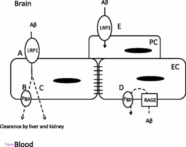
Figure 8