journal of hematology - link.springer.com · review open access signaling pathways in the...
TRANSCRIPT
Signaling pathways in the development ofinfantile hemangiomaJi et al.
JOURNAL OF HEMATOLOGY& ONCOLOGY
Ji et al. Journal of Hematology & Oncology 2014, 7:13http://www.jhoonline.org/content/7/1/13

JOURNAL OF HEMATOLOGY& ONCOLOGY
Ji et al. Journal of Hematology & Oncology 2014, 7:13http://www.jhoonline.org/content/7/1/13
REVIEW Open Access
Signaling pathways in the development ofinfantile hemangiomaYi Ji1*†, Siyuan Chen1,2†, Kai Li3, Li Li4, Chang Xu1 and Bo Xiang1*
Abstract
Infantile hemangioma (IH), which is the most common tumor in infants, is a benign vascular neoplasm resultingfrom the abnormal proliferation of endothelial cells and pericytes. For nearly a century, researchers have noted thatIH exhibits diverse and often dramatic clinical behaviors. On the one hand, most lesions pose no threat or potentialfor complication and resolve spontaneously without concern in most children with IH. On the other hand,approximately 10% of IHs are destructive, disfiguring and even vision- or life-threatening. Recent studies haveprovided some insight into the pathogenesis of these vascular tumors, leading to a better understanding of thebiological features of IH and, in particular, indicating that during hemangioma neovascularization, two mainpathogenic mechanisms prevail, angiogenesis and vasculogenesis. Both mechanisms have been linked to alterationsin several important cellular signaling pathways. These pathways are of interest from a therapeutic perspectivebecause targeting them may help to reverse, delay or prevent hemangioma neovascularization. In this review, weexplore some of the major pathways implicated in IH, including the VEGF/VEGFR, Notch, β-adrenergic, Tie2/angiopoietins,PI3K/AKT/mTOR, HIF-α-mediated and PDGF/PDGF-R-β pathways. We focus on the role of these pathways in thepathogenesis of IH, how they are altered and the consequences of these abnormalities. In addition, we review thelatest preclinical and clinical data on the rationally designed targeted agents that are now being directed againstsome of these pathways.
Keywords: Infantile hemangioma, Neovascularization, Angiogenesis, Vasculogenesis
BackgroundInfantile hemangioma (IH) is a common disorder in in-fancy, with an estimated prevalence of 5 to 10%. If leftuntreated, these tumors are characterized by a rapidgrowth phase during the first year of life, followed byslow involution, which may continue until the age of10–12 years (Figure 1) [1,2]. However, some IHs willleave residual changes, such as telangiectasias, fibro-fattytissue, scars, excessive atrophic skin and pigmentchanges. In 10% of cases, IHs grow dramatically and des-troy tissue, impair function or even threaten life [3]. Thestandard treatment options for IH include corticoste-roids or surgical excision, and the options in life- orsight-threatening cases include treatment with vincris-tine, interferon or cyclophosphamide. Unfortunately,
* Correspondence: [email protected]; [email protected]†Equal contributors1Division of Oncology, Department of Pediatric Surgery, West China Hospitalof Sichuan University, Chengdu 610041, ChinaFull list of author information is available at the end of the article
© 2014 Ji et al.; licensee BioMed Central Ltd. TCommons Attribution License (http://creativecreproduction in any medium, provided the orwaiver (http://creativecommons.org/publicdomstated.
none of these therapeutic modalities are ideal due to re-strictions or potential serious side effects [4-7]. β-blockers have recently been introduced as a safe and ef-fective treatment for IH [8-11]. However, their use is notwithout risk, and not all tumors respond to these drugs[12,13]. These issues have spurred extensive research toclarify the signaling pathways implicated in hemangiomaneovascularization in the hope that a greater under-standing of its molecular pathogenesis will reveal newstrategies to tackle IH.The initial histochemical work of Mulliken and Glowacki
[14], examining endothelial cell (EC) morphology, shedlight on the cellular components of IH. In the past decade,hemangioma-derived progenitor/stem cells (HemSCs), mes-enchymal stem cells (Hem-MSCs), endothelial progenitorcells (HemEPCs), ECs (HemECs) and perivascular cells(Hem-pericytes), all of which comprise the IH, have beenisolated (Table 1) [15-18]. In general, CD133 was used asa stem cell biomarker for the isolation of HemSCs fromIH tissues. HemEPCs were purified from HemSCs based
his is an Open Access article distributed under the terms of the Creativeommons.org/licenses/by/2.0), which permits unrestricted use, distribution, andiginal work is properly cited. The Creative Commons Public Domain Dedicationain/zero/1.0/) applies to the data made available in this article, unless otherwise

Figure 1 Hematoxylin and eosin (H&E) stained sections of proliferating, involuting and involuted phases of IH. The proliferating phase ischaracterized by densely packed tumor cells that form immature vessels (A). In the involuting phase, disorganized vasculature consists of flatendothelium and pericytes (B). The tumor is replaced by fat and/or connective tissues in the involuted phase (C). Scale bar = 100 μm.
Ji et al. Journal of Hematology & Oncology 2014, 7:13 Page 2 of 13http://www.jhoonline.org/content/7/1/13
on expression of the EC marker CD31. In contrast, Hem-MSCs didn’t express CD31 or CD34. In IH tissues, CD133expression was found to be located in both perivascularregion and endothelium [19]. Therefore, HemSCs maycontain both of Hem-MSCs and HemEPCs. Studies fromdifferent groups have demonstrated that HemSCs havethe ability to self-renew and can differentiate into endo-thelium, adipocytes and pericytes in vitro [15,20]. Whenimplanted subcutaneously into nude mice, HemSCs canproduce human glucose transporter-1 (GLUT-1) positivemicrovessels at 7–14 days [15,20-22].We now recognize that IH may be not only a disorder
of angiogenesis (i.e., the sprouting of new vessels fromexisting ones) but also – at least in part – a disorder ofvasculogenesis (i.e., the de novo formation of new bloodvessels from stem cells) [20,24,25]. Improved knowledgeof the signaling pathways that regulate angiogenesis andvasculogenesis has led to the identification of several pos-sible therapeutic targets that have driven the development
Table 1 Cellular components isolated from IH
Cell type Abbreviation Cell marker
Hemangioma-derived endothelial cell HemEC CD31/PECAM-1, vWFVEGFR-2, Tie-2 and V
Hemangioma-derived endothelialprogenitor cell
HemPEC CD133*, VEGFR-2, CDCD146, VE-cadherin
Hemangioma-derived mesenchymalstem cell
Hem-MSC SH2(CD105), SH3, SHCD29, α-SMA and C
Hemangioma-derived stem cell HemSC CD90, CD133, VEGFRneuroplin-1 and CD
Hemangioma-derived pericyte Hem-pericyte PDGFR-β, neural gliadesmin, calponin, smsmooth muscle α-acmuscle myosin heav
*CD133, a pentaspan membrane protein, is used as a stem cell biomarker for the isfor self-renewal, tumorigenesis, metabolism, differentiation, autophagy, apoptosis athe development of IH.
of molecularly targeted therapies. Because many of thesignaling pathways are implicated in the pathogenesis ofvarious tumor types, insight gained from these studies willenable the development of target-specific drugs, not onlyfor IH but also for malignant vascular tumors. This reviewwill highlight the most important of these findings. Al-though the signaling pathways involved in the develop-ment of IH are described separately below, there arenumerous interactions among them, indirectly reflectingthe complexity of IH pathogenesis.
VEGF/VEGFR pathwayThe human vascular endothelial growth factor (VEGF)family consists of VEGF-A, VEGF-B, VEGF-C, VEGF-Dand placental growth factor (PIGF). These growth fac-tors play pivotal roles in embryonic development andangiogenesis-dependent disease [26]. Many reports haveconfirmed that excessive VEGF expression in IH tissueparallels the proliferating phase of its growth. Conversely,
Characteristics
, E-selectin,E-cadherin
Immature endothelial cells; Clonal expansion;Increased proliferation, migration, tumorformation and survival ability.
34, CD31,and vWF
Immature endothelial cells; Increased adhesion,migration and proliferation in the presenceof endostatin or VEGF.
4, CD90,D133
Multilineage differentiation: adipogenic,osteoblastic and myoblastic differentiation
-1, VEGFR-2,146
Multilineage differentiation: ECs, neuronal cells,adipocytes, osteocytes and chondrocytes;Form hemangioma-like Glut-1+ blood vesselsin nude mice.
l antigen-2,ooth muscle 22α,tin, α-SMA, smoothy chain and CD90
Increased proliferation ability; Reduced contractility;Diminished ability to stabilize blood vessels in IH.
olation of progenitor/stem-like cells from IH tissues. CD133 is also responsiblend regeneration [23]. However, little is known about its biological functions in

Ji et al. Journal of Hematology & Oncology 2014, 7:13 Page 3 of 13http://www.jhoonline.org/content/7/1/13
in the involuting phase, VEGF expression rapidly de-creases, and many angiogenesis inhibitors become prom-inent [21,27,28].The functions of the different VEGF family members
are determined by their receptor specificity. Two recep-tors for VEGF are members of the tyrosine-kinase familyand conserved in ECs. These VEGF receptors (VEGFR)are VEGFR-Flt-1 (VEGFR-1) and VEGFR-Flk-1/KDR(VEGFR-2). VEGFR-1 and VEGFR-2 are located on ECs,bone-marrow derived hematopoietic cells and tumor cells,etc. [26,29]. The expression of these receptors is low innormal tissues and only upregulated during the develop-ment of those pathological states when neovascularizationoccurs [30]. Another receptor, VEGFR-3, is primarilyexpressed in lymph nodes and tumor blood vessels[31,32]. Neuropilin-1 and neuropilin-2 were discovered ascoreceptors that that enhanced the binding and effective-ness of the VEGF stimulation of their receptors [33].
Upregulated autocrine VEGF-A/VEGFR-2 loop in HemECOne of the most intensely studied factors involved inangiogenesis and vasculogenesis is VEGF-A. VEGFR-2is known to mediate the majority of the downstream
Figure 2 The VEGF signaling pathway in HemECs and HemSCs. Upon ligdifferent tyrosine residues. Phosphorylation in turn elicits differential downstre
angiogenic effects of VEGF-A, including microvascularpermeability, EC proliferation, migration and survival[34]. Upon the activation of VEGFR-2 in ECs, threemajor secondary messenger pathways trigger multipledownstream signals that promote angiogenesis. Thesepathways are the following: the mitogen-activated pro-tein/ERK kinase (MEK)/extracellular signal-regulatedkinase (ERK) cascade, the phosphatidylinostitol-3 kinase(PI3K)/serine-threonine protein kinase/Akt cascade andthe phospholipase C-γ/intracellular Ca2+/(protein kinaseC (PKC) cascade [35,36]. The genetic deletion of VEGF-Aor its primary signaling receptor VEGFR-2 results in earlyembryonic lethality, associated with a near-complete blockof hematopoietic and vascular development [37].High-VEGFR-2 cells are well documented to exhibit a
higher capacity of self-renewal and superior growthin vitro and in vivo compared with a low-VEGFR-2 cellpopulation [33,38]. HemECs demonstrate the phenotypeof a constitutively active autocrine VEGF-A/VEGFR-2loop (Figure 2), which renders the cells more sensitive toparacrine/external stimulation by VEGF-A and results inthe increased proliferation and migration of cells andtumor formation [39,40]. These characteristics likely result
and binding, VEGF receptors dimerize, leading to the phosphorylation ofam signaling events.
Ji et al. Journal of Hematology & Oncology 2014, 7:13 Page 4 of 13http://www.jhoonline.org/content/7/1/13
from the genetic instability of HemECs as somatic mis-sense mutations in the kinase insert of the VEGFR-2 genehave been found in IHs [41]. In addition, imbalances ingene expression have been reported in mesenchymal com-partments compared to normal tissues, suggesting a pos-sible reciprocal interaction between HemECs and thesurrounding cells [42-44]. Alternatively, HemECs may ori-ginate from progenitor/stem-like cells, which are knownto display robust proliferative and clonogenic capabilitiesand to express high levels of VEGF-A [15,21]. The expres-sion level of VEGF is also increased in HemECs, althoughthis increase is not dramatic [40]. Finally, COSMC was re-ported to be overexpressed in proliferating IHs, with anassociation with the enhanced VEGF-mediated phosphor-ylation of VEGFR-2 and its downstream signaling [45].The abnormal activation of VEGFR-2 on the cell sur-
face may also be beneficial to the survival of HemECs asVEGF-A plays a critical role in protecting ECs againstapoptotic cell death [46,47]. In addition, this inhibitionof EC apoptosis can improve angiogenesis and vasculo-genesis in patients with ischemia [30]. We recently indi-cated that maintaining Bcl-2 expression via VEGF-A/VEGFR-2 signaling in primary HemECs blocked the cellsfrom apoptotic death in the absence of external VEGF-A. Moreover, the inactivation of PI3K/Akt suppressedthe VEGF-A/VEGFR-2-mediated anti-apoptotic effectand unleashed the inhibitory effect of VEGF-A/VEGFR-2 signaling over the reduction of Bcl-2 expression,thereby amplifying the activation of the caspase cascade[48]. These findings suggest that HemECs may be ableto adapt to the abnormal physical environment of thetumor by undergoing a form of reprogramming that in-volves an increase in apoptosis resistance and by up-regulating a VEGF autocrine survival feedback loop tosustain these effects and stabilize the aberrant phenotype.
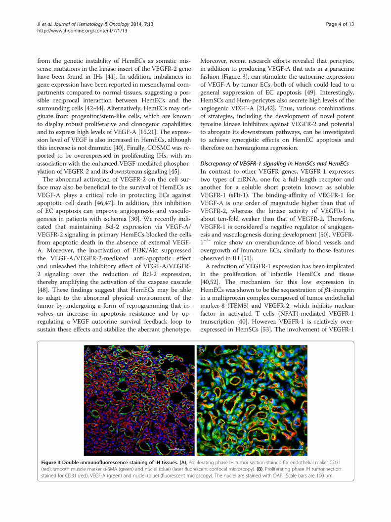
Figure 3 Double immunofluorescence staining of IH tissues. (A), Prolif(red), smooth muscle marker α-SMA (green) and nuclei (blue) (laser fluoresstained for CD31 (red), VEGF-A (green) and nuclei (blue) (fluorescent micro
Moreover, recent research efforts revealed that pericytes,in addition to producing VEGF-A that acts in a paracrinefashion (Figure 3), can stimulate the autocrine expressionof VEGF-A by tumor ECs, both of which could lead to ageneral suppression of EC apoptosis [49]. Interestingly,HemSCs and Hem-pericytes also secrete high levels of theangiogenic VEGF-A [21,42]. Thus, various combinationsof strategies, including the development of novel potenttyrosine kinase inhibitors against VEGFR-2 and potentialto abrogate its downstream pathways, can be investigatedto achieve synergistic effects on HemEC apoptosis andtherefore on hemangioma regression.
Discrepancy of VEGFR-1 signaling in HemSCs and HemECsIn contrast to other VEGFR genes, VEGFR-1 expressestwo types of mRNA, one for a full-length receptor andanother for a soluble short protein known as solubleVEGFR-1 (sFlt-1). The binding-affinity of VEGFR-1 forVEGF-A is one order of magnitude higher than that ofVEGFR-2, whereas the kinase activity of VEGFR-1 isabout ten-fold weaker than that of VEGFR-2. Therefore,VEGFR-1 is considered a negative regulator of angiogen-esis and vasculogenesis during development [50]. VEGFR-1−/− mice show an overabundance of blood vessels andovergrowth of immature ECs, similarly to those featuresobserved in IH [51].A reduction of VEGFR-1 expression has been implicated
in the proliferation of infantile HemECs and tissue[40,52]. The mechanism for this low expression inHemECs was shown to be the sequestration of β1-inergrinin a multiprotein complex composed of tumor endothelialmarker-8 (TEM8) and VEGFR-2, which inhibits nuclearfactor in activated T cells (NFAT)-mediated VEGFR-1transcription [40]. However, VEGFR-1 is relatively over-expressed in HemSCs [53]. The involvement of VEGFR-1
erating phase IH tumor section stained for endothelial maker CD31cent confocal microscopy). (B), Proliferating phase IH tumor sectionscopy). The nuclei are stained with DAPI. Scale bars are 100 μm.

Ji et al. Journal of Hematology & Oncology 2014, 7:13 Page 5 of 13http://www.jhoonline.org/content/7/1/13
in the progression of IH could involve at least onemechanism: the activation of HemSC function, with asubsequent increase in vasculogenesis. VEGF-A, either en-dogenous or exogenous, significantly induces VEGFR-1-mediated ERK1/2 phosphorylation in HemSCs andpromotes the differentiation of HemSCs to HemECs(Figure 2). Moreover, VEGF-B, which is the specific ligandfor VEGFR-1, is highly expressed in HemECs and inducessimilar effects [53]. These results clearly indicate that notonly the paracrine function of VEGF-B from HemECs butalso the persistent autocrine signaling through the VEGF-A/VEGFR-1 loop in HemSCs contributes to enhanced IHvasculogenesis in general.
Notch pathwayThe Notch pathway is a conserved ligand-receptor sig-naling mechanism that modulates cell fate and differen-tiation. The interaction of Notch receptors (Notch 1 to4) with their ligands (Delta-like 1, -3, -4, Jagged-1 and −2)leads to the cleavage of the transmembrane Notch recep-tor, giving rise to the Notch intracellular domain (NICD)that migrates into the nucleus. In the nucleus, the NICDassociates with a transcription factor, recombination signalbinding protein for immunoglobulin kappa J (RBP-Jk),and activates transcription from the RBP-Jk DNA bindingsite. The NICD-RBP-Jk complex upregulates the expres-sion of primary target genes of Notch signaling, such ashairy and enhancer of split (HES) and HES-related protein(HERP/HEY) family of transcription factors [54,55].
Notch expression in IHAlthough the expression levels of the Notch componentsare likely dynamic during development, making transientexpression difficult to detect, current data suggest thatmany known Notch components, mainly two ligands(Delta-like-4 and Jagged-1), three receptors (North-1, -3and −4) and four effectors (HES-1, HEY1, HEY2 andHEYL) are involved in the pathogenesis of IH. BothJagged-1 and Notch-4 are increased in proliferating IHs.All transcript levels of Notch-1, Notch-3, Notch-4,Jagged-1 and Delta-like-4 (Dll4) were higher in the IHthan in the placenta (a commonly used tissue for com-parisons). Conversely, Notch-2 is strongly decreased inboth proliferating and involuting IHs [44,56].
Notch signaling triggers cell-cell interactions in IHNotch signaling is initiated when the extracellular do-main of the receptor engages ligands found on neighbor-ing cells that are in close proximity to one another. Thus,Notch signaling depends on cell-contact-dependent inter-actions. In many cases, the cell that presents the ligand isa cell that does not have Notch signaling present, thus dis-tinguishing two neighboring cells into one with ligandwith little Notch signaling and one with receptor and high
Notch signaling [57]. In a study by Wu et al. [44], the in-vestigators demonstrated that HemSCs have distinctNotch expression patterns from HemECs. In HemSCs,where Notch3 is strongly expressed, HES1, HEY1, andHEYL were expressed at levels 10 to 100 times to that ofHEY-2. In HemECs, however, Notch-1, Notch-4 andJagged-1 have higher expression levels. HEY-2 proteinswere often found to be expressed in HemECs. However,HEY-2 was not uniformly present in all ECs, suggestingthat only a subset of IH ECs express the Notch target [56].These data suggest the possibility that the Notch pathwaymight also contribute to establishing two distinct subpop-ulations at different steps of angiogenesis in IH, such asECs versus smooth muscle cells (SMCs)/pericytes, arteriesversus veins and large vessels versus capillaries [54,58,59].We highlight the concept that ligand-receptor interactionsin Notch signaling depend on contact between two cells,which may be two different cell types. Notch ligands in-volved in IH angiogenesis may be presented by HemECs,pericytes or HemSCs. Interestingly, research by Boscololet al. [43] revealed that endothelial-derived Jagged-1 caninduce HemSCs to acquire a pericyte-like phenotype,which is a crucial step in the vasculogenesis of IH. Disrup-tion of the juxtacrine interaction between endothelialJagged-1 and Notch receptors on HemSCs inhibited bloodvessel formation in IH murine models. However, micehomozygous for a null mutation of several components ofthe Notch pathway, including Notch-1 and Jagged-1, re-sulted in embryonic lethality with vascular remodelingdefects. Vasculogenesis proceeded normally in these mu-tants, whereas the next step, angiogenesis, was disrupted[60,61]. These data suggest that the upregulated Jagged-1expression in the IH endothelium may provide a uniqueeffect to control the vascular development of IH.
Is there a specific relationship between VEGF and Notchpathways in IH?In vivo and in vitro studies have revealed several ways inwhich the VEGF and Notch pathways interact. Particu-larly, VEGF increases Dll4 expression [62,63]. Dll4 isstrongly expressed by the ECs of sprouting angiogenicvessels, which commonly respond to VEGF signals. Thereis evidence that the blockade of VEGF in tumors results ina rapid decrease of Dll4 expression in tumor ECs [64].Interestingly, although HemECs had higher VEGF-Alevels and increased activation of VEGFR-2 comparedwith normal ECs [40], the Dll4 levels in HemECs werelower than those found in normal ECs [44]. These dataargue against the idea that VEGF interacts with Notchsignaling in IH. However, several lines of evidence indicateotherwise. For example, the disruption of Dll4 orendothelium-specific loss of Notch1 increases the superfi-cial plexus vascular density and causes an excess of angio-genic sprouts. This loss of Notch signaling is associated

Ji et al. Journal of Hematology & Oncology 2014, 7:13 Page 6 of 13http://www.jhoonline.org/content/7/1/13
with an increase in VEGFR-2 activity [57]. Other studieshave suggested that reduced Notch activity resulted in re-duced VEGFR-1 expression and increased VEGFR-2 ex-pression in cultured ECs [65,66]. In addition, Hellstromet al. [67] demonstrated that Dll4-Notch signaling withinthe endothelial cell population serves to suppress thetip-cell phenotype. The retinal vascular abnormalities inDll4+/− mice and after long-term treatment with γ-secretase inhibitors might also result from changes in thepattern of VEGF-A expression [67]. In contrast to Dll4,Jagged-1 is proangiogenic protein that functions by down-regulating Dll4-Notch signaling. Jagged-1 also counteractsDll4-Notch signaling interactions between stalk ECs,which helps to sustain elevated VEGF receptor expressionin the newly formed and therefore immature vascularplexus at the angiogenic front [68]. By analogy to studiesof VEGF signaling in HemECs, Notch components maybe novel regulators for VEGF signaling in IH (Figure 4).
Figure 4 Tip/stalk cell specification during spouting angiogenesis andformed vessels in response to pro-angiogenic cues, such as hypoxia-induceDll4 on endothelial cells, which in turn activates Notch receptors on adjacesignaling, providing an additional feedback loop between the two pathwaexpression of VEGFR-1 and VEGFR-3 in those cells. In contrast, Notch activaconcomitant decrease in the proangiogenic response to exogenous VEGF. Boresponse in connector cells that exhibit Notch activation.
Until now, the involvement of Notch in IH develop-ment has remained poorly understood, and many issuesstill need to be addressed. How does the Notch pathwayplay a role in the interaction between HemECs andSMCs/pericytes? How are the different roles that theNotch pathway plays, such as arteriovenous patterning,tip cell differentiation and vessel wall formation, inte-grated during vascular development in IH? How dosome of the key downstream Notch target genes affectIH vessels in the presence of high VEGF levels? And fi-nally, does the expression and/or activity of VEGF com-ponents in IH depend on the nature of Notch signalingor vice versa? These questions should be addressed byfuture research efforts.
β-adrenergic signalingThe β-adrenergic receptors (β-ARs), a family of G-protein-coupled receptors that are activated by β-adrenergic
vascular development. Angiogenic sprouts emerge from the newlyd VEGF. VEGF stimulus, acting via VEGFR-2, increases the expression ofnt endothelial cells. Furthermore, VEGFRs are regulated by Notchys: activated Notch receptors on ECs can positively regulate thetion leads to the reduction of VEGFR-2 expression in cell culture and ath of these effects would likely lead to a lower migratory or proliferative

Ji et al. Journal of Hematology & Oncology 2014, 7:13 Page 7 of 13http://www.jhoonline.org/content/7/1/13
agonists (e.g., epinephrine or norepinephrine), can initiatea series of signaling cascades, thereby leading to multiple,cell-specific responses (Figure 5). The ligation of β-ARs byβ-adrenergic agonists triggers a G-protein coupled signal-ing cascade that stimulates cyclic AMP (cAMP) synthesis.This secondary messenger, cAMP, regulates many cellularfunctions through its effectors, such as cAMP-dependentprotein kinase (PKA) and EPAC (exchange proteins dir-ectly activated by cAMP) [69-71]. Preclinical studies have
Figure 5 β-adrenergic signaling modulates multiple cellular processeepinephrine or norepinephrine triggers a G-protein coupled signaling cascadcan mediate multiple signal pathways via the phosphorylation of various dowof EPAC leads to the Rap1A-mediated activation of Raf/MAPK signaling pathw
demonstrated that β-adrenergic signaling can regulatemultiple fundamental biological processes underlying theprogression and metastasis of tumors, including thepromotion of inflammation [72-74], angiogenesis [75-78],migration [79], invasion [80,81] and resistance to pro-grammed cell death [82-85]. Some evidence suggests thatthe stimulation of β-adrenergic signaling can also inhibitDNA damage repair and the cellular immune response[86,87] and promote surgery-induced metastasis [88,89].
s in tumor progression and metastasis. The ligation of β-ARs bye that stimulates cAMP synthesis. cAMP activates the PKA protein, whichnstream signal proteins. In another major pathway, the cAMP activationays and downstream effects on diverse cellular processes.

Ji et al. Journal of Hematology & Oncology 2014, 7:13 Page 8 of 13http://www.jhoonline.org/content/7/1/13
These findings have led to the hypothesis that commonlyprescribed β-blockers may favorably impact cancer pro-gression and metastasis in patients [90].In the six years since June 2008 when Leaute-Labreze
et al. [11] first described their serendipitous observationof the anti-proliferative effect of propranolol on severeIHs, many articles regarding β-blocker therapy for IHshave been published [8,10,91]. However, despite the ap-parent widespread use of β-blockers, their mechanism ofaction in IHs has not yet been elucidated. Agonists andantagonists of β-ARs are known to act antithetically viathe same intracellular pathways [92]. Given that the ex-pression of all three β-ARs has been demonstrated in IHtumors [93-96], does β-adrenergic signaling play a rolein the pathogenesis of IH? This hypothesis was immedi-ately and, to some degree, indirectly testable by Mayeret al. [97], who found that intrauterine exposure to β2-sympathomimetic hexoprenaline can increase the oc-currence of IH in preterm infants, suggesting a role forβ-AR stimulation in the initiation of IH. Furthermore,we recently demonstrated that the activation of β-ARsresulted in increased HemEC proliferation and upregula-tion of the ERK signaling cascade. VEGFR-2-mediatedERK signaling was also upregulated upon β-AR activa-tion to mediate the proliferation of HemECs [96]. Thesefindings unveil a functional connection between the β-ARs and IH development. However, confirmatory studiesin animal models of IH and mechanistic studies areneeded to clearly define the role of β-adrenergic signal-ing in the growth and involution of IH [98].
Tie-2/Angiopoietin signalingTie-2 and the angiopoietins (Ang), another receptor-ligand system involved in physiological and pathogenicangiogenesis, have also been reported to be associatedwith the development of IHs. Tie-2 tyrosine kinase re-ceptor is expressed specifically on vascular ECs and on acertain subtype of macrophages implicated in angiogen-esis. Ang-1 and Ang-2 have been identified as bona fideligands of the Tie-2 receptor. Ang-1, which is mainlyexpressed by pericytes, is a critical player in vessel mat-uration and mediates the migration, adhesion and sur-vival of ECs. Only tetrameric or higher multimeric formsof Ang-1 activate Tie-2, whereas oligomeric Ang-2 is aweak context-dependent agonist of Tie-2 and may evenantagonize the receptor [99]. Ang-1-mediated Tie-2 acti-vation stimulates a number of intracellular signalingpathways, such as the PI3K/Akt pathway, which pro-motes EC survival and nitric oxide (NO) synthesis bythe activation of the mitogen-activated protein kinase(MAPK) pathway [100,101]. The deletion of Ang-1 be-tween E10.5 and E12.5 results in an enlargement ofvessel diameter, mainly in the capillaries [102,103]. Itsphenotypes were comparable with those of IH, i.e.,
increased numbers of EC and overly covered by peri-cytes [25].Using laser capture microdissection, Calicchio et al.
[52] found Ang-2 was significantly increased in the IHendothelium compared with the placental vessels. Incontrast, Ang-1 was decreased in proliferating IH rela-tive to the placenta. Yu et al. [104] demonstrated thatTie-2 was specifically increased in HemECs and that thisincrease corresponds to enhanced cellular responses tothe Tie-2 agonist Ang-1. Consistent with these findings,Boscolo et al. [42] revealed that hemangioma-derivedpericytes exhibited low levels of Ang-1, resulting in a di-minished ability to stabilize blood vessels in IH.
HIF-α-mediated pathwayHypoxia is one of the most powerful inducers of angio-genesis and vasculogenesis. During tumorigenesis, whentumor cells outgrow the limiting diffusion distance tonearby blood vessels and become hypoxic, the balancebetween pro-angiogenic and anti-angiogenic moleculesis tipped towards pro-angiogenic molecules. This angio-genic switch provokes the expression of a variety of angio-genic factors by tumor cells and stromal cells, includingVEGF-A, stromal cell-derived factor-1α (SDF-1α), fibro-blast growth factor (FGF), platelet-derived growth factors(PDGFs), lysophosphatidic acid (LPA) and Ang [105].Although the initiating mechanism during the patho-
genesis of IH has yet to be discovered, there is evidencethat tissue hypoxia may contribute to their explosivegrowth. The initial clinical description of the promon-tory mark of IH as an ‘area of low blood flow’ suggeststhat tissue ischemia, a powerful stimulus for neovascu-larization, may be involved [106]. The hypothesis that is-chemia/hypoxia plays a crucial role is also supported bythe clinical observation of a blanched area of skin in theposition of the future hemangioma. This region may bean area of local ischemia in the skin, caused by some un-known events, that creates a hypoxic environment andthus triggers growth factor expression.In keeping with the observations described above, Ritter
et al. [107] proposed a mechanism for myeloid cell-facilitated IH growth involving the hypoxia-induced ex-pression of several growth factors (e.g., insulin-like growthfactor-2) that drive endothelial proliferation. Kleinmanet al. [108] demonstrated the presence of hypoxia-inducedmediators of progenitor/stem cell trafficking in proliferat-ing IH specimens and revealed that the combination ofhypoxia and estradiol results in a synergistic effect on theupregulation of matrix metalloproteinase (MMP-9) in ECsin vitro, a key factor in endothelial progenitor cells (EPCs).The transcription factor hypoxia inducible factor (HIF-1α)was also stabilized in proliferating hemangioma speci-mens. Subsequent investigations revealed that HemECsshow significantly a higher expression of HIF-1α than

Ji et al. Journal of Hematology & Oncology 2014, 7:13 Page 9 of 13http://www.jhoonline.org/content/7/1/13
normal ECs. This upregulated HIF-α is a major contribu-tor to the elevated VEGF levels produced in HemECs, andthe decreased expression of HIF-α reduces the prolifera-tion of these cells [39]. Moreover, the benefit observedduring IH treatment by propranolol has been suggested toalso be primarily due to the reduction of HIF-1α expres-sion [109]. This suggestion has been confirmed by Chimet al. [110], who demonstrated that propranolol exerts itssuppressive effects on HemECs through the HIF-1α-VEGF-A angiogenesis axis, the effects of which were me-diated through the PI3/Akt and p38/MAPK pathways.Altogether, these findings indicate a direct and causativeassociation between HIF-α signaling and the developmentof IH.An additional possible effect of HIF-1α signaling in the
pathogenesis of IH is mediation of EC autophagy. In theirrecent study, Chen et al. [111] revealed that a short expos-ure to hypoxia can induce HIF-α/BNIP3-dependent au-tophagy, which may promote EC survival growth. Incontrast, if the hypoxia stress is prolonged, the autophagyactivation may in turn become 5′-AMP-activated proteinkinase (AMPK)/mammalian target of rapamin (mTOR)dependent and therefore cause programmed EC death.However, the evidence from this study was weakened bynot being performed in IH-derived cells or in IH animalmodels.
PI3K/Akt/mTOR signalingPI3K generates 3-phosphorylated inositol lipids, causingthe activation of downstream signaling, resulting in theactivation of protein kinase B (PKB; also called c-Akt),which regulates, among others, mTOR, glycogen syn-thase kinase-3β and Forkhead box O transcription factoractivity. Downstream targets of mTOR include p70 ribo-somal protein 6S kinase (S6K). The overactivation of thePI3K/Akt/mTOR pathway, a signaling pathway thatplays a key role in cellular growth and survival, has beenimplicated in various tumor pathogeneses, and as such,the inhibition of the PI3K/Akt/mTOR pathway is oftherapeutic interest [112-114]. Medici and Olsen [39]found that HemECs had constitutively active PI3K/Akt/mTOR/p70S6K and tested their hypothesis that thesecells could be sensitive to mTOR inhibitors (e.g., rapa-mycin). Finally, they demonstrated that the treatment ofHemECs with rapamycin results in a significant decreasein HIF-1 and VEGF-A levels and in reduced prolifera-tion. Strikingly, in vivo and in vitro studies further dem-onstrated that rapamycin can reduce the self-renewalcapacity of the HemSCs, diminish the differentiation po-tential and inhibit the vasculogenic activity of these cellsin vivo [115]. These preclinical data provide us with apharmacological basis for the potential use of rapamycinin β-blocker-resistant IHs. Nonetheless, the mechanismthat accounts for the effects of rapamycin in IH is far
from clear, and a growing list of side effects make itdoubtful that rapamycin would ultimately be beneficialin pediatric patients [112].
PDGF-B/PDGFR-β signalingThe first evidence for a possible regulatory role of PDGF-B/PDGF receptor-β (PDGFR-β) signaling in IHs was pro-vided by Walter et al. [116]. These researchers establisheda genetic linkage with chromosome 5q in three familialhemangiomas. The region, 5q31-33, contains three candi-date genes involved in blood vessel growth. These geneswere fibroblast growth factor receptor-4 (FGFR4), PDGFR-β and VEGFR-3 [116]. Subsequently, a study examiningglobal gene expression changes between the IH growthphases by the genome-wide transcriptional profiling ofblood vessels showed a reduction in PDGFR-β expressionduring the involutive phase [52]. These findings providethe possibility that PDGF-B/PDGFR-β signaling may playa role in IH pathogenesis.The endothelium is a critical source of PDGF-B for
PDGF-β-positive mural cell recruitment, as demonstratedby the endothelium-specific ablation of PDGF-B, whichleads to pericyte deficiency [117]. The blockade of pericyterecruitment by abolishing PDGF-B/PDGFR-β signalingcauses a lack of basement membrane matrix depositionand concomitantly increased vessel widths [118]. Inaddition, the ectopic expression of PDGF-B by tumor cellsresults in the increased recruitment of mural cells toblood vessels on the establishment of subcutaneous tu-mors [119,120]. Unfortunately, despite the tight physicaland functional association between ECs and pericytes,there is a paucity of information about the signals ex-changed between the two cell types in IHs. Reassuringly,data from a separate study demonstrated that PDGF/PDGF-R-β signaling may act as an intrinsic negative regu-lator of IH involution. In this study, Roach and colleagues[121] found that PDGF is elevated during the proliferatingphase and may inhibit adipocyte differentiation. The ex-posure of HemSCs to exogenous PDGF results in an acti-vation of autocrine PDGF/PDGF-R-β signaling, therebyinhibiting IH involution. These findings highlight the in-volvement of PDGF/PDGF-R-β signaling in the develop-ment of IHs. Moreover, hemangioma-derived pericytesalso express PDGFR-β, although its effect has not beenelucidated in IH pathogenesis [42]. Thus, the possibility oftargeting HemSC and Hem-pericyte function, for ex-ample, via their PDGF receptors, to gain enhanced efficacyof antiangiogenic treatment regiments is supported by thereports of beneficial effects of combining PDGFR inhibi-tors with antiangiogenic drugs or regimens [118,122,123].
Conclusion and future challengesIn conclusion, the findings summarized above demon-strate that signaling pathways involved in the development

Ji et al. Journal of Hematology & Oncology 2014, 7:13 Page 10 of 13http://www.jhoonline.org/content/7/1/13
of IH are increasingly being clarified, underscoring theirsignificant relevance to understanding IH pathogenesis.However, similar to malignant tumors, there is extensivecrosstalk between individual signaling pathways in IH.This crosstalk is generally due to two factors. First, mul-tiple pathways often control a common process. Second,many signaling outcomes impact other processes throughfeedback loops and compensatory responses. Therefore,elucidating the molecular pathogenesis of IH presents anintriguing challenge. To solve this puzzle, an organized re-construction of the sequential molecular perturbationsduring IH neovascularization is required. Such an analysisneeds to combine data from different levels, includinggenetic aberrations, expression alterations and proteinmodification in a comprehensive set of tissue samples.These issues should highlight the important role that theincreased knowledge of the molecular pathways involvedin the pathogenesis of IH will have in guiding the develop-ment of effective, rationally designed therapeutic strat-egies. Future research efforts will not only provide us witha pharmacological basis of the therapeutic use of β-blocker in IHs but also a basis for the further investigationof other potential anti-hemangioma agents.
Competing interestsThe authors declare that they have no competing interests.
Authors’ contributionsYJ and SYC drafted the manuscript. All the authors have read and approvedthe final manuscript.
AcknowledgementsThis work was supported in part by grants from the National Nature ScienceFoundation of China (Grants 31201095, 81071903 and 81072069) and WestChina Hospital of Sichuan University. No institution was involved in datainterpretation, writing the article, or the decision to submit the paper forpublication. The authors are indebted to all reviewers for their kindlyreviewing of the manuscript.
Author details1Division of Oncology, Department of Pediatric Surgery, West China Hospitalof Sichuan University, Chengdu 610041, China. 2Pediatric Intensive Care Unit,West China Hospital of Sichuan University, Chengdu 610041, China. 3Divisionof Oncology, Department of Pediatric Surgery, Children’s Hospital of FudanUniversity, Shanghai 201102, China. 4Laboratory of Pathology, West ChinaHospital of Sichuan University, Chengdu 610041, China.
Received: 5 December 2013 Accepted: 28 January 2014Published: 31 January 2014
References1. Mulliken JB, Fishman SJ, Burrows PE: Vascular anomalies. Curr Probl Surg
2000, 37(8):517–584.2. Drolet BA, Esterly NB, Frieden IJ: Hemangiomas in children. N Engl J Med
1999, 341(3):173–181.3. Margileth AM, Museles M: Cutaneous hemangiomas in children. Diagnosis
and conservative management. JAMA 1965, 194(5):523–526.4. George ME, Sharma V, Jacobson J, Simon S, Nopper AJ: Adverse effects of
systemic glucocorticosteroid therapy in infants with hemangiomas.Arch Dermatol 2004, 140(8):963–969.
5. Goyal R, Watts P, Lane CM, Beck L, Gregory JW: Adrenal suppression andfailure to thrive after steroid injections for periocular hemangioma.Ophthalmology 2004, 111(2):389–395.
6. Neri I, Balestri R, Patrizi A: Hemangiomas: new insight and medicaltreatment. Dermatol Ther 2012, 25(4):322–334.
7. Chang LC, Haggstrom AN, Drolet BA, Baselga E, Chamlin SL, Garzon MC,Horii KA, Lucky AW, Mancini AJ, Metry DW, et al: Growth characteristics ofinfantile hemangiomas: implications for management. Pediatrics 2008,122(2):360–367.
8. Chan H, McKay C, Adams S, Wargon O: RCT of timolol maleate gel forsuperficial infantile hemangiomas in 5- to 24-week-olds. Pediatrics 2013,131(6):e1739–e1747.
9. Malik MA, Menon P, Rao KL, Samujh R: Effect of propranolol vsprednisolone vs propranolol with prednisolone in the management ofinfantile hemangioma: a randomized controlled study. J Pediatr Surg2013, 48(12):2453–2459.
10. Hogeling M, Adams S, Wargon O: A randomized controlled trial ofpropranolol for infantile hemangiomas. Pediatrics 2011, 128(2):e259–e266.
11. Leaute-Labreze C, Dumas DLRE, Hubiche T, Boralevi F, Thambo JB, Taieb A:Propranolol for severe hemangiomas of infancy. N Engl J Med 2008,358(24):2649–2651.
12. Causse S, Aubert H, Saint-Jean M, Puzenat E, Bursztejn AC, Eschard C, Mahe E,Maruani A, Mazereeuw-Hautier J, Dreyfus I, et al: Propranolol-resistant infantilehaemangiomas. Br J Dermatol 2013, 169(1):125–129.
13. Shehata N, Powell J, Dubois J, Hatami A, Rousseau E, Ondrejchak S, McCuaig C:Late rebound of infantile hemangioma after cessation of oral propranolol.Pediatr Dermatol 2013, 30(5):587–591.
14. Mulliken JB, Glowacki J: Hemangiomas and vascular malformations ininfants and children: a classification based on endothelial characteristics.Plast Reconstr Surg 1982, 69(3):412–422.
15. Khan ZA, Boscolo E, Picard A, Psutka S, Melero-Martin JM, Bartch TC, Mulliken JB,Bischoff J: Multipotential stem cells recapitulate human infantilehemangioma in immunodeficient mice. J Clin Invest 2008, 118(7):2592–2599.
16. Yu Y, Fuhr J, Boye E, Gyorffy S, Soker S, Atala A, Mulliken JB, Bischoff J:Mesenchymal stem cells and adipogenesis in hemangioma involution.Stem Cells 2006, 24(6):1605–1612.
17. Yu Y, Flint AF, Mulliken JB, Wu JK, Bischoff J: Endothelial progenitor cells ininfantile hemangioma. Blood 2004, 103(4):1373–1375.
18. Dosanjh A, Chang J, Bresnick S, Zhou L, Reinisch J, Longaker M, Karasek M:In vitro characteristics of neonatal hemangioma endothelial cells:similarities and differences between normal neonatal and fetalendothelial cells. J Cutan Pathol 2000, 27(9):441–450.
19. Yuan SM, Chen RL, Shen WM, Chen HN, Zhou XJ: Mesenchymal stem cellsin infantile hemangioma reside in the perivascular region. Pediatr DevPathol 2012, 15(1):5–12.
20. Xu D, TM O, Shartava A, Fowles TC, Yang J, Fink LM, Ward DC, Mihm MC,Waner M, Ma Y: Isolation, characterization, and in vitro propagation ofinfantile hemangioma stem cells and an in vivo mouse model. J HematolOncol 2011, 4:54.
21. Greenberger S, Boscolo E, Adini I, Mulliken JB, Bischoff J: Corticosteroidsuppression of VEGF-A in infantile hemangioma-derived stem cells.N Engl J Med 2010, 362(11):1005–1013.
22. Mai HM, Zheng JW, Wang YA, Yang XJ, Zhou Q, Qin ZP, Li KL: CD133selected stem cells from proliferating infantile hemangioma andestablishment of an in vivo mice model of hemangioma. Chin Med J(Engl) 2013, 126(1):88–94.
23. Li Z: CD133: a stem cell biomarker and beyond. Exp Hematol Oncol 2013,2(1):17.
24. Greenberger S, Bischoff J: Pathogenesis of infantile haemangioma.Br J Dermatol 2013, 169(1):12–19.
25. Boscolo E, Bischoff J: Vasculogenesis in infantile hemangioma.Angiogenesis 2009, 12(2):197–207.
26. Verheul HM, Pinedo HM: The role of vascular endothelial growth factor(VEGF) in tumor angiogenesis and early clinical development of VEGF-receptor kinase inhibitors. Clin Breast Cancer 2000, 1(Suppl 1):S80–S84.
27. Przewratil P, Sitkiewicz A, Andrzejewska E: Local serum levels of vascularendothelial growth factor in infantile hemangioma: intriguingmechanism of endothelial growth. Cytokine 2010, 49(2):141–147.
28. Zhang L, Lin X, Wang W, Zhuang X, Dong J, Qi Z, Hu Q: Circulatinglevel of vascular endothelial growth factor in differentiatinghemangioma from vascular malformation patients. Plast Reconstr Surg2005, 116(1):200–204.
29. Shibuya M: Vascular endothelial growth factor receptor-1 (VEGFR-1/Flt-1):a dual regulator for angiogenesis. Angiogenesis 2006, 9(4):225–230. 231.

Ji et al. Journal of Hematology & Oncology 2014, 7:13 Page 11 of 13http://www.jhoonline.org/content/7/1/13
30. Dimmeler S, Zeiher AM: Endothelial cell apoptosis in angiogenesis andvessel regression. Circ Res 2000, 87(6):434–439.
31. Huang HY, Ho CC, Huang PH, Hsu SM: Co-expression of VEGF-C and itsreceptors, VEGFR-2 and VEGFR-3, in endothelial cells of lymphangioma.Implication in autocrine or paracrine regulation of lymphangioma.Lab Invest 2001, 81(12):1729–1734.
32. Shawber CJ, Funahashi Y, Francisco E, Vorontchikhina M, Kitamura Y, Stowell SA,Borisenko V, Feirt N, Podgrabinska S, Shiraishi K, et al: Notch alters VEGFresponsiveness in human and murine endothelial cells by direct regulationof VEGFR-3 expression. J Clin Invest 2007, 117(11):3369–3382.
33. Hamerlik P, Lathia JD, Rasmussen R, Wu Q, Bartkova J, Lee M, Moudry P,Bartek JJ, Fischer W, Lukas J, et al: Autocrine VEGF-VEGFR2-Neuropilin-1signaling promotes glioma stem-like cell viability and tumor growth.J Exp Med 2012, 209(3):507–520.
34. Olsson AK, Dimberg A, Kreuger J, Claesson-Welsh L: VEGF receptorsignalling - in control of vascular function. Nat Rev Mol Cell Biol 2006,7(5):359–371.
35. Gupta K, Kshirsagar S, Li W, Gui L, Ramakrishnan S, Gupta P, Law PY, HebbelRP: VEGF prevents apoptosis of human microvascular endothelial cellsvia opposing effects on MAPK/ERK and SAPK/JNK signaling. Exp Cell Res1999, 247(2):495–504.
36. Gerber HP, McMurtrey A, Kowalski J, Yan M, Keyt BA, Dixit V, Ferrara N:Vascular endothelial growth factor regulates endothelial cell survivalthrough the phosphatidylinositol 3′-kinase/Akt signal transductionpathway. Requirement for Flk-1/KDR activation. J Biol Chem 1998,273(46):30336–30343.
37. Ferrara N, Gerber HP, LeCouter J: The biology of VEGF and its receptors.Nat Med 2003, 9(6):669–676.
38. Gerber HP, Malik AK, Solar GP, Sherman D, Liang XH, Meng G, Hong K,Marsters JC, Ferrara N: VEGF regulates haematopoietic stem cellsurvival by an internal autocrine loop mechanism. Nature 2002,417(6892):954–958.
39. Medici D, Olsen BR: Rapamycin inhibits proliferation of hemangiomaendothelial cells by reducing HIF-1-dependent expression of VEGF.PLoS One 2012, 7(8):e42913.
40. Jinnin M, Medici D, Park L, Limaye N, Liu Y, Boscolo E, Bischoff J, Vikkula M,Boye E, Olsen BR: Suppressed NFAT-dependent VEGFR1 expression andconstitutive VEGFR2 signaling in infantile hemangioma. Nat Med 2008,14(11):1236–1246.
41. Walter JW, North PE, Waner M, Mizeracki A, Blei F, Walker JW, Reinisch JF,Marchuk DA: Somatic mutation of vascular endothelial growth factorreceptors in juvenile hemangioma. Genes Chromosomes Cancer 2002,33(3):295–303.
42. Boscolo E, Mulliken JB, Bischoff J: Pericytes from infantile hemangiomadisplay proangiogenic properties and dysregulated angiopoietin-1.Arterioscler Thromb Vasc Biol 2013, 33(3):501–509.
43. Boscolo E, Stewart CL, Greenberger S, Wu JK, Durham JT, Herman IM,Mulliken JB, Kitajewski J, Bischoff J: JAGGED1 signaling regulateshemangioma stem cell-to-pericyte/vascular smooth muscle celldifferentiation. Arterioscler Thromb Vasc Biol 2011, 31(10):2181–2192.
44. Wu JK, Adepoju O, De Silva D, Baribault K, Boscolo E, Bischoff J, Kitajewski J:A switch in Notch gene expression parallels stem cell to endothelialtransition in infantile hemangioma. Angiogenesis 2010, 13(1):15–23.
45. Lee JJ, Chen CH, Chen YH, Huang MJ, Huang J, Hung JS, Chen MT, Huang MC:COSMC is overexpressed in proliferating infantile hemangioma andenhances endothelial cell growth via VEGFR2. PLoS One 2013, 8(2):e56211.
46. Lichtenberger BM, Tan PK, Niederleithner H, Ferrara N, Petzelbauer P, Sibilia M:Autocrine VEGF signaling synergizes with EGFR in tumor cells to promoteepithelial cancer development. Cell 2010, 140(2):268–279.
47. Lee S, Chen TT, Barber CL, Jordan MC, Murdock J, Desai S, Ferrara N, Nagy A,Roos KP, Iruela-Arispe ML: Autocrine VEGF signaling is required for vascularhomeostasis. Cell 2007, 130(4):691–703.
48. Ji Y, Chen S, Li K, Xiao X, Xu T, Zheng S: Upregulated autocrine vascularendothelial growth factor (VEGF)/VEGF receptor-2 loop preventsapoptosis in haemangioma-derived endothelial cells. Br J Dermatol 2014,170(1):78–86.
49. Franco M, Roswall P, Cortez E, Hanahan D, Pietras K: Pericytes promoteendothelial cell survival through induction of autocrine VEGF-A signalingand Bcl-w expression. Blood 2011, 118(10):2906–2917.
50. Kearney JB, Ambler CA, Monaco KA, Johnson N, Rapoport RG, Bautch VL:Vascular endothelial growth factor receptor Flt-1 negatively regulates
developmental blood vessel formation by modulating endothelial celldivision. Blood 2002, 99(7):2397–2407.
51. Fong GH, Rossant J, Gertsenstein M, Breitman ML: Role of the Flt-1 receptortyrosine kinase in regulating the assembly of vascular endothelium.Nature 1995, 376(6535):66–70.
52. Calicchio ML, Collins T, Kozakewich HP: Identification of signaling systemsin proliferating and involuting phase infantile hemangiomas bygenome-wide transcriptional profiling. Am J Pathol 2009,174(5):1638–1649.
53. Boscolo E, Mulliken JB, Bischoff J: VEGFR-1 mediates endothelialdifferentiation and formation of blood vessels in a murine model ofinfantile hemangioma. Am J Pathol 2011, 179(5):2266–2277.
54. Iso T, Hamamori Y, Kedes L: Notch signaling in vascular development.Arterioscler Thromb Vasc Biol 2003, 23(4):543–553.
55. Gridley T: Notch signaling during vascular development. Proc Natl AcadSci U S A 2001, 98(10):5377–5378.
56. Adepoju O, Wong A, Kitajewski A, Tong K, Boscolo E, Bischoff J, Kitajewski J,Wu JK: Expression of HES and HEY genes in infantile hemangiomas.Vasc Cell 2011, 3:19.
57. Dufraine J, Funahashi Y, Kitajewski J: Notch signaling regulates tumorangiogenesis by diverse mechanisms. Oncogene 2008, 27(38):5132–5137.
58. Lanner F, Sohl M, Farnebo F: Functional arterial and venous fate isdetermined by graded VEGF signaling and notch status during embryonicstem cell differentiation. Arterioscler Thromb Vasc Biol 2007, 27(3):487–493.
59. Li JL, Harris AL: Notch signaling from tumor cells: a new mechanism ofangiogenesis. Cancer Cell 2005, 8(1):1–3.
60. Krebs LT, Xue Y, Norton CR, Shutter JR, Maguire M, Sundberg JP, Gallahan D,Closson V, Kitajewski J, Callahan R, et al: Notch signaling is essential forvascular morphogenesis in mice. Genes Dev 2000, 14(11):1343–1352.
61. Xue Y, Gao X, Lindsell CE, Norton CR, Chang B, Hicks C, Gendron-Maguire M,Rand EB, Weinmaster G, Gridley T: Embryonic lethality and vasculardefects in mice lacking the Notch ligand Jagged1. Hum Mol Genet 1999,8(5):723–730.
62. Hainaud P, Contreres JO, Villemain A, Liu LX, Plouet J, Tobelem G, Dupuy E:The role of the vascular endothelial growth factor-Delta-like 4 ligand/Notch4-ephrin B2 cascade in tumor vessel remodeling and endothelialcell functions. Cancer Res 2006, 66(17):8501–8510.
63. Patel NS, Li JL, Generali D, Poulsom R, Cranston DW, Harris AL: Up-regulation of delta-like 4 ligand in human tumor vasculature and therole of basal expression in endothelial cell function. Cancer Res 2005,65(19):8690–8697.
64. Noguera-Troise I, Daly C, Papadopoulos NJ, Coetzee S, Boland P, Gale NW,Lin HC, Yancopoulos GD, Thurston G: Blockade of Dll4 inhibits tumourgrowth by promoting non-productive angiogenesis. Nature 2006,444(7122):1032–1037.
65. Williams CK, Li JL, Murga M, Harris AL, Tosato G: Up-regulation of theNotch ligand Delta-like 4 inhibits VEGF-induced endothelial cell function.Blood 2006, 107(3):931–939.
66. Zhang J, Ye J, Ma D, Liu N, Wu H, Yu S, Sun X, Tse W, Ji C: Cross-talkbetween leukemic and endothelial cells promotes angiogenesis by VEGFactivation of the Notch/Dll4 pathway. Carcinogenesis 2013, 34(3):667–677.
67. Hellstrom M, Phng LK, Hofmann JJ, Wallgard E, Coultas L, Lindblom P, Alva J,Nilsson AK, Karlsson L, Gaiano N, et al: Dll4 signalling through Notch1regulates formation of tip cells during angiogenesis. Nature 2007,445(7129):776–780.
68. Benedito R, Roca C, Sorensen I, Adams S, Gossler A, Fruttiger M, Adams RH:The notch ligands Dll4 and Jagged1 have opposing effects onangiogenesis. Cell 2009, 137(6):1124–1135.
69. Zhang X, Odom DT, Koo SH, Conkright MD, Canettieri G, Best J, Chen H,Jenner R, Herbolsheimer E, Jacobsen E, et al: Genome-wide analysis ofcAMP-response element binding protein occupancy, phosphorylation,and target gene activation in human tissues. Proc Natl Acad Sci U S A2005, 102(12):4459–4464.
70. Luttrell LM, Ferguson SS, Daaka Y, Miller WE, Maudsley S, Della RG, Lin F,Kawakatsu H, Owada K, Luttrell DK, et al: Beta-arrestin-dependentformation of beta2 adrenergic receptor-Src protein kinase complexes.Science 1999, 283(5402):655–661.
71. Ji Y, Chen S, Xiao X, Zheng S, Li K: beta-blockers: a novel class ofantitumor agents. Onco Targets Ther 2012, 5:391–401.
72. Shahzad MM, Arevalo JM, Armaiz-Pena GN, Lu C, Stone RL, Moreno-Smith M,Nishimura M, Lee JW, Jennings NB, Bottsford-Miller J, et al: Stress effects on
Ji et al. Journal of Hematology & Oncology 2014, 7:13 Page 12 of 13http://www.jhoonline.org/content/7/1/13
FosB- and interleukin-8 (IL8)-driven ovarian cancer growth and metastasis. JBiol Chem 2010, 285(46):35462–35470.
73. Bernabe DG, Tamae AC, Biasoli ER, Oliveira SH: Stress hormones increasecell proliferation and regulates interleukin-6 secretion in human oralsquamous cell carcinoma cells. Brain Behav Immun 2011, 25(3):574–583.
74. Cole SW, Arevalo JM, Takahashi R, Sloan EK, Lutgendorf SK, Sood AK,Sheridan JF, Seeman TE: Computational identification of gene-socialenvironment interaction at the human IL6 locus. Proc Natl Acad Sci U S A2010, 107(12):5681–5686.
75. Chakroborty D, Sarkar C, Basu B, Dasgupta PS, Basu S: Catecholaminesregulate tumor angiogenesis. Cancer Res 2009, 69(9):3727–3730.
76. Thaker PH, Han LY, Kamat AA, Arevalo JM, Takahashi R, Lu C, Jennings NB,Armaiz-Pena G, Bankson JA, Ravoori M, et al: Chronic stress promotestumor growth and angiogenesis in a mouse model of ovarian carcinoma.Nat Med 2006, 12(8):939–944.
77. Pasquier E, Street J, Pouchy C, Carre M, Gifford AJ, Murray J, Norris MD,Trahair T, Andre N, Kavallaris M: beta-blockers increase response tochemotherapy via direct antitumour and anti-angiogenic mechanisms inneuroblastoma. Br J Cancer 2013, 108(12):2485–2494.
78. Ji Y, Chen S: Comment on ‘Beta-blockers increase response tochemotherapy via direct anti-tumour and anti-angiogenic mechanismsin neuroblastoma’. Br J Cancer 2013, 109(7):2022–2023.
79. Entschladen F, Drell TT, Lang K, Joseph J, Zaenker KS: Tumour-cellmigration, invasion, and metastasis: navigation by neurotransmitters.Lancet Oncol 2004, 5(4):254–258.
80. Sood AK, Bhatty R, Kamat AA, Landen CN, Han L, Thaker PH, Li Y,Gershenson DM, Lutgendorf S, Cole SW: Stress hormone-mediatedinvasion of ovarian cancer cells. Clin Cancer Res 2006, 12(2):369–375.
81. Armaiz-Pena GN, Allen JK, Cruz A, Stone RL, Nick AM, Lin YG, Han LY,Mangala LS, Villares GJ, Vivas-Mejia P, et al: Src activation by beta-adrenoreceptors is a key switch for tumour metastasis. Nat Commun2013, 4:1403.
82. Sastry KS, Karpova Y, Prokopovich S, Smith AJ, Essau B, Gersappe A, Carson JP,Weber MJ, Register TC, Chen YQ, et al: Epinephrine protects cancer cells fromapoptosis via activation of cAMP-dependent protein kinase and BAD phos-phorylation. J Biol Chem 2007, 282(19):14094–14100.
83. Sood AK, Armaiz-Pena GN, Halder J, Nick AM, Stone RL, Hu W, Carroll AR,Spannuth WA, Deavers MT, Allen JK, et al: Adrenergic modulation of focaladhesion kinase protects human ovarian cancer cells from anoikis. J ClinInvest 2010, 120(5):1515–1523.
84. Hassan S, Karpova Y, Baiz D, Yancey D, Pullikuth A, Flores A, Register T, Cline JM,D’Agostino RJ, Danial N, et al: Behavioral stress accelerates prostate cancerdevelopment in mice. J Clin Invest 2013, 123(2):874–886.
85. Ji Y, Li K, Xiao X, Zheng S, Xu T, Chen S: Effects of propranolol on theproliferation and apoptosis of hemangioma-derived endothelial cells.J Pediatr Surg 2012, 47(12):2216–2223.
86. Hara MR, Kovacs JJ, Whalen EJ, Rajagopal S, Strachan RT, Grant W, Towers AJ,Williams B, Lam CM, Xiao K, et al: A stress response pathway regulates DNAdamage through beta2-adrenoreceptors and beta-arrestin-1.Nature 2011, 477(7364):349–353.
87. Glaser R, Kiecolt-Glaser JK: Stress-induced immune dysfunction: implicationsfor health. Nat Rev Immunol 2005, 5(3):243–251.
88. Goldfarb Y, Sorski L, Benish M, Levi B, Melamed R, Ben-Eliyahu S: Improvingpostoperative immune status and resistance to cancer metastasis: acombined perioperative approach of immunostimulation and preventionof excessive surgical stress responses. Ann Surg 2011, 253(4):798–810.
89. Glasner A, Avraham R, Rosenne E, Benish M, Zmora O, Shemer S, Meiboom H,Ben-Eliyahu S: Improving survival rates in two models of spontaneous post-operative metastasis in mice by combined administration of a beta-adrenergic antagonist and a cyclooxygenase-2 inhibitor. J Immunol 2010,184(5):2449–2457.
90. Ji Y, Chen S: Do antihypertensive medications influence breast cancerrisk? JAMA Intern Med 2014: . In press.
91. Pope E, Chakkittakandiyil A, Lara-Corrales I, Maki E, Weinstein M: Expandingthe therapeutic repertoire of infantile haemangiomas: cohort-blindedstudy of oral nadolol compared with propranolol. Br J Dermatol 2013,168(1):222–224.
92. Powe DG, Entschladen F: Targeted therapies: using beta-blockers toinhibit breast cancer progression. Nat Rev Clin Oncol 2011, 8(9):511–512.
93. Hadaschik E, Scheiba N, Engstner M, Flux K: High levels of beta2-adrenoceptors are expressed in infantile capillary hemangiomas and
may mediate the therapeutic effect of propranolol. J Cutan Pathol 2012,39(9):881–883.
94. Chisholm KM, Chang KW, Truong MT, Kwok S, West RB, Heerema-McKenney AE:beta-Adrenergic receptor expression in vascular tumors. Mod Pathol 2012,25(11):1446–1451.
95. Rossler J, Haubold M, Gilsbach R, Juttner E, Schmitt D, Niemeyer CM, Hein L:beta1-Adrenoceptor mRNA level reveals distinctions between infantilehemangioma and vascular malformations. Pediatr Res 2013,73(4 Pt 1):409–413.
96. Ji Y, Chen S, Li K, Xiao X, Zheng S, Xu T: The role of beta-adrenergic receptorsignaling in the proliferation of hemangioma-derived endothelial cells.Cell Div 2013, 8(1):1.
97. Mayer M, Minichmayr A, Klement F, Hroncek K, Wertaschnigg D, Arzt W,Wiesinger-Eidenberger G, Lechner E: Tocolysis with the beta-2-sympathomimetic hexoprenaline increases occurrence of infantilehaemangioma in preterm infants. Arch Dis Child Fetal Neonatal Ed 2013,98(2):F108–F111.
98. Ji Y, Chen S, Li K, Xiao X, Zheng S: Propranolol: a novel anti-hemangiomaagent with multiple potential mechanisms of action. Ann Surg 2013.In press.
99. Kim KT, Choi HH, Steinmetz MO, Maco B, Kammerer RA, Ahn SY, Kim HZ,Lee GM, Koh GY: Oligomerization and multimerization are critical forangiopoietin-1 to bind and phosphorylate Tie2. J Biol Chem 2005,280(20):20126–20131.
100. Jones N, Master Z, Jones J, Bouchard D, Gunji Y, Sasaki H, Daly R, Alitalo K,Dumont DJ: Identification of Tek/Tie2 binding partners. Binding to amultifunctional docking site mediates cell survival and migration. J BiolChem 1999, 274(43):30896–30905.
101. Jones N, Chen SH, Sturk C, Master Z, Tran J, Kerbel RS, Dumont DJ:A unique autophosphorylation site on Tie2/Tek mediates Dok-Rphosphotyrosine binding domain binding and function. Mol Cell Biol2003, 23(8):2658–2668.
102. Jeansson M, Gawlik A, Anderson G, Li C, Kerjaschki D, Henkelman M,Quaggin SE: Angiopoietin-1 is essential in mouse vasculature duringdevelopment and in response to injury. J Clin Invest 2011,121(6):2278–2289.
103. Koh GY: Orchestral actions of angiopoietin-1 in vascular regeneration.Trends Mol Med 2013, 19(1):31–39.
104. Yu Y, Varughese J, Brown LF, Mulliken JB, Bischoff J: Increased Tie2expression, enhanced response to angiopoietin-1, and dysregulatedangiopoietin-2 expression in hemangioma-derived endothelial cells.Am J Pathol 2001, 159(6):2271–2280.
105. Boye E, Olsen BR: Signaling mechanisms in infantile hemangioma.Curr Opin Hematol 2009, 16(3):202–208.
106. Chen TS, Eichenfield LF, Friedlander SF: Infantile hemangiomas: an updateon pathogenesis and therapy. Pediatrics 2013, 131(1):99–108.
107. Ritter MR, Reinisch J, Friedlander SF, Friedlander M: Myeloid cells ininfantile hemangioma. Am J Pathol 2006, 168(2):621–628.
108. Kleinman ME, Greives MR, Churgin SS, Blechman KM, Chang EI, Ceradini DJ,Tepper OM, Gurtner GC: Hypoxia-induced mediators of stem/progenitorcell trafficking are increased in children with hemangioma.Arterioscler Thromb Vasc Biol 2007, 27(12):2664–2670.
109. Storch CH, Hoeger PH: Propranolol for infantile haemangiomas: insightsinto the molecular mechanisms of action. Br J Dermatol 2010,163(2):269–274.
110. Chim H, Armijo BS, Miller E, Gliniak C, Serret MA, Gosain AK: Propranololinduces regression of hemangioma cells through HIF-1alpha-mediatedinhibition of VEGF-A. Ann Surg 2012, 256(1):146–156.
111. Chen G, Zhang W, Li YP, Ren JG, Xu N, Liu H, Wang FQ, Sun ZJ, Jia J, Zhao YF:Hypoxia-induced autophagy in endothelial cells: a double-edged sword inthe progression of infantile haemangioma? Cardiovasc Res 2013,98(3):437–448.
112. Lamming DW, Ye L, Sabatini DM, Baur JA: Rapalogs and mTOR inhibitorsas anti-aging therapeutics. J Clin Invest 2013, 123(3):980–989.
113. Benjamin D, Colombi M, Moroni C, Hall MN: Rapamycin passes the torch:a new generation of mTOR inhibitors. Nat Rev Drug Discov 2011,10(11):868–880.
114. Slomovitz BM, Coleman RL: The PI3K/AKT/mTOR pathway as a therapeutictarget in endometrial cancer. Clin Cancer Res 2012, 18(21):5856–5864.
115. Greenberger S, Yuan S, Walsh LA, Boscolo E, Kang KT, Matthews B, Mulliken JB,Bischoff J: Rapamycin suppresses self-renewal and vasculogenic potential of

Ji et al. Journal of Hematology & Oncology 2014, 7:13 Page 13 of 13http://www.jhoonline.org/content/7/1/13
stem cells isolated from infantile hemangioma. J Invest Dermatol 2011,131(12):2467–2476.
116. Walter JW, Blei F, Anderson JL, Orlow SJ, Speer MC, Marchuk DA: Geneticmapping of a novel familial form of infantile hemangioma. Am J MedGenet 1999, 82(1):77–83.
117. Bjarnegard M, Enge M, Norlin J, Gustafsdottir S, Fredriksson S, Abramsson A,Takemoto M, Gustafsson E, Fassler R, Betsholtz C: Endothelium-specificablation of PDGFB leads to pericyte loss and glomerular, cardiac andplacental abnormalities. Development 2004, 131(8):1847–1857.
118. Stratman AN, Schwindt AE, Malotte KM, Davis GE: Endothelial-derivedPDGF-BB and HB-EGF coordinately regulate pericyte recruitment duringvasculogenic tube assembly and stabilization. Blood 2010,116(22):4720–4730.
119. Sennino B, Falcon BL, McCauley D, Le T, McCauley T, Kurz JC, Haskell A,Epstein DM, McDonald DM: Sequential loss of tumor vessel pericytes andendothelial cells after inhibition of platelet-derived growth factor B byselective aptamer AX102. Cancer Res 2007, 67(15):7358–7367.
120. Pietras K, Hanahan D: A multitargeted, metronomic, and maximum-tolerated dose “chemo-switch” regimen is antiangiogenic, producingobjective responses and survival benefit in a mouse model of cancer.J Clin Oncol 2005, 23(5):939–952.
121. Roach EE, Chakrabarti R, Park NI, Keats EC, Yip J, Chan NG, Khan ZA: Intrinsicregulation of hemangioma involution by platelet-derived growth factor.Cell Death Dis 2012, 3:e328.
122. Wnuk M, Hlushchuk R, Tuffin G, Huynh-Do U, Djonov V: The effects ofPTK787/ZK222584, an inhibitor of VEGFR and PDGFRbeta pathways, onintussusceptive angiogenesis and glomerular recovery from Thy1.1nephritis. Am J Pathol 2011, 178(4):1899–1912.
123. Erber R, Thurnher A, Katsen AD, Groth G, Kerger H, Hammes HP, Menger MD,Ullrich A, Vajkoczy P: Combined inhibition of VEGF and PDGF signalingenforces tumor vessel regression by interfering with pericyte-mediatedendothelial cell survival mechanisms. Faseb J 2004, 18(2):338–340.
doi:10.1186/1756-8722-7-13Cite this article as: Ji et al.: Signaling pathways in the development ofinfantile hemangioma. Journal of Hematology & Oncology 2014 7:13.
Submit your next manuscript to BioMed Centraland take full advantage of:
• Convenient online submission
• Thorough peer review
• No space constraints or color figure charges
• Immediate publication on acceptance
• Inclusion in PubMed, CAS, Scopus and Google Scholar
• Research which is freely available for redistribution
Submit your manuscript at www.biomedcentral.com/submit