journal of ethnopharmacology - university of malaya · research paper chemoprevention of colonic...
TRANSCRIPT
Research Paper
Chemoprevention of colonic aberrant crypt foci by Gynura procumbensin rats
Abdrabuh N. Shwter a, Nor Azizan Abdullah b, Mohammed A. Alshawsh b,Abdulsamd Alsalahi a, Maryam Hajrezaei a, Amel A. Almaqrami a, Sameer D. Salem c,Mahmood A. Abdulla a,n
a Department of Biomedical Science, Faculty of Medicine, University of Malaya, 50603 Kuala Lumpur, Malaysiab Department of Pharmacology, Faculty of Medicine, University of Malaya, 50603 Kuala Lumpur, Malaysiac Department of Molecular Medicine, Faculty of Medicine, University of Malaya, 50603 Kuala Lumpur, Malaysia
a r t i c l e i n f o
Article history:Received 3 October 2013Received in revised form25 November 2013Accepted 20 December 2013Available online 3 January 2014
Keywords:Gynura procumbensAzoxymethaneAberrant crypt fociAntioxidant activityPCNABcl-2
a b s t r a c t
Ethnopharmacological relevance: Gynura procumbens is commonly used as a traditional medicinal plantin Malaysia for treatment of many diseases. To investigate the chemopreventive properties of Gynuraprocumbens on azoxymethane (AOM)-induced aberrant crypt foci (ACF) in rats.Methods: Five groups of adult male rats were used in this experiment. Normal/control group; the ratswere injected subcutaneously with 15 mg/kg of sterile normal saline once a week for two weeks, andorally administered with 10% Tween 20 (5 mL/kg). Carcinogen and treatment groups; the rats wereinjected subcutaneously each with 15 mg/kg body weight AOM once a week for 2 weeks and werecontinued to be fed for two months, respectively with 10% Tween 20, 500 and 250 mg/kg body weightplant extracts. Reference group; the rats were injected subcutaneously with 15 mg/kg body weight AOMonce a week for 2 weeks, and injected intraperitoneally with fluorouracil 35 mg/kg body weight for fiveconsecutive days.Result: Total ACF detected in methylene blue stained whole mounts of rat colon were 21, 23and 130 inrats fed with 500, 250 mg/kg body weight treatment and carcinogen groups, respectively. Treatmentwith high and low doses of the plant extract led to83.6% and 82.2% decrease in the total crypts in thegroups fed 500 mg/kg and 250 mg/kg Gynura procumbens respectively compared to carcinogen group.Immunohistochemical staining of ACF showed suppressed azoxymethane induced colonic cell prolifera-tion and Bcl-2 expression. Glutathione-S-transfarase and superoxide dismutase activities were higher intreated rats compared to carcinogen groups.Conclusion: Gynura procumbens reduced the incidence of AOM induced ACF. The findings showed thatGynura procumbens may have antiproliferative and antioxidative properties. Moreover, Gynura procumbenspossesses the medicinal properties to prevent colon cancer.
& 2014 Elsevier Ireland Ltd. All rights reserved.
1. Introduction
Colon cancer is the third-leading cause of cancer deaths world-wide (Yamashita et al., 1994). It is estimated that about one millionnew cases of colorectal cancer (CRC) are diagnosed, and about halfa million deaths caused by colon cancer occurred each yearworldwide (Tenesa and Dunlop, 2009). In 2012, there wereapproximately 103,170 cases of colon cancer and 40,290 cases ofrectal cancer are reported (Saslow et al., 2012). Recently, the use ofherbal medicines for complementary treatments of some diseases
has been popular and researchers have shown that medicinalplants are commonly used by cancer patients to manage theirdiseases (Riboli and Norat, 2003; Van Duijnhoven et al., 2009).Gynura procumbens (Lour.)Merr. Compositae is an annual ever-green shrub with a fleshy stem and purple tint. In Malaysia (Lour.)Merr. Medicinal plant is called as Sambung nyawa, and it iscommonly used in South-East Asia, particularly in Malaysia,Indonesia, and Thailand. It has been widely used for the remedyof rash, eruptive fevers, migraines, kidney disorder0s, constipation,mellitus, diabetes hypertension, and cancer (Perry and Metzger, 1980).Pharmacologic studies have shown that Gynura procumbens possessesanti-herpes simplex virus, (Nawawi et al., 1999), anti-inflammatory(Iskander et al., 2002), anti-ulcerogenic activities (Mahmood et al.,2010) and anticancer properties (Agustina et al., 2006). The advantages
Contents lists available at ScienceDirect
journal homepage: www.elsevier.com/locate/jep
Journal of Ethnopharmacology
0378-8741/$ - see front matter & 2014 Elsevier Ireland Ltd. All rights reserved.http://dx.doi.org/10.1016/j.jep.2013.12.044
n Corresponding author. Tel./fax: þ6 379676600.E-mail address: [email protected] (M.A. Abdulla).
Journal of Ethnopharmacology 151 (2014) 1194–1201

of the traditional medicinal use of Gynura procumbens have supportedby the investigation of possible pharmaceutically active ingredientscontaining sapnins, flavonoids and terpenoids (Akowuah et al., 2002).Previous studies reported that consumption of the ethanol extract ofGynura procumbens leaves inhibits tongue carcinogenesis in rats(Agustina et al., 2006). Moreover, it showed an inhibitory effect onthe carcinogenicity of mice lung tumour induced by benzo (a)pyrene(BAP) (Nisa et al., 2012). A previous in vitro study showed that theacetate fraction of Gynura procumbens leaves had a cytotoxic effect onbreast cancer cells (Jenie et al., 2007). Studies about the effects ofGynura procumbens on colon cancer are scarce, particularly dietarychemoprevention studies with respect to colon carcinogenesis. Themajor problem of chemotherapy treatment its side effect which mayresult in other undesirable effects to human body such as hairfall, skingetting darker (Valeriote et al., 2002; Jiang et al., 2004). This problemhas lead to the use of medicinal plants that have less side effects(Sugiyanto et al., 2003). The purpose of the present study was toinvestigate the chemopreventive properties of Gynura procumbensleaves on azoxymethane (AOM)-induced aberrant crypt foci (ACF) inSprague-Dawley rats. This may help in the development of subsequentresearch in cancer prevention.
2. Materials and methods
2.1. Plant sample and extract preparation
Fresh leaves of Gynura procumbens plant were purchased fromEthno Resources SdnBhd, Malaysia, and the voucher specimen wasdeposited at the Herbarium of RimbaIlmu, University of Malaya,Kuala Lumpur. The leaves were washed by tap water and thendried up; ground into a fine powder by electrical blender. Onehundred grams of the powder were soaked in 900 mL of ethanol(95%) for 48 h. Subsequently, the ethanol extract was filtered using(Whatman No. 1) filter paper and evaporated to obtain the crudeextract using (Buchi) type rotary evaporator. The percent yield ofGynura procumbens ethanol extracts was 4.0% (w/w). The ethanolextract was dissolved in 10% Tween 20 and feed by oral routeto the rats in a dosage of 250 and500 mg/kg body weightrespectively.
2.2. Chemicals
Azoxymethane (AOM) is a colon carcinogen and used com-monly to induce aberrant crypt foci in the rat colon (Sigma-Aldrich, Switzerland) in 100 mg vial and stored at �20 1C untilfurther use. Before administration, it was diluted with normalsaline to 10 mL and given subcutaneously at 15 mg/kg body weightto the rats once a week for two weeks (Andersson et al., 2008).5-Fluorouracil (Sigma Chemical Co., St. Louis, MO, USA) as stan-dard drug was dissolved in normal saline and injected intraper-itoneally in the rats at a dosage of 35 mg/kg body weight (Tanakaet al., 2001).
2.3. Antioxidant measurement in vitro
2.3.1. Total phenolic content(TPC) of the plant extract was assessed according to the Folin–
Ciocalteu spectrophotometric method with slight modifications(Gan et al., 2010). Briefly, the measurement was performed bymixing equal volumes of sample (1 mg/mL DMSO) and 10% Folin–Ciocalteu reagent in a 96-well plate, incubated for 5 min then10% sodium carbonate solution was added. After 90 min incuba-tion, the TPC was evaluated at 750 nm and the measurement ofTPC was compared to a standard curve of Gallic acid (GA) solution,
and TPC was expressed as milligrams of Gallic acid equivalents(GAE) per gram of plant extract (mg GAE/g db) (AOAC, 1995).
2.3.2. Total flavonoids measurement was assessed by using thealuminum chloride colorimetric protocol
Sample was prepared 1 mg/mL DMSO and mixed with equalvolume of 0.2% AlCl3. After 10 min incubation the supernatantplaced on 96-well plate and read at 510 nm. The measurement oftotal flavonoids was expressed as quercetin equivalents in mg(QE)/g of plant extract. The assay was carried out in triplicates(Dowd, 1959).
2.4. Acute toxicity study
To evaluate a safe dosage for the Gynura procumbens, a total of36 healthy Sprague Dawley (SD) rats (18 females and 18 males)were purchased from the animal experimental unit, Faculty ofMedicine. They were allocated equally into three groups andtreated with vehicle (Tween20), 2 g/kg or 5 g/kg of Gynuraprocumbens preparation, respectively. After overnight fasting, therats were administered appropriate doses, and observed for30 min, 2, 4, 8, 24 and 48 h for behavioural and toxicologicalsigns. No death occurred during the period of 14 days. Animalswere fasted on day 14th and sacrificed on day 15th under over-dose of ketamine and xylazine. Biochemistry and histology para-meters were examined according to standard protocol.
2.5. Experimental animals for colon chemoprotective
Adult healthy male SD male rats (120–150 g weight) werepurchased from animal experimental unit. Thirty rats (six-weeksold) were randomly allocated into 5 groups of 6 rats each. Theanimals were housed individually in separate cages and were fedwith rat0s pellet and allowed excess of tap water. The protocol ofthis experiment was accepted by committee members’ ethics forlaboratory animal experimentation Ethic No. PM/07/05/2012/MMA (b) (R).
2.5.1. Experimental protocolsA total of 30 healthy male SD rats were divided randomly into
5 groups. Group 1 rats, which served as the control/normal groupreceived subcutaneous injections of normal saline (5 mL/kg) andorally administered with (5 mL/kg) 10% Tween 20 daily for10 weeks.
Group 2 received subcutaneous injections of AOM at a dosageof 15 mg/kg body weight, once per week, for two consecutiveweeks (Robles et al., 2010). They were daily gavage with 10%Tween 20 (5 mL/kg) daily for 10 weeks and served as carcinogengroups.
Groups 3 and 4 (Gynura procumbens treatment groups)received subcutaneous injections of AOM15 mg/kg body weightonce per week, for two consecutive weeks and administered orallywith the Gynura procumbens ethanol extract 500 mg/kg and250 mg/kg body weight, respectively, daily for 10 weeks. Group5 (reference group) rats were injected subcutaneously with AOM15 mg/kg once per week, for two consecutive weeks and injectedintraperitoneally with 35 mg/kg body weight daily of 5-FU for fiveconsecutive days (Tanaka et al., 2001). All rats were weightedbiweekly during the experiment.
2.5.2. Gross evaluation of clone mucosaThe rats were sacrificed after 10 weeks under high dose of
Xylazine (5 and 10 mg/mL) and Ketamine (50 and 100 mg/mL)anaesthesia. Complete autopsies were performed and the colonsfrom caecum to rectumwere immediately removed, gently flushed
A.N. Shwter et al. / Journal of Ethnopharmacology 151 (2014) 1194–1201 1195
with PBS, and open longitudinally, Ultimately, the tissue sampleswere fixed in phosphate buffered formalin and stained withmethylene blue (0.2%) in phosphate buffered saline for 20 min atroom temperature and rinsed twice in phosphate buffered saline,using a light microscope to score based on the number of ACF,which are defined as foci containing more than two aberrantcrypts. The number of ACF per colon and the number of aberrantcrypts in each focus were determined with the aid of a lightmicroscope. ACF were readily identified from zone relative tonormal crypts by their swelling and noticeable pericryptal zone(Kawamori et al., 1994). Scoring was based on number of ACFidentified in colon.
2.5.3. Histopathological examinationColon tissues were cut, and samples were fixed in 10% buffered
formalin for 24 h and processed by programmed tissue processingautomated machine. The colon sections were embedded in paraf-fin and tissue blocks sectioned at thickness of 5 μm and stainedwith routine hematoxylin and eosin to evaluate tumor histology.
2.5.4. Immunohistochemical stainingImmunohistochemical analysis was performed using the com-
mercial kit of streptavidin–biotin and peroxidase protocol accord-ing to the manufacturer0s structure (Dako ARK™ USA) to detectproliferating cell nuclear antigen (PCNA), and Bcl-2 protein. Tissuesections were fixed with 10% phosphate buffered formalin, dehy-drated using series grade of ethanol, and cut at 5 μm sections.These sections were deparaffinised and rehydrated in gradedseries of ethanol. Then, incubated in a microwave for antigenretrieval using (10 mM sodium citrate buffer).The processed tis-sues were then rinsed in phosphate buffered saline (PBS) andendogenous peroxidise; and were blocked using 0.3% H2O2 for20–30 min. Tissue samples were washed gently using wash bufferand incubated with PCNA (1:100) and Bcl-2 (1:100) biotinylatedprimary antibodies for 15 min and washed with PBS. Then,the samples were kept in buffer bath in a humid chamber.Streptavidin-peroxidase (streptavidin conjugated to horseradishperoxidase in PBS containing an anti-microbial agent) was addedand then incubated for 15 min followed by gentile rinsing. Diami-nobenzidine (DAB)-substrate chromagen was added to the sam-ples, then incubated for over 5 min followed by rinsing withphosphate buffered saline and immersed in hematoxylin for 5 s.The slides were rinsed then dipped for 10 times in 0.037 M/L ofammonia. Negative control sections were processed similarly butwith the omission of the primary antibodies. The slides wererinsed in a bath of deionized water for 2–5 min. Positive antigensgiven brown staining under optical microscope. The PCNA labelingindex (PI) was calculated as the [(number of positive cells)/(totalnumber of cells)]�100 for each field, which is averaged to get thePI for each section (Yamashita et al., 1994).
2.6. Antioxidant activity
One gram of colon tissue was mixed with 10 mL of phosphatebuffer solution (10% w/v), then tissue homogenized and centrifuged
at 4000 rpm for 10 min at �4 1C. The colon homogenate super-natant was used in the measurement of thiobarbituric acid reactivesubstances TBARS assay that measured malondialdehyde (MDA)for lipid peroxidation levels. Glutathione-S-transfer (GST) andsuperoxide dismutase (SOD) were also assessed in colon tissuehomogenate. These enzymes were selected due to their role inchemoprevention (Asiamah et al., 2011). All measurements wereperformed by using commercial kits (Cayman Chemical Company,U.S.A).
2.7. Biochemical analysis
Blood were collected from animals in gel activating tubes andcentrifuged at 3400 rpm for 10 min. The serums was separatedand sent for the determination of glucose, albumin, alanineaminotransferase (ALT), alkaline phosphatase (ALP), aspartateaminotransferase (AST), creatinine and urea levels. These enzymeswere measured by standard automated techniques in the CentralDiagnostic Laboratory.
2.8. Statistical analysis
Statistical analysis of the values was performed using SPSS(version 19). One-way ANOVA followed by Tukey0s post-hoc testwere used to compare the measurements between groups. Thedata were examined for normality and reported as the mean7-standard deviation of the mean. All values at Po0.05 wereconsidered significant.
3. Results and discussion
3.1. Antioxidant in vitro result
Total phenolic content of the ethanolic extract of Gynuraprocumbens were estimated by the Folin–Ciocalteu colorimetricmethod using gallic acid to generate the standard curve and wasdetermined to be 64.7 mg GAE/g of ethanol extracts (standardcurve equation: y¼0.0077xþ0.0036, R2¼0.994). At the same time,flavonoids were 40.771.94 mg (Quercetin equivalents) per g ofethanol extracts) and a ratio flavonoids/phenolics of 0.62. Thus,phenolic compounds were the predominant antioxidant constitu-ents in Gynura procumbens extracts, which lead to more potentradical scavenging effects. Phenolic may inhibit cancer throughdifferent mechanisms such as antioxidant effects, activation ofdetoxifying enzymes and increasing cell to cell communication(Malin et al., 2003). In the current study, was showed that Gynuraprocumbens has phenolic content, indicating that it may play a rolein the cancer prevention.
3.2. Acute toxicity test
No mortality was observed in the animals that were orallyadministered Gynura procumbens extract at doses 2 g/kg and 5 g/kg.In addition, there were no visible manifestations of hepatotoxic and
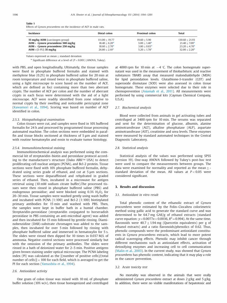
Table 1Effects of Gynura procumbens on the incidence of ACF in male rats.
Incidance Distal colon Proximal colon Total
15 mg/kg AOM (carcinogen group) 111.00719.77 19.6075.90 130.60721.93AOMþ Gynura procumbens 500 mg/kg 18.4076.20n 3.0071.40n 21.4077.09n
AOMþ Gynura procumbens 250 mg/kg 18.6072.70n 3.8070.83n 23.2074.70n
AOMþ(5-FU) 35 mg/kg 26.8072.10n 5.2071.78n 32.0072.20n
Values expressed as mean7standard deviationn Significant difference at a level of (Po0.001) (ANOVA, Tukey).
A.N. Shwter et al. / Journal of Ethnopharmacology 151 (2014) 1194–12011196

nephrotoxic effects, based on outward abnormal behaviour at thesedoses. Thus there were no significant differences in biochemical andhistopathological data between carcinogen group and treated groups.These findings indicated that the oral gavage of Gynura procumbensextract was concluded to be safe, and there no kidney and liver toxicitydetected even at the highest dosage administered.
3.3. Body and organs weight
Body weight of all rats were taken at the beginning (0 week) ofthe experiment then weekly up to 10 weeks. Colon, liver andkidney were weighed after rats were sacrificed. Although there aredifferences in weight between groups, but these differences werenot statistically significant.
3.4. Effects of Gynura procumbens on the incidence of ACF in malerats
The incidence of aberrant crypt foci is shown in Table 1. Ratsfed with Gynura procumbens showed significantly lower ACFnumbers comparing with carcinogen groups (Po0.001). In allexperimental groups, ACF incidence in the distal colon wassignificantly higher compared with proximal colon (Po0.001).However, the number of ACF in both the distal and proximal colonin carcinogen group (130.60721.93) were significantly highercompared to the 500 mg/kg Gynura procumbens, 250 mg/kgGynura procumbens and reference groups (21.4077.09,23.2074.7, and 32.072.2, respectively; Po0.001). There are about83.6%and 82.2% reduction in the total crypts in the groups fed with500 mg/kg and 250 mg/kg Gynura procumbens, respectively, com-pared to carcinogen group. Previous studies have approved the useof ACF as a biomarker indicator in colon cancer (Boateng et al.,2007). ACF has been reported by Bird in 1987 in rodents injectedwith AOM, and similar lesions were described in humans in 1991
by Pretlow, Since then, the AOM-induced ACF model has been themost widely used animal model for colon cancer (Zhang et al.,2000). Due to the presence of phytochemical content in Gynuraprocumbens extract, this plant might be able to reduce theincidence of ACF and prevent cancer development (Agustinaet al., 2006).
3.4.1. Effects of Gynura procumbens on the crypt multiplicity in malerats
The carcinogen group had a higher incidence of ACF with largercrypts compared to the experimental groups (Table 2).
3.5. Effects of Gynura procumbens on the antioxidant in vivoin AOM -induced colon Cancer rats
3.5.1. Effects of Gynura procumbens on the GST in AOM-inducedcolon cancer rats
Glutathione-S-transferase is a phase II detoxification enzyme.Our result showed that colon GST activity (nmoL/min/mL) wassignificantly high in the rats fed with 500 mg/kg and 250 mg/kgGynura procumbens followed with the carcinogen group fed rats;3475.8, 26.39870.92, 16.6574.57, respectively, Po0.05 (Fig. 1).One of the most common known chemopreventive activities, theindirect defensive mechanism of phytochemicals is the inductionof phase II metabolizing enzymes such as glutathione-S-transfer-ase (GST). One of the strategies for polyphenol to prevent cancer isthat might be interact with phase I and phase II enzyme systems,phase II enzymes catalyze the conjugation reactions betweenglutathione and Phase I reactive electrophilic intermediates tofacilitate their elimination from the body (Wilkinson and Clapper,1997; Moskaug et al., 2005). Our results are also consistent withprevious studies where GST activity was significantly inducedwhen the rats were administrated with (prebioticsþSM), flax seed
Table 2Effects of Gynura procumbens on the crypt multiplicity in male rats.
Groups Number of foci containing
1 Crypt 2 Crypts 3 Crypts 4 Crypts Z5 Crypt
15 mg/kg AOM (carcinogen group) 13.077.4 31.0713.6 22.078.4 25.075.9 38.073.1AOMþ500 mg/kg Gynura procumbens 3.071.9n 6.872.9n 5.871.9n 2.271.3n 3.672.3n
AOMþ250 mg/kg Gynura procumbens 2.471.7n 4.272.0n 6.271.3n 5.072.1n 5.471.8n
AOMþ(5-FU) 35 mg/kg 8.074.3n 7.273.0n 8.372.5n 3.271.8n 5.271.7n
Values expressed as mean7standard deviation.n Significant difference at a level of (Po0.001) (ANOVA, Tukey).
Fig. 1. Effect of Gynura procumbens on G-ST (nmoL/min/mL), in AOM-induced coloncancer rats. Effect of feeding GPE on colonic GST activity (nmoL/min/mL) in SD malerats. Abbreviations: GPE: Gynura procumbens extract, GST: glutathione-S-transferase.Values expressed as mean7standard deviation. nSignificant difference at a level at(Po0.001) (ANOVA, Tukey).
Fig. 2. Effect of Gynura procumbens ethanol extracts on SOD (U/mL), in AOM-induced colon cancer rats. Effect of feeding GPE antioxidative enzyme activitiesSOD activities (U/mL) in SD male rats. Abbreviations: GPE: Gynura procumbensextract, SOD: superoxide-dismutase, values expressed as mean7standard devia-tion. nSignificant difference at a level at (Po0.001) (ANOVA, Tukey).
A.N. Shwter et al. / Journal of Ethnopharmacology 151 (2014) 1194–1201 1197

meal and silymarin (Kohno et al., 2002; Williams et al., 2007;Gourineni et al., 2011).
3.5.2. Effects of Gynura procumbens on the SOD in AOM-inducedcolon cancer rats
Superoxide dismutase activity (U/mL) ranged from a high of11.5570.331in rats fed with Gynura procumbens 250 mg/kg and8.4170.941 in rats fed with Gynura procumbens500 mg/kg to low5.8770.311 in carcinogen group rats fed with AOM (Fig. 2). Theresults showed that rats fed with treatment diets showed sig-nificantly higher superoxide dismutase activity (SOD) (U/mL)compared with the carcinogen group (Fig. 2). Superoxide dismu-tase catalyzes the dismutation of superoxides anion radical, whichare potent carcinogens (Hei and Filipic, 2004). They are convertedto hydrogen peroxide (H2O2) and molecular oxygen. SOD work insync in the prevention of ACF formation, where the H2O2 producedby the SOD is further reduced to molecules of water by catalase.The results of current study were similar to the previous studiesthat showed a significant lower activity of SOD enzyme in differenttumor cell lines (Oliva et al., 1997; Sanchez et al., 2006).
3.5.3. Effects of Gynura procumbens on the MDA in AOM-inducedcolon cancer rats
Malonialdehyde level (MDA) in carcinogen group was signifi-cantly higher compared to treatment groups Gynura procumbens250 mg/kg and 500 mg/kg (Fig. 3). TBARS assay indicated the levelof lipid peroxidation that is present by measuring the level of MDAin all groups. In this study, there was a significant reduction ofMDA in groups treated with Gynura procumbens ethanol extractboth doses compared to the untreated AOM-induced group(carcinogen group). This reduction in the MDA level of extract-treated rats was comparable with 5-FU treated group, suggestingthat the Gynura procumbens reduce lipid peroxidation and pro-tected colon cells as 5-FU. A reduced lipid peroxidation is in turnassociated with a reduction in oxidative stress. Oxidative stressplay a significant role in the molecular mechanism of CRC initia-tion as well as progression (Seril et al., 2003). The increase of theoxidation products MDA in tumor tissue is accompanied bya significant decrease of the antioxidant enzyme SOD (Olivaet al., 1997; Sanchez et al., 2006).
3.6. Detection of ACF by methylene blue stain
The aberrant crypt foci were visualized and counted usingmethylene blue (0.2%). The ACF were slightly elevated above thesurrounding epithelial mucosa and demonstrated characteristicoval or slit-like opening. Some ACF were stained more densely bymethylene blue and detected as almost normal-sized lesions(Fig. 4).
3.7. Histopathological study
The histological features of dysplastic ACF and normal coloncells have also been illustrated by H&E staining. Histology sectionof normal colon and treated groups showed normal architecture ofthe mucosal and submucosal layers. AOM-induced ACF groupshowed epithelial mucosal glands proliferation with severe dys-plastic alteration changes which represent transformation tocarcinoma (Fig. 5).
Fig. 3. Effect of Gynura procumbens ethanol extracts on MDA (U/m), in AOM-induced colon cancer rats. Effect of feeding GPE antioxidative enzyme activitiesMDA activities (mM) in SD male rats. Abbreviations: GPE: Gynura procumbens, MDA:malonialdehyde level, Values expressed as mean7standard deviation. nSignificantdifference at a level at (Po0.001) (ANOVA, Tukey).
Fig. 4. Effects of Gynura procumbens on AOM-induced ACF in rat0s colon (methylene blue staining). Normal crypts of normal group (a). ACF with multiple crypts inCarcinogen group (b). 5-FU, high and low doses of Gynura procumbens treated groups respectively (c)–(e), with normal crypts and few ACF with crypts (o5) due to thepreventive effects of plant extract. (For interpretation of the references to color in this figure legend, the reader is referred to the web version of this article.)
A.N. Shwter et al. / Journal of Ethnopharmacology 151 (2014) 1194–12011198

3.8. Immunohistochemical staining
PCNA andBcl-2 staining of colons: The proliferating cell nuclearantigen (PCNA) was evaluated as an important marker for cellproliferation in the colon sections, and the results showed that thecarcinogen group was much higher expression of PCNA protein thantreated groups (Fig. 6). The percentage of PCNA -positive cells of thecolon sections in the carcinogen group were 48.83%, whereas PCNA-positive cells (%) from the dose Gynura procumbens fed 500 mg/kg,250 mg/kg and reference groups were 17%, 20% and 10%, respectively.Immunohistochemical staining of Bcl-2protein demonstrated thatrats in the treated groups had low expression of Bcl-2 protein withrespect to AOM group that showed higher expression (Fig. 7). Thecellular proliferation rate plated a significant prognostic marker forvariety of cancer disorders, including colon cancer (Violette et al.,2002; Yamaguchi et al., 2003). The immunohistochemical staining
assay of PCNA, and Bcl-2 proteins was carried out to assess theproliferation and apoptotic alterations of colorectal mucosa. Ourresults showed that Gynura procumbens ethanol extract suppressedthe expression of PCNA. Most of the possible chemopreventive drugsused against chemically induced colon carcinogenesis inhibit theproliferation of cell activity through PCNA index (Tanaka et al., 2001).Previous studies reported such findings during the administration ofAOM (Yamashita et al., 1994; Tanaka et al., 1999). Our findings alsoshowed increased expression of PCNA, and this may reflect anincreased cell proliferation in colon tumors.
The anti-apoptotic protein Bcl-2 was down-regulated in alltreated groups. Enhanced Bcl-2 expression has been observed inrats with chemically induced colonic adenocarcinomas. The anti-apoptotic function of bcl-2 oncogene may be played important rolein tumorigenesis by raising the threshold for apoptosis (Adamsand Cory, 1998). Our findings are in agreement with similar studies
Fig. 5. Effects of Gynura procumbens on AOM-induced ACF in rat0s colon. Haematoxylin staining. (a) Normal group, (b) carcinogen group (c)–(e) 5-FU, high dose & low dose ofGynura procumbens treated groups respectively (100� ).
Fig. 6. Expression of PCNA protein in the colon tissue of rats. Expression of PCNA protein in the colon tissue of rats. (a) Normal group, (b) carcinogen group (c) 5-FU treatedgroup (d) and (e) high and low doses treated groups. PCNA protein showed down-expression of PCNA protein in rats treated with 5-FU and plant extract comparing tocarcinogen group.
A.N. Shwter et al. / Journal of Ethnopharmacology 151 (2014) 1194–1201 1199

on bcl-2 expression in human normal mucosa and colorectalcancer (Maurer et al., 1998).
3.9. Effect of Gynura procumbens on biochemical parameters
There were no statistically significant differences in the level ofurea, creatinine, total protein and (ALT, AST, ALP) for the AOMgroup compared with the normal and treated groups (Tables 3 and4).
4. Conclusion
According to the results of this study, it can be expected thatGynura procumbens may have antiproliferative and antioxidative
properties. Reduction in ACF incidence could be due to the directeffects of treatment diets indirect mechanism such as stimulatingof detoxifying (GST) and antioxidative enzymes or by acting asantiproliferative. Gynura procumbens proves to be a medicinalplant which possesses the medicinal properties to prevent coloncancer as results from this study have made obvious.
Acknowledgments
The authors express their gratitude to the staffs of the Facultyof Medicine Animal House for the care and supply of rats. Thisresearch was supported by the University of Malaya Grant (PV046/2012) and University of Malaya High Impact Research Grant (UM/M0HE/HIR Grant E000045-20001).
Fig. 7. Expression of Bcl-2 protein in the colon tissue of rats. Expression of Bcl-2 protein in the colon tissue of rats. (a) Normal group, (b) carcinogen group (c) 5-FU treatedgroup (d), and (e) high and low doses treated groups. Immunohistochemical analysis of Bcl-2 protein showed down-expression of Bcl-2 protein in rats treated with 5-FU andplant extract comparing to carcinogen group.
Table 3Effects of 500 mg/kg and 250 mg/kg Gynura procumbens extract on liver biochemical parameters.
Groups ALT (IU/L) AST (IU/L) ALP (IU/L) Total bilirubin (μmol/L) Total protein (g/L)
Tween 20 (10%) 49.673.3 16872.2 130.673.8 2.870.20 64.270.66Gynura procumbens 500 mg/kg 61.00712.97 189.473.6 149.2710.2 2.670.24 67.671.0Gynura procumbens 250 mg/kg 61.6712.9 161.8726.7 161726.6 2.470.24 7070.8315 mg/kg AOM (carcinogen group) 47.678.2 203723.43 106.8724.7 2.470.54 64.471.8AOMþ(5-FU) 35 mg/kg 53.277.25 163.8729.1 135.4713.7 2.470.55 66.473.7
Values expressed as mean7standard deviation. No significant difference at (Po0.05) (ANOVA, Tukey).
Table 4Effects of 500 mg/kg and 250 mg/kg Gynura procumbens on kidney biochemical parameters.
Groups Sodium (mmol/L) Potassium (mmol/L) Urea (mmol/L) Creatinine (μmol/L)
Tween 20 (10%) 144.671.1 4.627 .012 5.0670.22 28.874.66Gynura procumbens 500 mg/kg 140.471.8 4.467 .012 6.0470.4 29.673.17Gynura procumbens 250 mg/kg 14170.4 4.8570.15 5.9870.52 28.872.615 mg/kg AOM (carcinogen group) 13873.7 4.570.16 5.0170.51 28.876.01AOMþ(5-FU) 35 mg/kg 140.873.5 4.370.27 5.570.7 32.672.5
Values expressed as mean7standard deviation. No significant difference at (Po0.05) (ANOVA, Tukey).
A.N. Shwter et al. / Journal of Ethnopharmacology 151 (2014) 1194–12011200

References
Adams, J.M., Cory, S., 1998. The Bcl-2 protein family: arbiters of cell survival. Science281, 1322–1326.
Agustina, D., Wasito, H.S., Supatinah, A., 2006. Anticarcinogenesis effect of Gynuraprocumbens (Lour) Merr on tongue carcinogenesis in 4NQO-induced rat. Dental J.39, 126–132.
Akowuah, G., Sadikun, A., Mariam, A., 2002. Flavonoid identification and hypogly-caemic studies of the butanol fraction from Gynura procumbens. Pharm. Biol. 40,405–410.
Andersson, D., Cheng, Y., Duan, R.-D., 2008. Ursolic acid inhibits the formation ofaberrant crypt foci and affects colonic sphingomyelin hydrolyzing enzymes inazoxymethane-treated rats. J. Cancer Res. Clin. Oncol. 134, 101–107.
AOAC., 1995. Association of Official Analytical Chemists, Official Methods of Analysisof the Association of the Analytical Chemists, Arlington, WA, USA, Arlington,Arlington, WA, USA, 16th ed.
Asiamah, D., Verghese, M., Boateng, J., Kanda, B., Shackelford, L., Walker, L., 2011.Chemopreventive potential of bitter melon (Momordica charantia) againstprecancerous lesions in the colon of fisher 344 male rats. Int. J. Cancer Res. 7,36–46.
Boateng, J., Verghese, M., Shackelford, L., Walker, L., Khatiwada, J., Ogutu, S.,Williams, D., Jones, J., Guyton, M., Asiamah, D., 2007. Selected fruits reduceazoxymethane (AOM)-induced aberrant crypt foci (ACF) in Fisher 344 malerats. Food Chem. Toxicol. 45, 725–732.
Dowd, L.E., 1959. Spectrophotometric determination of quercetin. Anal. Chem. 31,1184–1187.
Gan, R.Y., Xu, X.R., Song, F.L., Kuang, L., Li, H.B., 2010. Antioxidant activity and totalphenolic content of medicinal plants associated with prevention and treatmentof cardiovascular and cerebrovascular diseases. J. Med. Plants Res. 4,2438–2444.
Gourineni, V., Verghese, M., Boateng, J., Shackelford, L., Bhat, N., Walker, L., 2011.Combinational effects of prebiotics and soybean against azoxymethane-induced colon cancer in vivo. J. Nutr. Metab. 2011, 9.
Hei, T.K., Filipic, M., 2004. Role of oxidative damage in the genotoxicity of arsenic.Free Radical Biol. Med. 37, 574–581.
Iskander, M., Song, Y., Coupar, I., Jiratchariyakul, W., 2002. Antiinflammatoryscreening of the medicinal plant Gynura procumbens. Plant Foods Hum. Nutr.(Formerly Qualitas Plantarum) 57, 233–244.
Jenie, R.I., Meiyanto, E., Si, M., 2007. Aplikasi Ko-Kemoterapi Doxorubicin-Fraksi EtilAsetat daun Sambung Nyawa (Gynura procumbens (Lour.) Merr.) terhadap selkanker payudara MCF-7 dan T47D. Universitas Gadjah Mada.
Jiang, Q., Wong, J., Fyrst, H., Saba, J.D., Ames, B.N., 2004. gamma-Tocopherol orcombinations of vitamin E forms induce cell death in human prostate cancercells by interrupting sphingolipid synthesis. Proc. Nat. Acad. Sci. U.S.A. 101,17825–17830.
Kawamori, T., Tanaka, T., Kojima, T., Suzui, M., Ohnishi, M., Mori, H., 1994.Suppression of azoxymethane‐induced rat colon aberrant crypt foci by dietaryprotocatechuic acid. Cancer Sci. 85, 686–691.
Kohno, H., Tanaka, T., Kawabata, K., Hirose, Y., Sugie, S., Tsuda, H., Mori, H., 2002.Silymarin, a naturally occurring polyphenolic antioxidant flavonoid, inhibitsazoxymethane‐induced colon carcinogenesis in male F344 rats. Int. J. Cancer101, 461–468.
Mahmood, A., Mariod, A.A., Al-Bayaty, F., Abdel-Wahab, S.I., 2010. Anti-ulcerogenicactivity of Gynura procumbens leaf extract against experimentally-inducedgastric lesions in rats. J. Med. Plants Res. 4, 685–691.
Malin, A.S., Qi, D., Shu, X.O., Gao, Y.T., Friedmann, J.M., Jin, F., Zheng, W., 2003. Intakeof fruits, vegetables and selected micronutrients in relation to the risk of breastcancer. Int. J. Cancer 105, 413–418.
Maurer, C.A., Friess, H., Buhler, S.S., Wahl, B.R., Graber, H., Zimmermann, A., Buchler,M.W., 1998. Apoptosis inhibiting factor Bcl-xL might be the crucial member ofthe Bcl-2 gene family in colorectal cancer. Dig. Dis. Sci. 43, 2641–2648.
Moskaug, J., Carlsen, H., Myhrstad, M.C., Blomhoff, R., 2005. Polyphenols andglutathione synthesis regulation. Am. J. Clin. Nutr. 81, 277S–283S.
Nawawi, A.a., Nakamura, N., Hattori, M., Kurokawa, M., Shiraki, K., 1999. Inhibitoryeffects of Indonesian medicinal plants on the infection of herpes simplex virustype 1. Phytother. Res. 13, 37–41.
Nisa, F., Hermawan, A., Murwanti, R., Meiyanto, E., 2012. Antiproliferative effect ofGynura procumbens (lour.) Merr. Leaves etanolic extract on 7,12-dimethylbenz(a) antracene induced male rat liver. Adv. Pharm. Bull. 2, 99–106.
Oliva, M.R., Ripoll, F., Muñiz, P., Iradi, A., Trullenque, R., Valls, V., Drehmer, E.,Saez, G.T., 1997. Genetic alterations and oxidative metabolism in sporadiccolorectal tumors from a Spanish community. Mol. Carcinog. 18, 232–243.
Perry, L.M., Metzger, J., 1980. Medicinal Plants of East and Southeast Asia:Attributed Properties and Uses. MIT Press.
Riboli, E., Norat, T., 2003. Epidemiologic evidence of the protective effect of fruitand vegetables on cancer risk. Am. J. Clin. Nutr. 78, 559S–569S.
Robles, F.E., Zhu, Y., Lee, J., Sharma, S., Wax, A., 2010. Detection of early colorectalcancer development in the azoxymethane rat carcinogenesis model withFourier domain low coherence interferometry. Biomed. Opt. Exp. 1, 736–745.
Sanchez, M., Torres, J.V., Tormos, C., Iradi, A., Muñiz, P., Espinosa, O., Salvador, A.,Rodriguez-Delgado, J., Fandos, M., Sáez, G.T., 2006. Impairment of antioxidantenzymes, lipid peroxidation and 8-oxo-20-deoxyguanosine in advanced epithe-lial ovarian carcinoma of a Spanish community. Cancer Lett. 233, 28–35.
Saslow, D., Solomon, D., Lawson, H.W., Killackey, M., Kulasingam, S.L., Cain, J.,Garcia, F.A., Moriarty, A.T., Waxman, A.G., Wilbur, D.C., 2012. American CancerSociety, American Society for Colposcopy and Cervical Pathology, and AmericanSociety for Clinical Pathology screening guidelines for the prevention and earlydetection of cervical cancer. CA Cancer J. Clin. 62, 147–172.
Seril, D.N., Liao, J., Yang, G.Y., Yang, C.S., 2003. Oxidative stress and ulcerative colitis-associated carcinogenesis: studies in humans and animal models. Carcinogen-esis 24, 353–362.
Sugiyanto, B.S., Meiyanto, E., Nugroho, A.E., Jenie, U.A., 2003. The anticarcinogenicactivity of plants compounds. Majalah Farmasi Indonesia 14, 216–225.
Tanaka, T., Kawabata, K., Kakumoto, M., Makita, H., Ushida, J., Honjo, S., Hara, A.,Tsuda, H., Mori, H., 1999. Modifying effects of a flavonoid morin onazoxymethane-induced large bowel tumorigenesis in rats. Carcinogenesis 20,1477–1484.
Tanaka, T., Kohno, H., Mori, H., 2001. Chemoprevention of colon carcinogenesis bydietary non-nutritive compounds. Asian Pac. J. Cancer Prev. 2, 165–177.
Tenesa, A., Dunlop, M.G., 2009. New insights into the aetiology of colorectal cancerfrom genome-wide association studies. Nat. Rev. Genet. 10, 353–358.
Valeriote, F., Grieshaber, C.K., Pietraszkewicz, H., Hoffmann, J., Pan, M., McLaughlin, S.,2002. Discovery and development of anticancer agents from plants. J. Exp. Ther.Oncol. 2, 228–236.
Van Duijnhoven, F.J., Bueno-De-Mesquita, H.B., Ferrari, P., Jenab, M., Boshuizen, H.C.,Ros, M.M., Casagrande, C., Tjønneland, A., Olsen, A., Overvad, K., 2009. Fruit,vegetables, and colorectal cancer risk: the European Prospective Investigationinto Cancer and Nutrition. Am. J. Clin. Nutr. 89, 1441–1452.
Violette, S., Poulain, L., Dussaulx, E., Pepin, D., Faussat, A.M., Chambaz, J., Lacorte, J.M.,Staedel, C., Lesuffleur, T., 2002. Resistance of colon cancer cells to long‐term 5‐fluorouracil exposure is correlated to the relative level of Bcl‐2 and Bcl‐XL inaddition to Bax and p53 status. Int. J. Cancer 98, 498–504.
Wilkinson, J., Clapper, M.L., 1997. Detoxication enzymes and chemoprevention. In:Proceedings of the Society for Experimental Biology and Medicine. Society forExperimental Biology and Medicine (New York, NY). Royal Society of Medicine,pp. 192–200.
Williams, D., Verghese, M., Walker, L., Boateng, J., Shackelford, L., Chawan, C., 2007.Flax seed oil and flax seed meal reduce the formation of aberrant crypt foci(ACF) in azoxymethane-induced colon cancer in Fisher 344 male rats. FoodChem. Toxicol. 45, 153–159.
Yamaguchi, H., Bhalla, K., Wang, H.G., 2003. Bax plays a pivotal role in thapsigargin-induced apoptosis of human colon cancer HCT116 cells by controlling Smac/Diablo and Omi/HtrA2 release from mitochondria. Cancer Res. 63, 1483–1489.
Yamashita, N., Minamoto, T., Onda, M., Esumi, H., 1994. Increased cell proliferationof azoxymethane‐induced aberrant crypt foci of rat colon. Cancer Sci. 85,692–698.
Zhang, L., Yu, J., Park, B.H., Kinzler, K.W., Vogelstein, B., 2000. Role of BAX in theapoptotic response to anticancer agents. Sci. Signaling 290, 989.
A.N. Shwter et al. / Journal of Ethnopharmacology 151 (2014) 1194–1201 1201