jonathan l. rees - powering silicon valley of... · jonathan l. rees systems group, dermatology,...
TRANSCRIPT

11 Sep 2003 14:51 AR AR201-GE37-04.tex AR201-GE37-04.sgm LaTeX2e(2002/01/18)P1: GCE10.1146/annurev.genet.37.110801.143233
Annu. Rev. Genet. 2003. 37:67–90doi: 10.1146/annurev.genet.37.110801.143233
Copyright c© 2003 by Annual Reviews. All rights reservedFirst published online as a Review in Advance on June 17, 2003
GENETICS OF HAIR AND SKIN COLOR
Jonathan L. ReesSystems Group, Dermatology, University of Edinburgh, Lauriston Buildings, LauristonPlace, Edinburgh, EH3 9YW, United Kingdom; email: [email protected]
Key Words melanin, melanocortin 1 receptor (MC1R), eumelanin, pheomelanin,red hair
■ Abstract Differences in skin and hair color are principally genetically deter-mined and are due to variation in the amount, type, and packaging of melanin polymersproduced by melanocytes secreted into keratinocytes. Pigmentary phenotype is genet-ically complex and at a physiological level complicated. Genes determining a numberof rare Mendelian disorders of pigmentation such as albinism have been identified,but only one gene, the melanocortin 1 receptor (MCR1), has so far been identified toexplain variation in the normal population such as that leading to red hair, freckling,and sun-sensitivity. Genotype-phenotype relations of theMC1Rare reviewed, as wellas methods to improve the phenotypic assessment of human pigmentary status. It isargued that given advances in model systems, increases in technical facility, and thelower cost of genotype assessment, the lack of standardized phenotype assessment isnow a major limit on advance.
CONTENTS
INTRODUCTION . . . . . . . . . . . . . . . . . . . . . . . . . . . . . . . . . . . . . . . . . . . . . . . . . . . . . 68BIOLOGY OF HUMAN PIGMENTATION . . . . . . . . . . . . . . . . . . . . . . . . . . . . . . . . 69
Melanocytes and Melanogenesis. . . . . . . . . . . . . . . . . . . . . . . . . . . . . . . . . . . . . . . . 69Types of Melanin . . . . . . . . . . . . . . . . . . . . . . . . . . . . . . . . . . . . . . . . . . . . . . . . . . . . 70Body Site and Temporal Variation. . . . . . . . . . . . . . . . . . . . . . . . . . . . . . . . . . . . . . . 70Functions of Melanin. . . . . . . . . . . . . . . . . . . . . . . . . . . . . . . . . . . . . . . . . . . . . . . . . 71Defining Pigmentary Phenotypes. . . . . . . . . . . . . . . . . . . . . . . . . . . . . . . . . . . . . . . 72
ALBINISM AND RELATED DISORDERS . . . . . . . . . . . . . . . . . . . . . . . . . . . . . . . . 74Oculocutaneous Albinism Type 1 (OCA1, MIM 203100). . . . . . . . . . . . . . . . . . . . 74Oculocutaneous Albinism Type 2 (OCA2, MIM 203200). . . . . . . . . . . . . . . . . . . . 74Oculocutaneous Albinism Type 3 (OCA3, MIM 203290). . . . . . . . . . . . . . . . . . . . 75
THE MELANOCORTIN 1 RECEPTOR (MC1R), (MIM 155555) . . . . . . . . . . . . . . . 75Red Versus Yellow. . . . . . . . . . . . . . . . . . . . . . . . . . . . . . . . . . . . . . . . . . . . . . . . . . . 75αMSH and Human Pigmentation. . . . . . . . . . . . . . . . . . . . . . . . . . . . . . . . . . . . . . . 76Genotype-Phenotype Correlation at theMC1R . . . . . . . . . . . . . . . . . . . . . . . . . . . . . 77Spectrum of Sequence Diversity at the MC1R. . . . . . . . . . . . . . . . . . . . . . . . . . . . . 78Role of Agouti in Man . . . . . . . . . . . . . . . . . . . . . . . . . . . . . . . . . . . . . . . . . . . . . . . . 79Evolution at the MC1R . . . . . . . . . . . . . . . . . . . . . . . . . . . . . . . . . . . . . . . . . . . . . . . 79
0066-4197/03/1215-0067$14.00 67
Ann
u. R
ev. G
enet
. 200
3.37
:67-
90. D
ownl
oade
d fr
om w
ww
.ann
ualr
evie
ws.
org
by S
an J
ose
Stat
e U
nive
rsity
on
10/0
5/10
. For
per
sona
l use
onl
y.

11 Sep 2003 14:51 AR AR201-GE37-04.tex AR201-GE37-04.sgm LaTeX2e(2002/01/18)P1: GCE
68 REES
FUTURE STUDIES AND THE DEFINITION OF PHENOTYPE. . . . . . . . . . . . . . . 80Phenotype of MC1R Variants Against “Black Skin”. . . . . . . . . . . . . . . . . . . . . . . . 81
INTRODUCTION
Variation in skin pigmentation–skin and hair color—between people of differentgenetic ancestries is one of the most striking human characteristics (84). Study,and selection, of animals with particular pigmentary phenotypes has been of eco-nomic importance (5, 54, 55); pigmentation in the mouse and birds are classicalexperimental systems to study gene action (5, 54, 55, 104); and at the same time,even among nonexperts, it is widely understood that human skin color and haircolor are largely under genetic control, reflecting a person’s genetic heritage (97).One would have expected, therefore, the study of the genetics of skin and haircolor in man to be a subject of much study: it isn’t. For instance, we remain almostcompletely ignorant of such simple issues as the mode of inheritance of blondehair. Indeed, although textbooks frequently refer to hair or eye color as an exampleto illustrate the role of genetics in understanding human diversity of form, until re-cently little was known of the genetic mechanisms underpinning normal variationin skin and hair color (14, 97).
Over the past ten years this situation has begun to change (5, 54, 56, 93, 106).Advances based on the asset of the mouse fancy (5, 55, 104), coupled with the fa-cility of modern molecular technology, have allowed the identification of a numberof genes important in the determination of skin and hair color in man. The geneticsof many Mendelian disorders of medical importance such as albinism (63) havebecome clearer: Existing clinical classifications have been shown to be inadequate,and mechanistic likenesses between what were once thought to be distinct pro-cesses outlined (63). This review briefly discusses these conditions, but takes as itsfocus advances in our understanding of pigmentary variation within what may bearbitrarily, but usefully, defined as the normal population, describing in some detailthe role of the melanocortin 1 receptor in human pigmentation (MC1R)—the onlygene identified to date that appears to underpin variation in the normal population(93).
The review is structured into four parts. The first outlines the biology of humanpigmentation, highlighting methodological issues in the assessment of pigmentaryphenotype. The emphasis is on the assessment of phenotype and presentation ofan outline of how complicated (rather than necessarily complex) phenotype canbe. In the second section, the major Mendelian disorders of pigmentation—chieflyalbinism—are briefly summarized. The third section deals in some detail withthe melanocortin 1 receptor (MC1R), its genetics and molecular physiology, andwhat we know of the relation betweenMC1Rgenotype and human phenotype.Finally, I discuss areas that need to be developed and explored further, payingparticular attention to the need to develop appropriate assay systems to understandthe genetics of human pigment diversity.
Ann
u. R
ev. G
enet
. 200
3.37
:67-
90. D
ownl
oade
d fr
om w
ww
.ann
ualr
evie
ws.
org
by S
an J
ose
Stat
e U
nive
rsity
on
10/0
5/10
. For
per
sona
l use
onl
y.

11 Sep 2003 14:51 AR AR201-GE37-04.tex AR201-GE37-04.sgm LaTeX2e(2002/01/18)P1: GCE
HAIR AND SKIN COLOR 69
BIOLOGY OF HUMAN PIGMENTATION
Skin color is, except in rare pathological instances, the result of three pigmentsor chromophores: melanin, a brown/black or red/yellow polymer produced bymelanocytes; hemoglobin in red blood cells in the superficial vasculature; andthird, and to a much lesser degree, dietary carotenoids, sometimes most evidentas a yellow color on the palms (90). Systematic differences in skin and hair colorworldwide are principally the result of differences in the melanin content of skin.1
Melanocytes and Melanogenesis
Melanin is a complex quinone/indole-quinone-derived mixture of biopolymersproduced in melanocytes from tyrosine (51, 52, 88). Melanocytes are dendriticneural crest-derived cells that migrate into epidermis in the first trimester. Melaninproduction is associated with the production of a number of toxic intermedi-aries and largely takes place within a lysosomal-like granule, the melanosome.Melanosomes are secreted via a poorly characterized process into adjacent ker-atinocytes. Unlike iris melanocytes, epidermal melanocytes are therefore said tobe incontinent, i.e., they secrete their melanin. Melanin chemistry is complex andremains poorly understood for a number of reasons that make chemical charac-terization difficult: It is a mixture of polymers; many intermediates are unstableand rapidly autooxidize; methods to solubilize melanin alter its primary structure(51, 88).
Hair and epidermal pigmentation are, in so far as melanocytes are concerned,similar processes: in interfollicular skin, pigment is passed from the melanocytesto the adjacent keratinocytes; in hair, a similar process exists, with pigment beingadded to the growing keratinocytes that will make up the shaft of the hair (hair is justone form of epidermis). In interfollicular skin, the melanocytes are found within theepidermal compartment immediately adjacent or close to the basement membrane.Melanosomes passed to adjacent keratinocytes tend to produce caps over the nuclei,shielding the nuclear material from ultraviolet radiation (UVR). This melanin ismost evident in the basal compartment, the site of the keratinocyte proliferativecompartment, but melanin remains with keratinocytes as they differentiate andmove upwards—this melanin will still exert photoprotective activity on the cellsbeneath it (by casting a UVR shadow). In hair, the majority of what is visible tothe eye is a dead structure, the color is the result of melanocytes in the hair bulb
1Abbreviations: αMSH, α-melanocyte stimulating hormone; ACTH, adrenocorti-cotrophic hormone; AHP, aminohydroxyphenylalanine; BAC, bacterial artificialchromosome; cAMP, cyclic adenosine monophosphate; CIE, Commission Inter-nationale de L’Eclairage; DHICA-melanin, 5,6-dihydroxyindole-2-carboxylic acid;DOPA,3,4,dihydroxyphenylalanine; HPLC, high performance liquid chromatography;MC1R, melanocortin 1 receptor; MIM, Mendelian Inheritance in Man; OCA, occulo cu-taneous albinism; POMC, pro-opiomelanocortin; PTCA, pyrrole-2,3,5-tricarboxylic acid;TYRP1, tyrosinase related protein 1; UVR, ultraviolet radiation.
Ann
u. R
ev. G
enet
. 200
3.37
:67-
90. D
ownl
oade
d fr
om w
ww
.ann
ualr
evie
ws.
org
by S
an J
ose
Stat
e U
nive
rsity
on
10/0
5/10
. For
per
sona
l use
onl
y.

11 Sep 2003 14:51 AR AR201-GE37-04.tex AR201-GE37-04.sgm LaTeX2e(2002/01/18)P1: GCE
70 REES
passing their melanin to the adjacent keratinocytes as they undergo high rates ofproliferation, stream pass the melanocytes, and then cornify (84, 90).
Types of Melanin
Although the nature of melanin has frustrated precise chemical description, geneticapproaches coupled with a number of available chemical assays have allowed someuseful insights (5, 93). Melanin, with particular relevance to the current context, iscommonly described as being of two principal classes: eumelanin, which is brownor black, and pheomelanin, which results from the incorporation of cysteine, isyellow or red [for reviews see (51, 80, 88, 89, 124, 125)]. One simple classificationor description of pigment status is to consider two axes of melanin production: theamount of melanin(s) produced, and the relative amounts of either eumelanin orpheomelanin. The absence or relative absence of both melanin types is associatedwith white hair; a preponderance of eumelanin, with brown or black hair; and apreponderance of pheomelanin with red or yellow hair. A more precise chemicalcharacterisation of the brown melanin polymers is possible (89).
Finally, color is not merely the result of the chemical composition of the variousmelanin polymers. Melanin is packaged into melanosomes, and melanosomes varyin shape and size (90). Such differences, by way of light scattering, will influencecolor, a fact colorfully illustrated by the way amphibians and fish disperse theirmelanosomes to influence their skin’s color characteristics (3).
Body Site and Temporal Variation
Differences in pigmentation between people are largely the result of differences inthe amount and types of melanin produced, and the macromolecular structure andpackaging of melanin, and not the number of melanocytes. There are, however,differences in pigmentation between different regions of the body as well as differ-ences between people in the respective color of their hair and (interfollicular) skin(90). For instance, people from Northern Europe, such as many Scandinavians,have light or blonde hair and pale skin but their skin pigment increases in responseto UVR to a moderate degree (94). Conversely, many people from Western Europe,such as the Irish, have pale skin, red hair, and a skin that tans little in response torepeated UVR irradiation. In the (relatively) unexposed body sites, such as the but-tock, their skin may be similarly colored to that of Scandinavians, but in responseto UVR it tans less, and the hair color may well be different—red as comparedwith blonde (94). Similarly, the color of some Caucasian skin may be similar tothat of some Asians and yet the latter appears to have a greater propensity to tan inresponse to UVR or develop pigmentation after other inflammatory insults (suchas from skin disease).
Furthermore, any idea of a unitary pigmentary phenotype has to take into ac-count not only that skin color may vary between different body regions, but alsothat hair color may vary both in time and site. Scalp hair may be blonde in child-hood and become brown or black in adolescence, before becoming white again inmiddle or old age. Beard or pubic hair may be red, and the scalp hair black or dark
Ann
u. R
ev. G
enet
. 200
3.37
:67-
90. D
ownl
oade
d fr
om w
ww
.ann
ualr
evie
ws.
org
by S
an J
ose
Stat
e U
nive
rsity
on
10/0
5/10
. For
per
sona
l use
onl
y.

11 Sep 2003 14:51 AR AR201-GE37-04.tex AR201-GE37-04.sgm LaTeX2e(2002/01/18)P1: GCE
HAIR AND SKIN COLOR 71
brown. Skin color on sun-protected sites such as the buttock will be paler than onexposed sites but, even in those not exposed to UVR (such as the newborn), areasof skin such as the outside of the arms are a darker color than the inside.
A number of influences are at work to explain this diversity. First, in mostindividuals, repeated exposure to UVR causes an increase in facultative melaninproduction and possibly an increase in melanocyte number (36). Second, differ-ent body sites are preprogrammed to have differing numbers of melanocytes andconstitutive melanin production (90). Third, the amounts and type of melanin pro-duction vary with age and by site, with children being paler skinned than adults,and females paler than males. Fourth, hair and skin melanocytes may show somedegree of independence, that is the skin may be highly pigmented, the hair less so,although even this depends on body site. Finally, with age, melanocyte activity inhair, although apparently not in skin, may diminish, leading initially to a mixture ofwhite and darker hairs (gray is a misnomer, as hairs are either white, or brown/black,rather than gray). The mixture of hair that is called gray may, under pathologicalconditions, revert to brown or black. The genetic factors responsible for virtuallyall these variations are poorly understood, if at all, and place important constraintson the precision of our current models of understanding of normal pigmentaryvariation. In general, they would be expected to lead to an underestimate of thestrength of genotype-phenotype correlations in the absence of appropriate designs.
Functions of Melanin
Many theories have been advanced for the biological role played by melanin (45).In other species pigmentation may either play an important role in avoiding atten-tion by predators (camouflage) or, conversely, in drawing attention to particularbiological forms [such as the blue scrotum of the vervet monkey, which appearsblue because of light scattering of long-wavelength light from deep-seated “brown”melanin (87)]. In man, melanin plays particular roles in the eye and ear, but in theintegument its roles are limited to (a) photoprotection against ultraviolet radiationand (b) sociocultural.
These functions are illustrated by the phenotype of albinos living in areas withhigh ambient UVR, such as Tanzania: Such individuals burn in the sun with pain,blistering, and an increased risk of infection and fluid loss; develop signs of pre-mature aging of the skin (in reality, signs of excess UVR damage for their chrono-logical age); and develop a range of skin cancers that may kill them as teenagersor in early adulthood (73). Melanin is an extremely effective sunblock, protect-ing against the harmful effects of electromagnetic radiation above∼300 nm (98)(shorter wavelengths fail to pass through the atmosphere). UVR absorption, andhence protection, by melanin is greatest at the shorter wavelengths, where damageto nucleic acids and protein is maximal, and declines as a function of wavelengthwell into the visible spectrum (>400 nm). Although experimental studies are lack-ing, and therefore evidence is limited, differences in pigmentation, with differencesin eumelanin of two- to threefold, can account for up to a 100-fold variation insensitivity measured as the propensity to develop erythema in response to UVR
Ann
u. R
ev. G
enet
. 200
3.37
:67-
90. D
ownl
oade
d fr
om w
ww
.ann
ualr
evie
ws.
org
by S
an J
ose
Stat
e U
nive
rsity
on
10/0
5/10
. For
per
sona
l use
onl
y.

11 Sep 2003 14:51 AR AR201-GE37-04.tex AR201-GE37-04.sgm LaTeX2e(2002/01/18)P1: GCE
72 REES
(2). The action spectrum, or propensity for UVR of any particular wavelength tocause erythema, is strikingly similar to that of DNA, suggesting that the formermay be a useful proxy for the latter (27, 115). By contrast, ambient UVR variesless so, with total ambient UVR being∼3.5 times higher in equatorial regions thanin areas such as Scotland (26). These total ambient UVR levels may be misleadingin that a major determinant of individual dose will be ambient temperature in thatin colder climates protective clothing as well as providing insulation will shieldfrom UVR (25). The additional heat stress of a dark skin (black object) may alsobe relevant to any evolutionary tradeoffs.
For completion, various theories have been advanced to explain the lighteningof skin as distance from the equator increases. The most popular is that sincevitamin D is partly biosynthesized by keratinocytes in response to UVR (in theabsence of abundant dietary sources), dark skin may lead to vitamin D deficiencyin Northern climates (9, 53, 59). Rickets, with disastrous effects on reproductivefitness, would be one result, but other roles for vitamin D in protection againstinfection have also been posited as relevant (47, 53). Other explanations have alsobeen suggested (19).
Defining Pigmentary Phenotypes
It might be thought straightforward to measure skin or hair color in a biologicallymeaningful way (38, 94). In reality, the various disciplines concerned with skincolor such as anthropologists, dermatologists, and cosmetic scientists have tendedto develop their own methodologies. From the perspective of genetics, whereascolor may be one realistic endpoint by which for instance assortive mating maybe mediated (24, 119), resistance to UVR, albeit dependent on skin color, is notsynonymous with it. Thus, although the amount and type of melanin (as explainedabove) may be a major determinant of UVR susceptibility, other factors also play arole. Even skin that has no pigmentation can develop photoprotection, defined as areduced biological response to a subsequent dose of UVR (65, 71). This is thoughtto be due to the development of epidermal thickening and possibly qualitativechanges within the epidermal layer (65, 71). Second, skin color may be similarbetween individuals, but their response to UVR may vary several fold (32). Suchdifferences may account for some of the low skin cancer rates seen in some Asianseven though they do not seem very brown; however, alternative and more mundaneexplanations such as behavioral differences may be more likely (18). Skin coloris a key determinant of UVR sensitivity, but not the only one, nor should weequate color completely with our current understanding of melanin structure. Theevolutionary determinants of skin color and resistance to UVR may overlap a greatdeal but they are not completely synonymous.
SKIN AND HAIR COLOR Skin color has been assessed in a number of ways. Broad-band spectrophotometry allows the sampling of reflectance (strictly speaking re-mittance) at a number of wavelengths such that wavelength-dependent curves
Ann
u. R
ev. G
enet
. 200
3.37
:67-
90. D
ownl
oade
d fr
om w
ww
.ann
ualr
evie
ws.
org
by S
an J
ose
Stat
e U
nive
rsity
on
10/0
5/10
. For
per
sona
l use
onl
y.
11 Sep 2003 14:51 AR AR201-GE37-04.tex AR201-GE37-04.sgm LaTeX2e(2002/01/18)P1: GCE
HAIR AND SKIN COLOR 73
can be plotted (66, 103, 122, 123). How to manipulate these readings may not bestraightforward, although most authors choose ratios or absolute values of oneor more readings (70). Dermatologists have tended to use the tristimulus systemswhereby color is represented according to one of a number of CIE indexes such asthe L∗a∗b∗ score, where color is defined on three axes, light/dark, red/green, andyellow/blue (103). This color system is easy to manipulate and can be computedfrom broadband spectrophotometer readings, although there is a tendency to imag-ine that individual aspects of the index represent distinct biological qualities (e.g.,melanin or blood), which may not be the case (15). Simpler instruments based on afew reference points using laser irradiation have also been used (28, 32). Any suchapproaches have to take notice of site, age, and sex variation, and changes due toambient UVR. For instance, there are seasonal changes in virtually all body sitesalthough they are least for the buttock (72). Second, it is questionable whether dif-ferences in erythema can be separated from those due to melanin because althoughdistinct, the spectra overlap (66).
Hair color too can be assessed colorimetrically, as described above. Addedpigments (hair dyes) will invalidate readings. Hair color charts have also beenused but have obvious drawbacks in that ambient lighting is not controlled for, andthey are subjective (31). The hair samples used are usually not natural, and matchsome populations better than others. As for skin, seasonal variation, although inthis case due to bleaching caused by sunlight, is also a confounder.
CHEMICAL MELANIN ASSAYS The major classes of melanin, eumelanins andpheomelanins, can be assayed based on chemical degradation methods and HPLCanalysis of specific degradation products using UVR and electrochemical de-tectors (51, 52, 124). Eumelanin is degraded into pyrrole-2,3,5-tricarboxylic acid(PTCA) by potassium permanganate oxidation of chiefly 5,6-dihydroxyindole-2-carboxylic acid melanin (DHICA melanin). Reductive hydrolysis of pheomelaninwith hydriodic acid splits the sulphur-carbon bonds in pheomelanin to give riseto aminohydroxyphenylalanine (AHP) isomers. There have been relatively fewstudies in man of skin melanins and even systematic studies of hair are lacking(48, 85, 113). Dyed hair may interfere with the assays. To obtain appropriate sam-ples of skin, biopsy is necessary. The assays are thus invasive and time-consuming.
SKIN SENSITIVITY TO UVR Another approach to defining pigmentary phenotypeis to assay the skin’s response to ultraviolet radiation in an experimental setting,using erythema as an endpoint (32). This is not as invasive as biopsy, is obviouslydependent on skin melanin content, but also may reflect other factors includingdifferences in inflammatory responses and in mechanisms for skin repair betweenpersons. As changes in both pigmentation and blood flow may occur, reflectanceinstruments are invalidated except if used early following response (28, 29), ne-cessitating use of a Doppler flow instrument or alternative methods to assess bloodflow.
Ann
u. R
ev. G
enet
. 200
3.37
:67-
90. D
ownl
oade
d fr
om w
ww
.ann
ualr
evie
ws.
org
by S
an J
ose
Stat
e U
nive
rsity
on
10/0
5/10
. For
per
sona
l use
onl
y.
11 Sep 2003 14:51 AR AR201-GE37-04.tex AR201-GE37-04.sgm LaTeX2e(2002/01/18)P1: GCE
74 REES
EPIDERMAL TRANSMISSION Finally, if biopsy is feasible, direct spectrophotomet-ric examination of epidermis can be carried out (12, 13, 46). This has the possibilityto dissociate pigmentary and nonpigmentary elements of photoprotection and pro-vide more direct measures of biological dose than that obtained from reflectance.
PHOTOTYPE Originally introduced to allow better dosing of patients receivingphotochemotherapy, this, at best, ordinal scale has found widespread use in clinicaldermatology (30). It is based on the answers to questions about whether a personburns or tans in response to natural sunshine and to what degree. As a validinstrument it is subject to a number of limitations but is used widely (91).
Few studies have made use of the full range of technologies to define pigmentaryphenotypes or to compare the various methods appropriately (94).
ALBINISM AND RELATED DISORDERS
Albinism is one of the archetypal inborn errors of metabolism described by Archi-bald Garrod, with a frequency of around 1:20,000. It is usually defined as a congen-ital hypopigmentation of the skin, hair, or eyes (63). Ocular manifestations includereduced retinal pigmentation, abnormal decussation of the optic tract, nystagmus,and translucent irises (and hence reduced visual acuity) (63).
There are a number of different forms of albinism but all have in commona normal number of melanocytes but an impairment (to varying degrees) of theproduction of melanin. Some of the major forms of albinism are described brieflybelow but more authoritative reviews are recommended for a more detailed treat-ment (63).
Oculocutaneous Albinism Type 1 (OCA1, MIM 203100)
This is the second most common form of albinism and usually produces a strikingphenotype (63). It is due to mutations in the copper-containing enzyme tyrosinase,the rate-limiting enzyme in the hydroxylation of tyrosine to DOPA (3,4,dihy-doxyphenylalanine) and dopaquinone. OCA1 is divided into two groups based onthe phenotype, with OCA1 being more severe than OCA1B. Classically, the formerpatients show a complete absence of melanin in hair or skin at birth, with failure totan or develop pigmented nevi. OCA1B may show freckles (a focal overproductionof melanin) and nevi (a focal increase in melanocyte numbers) and may tan a littlein later life.
Oculocutaneous Albinism Type 2 (OCA2, MIM 203200)
This disorder is due to mutations in the human homologue (P) of the pink-eyedilution mouse gene, and maps to 15q11.2-q12 (63). There are a number of phe-notypes associated with mutations of this gene including that seen in most albinosin sub-Saharan Africa, with nevi, freckling, and lightly colored irises due to alarge intragenic deletion (63).P may have relevance beyond this clinical group:P
Ann
u. R
ev. G
enet
. 200
3.37
:67-
90. D
ownl
oade
d fr
om w
ww
.ann
ualr
evie
ws.
org
by S
an J
ose
Stat
e U
nive
rsity
on
10/0
5/10
. For
per
sona
l use
onl
y.
11 Sep 2003 14:51 AR AR201-GE37-04.tex AR201-GE37-04.sgm LaTeX2e(2002/01/18)P1: GCE
HAIR AND SKIN COLOR 75
mutation carriers have lighter skin than controls, and patients with Prader-Willi orAngelman syndromes, which may affect theP gene, have lighter skin than otherunaffected family members (96, 106, 107).
Oculocutaneous Albinism Type 3 (OCA3, MIM 203290)
At least in African populations this is characterized by reddish-brown skin, gingerhair, and brown irises. It is due to mutation of the tyrosinase-related protein 1(TYRP1). Not all of the usual ocular manifestations of albinism may be present(76, 106, 107).
Other types of albinism and related disorders are reviewed by King (63). The ex-ample of OCA2 and OCA1B suggest, at least to this author, that sequence variationin some of these loci may underpin pigmentary variation in the normal population.It is worth contemplating that in individuals from pale-skinned populations, suchas those in Northern or Western Europe, forme fruste phenotypes may be harderto recognize than in African populations.
THE MELANOCORTIN 1 RECEPTOR (MC1R),(MIM 155555)
In 1993, Cone and co-workers identified the extension locus as the melanocortin 1receptor (Mc1r) (95). A number of mouse mutations had previously been mappedto this locus, with recessive mutations producing yellow or pheomelanin hair, ascompared with a brown/black eumelanic wild type (20, 104). Dominant gain-of-function mutations resulted in a black color due to increased eumelanin (104). TheMc1r encodes a seven-pass-transmembrane receptor that, when activated, signalsvia cAMP to increase the eumelanin/pheomelanin ratio (20). The receptor hasbeen likened to a pigmentary switch: activation leading to brown or black melaninat the expense of yellow or red pheomelanin. In mouse the endogenous ligand isα-melanocyte stimulating hormone (αMSH), although other related peptides suchas adrenocorticotrophic hormone (ACTH) also show activity at the receptor (20).In mouse there is a also a physiological antagonist, AGOUTI, produced in skinand acting in a paracrine manner to oppose the effects ofαMSH (79).
The humanMC1Rwas cloned (17, 82) and is located at 16q24.3 (35). Conepresciently predicted thatMC1Rvariation may underpin human pigmentary vari-ation, mentioning red hair as an example (95). It is at this stage worth explainingthe differing nature and color ofMC1Rmutations.
Red Versus Yellow
In mouse, mutations reducing function at theMc1rcause yellow hair with melaninassays showing a high phaeomelanic/eumelanic ratio (95). However, in otherspecies and in man, the phenotype is different, with a red rather than yellow colorbeing the dominant phenotype (93, 117). The situation in dogs is illustrative (83):
Ann
u. R
ev. G
enet
. 200
3.37
:67-
90. D
ownl
oade
d fr
om w
ww
.ann
ualr
evie
ws.
org
by S
an J
ose
Stat
e U
nive
rsity
on
10/0
5/10
. For
per
sona
l use
onl
y.

11 Sep 2003 14:51 AR AR201-GE37-04.tex AR201-GE37-04.sgm LaTeX2e(2002/01/18)P1: GCE
76 REES
Red setters are—given the name, not surprisingly—red, but the sameMC1Rmuta-tion in labradors is accompanied by a yellow color. The reason for this difference,presumably due to interaction with other loci, is not known.
In man, theMC1Rencodes a predicted 317-amino acid protein (40). Like manyG-coupled receptors, there are no introns within theMC1R, although claims havebeen made for another exon 5′ although (as yet) it appears of little physiologicalsignificance (37, 110). There are four other known receptors in the melanocortinfamily (20, 21, 82): the MC2R, better known as the ACTH receptor; the MC3Rand MC4R, which are found particularly within the central nervous system, withthe latter playing a key role in energy homeostasis; and the MC5R, which is widelyexpressed but in mouse plays a key role in exocrine gland function, particularly inthe sebaceous glands of skin (and possibly in human) (16, 39). In terms of pharma-cology, the various receptors show some similarity of response to ligands but theactivity characteristics for the MC2R appear distinct from the others (20, 99–101),and there appear differences between mouse and human. ACTH, another cleavageproduct of pro-opiomelanocortin (POMC), is also active at the MC1R in human.
Initial sequencing of theMC1Rin human showed that sequence diversity wascommon and that some changes seemed to be associated with people with red hairand pale skin (117). Before describing in more detail these sequence variants, otherevidence is cited about the role of this signaling pathway in human pigmentaryphysiology.
αMSH and Human Pigmentation
AlthoughαMSH, a cleavage product of POMC, was named because of its pigmen-tary activity, and despite early experiments showing a role for exogenousαMSHand the closely related peptide ACTH in increasing pigment formation (eumelanin)in human, there was still doubt about whether this pathway had any function inhuman (as compared with mouse) (69). In part, this was because circulating levelsof these hormones were thought to be below physiological significance, but alsobecause in some cell culture conditions, addition of melanocortins did not increasemelanocyte pigmentation (34). On the other hand, other experimenters did see aneffect ofαMSH in melanocyte cultures (23, 49) andαMSH had been identified inskin previously (114), raising the possibility that local control was more importantthan circulating levels. This latter point only exemplifies the everyday observationthat tanning in response to UVR, which is thought to be in part mediated byαMSH,is a local rather than circulating effect. Another question relating to this signalingpathway was whether a ligand was physiologically necessary in human, or whetherthe receptor possessed intrinsic activity. However, subsequent to the reporting ofassociations between sequence diversity at the MC1R, and pigmentary phenotypein human (117), two siblings were reported from Germany, with bright red hair(in the absence of a family history) and a complex endocrine phenotype resultingfrom a mutation at the (POMC) gene (MIM 176830) (68). The sibs were thereforeunable to produce ACTH orαMSH, demonstrating that at least one of them is
Ann
u. R
ev. G
enet
. 200
3.37
:67-
90. D
ownl
oade
d fr
om w
ww
.ann
ualr
evie
ws.
org
by S
an J
ose
Stat
e U
nive
rsity
on
10/0
5/10
. For
per
sona
l use
onl
y.

11 Sep 2003 14:51 AR AR201-GE37-04.tex AR201-GE37-04.sgm LaTeX2e(2002/01/18)P1: GCE
HAIR AND SKIN COLOR 77
necessary for activation at the MC1R. The relation between ligand and receptormay differ between mouse and human (4, 42).
Genotype-Phenotype Correlation at the MC1R
Studies have shown that theMC1Rcoding region is highly polymorphic, with over35 segregating sites identified to date (11, 37, 92, 105, 107, 117, 118). An initial(and remaining) problem was to define which alleles were functionally significant.No gain-of-function mutations have yet been identified although the status ofthe R163Q, common in Asian populations, has been poorly studied (11, 37, 92).No sequence variation outside the coding region has been shown to account forphenotypic variation in man (37; A.J. Ray & J.L.R., unpublished observations).
Human, family, population, and disease association studies have shown a num-ber of repeatable phenotype associations with MC1R diversity. The R151C,R160W, and D294H changes are clearly associated with red hair and pale skin(11, 31, 41, 105). Most persons with red hair are either homozygous or compoundheterozygous for a combination of these changes (11, 31, 105). Perhaps 10–20%of individuals with red hair show only a change on one allele, although these indi-viduals tend to have lighter red-colored hair than those harboring two diminishedfunction alleles (31) (strawberry blonde rather than carrot). If chimp sequence istreated as the root haplotype, some human alleles carry more than one change fromconsensus but only a single change on a singular-allele has been causally impli-cated in red hair (37). In a Northern UK population, over 40% of the populationharbor known functionally significant alleles with diminished function such as theR151C or R160W (41).
To support a causal role of these specific changes in the phenotype, heterologoustransfection assays with cAMP as a read out in response toαMSH or similarligand show diminished, but not absent, activity (33, 102). All these variants areable to bind the receptor (33, 102). Bacterial artificial chromosome (BAC) rescueof homozygousMc1re (null) mice has also been carried out to further define thenature of these sequence variants (42). In this assay, the different alleles were alsonot complete loss-of-function alleles and appeared to impair signaling to differentdegrees, with a greater impairment of function with D294H than with R151C orR160W (42).
Other studies have also highlighted the important role of these diminished func-tion alleles. Thus, whereas all the human studies alluded to have classed hair coloraccording to author-defined criteria, associations have also been seen with a rangeof other phenotypic characteristics. This includes a tendency to burn rather thanto tan in response to repeated irradiation (based on subject recall) (41); objectiveassessment of freckling (6, 31, 105) and solar lentigines (6); and associations withboth melanoma and nonmelanoma skin cancer (7, 10, 59, 61, 86, 105). Risk ratioshave typically been in the range of 2–5, with clear evidence of a heterozygote effect.
The relation betweenMC1Rsequence change and skin cancer has been the sub-ject of different interpretations. What is not in dispute is that the alleles
Ann
u. R
ev. G
enet
. 200
3.37
:67-
90. D
ownl
oade
d fr
om w
ww
.ann
ualr
evie
ws.
org
by S
an J
ose
Stat
e U
nive
rsity
on
10/0
5/10
. For
per
sona
l use
onl
y.
11 Sep 2003 14:51 AR AR201-GE37-04.tex AR201-GE37-04.sgm LaTeX2e(2002/01/18)P1: GCE
78 REES
mentioned above are associated with a variety of human skin cancers includingbasal cell carcinoma, squamous cell carcinoma, and melanoma. (The situation withrespect to other alleles is discussed below.) The most immediate, and to this authormost plausible, explanation is that since theMC1Ris a determinant of pigmentaryphenotype with wild-type function enhancing eumelanin rather than pheomelaninproduction, and since eumelanin is more protective than pheomelanin against theharmful effects of UVR, then it is only to be expected that associations betweenMC1Rand skin cancers are seen. Since UVR is the major environmental causeof skin cancer, and pigmentation the major genetic determinant of UVR protec-tion, it is no surprise to see such an association. Indeed, the available logisticregression equations show such an effect. However, in many studies, even whenclinical measures of Fitzpatrick phototype (30) are taken into account, effects oftheMC1Rstill persist. Two explanations have been advanced. The first, favored bythis author, is that this simply reflects the inadequacy of the Fitzpatrick phototypeas a measure of pigmentary status (94), flattening the regression fit and therebyallowing the effect ofMC1Rstill to be seen. From this standpoint, what is seen isa methodological rather than a biological issue. An alternative viewpoint is sug-gested by others (61, 86). There is a body of literature ascribing effects to variousmelanotrophic peptides acting via the MC1R on melanocyte growth (rather thanon melanogenesis) and on cells other than melanocytes such as endothelial cellsor immunocompetent cells (8, 44, 74, 81, 108, 112). From this viewpoint, it can beconsidered that theMC1Rexerts its effects not just through the amount and type ofmelanin protecting against UVR but via other nonpigmentary mechanisms. Sincethe relation is with tumors derived from different cell types, both of which are UVRinduced, any common mechanism proposed cannot be cell-type specific (such asinvolving the melanocyte alone).
Spectrum of Sequence Diversity at the MC1R
Initial association studies were confusing because of the degree of diversity at theMC1R(43, 50, 67, 117, 118), even though the absolute degree of diversity may notbe very unusual (37). Thus in the initial publication, although some variants ap-peared associated with a red hair/pale skin phenotype, it was unclear which of themany alleles were significant, as only a minority of the population had the consen-sus sequence, and any mode of inheritance was unclear. Subsequent associationand family studies and functional studies have, in part, resolved the role playedby several alleles including the R151C, R160W, and D294H as described above(and some others, 57). There are, however, uncertainties remaining. For instance,the V60L allele is very common (15% of a UK population) (31), heterologoustransfection assays show it to possess diminished signaling (102), and some as-sociation studies have shown associations with cancer phenotypes (7, 61); somefamily studies have also been reported modeling the V60L as a low-penetrantdiminished-function allele (31). For some other alleles, such as the D84E, thefunctional status remains unclear, with discrepant findings (7, 43, 61, 118).
Ann
u. R
ev. G
enet
. 200
3.37
:67-
90. D
ownl
oade
d fr
om w
ww
.ann
ualr
evie
ws.
org
by S
an J
ose
Stat
e U
nive
rsity
on
10/0
5/10
. For
per
sona
l use
onl
y.

11 Sep 2003 14:51 AR AR201-GE37-04.tex AR201-GE37-04.sgm LaTeX2e(2002/01/18)P1: GCE
HAIR AND SKIN COLOR 79
Reference was made earlier to the fact that in BAC rescue ofMc1re mice,the R151C, R160W, and D294H alleles were not functionally equivalent (42). Itmay make sense therefore to think of a broad range of alleles possessing variousdegrees of activity, with the observed phenotype depending on other genetic (orother) influences as well. Because there are a large number of rarer alleles, currentassociation studies have limited power to test associations, and functional studieson many alleles have not been carried out. Association studies that classify allelesas either wild-type (or pseudo wild-type) or functionally significant are likely tocontain nonrandom classification errors. It also remains unclear how sensitive thein vitro assays are for variants with minor differences in activity.
Role of Agouti in Man
In mouse, AGOUTI signaling protein, produced in a paracrine manner from thedermal papilla, antagonizes the effects ofαMSH at the MC1R (1, 4). Two studieshave looked for a role of this protein in human by relating sequence diversity topigmentary phenotype (60, 120). No nonsynonymous coding region diversity wasfound in either study, but a weak association was seen in the 3′ untranslated regionwith dark hair color in one study (60). Expression studies of agouti have not beenpublished in man either, so resolving this issue will require larger studies, moreprecise estimates of phenotype, and a better understanding of the relation betweensequence change and function around the agouti locus.
Some other indirect evidence about any putative role of agouti in human comesfrom the rescue ofMc1re mice with humanMCIR (but under a mouse promoter)(42). These experiments show that wild-type human MC1R is relatively resistantto the effects of AGOUTI and that mouse AGOUTI is still able to bind mutanthuman MC1R.
In some animals agouti is under the control of promoters with body site speci-ficity giving, for instance, a lighter coat ventrally than dorsally (121). In human,there is also body site variation with red beards being present in some men withdark scalp hair. It is tempting to imagine analogous mechanisms with differentagouti promoters being active in different body sites but there is no evidence asyet to support this speculation.
Evolution at the MC1R
Mutation at theMC1Runderpins integument color changes in a wide range of an-imals apart from human, including dog (83), fox (116), bird (109, 111), pig (62),horse (77), and cow (58, 64). Whereas in many species such changes reflect usesof pigment in camouflage or sexual behavior, or selection by human for particularphenotypes (such as in dog), pigment in man has largely taken on different func-tions. In many species, such as mouse, interfollicular skin has few, if any, epidermalmelanocytes. Protection against UVR is provided by hair—of any color–except,for instance, on sites such as the ear of mice that have only scant hair, wheremelanocytes are not confined to the follicle but are found in the interfollicular
Ann
u. R
ev. G
enet
. 200
3.37
:67-
90. D
ownl
oade
d fr
om w
ww
.ann
ualr
evie
ws.
org
by S
an J
ose
Stat
e U
nive
rsity
on
10/0
5/10
. For
per
sona
l use
onl
y.

11 Sep 2003 14:51 AR AR201-GE37-04.tex AR201-GE37-04.sgm LaTeX2e(2002/01/18)P1: GCE
80 REES
epidermis. In human, melanocytes are found in both follicular and interfollicularskin where they play a major role in protection against UVR. How is the variationin human skin and hair color to be explained?
Several studies have taken advantage of sequence diversity to answer this ques-tion (37, 75, 92). Although the data produced, based on sequencing ofMC1R indiverse human populations, have many similarities, the conclusions are different(37, 75, 92). It is worth thinking through what questions can be asked of the data.
One possibility is that variation at the humanMC1Ris a result of selection. Inturn, selection could be accounted for by the need to protect against burning fromUVR in equatorial regions. By contrast, in areas with low ambient UVR, such asNorthern Europe, the need to avoid rickets by maximizing the amount of UVRthat reaches the keratinocytes that synthesize vitamin D could be another factorfavoring loss of pigment (53). Other reasons for selection could include activechoice of mate with particular phenotypes (24). An alternative view is that muchvariation at the MC1R is due to random change rather than to selection.
Published studies show that sequence diversity is greatest at theMC1R innon-African populations: European populations show much higher diversity thanAfrican or Asian populations (37, 92). Harding et al. (37), based on a study of224 individuals from around the world, argue that given the diversity of sequencebetween chimp and human and the time of evolutionary divergence, the low rate ofsequence diversity in African populations reflects functional constraint. By con-trast, they argue for release of this constraint in European populations (they find nostatistically significant evidence in their dataset for departure from neutral theoryto explain European diversity). Using a coalescent model they can now date thesemutations to around 20–40,000 years ago, earlier than their published estimates(37) (R. Harding, personal communication). Conversely, Rana et al. (92), basedon a study of 121 individuals from around the world, argue, chiefly on the basis ofthe ratio of synonymous and nonsynonymous changes, for selection operating attheMC1Rin European populations. What is not in doubt are the broad similaritiesbetween the datasets and the contrast to many other loci where diversity is greaterin African populations. The robustness of the statistical models and tests need to bereviewed in the light of future studies. Although it does not help choose betweenthe competing interpretations, the differences in pigmentary phenotype betweenthe populations studied cannot be solely explained by changes at theMC1R. Redhair and blonde hair, the latter of which cannot be explained by changes at theMC1R, are both more common in Northern European populations, as is pale skin.
FUTURE STUDIES AND THE DEFINITIONOF PHENOTYPE
The original studies on red hair inheritance were published almost a century ago byDavenport (22). Red hair approximates to a recessive trait: the presence of two of alimited number of sequence variants in Northern European populations results in a
Ann
u. R
ev. G
enet
. 200
3.37
:67-
90. D
ownl
oade
d fr
om w
ww
.ann
ualr
evie
ws.
org
by S
an J
ose
Stat
e U
nive
rsity
on
10/0
5/10
. For
per
sona
l use
onl
y.

11 Sep 2003 14:51 AR AR201-GE37-04.tex AR201-GE37-04.sgm LaTeX2e(2002/01/18)P1: GCE
HAIR AND SKIN COLOR 81
very high chance of having red hair (∼0.95). The focus on theMC1Rin this reviewreflects that it is the only gene known so far that plays a part in normal variationin pigmentation. There are several reasons for this. There was a powerful animalmodel; the gene was one of a known family of receptors whose pharmacology waswell understood; sequence variants were confined to the coding region of a smallintronless gene; and many variants at the locus had a large effect on phenotype.
Even so, statements that this particular allele causes red hair or pale skin areimprecise and inadequate. Where does red hair begin and end; how do we mecha-nistically think of the heterozygote effect on phototype and cancer risk; how muchof the variation in human pigmentation can we attribute to this locus? Most of thestudies described above (including those from the author’s laboratory) are inade-quate to answer these questions with the necessary precision: to take the subjectfurther we will, among other things, require more appropriate quantitative tech-niques to document phenotype and allow comparison between different groups.Put simply, how can we critically review different studies when their authors pro-vide no transferable definition of red (hair) or of pale skin? In the remainder of thisreview, I anticipate one way in which progress can be made, using early resultsfrom my own laboratory to illustrate the approach.
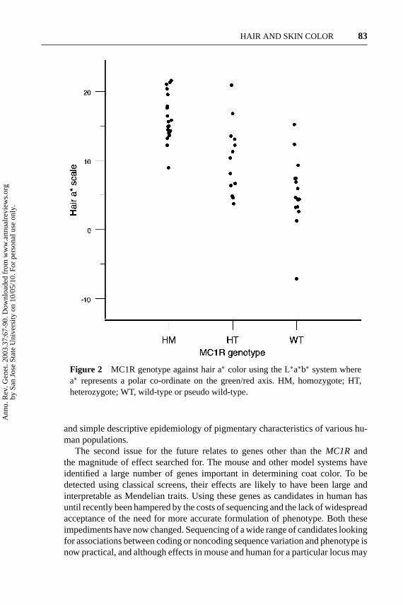
Early studies of theMC1Rand red hair provided no definition of red beyond asimple operational definition used by the investigator (11, 117). Even so, a single(or composite?) group “red” emerged after bootstrapping back toMC1Rsequenceso that shades such as carrot red, strawberry blonde, and auburn were includedtogether (31). A chart linking genotype with crude color assessment using thismethod and objective HPLC analysis of melanins is revealing (Figure 1): Basedon hair melanin measures the “color” varies considerably and the sequence datalend credence to the idea that there is greater clustering of phenotype if melanin dataare used rather than categorical classification of hair. Indeed, simple colorimetricmeasures may be useful as there will be a relation between melanins, hair colorusing spectrophotometry, and sequence (Figure 2). The advantage of such methodsis objectivity and the ability to handle the trait quantitatively. The disadvantage isthat rather than relying on recall of hair color at a certain age, subject age needsto be taken into account in the study design. The possible advantage of such anapproach is illustrated by a recent report (78).
Phenotype of MC1R Variants Against “Black Skin”
Observation would suggest (to this author) that the effects of the MC1R wouldvary on different genetic backgrounds: That this is so would hardly be surprising.How do we measure this effect? For instance, would we expect to see an effectof a single R151C, R160W, or D294H allele on a black African? Would we ex-pect to see the same effect on hair and skin? We have recently, in collaborationwith Colin McKenzie in Jamaica, studied three red-headed children born to self-described black parents (78). The children harbor some of theMC1Rmutationsseen in Northern Europeans. In photographs their hair color is obviously red, but
Ann
u. R
ev. G
enet
. 200
3.37
:67-
90. D
ownl
oade
d fr
om w
ww
.ann
ualr
evie
ws.
org
by S
an J
ose
Stat
e U
nive
rsity
on
10/0
5/10
. For
per
sona
l use
onl
y.

11 Sep 2003 14:51 AR AR201-GE37-04.tex AR201-GE37-04.sgm LaTeX2e(2002/01/18)P1: GCE
82 REES
Figure 1 Hair color classified using a hair chart against log of eumelanin andpheomelanin ratio by genotype (closed circles, homozygous diminished function;triangles, heterozygote;open squares, wild-type or pseudo wild-type).
more usefully, hair melanins were assessed quantitatively showing values withinthe range seen in persons with red hair from Northern Europe (J.L.R., unpub-lished). These results do not allow for differences in age, but at face value suggestthat the homozygous effects of MC1R, even on a very different pigmentary back-ground, are large and observable. Perhaps the effect is greater on hair than skin?One can imagine a similar experiment in European populations. Traditionally,there is a pocket of red-headed persons in Naples. How much difference wouldwe expect betweenMC1Rgenotypically identical individuals between Naples andEdinburgh? When studied quantitatively, estimates of the relative contributions ofthis locus and that not due to this locus can be formulated. To carry out this plan re-quires standardized methods to assess hair color, agreed by different investigators,
Ann
u. R
ev. G
enet
. 200
3.37
:67-
90. D
ownl
oade
d fr
om w
ww
.ann
ualr
evie
ws.
org
by S
an J
ose
Stat
e U
nive
rsity
on
10/0
5/10
. For
per
sona
l use
onl
y.
11 Sep 2003 14:51 AR AR201-GE37-04.tex AR201-GE37-04.sgm LaTeX2e(2002/01/18)P1: GCE
HAIR AND SKIN COLOR 83
Figure 2 MC1R genotype against hair a∗ color using the L∗a∗b∗ system wherea∗ represents a polar co-ordinate on the green/red axis. HM, homozygote; HT,heterozygote; WT, wild-type or pseudo wild-type.
and simple descriptive epidemiology of pigmentary characteristics of various hu-man populations.
The second issue for the future relates to genes other than theMC1R andthe magnitude of effect searched for. The mouse and other model systems haveidentified a large number of genes important in determining coat color. To bedetected using classical screens, their effects are likely to have been large andinterpretable as Mendelian traits. Using these genes as candidates in human hasuntil recently been hampered by the costs of sequencing and the lack of widespreadacceptance of the need for more accurate formulation of phenotype. Both theseimpediments have now changed. Sequencing of a wide range of candidates lookingfor associations between coding or noncoding sequence variation and phenotype isnow practical, and although effects in mouse and human for a particular locus may
Ann
u. R
ev. G
enet
. 200
3.37
:67-
90. D
ownl
oade
d fr
om w
ww
.ann
ualr
evie
ws.
org
by S
an J
ose
Stat
e U
nive
rsity
on
10/0
5/10
. For
per
sona
l use
onl
y.

11 Sep 2003 14:51 AR AR201-GE37-04.tex AR201-GE37-04.sgm LaTeX2e(2002/01/18)P1: GCE
84 REES
not be comparable, the mouse allows subsequent functional assays to be planned.The experimental caution remains that without family studies or functional assays,the causality of any haplotype changes with pigmentation may be confounded byany relation between pigmentary characteristics and evolutionary ancestry.
ACKNOWLEDGMENTS
Thanks to many colleagues, notably Ian Jackson (Edinburgh), Rosalind Harding(Oxford), Brian Diffey (Newcastle), and Kazu Wakamatsu and Shisuke Ito (Japan).My work on the MC1R and skin phenotype is supported by the Wellcome Trust.
The Annual Review of Geneticsis online at http://genet.annualreviews.org
LITERATURE CITED
1. Abdel-Malek ZA, Scott MC, FurumuraM, Lamoreux ML, Ollmann M, et al.2001. The melanocortin 1 receptor isthe principal mediator of the effects ofagouti signaling protein on mammalianmelanocytes.J. Cell Sci.114:1019–24
2. Alaluf S, Atkins D, Barrett K, Blount M,Carter N, Heath A. 2002. Ethnic vari-ation in melanin content and composi-tion in photoexposed and photoprotectedhuman skin.Pigment Cell Res.15:112–18
3. Bagnara JT. 1998. Comparative anatomyand physiology of pigment cells in non-mammalian tissues. See Ref. 84, 2:9–40
4. Barsh G. 1999. From Agouti to Pomc—100 years of fat blonde mice.Nat. Med.5:984–85
5. Barsh GS. 1996. The genetics of pigmen-tation: from fancy genes to complex traits.Trends Genet.12:299–305
6. Bastiaens M, ter Huurne J, Gruis N,Bergman W, Westendorp R, et al. 2001.The melanocortin-1-receptor gene is themajor freckle gene.Hum. Mol. Genet.10:1701–8
7. Bastiaens MT, Huurne JA, Kielich C,Gruis NA, Westendorp RG, et al. 2001.Melanocortin-1 receptor gene variants de-termine the risk of nonmelanoma skincancer independently of fair skin and redhair.Am. J. Hum. Genet.68:884–94
8. Bhardwaj RS, Luger TA. 1994. Proopio-melanocortin production by epidermalcells: evidence for an immune neuroen-docrine network in the epidermis.Arch.Dermatol. Res.287:85–90
9. Bodmer WF, Cavalli-Sforza LL. 1976.Racial differentiation. InGenetics, Evo-lution, and Man. 19:559–604. San Fran-cisco: Freeman
10. Box NF, Duffy DL, Irving R, Russell A,Chen W, et al. 2001. Melanocortin 1 re-ceptor genotype is a risk factor for basaland squamous cell carcinoma.J. Invest.Dermatol.116:224–29
11. Box NF, Wyeth JR, O’Gorman LE, MartinNG, Sturm RA. 1997. Characterization ofmelanocyte stimulating hormone receptorvariant alleles in twins with red hair.Hum.Mol. Genet.6:1891–97
12. Bruls WA, Slaper H, van der Leun JC,Berrens L. 1984. Transmission of humanepidermis and stratum corneum as a func-tion of thickness in the ultraviolet and vis-ible wavelengths.Photochem. Photobiol.40:485–94
13. Bruls WA, van Weelden H, van der LeunJC. 1984. Transmission of UV-radiationthrough human epidermal layers as afactor influencing the minimal erythemadose.Photochem. Photobiol.39:63–67
14. Byard PJ, Lees FC. 1981. Estimating thenumber of loci determining skin colour in
Ann
u. R
ev. G
enet
. 200
3.37
:67-
90. D
ownl
oade
d fr
om w
ww
.ann
ualr
evie
ws.
org
by S
an J
ose
Stat
e U
nive
rsity
on
10/0
5/10
. For
per
sona
l use
onl
y.

11 Sep 2003 14:51 AR AR201-GE37-04.tex AR201-GE37-04.sgm LaTeX2e(2002/01/18)P1: GCE
HAIR AND SKIN COLOR 85
a hybrid population.Ann. Hum. Biol.8:49–58
15. Chardon A, Cretois I, Hourseau C. 1991.Skin colour typology and suntanningpathways.Int. J. Cosmet. Sci.13:191–208
16. Chen WB, Kelly MA, Opitz-Araya X,Thomas RE, Low MJ, Cone RD. 1997.Exocrine gland dysfunction in MC5-R-deficient mice: evidence for coordinatedregulation of exocrine gland function bymelanocortin peptides.Cell 91:789–98
17. Chhajlani V, Muceniece R, Wikberg JE.1993. Molecular cloning of a novel humanmelanocortin receptor.Biochem. Bio-phys. Res. Commun.195:866–73. Erra-tum. 1996.Biochem. Biophys. Res. Com-mun.218 (2):638
18. Chuang TY, Reizner GT, Elpern DJ, StoneJL, Farmer ER. 1995. Nonmelanoma skincancer in Japanese ethnic Hawaiians inKauai, Hawaii: an incidence report.J. Am.Acad. Dermatol.33:422–26
19. Cohn B. 2002. Sunlight, skin color, andfolic acid. J. Am. Acad. Dermatol.46:317–18
20. Cone RD, Lu D, Koppula S, Vage DI,Klungland H, et al. 1996. The melano-cortin receptors: agonists, antagonists,and the hormonal control of pigmentation.Rec. Prog. Horm. Res.51:287–317
21. Cone RD, Mountjoy KG, Robbins LS,Nadeau JH, Johnson KR, et al. 1993.Cloning and functional characterizationof a family of receptors for the melan-otropic peptides.Ann. NY Acad. Sci.680:342–63
22. Davenport CC, Davenport CB. 1909.Heredity of hair color in man.Am. Nat.43:193–211
23. De Luca M, Siegrist W, Bondanza S,Mathor M, Cancedda R, Eberle AN. 1993.Alpha melanocyte stimulating hormone(alpha MSH) stimulates normal humanmelanocyte growth by binding to high-affinity receptors.J. Cell Sci.105:1079–84
24. Diamond JM. 1994. Race without color.Discovery.Nov. 83–89
25. Diffey BL. 1999. Human exposure to ul-traviolet radiation. See Ref. 39a, 2:5–24
26. Diffey BL, Elwood JM. 1994. Tables ofambient solar ultraviolet radiation for usein epidemiological studies of malignantmelanoma and other diseases. InEpidemi-ological Aspects of Cutaneous MalignantMelanoma, ed. RP Gallagher, JM Elwood.19:81–105. Boston: Kluwer
27. Diffey BL, Jansen CT, Urbach F, WulfHC. 1997. The standard erythema dose:a new photobiological concept.Photoder-matol. Photoimmunol. Photomed.13:64–66
28. Diffey BL, Oliver RJ, Farr PM. 1984. Aportable instrument for quantifying ery-thema induced by ultraviolet radiation.Br.J. Dermatol.111:663–72
29. Farr PM, Diffey BL. 1986. The vascularresponse of human skin to ultraviolet ra-diation. Photochem. Photobiol.44:501–7
30. Fitzpatrick TB. 1988. The validity andpracticality of sun-reactive skin types Ithrough VI.Arch. Dermatol.124:869–71
31. Flanagan N, Healy E, Ray A, Philips S,Todd C, et al. 2000. Pleiotropic effectsof the melanocortin 1 receptor (MC1R)gene on human pigmentation.Hum. Mol.Genet.9:2531–37
32. Flanagan N, Ray AJ, Todd C, Birch-Machin MA, Rees JL. 2001. The re-lation between melanocortin 1 recep-tor genotype and experimentally assessedultraviolet radiation sensitivity.J. Invest.Dermatol.117:1314–17
33. Frandberg PA, Doufexis M, Kapas S,Chhajlani V. 1998. Human pigmentationphenotype: A point mutation generatesnonfunctional MSH receptor.Biochem.Biophys. Res. Commun.245:490–92
34. Friedmann PS, Wren F, Buffey J, Mac-Neil S. 1990. Alpha-MSH causes a smallrise in cAMP but has no effect on basalor ultraviolet-stimulated melanogenesisin human melanocytes.Br. J. Dermatol.123:145–51
35. Gantz I, Yamada T, Tashiro T, Konda
Ann
u. R
ev. G
enet
. 200
3.37
:67-
90. D
ownl
oade
d fr
om w
ww
.ann
ualr
evie
ws.
org
by S
an J
ose
Stat
e U
nive
rsity
on
10/0
5/10
. For
per
sona
l use
onl
y.

11 Sep 2003 14:51 AR AR201-GE37-04.tex AR201-GE37-04.sgm LaTeX2e(2002/01/18)P1: GCE
86 REES
Y, Shimoto Y, et al. 1994. Mapping ofthe gene encoding the melanocortin-1(alpha-melanocyte stimulating hormone)receptor (MC1R) to human chromosome16q24.3 by fluorescence in situ hybridiza-tion. Genomics19:394–95
36. Gilchrest BA, Blog FB, Szabo G. 1979.Effects of ageing and chronic sun expo-sure on melanocytes in human skin.J. In-vest. Dermatol.73:141–43
37. Harding RM, Healy E, Ray AJ, Ellis NS,Flanagan N, et al. 2000. Evidence forvariable selective pressures at the humanpigmentation locus, MC1R.Am. J. Hum.Genet.66:1351–61
38. Harrison GA. 1973. Differences in humanpigmentation: measurement, geographicvariation, and causes.J. Invest. Dermatol.60:418–26
39. Hatta N, Dixon C, Ray AJ, Phillips SR,Cunliffe WJ, et al. 2001. Expression, can-didate gene, and population studies of themelanocortin 5 receptor.J. Invest. Derma-tol. 116:564–70
39a. Hawk JLM, ed. 1999.Photodermatology.London: Arnold
40. Healy E, Birch-Machin MA, Rees JL.2000. The human melanocortin-1 recep-tor. InThe Human Melanocortin-1 Recep-tor, ed. RD Cone. 11:341–60. Totawa, NJ:Humana
41. Healy E, Flannagan N, Ray A, Todd C,Jackson IJ, et al. 2000. Melanocortin-1-receptor gene and sun sensitivity inindividuals without red hair. Lancet355:1072–73
42. Healy E, Jordan SA, Budd PS, SuffolkR, Rees JL, Jackson IJ. 2001. Functionalvariation of MC1R alleles from red-hairedindividuals.Hum. Mol. Genet.10:2397–402
43. Healy E, Todd C, Jackson IJ, Birch-Machin M, Rees JL. 1999. Skin type,melanoma, and melanocortin 1 receptorvariants.J. Invest. Dermatol.112:512–13
44. Hedley SJ, Gawkrodger DJ, WeetmanAP, Morandini R, Boeynaems JM, et al.1998.α-melanocyte stimulating hormone
inhibits tumour necrosis factor—a stim-ulated intercellular adhesion molecule-1expression in normal cutaneous humanmelanocytes and in melanoma cell lines.Br. J. Dermatol.138:536–43
45. Hill HZ. 1992. The function of melaninor six blind people examine an elephant.BioEssays14(1):49–56
46. Hoffmann K, Kaspar K, Altmeyer P,Gambichler T. 2000. UV transmissionmeasurements of small skin specimenswith special quartz cuvettes.Dermatology201:307–11
47. Holick MF. 2001. Sunlight “D” ilemma:risk of skin cancer or bone disease andmuscle weakness.Lancet357:4–6
48. Hunt G, Kyne S, Ito S, Wakamatsu K,Todd C, Thody A. 1995. Eumelanin andpheomelanin contents of human epider-mis and cultured melanocytes.PigmentCell Res.8:202–8
49. Hunt G, Todd C, Cresswell JE, Thody AJ.1994. Alpha-melanocyte stimulating hor-mone and its analogue Nle4DPhe7 alpha-MSH affect morphology, tyrosinase activ-ity and melanogenesis in cultured humanmelanocytes.J. Cell Sci.107(Pt. 1):205–11
50. Ichii-Jones F, Lear JT, Heagerty AHM,Smith AG, Hutchinson PE, et al. 1998.Susceptibility to melanoma: influenceof skin type and polymorphism in themelanocyte stimulating hormone receptorgene.J. Invest. Dermatol.111:218–21
51. Ito S. 1998. Advances in chemical analy-sis of melanins. See Ref. 84, pp. 439–50
52. Ito S, Ozeki H, Wakamatsu K. 2000. Spec-trophotometric and HPLC characteriza-tion of hair melanins. InMelanogenesisand Malignant Melanoma: Biochemistry,Cell Biology, Molecular Biology, Patho-physiology, Diagnosis and Treatment, ed.K Hori, VJ Hearing, J Nakayama, pp. 63–72. Oxford: Oxford Univ. Press
53. Jablonski NG, Chaplin G. 2000. The evo-lution of human skin coloration.J. Hum.Evol.39:57–106
54. Jackson IJ. 1991. Mouse coat color
Ann
u. R
ev. G
enet
. 200
3.37
:67-
90. D
ownl
oade
d fr
om w
ww
.ann
ualr
evie
ws.
org
by S
an J
ose
Stat
e U
nive
rsity
on
10/0
5/10
. For
per
sona
l use
onl
y.

11 Sep 2003 14:51 AR AR201-GE37-04.tex AR201-GE37-04.sgm LaTeX2e(2002/01/18)P1: GCE
HAIR AND SKIN COLOR 87
mutations: a molecular genetic resourcewhich spans the centuries.BioEssays13:439–46
55. Jackson IJ. 1994. Molecular and devel-opmental genetics of mouse coat color.Annu. Rev. Genet.28:189–217
56. Jackson IJ. 1997. Homologous pigmen-tation mutations in human, mouse andother model organisms.Hum. Mol. Genet.6:1613–24
57. Jimenez-Cervantes C, Olivares C, Gon-zalez P, Morandini R, Ghanem G, CarlosGarcia-Borron J. 2001. The pro162 vari-ant is a loss-of-function mutation of thehuman melanocortin 1 receptor gene.J.Invest. Dermatol.117:156–58
58. Joerg H, Fries HR, Meijerink E, Stran-zinger GF. 1996. Red coat color in Hol-stein cattle is associated with a deletion inthe MSHR gene.Mamm. Genome7:317–18
59. Jones FI, Ramachandran S, Lear J, SmithA, Bowers B, et al. 1999. The melanocytestimulating hormone receptor polymor-phism: association of the V92M andA294H alleles with basal cell carcinoma.Clin. Chim. Acta282:125–34
60. Kanetsky PA, Swoyer J, Panossian S,Holmes R, Guerry D, Rebbeck TR. 2002.A polymorphism in the agouti signalingprotein gene is associated with human pig-mentation.Am. J. Hum. Genet.70:770–75
61. Kennedy C, ter Huurne J, BerkhoutM, Gruis N, Bastiaens M, et al. 2001.Melanocortin 1 receptor (MC1R) genevariants are associated with an increasedrisk for cutaneous melanoma which islargely independent of skin type and haircolor.J. Invest. Dermatol.117:294–300
62. Kijas JMH, Wales R, T¨ornsten A,Chardon P, Moller M, Andersson L. 1998.Melanocortin receptor 1 (MC1R) muta-tions and coat color in pigs.Genetics150:1177–85
63. King RA, Hearing VJ, Creel DJ, OettingWS. 2001. Albinism. InThe Metabolicand Molecular Bases of Inherited Dis-ease, ed. CR Scriver, AL Beaudet, WS Sly,
D Valle, B Childs, KW Kinzler, B Vogel-stein. 200:5587–28. New York: McGrawHill. 8th ed.
64. Klungland H, Vage DI, Gomez-Raya L,Adalsteinsson S, Lien S. 1995. The role ofmelanocyte-stimulating hormone (MSH)receptor in bovine coat color determina-tion. Mamm. Genome6:636–39
65. Kollias N, Sayre RM, Zeise L, ChedekelMR. 1991. Photoprotection by melanin.J.Photochem. Photobiol. B9:135–60
66. Kollias N, Stamatas GN. 2002. Opticalnon-invasive approaches to diagnosis ofskin diseases.J. Investig. Dermatol. Symp.Proc.7:64–75
67. Koppula SV, Robbins LS, Lu D, BaackE, White CRJ, et al. 1997. Identificationof common polymorphisms in the cod-ing sequence of the human MSH receptor(MCIR) with possible biological effects.Hum. Mutat.9:30–36
68. Krude H, Biebermann H, Luck W, Horn R,Brabant G, Gr¨uters A. 1998. Severe early-onset obesity, adrenal insufficiency andred hair pigmentation caused byPOMCmutations in humans.Nat. Genet.19:155–57
69. Lerner AB. 1993. The discovery of themelanotropins.Ann. NY Acad. Sci.680:1–12
70. Little MA, Wolff ME. 1981. Skin and hairreflectance in women with red hair.Ann.Hum. Biol.8:231–41
71. Lock-Andersen J, Therkildsen P, de FineO, Gniadecka M, Dahlstrom K, et al.1997. Epidermal thickness, skin pig-mentation and constitutive photosensitiv-ity. Photodermatol. Photoimmunol. Pho-tomed.13:153–58
72. Lock-Andersen J, Wulf HC. 1997. Sea-sonal variation of skin pigmentation.ActaDerm. Venereol.77:219–21
73. Lookingbill DP, Lookingbill GL, LeppardB. 1995. Actinic damage and skin can-cer in albinos in northern Tanzania: find-ings in 164 patients enrolled in an outreachskin care program.J. Am. Acad. Derma-tol. 32:653–58
Ann
u. R
ev. G
enet
. 200
3.37
:67-
90. D
ownl
oade
d fr
om w
ww
.ann
ualr
evie
ws.
org
by S
an J
ose
Stat
e U
nive
rsity
on
10/0
5/10
. For
per
sona
l use
onl
y.
11 Sep 2003 14:51 AR AR201-GE37-04.tex AR201-GE37-04.sgm LaTeX2e(2002/01/18)P1: GCE
88 REES
74. Luger TA, Bhardwaj RS, Grabbe S, Sch-warz T. 1996. Regulation of the immuneresponse by epidermal cytokines and neu-rohormones.J. Dermatol. Sci.13:5–10
75. Makova KD, Ramsay M, Jenkins T, LiWH. 2001. Human DNA sequence vari-ation in a 6.6-kb region containing themelanocortin 1 receptor promoter.Genet-ics158:1253–68
76. Manga P, Kromberg JGR, Box NF, SturmRA, Jenkins T, Ramsay M. 1997. Ru-fous oculocutaneous albinism in southernAfrican Blacks is caused by mutations intheTYRP1gene.Am. J. Hum. Genet.61:1095–101
77. Marklund L, Moller MJ, Sandberg K, An-dersson L. 1996. A missense mutation inthe gene for melanocyte-stimulating hor-mone receptor (MC1R) is associated withthe chestnut coat color in horses.Mamm.Genome7:895–99
78. McKenzie CA, Harding RM, TomlinsonJB, Ray AJ, Wakamatsu K, Rees JL. 2003.Phenotypic expression of melanocortin-1receptor (MC1R) mutations in Black Ja-maicans.J. Invest. Dermatol.In press
79. Miller MW, Duhl DM, Vrieling H, CordesSP, Ollmann MM, et al. 1993. Cloningof the mouse agouti gene predicts a se-creted protein ubiquitously expressed inmice carrying the lethal yellow mutation.Genes Dev.7:454–67
80. Montagna W, Prota G, Kenney JA. 1993.Black Skin: Structure and Function.Lon-don: Academic
81. Morandini R, Boeynaems JM, Hedley SJ,MacNeil S, Ghanem G. 1998. Modulationof ICAM-1 expression by a-MSH in hu-man melanoma cells and melanocytes.J.Cell. Physiol.175:276–82
82. Mountjoy KG, Robbins LS, Mortrud MT,Cone RD. 1992. The cloning of a familyof genes that encode the melanocortin re-ceptors.Science257:1248–51
83. Newton JM, Wilkie AL, He L, Jordan SA,Metallinos DL, et al. 2000. Melanocortin1 receptor variation in the domestic dog.Mamm. Genome11:24–30
84. Nordlund JJ, Boissy RE, Hearing VJ,King RA, Ortonne JP, eds. 1998.The Pig-mentary System: Physiology and Patho-physiology.New York: Oxford Univ. Press
85. Ozeki H, Ito S, Wakamatsu K, ThodyAJ. 1996. Spectrophotometric characteri-zation of eumelanin and pheomelanin inhair.Pigment Cell Res.9:265–70
86. Palmer JS, Duffy DL, Box NF, Aitken JF,O’Gorman LE, et al. 2000. Melanocortin-1 receptor polymorphisms and risk ofmelanoma: Is the association explainedsolely by pigmentation phenotype?Am.J. Hum. Genet.66:176–86
87. Price JS, Burton JL, Shuster S, WolffK. 1976. Control of scrotal colour in thevervet monkey.J. Med. Primatol.5:296–304
88. Prota G. 1992.Melanins and Melanogen-esis.San Diego: Academic
89. Prota G, d’Ischia M, Napolitano A. 1998.The chemistry of melanins and relatedmetabolites. See Ref. 84, pp. 307–32
90. Quevedo WC, Holstein TJ. 1998. Generalbiology of mammalian pigmentation. SeeRef. 84, pp. 43–58
91. Rampen FH, Fleuren BA, de Boo TM,Lemmens WA. 1988. Unreliability of self-reported burning tendency and tanningability. Arch. Dermatol.124:885–88
92. Rana BK, Hewett-Emmett D, Jin L,Chang BHJ, Sambuughin N, et al.1999. High polymorphism at the humanmelanocortin 1 receptor locus.Genetics151:1547–57
93. Rees JL. 2000. The melanocortin 1 recep-tor (MC1R): more than just red hair.Pig-ment Cell Res.13:135–40
94. Rees JL. 2002. Molecular phototypes.In Mechanisms of Suntanning, ed. Jean-Paul Ortonne, Robert Ballotti. 31:333–339. London: Martin Dunitz
95. Robbins LS, Nadeau JH, Johnson KR,Kelly MA, Roselli-Rehfuss L, et al. 1993.Pigmentation phenotypes of variant ex-tension locus alleles result from point mu-tations that alter MSH receptor function.Cell 72:827–34
Ann
u. R
ev. G
enet
. 200
3.37
:67-
90. D
ownl
oade
d fr
om w
ww
.ann
ualr
evie
ws.
org
by S
an J
ose
Stat
e U
nive
rsity
on
10/0
5/10
. For
per
sona
l use
onl
y.

11 Sep 2003 14:51 AR AR201-GE37-04.tex AR201-GE37-04.sgm LaTeX2e(2002/01/18)P1: GCE
HAIR AND SKIN COLOR 89
96. Roberts DF, Kromberg JG, Jenkins T.1986. Differentiation of heterozygotes inrecessive albinism.J. Med. Genet.23:323–27
97. Robins AH. 1991. The evolution of skincolour. InBiological Perspectives on Hu-man Pigmentation, pp. 187–212. Cam-bridge: Cambridge Univ. Press
98. Sarna T, Swartz HM. 1998. The physi-cal properties of melanins. See Ref. 84,25:333–57
99. Schioth HB, Muceniece R, Larsson M,Mutulis F, Szardenings M, et al. 1997.Binding of cyclic and linear MSH corepeptides to the melanocortin receptorsubtypes.Eur. J. Pharmacol.319:369–73
100. Schioth HB, Muceniece R, Wikberg JE.1996. Characterisation of the melano-cortin 4 receptor by radioligand binding.Pharmacol. Toxicol.79:161–65
101. Schioth HB, Muceniece R, Wikberg JE,Chhajlani V. 1995. Characterisation ofmelanocortin receptor subtypes by radi-oligand binding analysis.Eur. J. Pharma-col. 288:311–17
102. Schioth HB, Phillips S, Rudzish R, Birch-Machin M, Wikberg J, Rees JL. 1999.Loss of function mutations of the humanmelanocortin 1 receptor are common andassociated with red hair.Biochem. Bio-phys. Res. Commun.260:488–91
103. Shriver MD, Parra EJ. 2000. Comparisonof narrow-band reflectance spectroscopyand tristimulus colorimetry for measure-ments of skin and hair color in persons ofdifferent biological ancestry.Am. J. Phys.Anthropol.112:17–27
104. Silvers WK. 1979.Coat Colors of Mice.New York: Springer-Verlag
105. Smith R, Healy E, Siddiqui S, FlanaganN, Steijlen PM, et al. 1998. Melanocortin1 receptor variants in an Irish population.J. Invest. Dermatol.111:119–22
106. Sturm RA, Box NF, Ramsay M. 1998.Human pigmentation genetics: the differ-ence is only skin deep.BioEssays20:712–21
107. Sturm RA, Teasdale RD, Box NF. 2001.Human pigmentation genes: identifica-tion, structure and consequences of poly-morphic variation.Gene277:49–62
108. Suzuki I, Cone RD, Im S, NordlundJ, Abdel-Malek ZA. 1996. Binding ofmelanotropic hormones to the melano-cortin receptor MC1R on human melano-cytes stimulates proliferation and melano-genesis.Endocrinology137:1627–33
109. Takeuchi S, Suzuki H, Yabuuchi M, Taka-hashi S. 1996. A possible involvementof melanocortin 1-receptor in regulatingfeather color pigmentation in the chicken.Biochim. Biophys. Acta1308:164–68
110. Tan CP, McKee KK, Weinberg DH, Mac-Neil T, Palyha OC, et al. 1999. Molecularanalysis of a new splice variant of the hu-man melanocortin-1 receptor.FEBS Lett.451:137–41
111. Theron E, Hawkins K, Bermingham E,Ricklefs RE, Mundy NI. 2001. The molec-ular basis of an avian plumage poly-morphism in the wild: a melanocortin-1-receptor point mutation is perfectly asso-ciated with the melanic plumage morphof the bananaquit,Coereba flaveola. Curr.Biol. 11:550–57
112. Thody AJ, Graham A. 1998. Doesα-MSHhave a role in regulating skin pigmentationin humans.Pigment Cell Res.11:265–74
113. Thody AJ, Higgins EM, Wakamatsu K, ItoS, Burchill SA, Marks JM. 1991. Pheome-lanin as well as eumelanin is present inhuman epidermis.J. Invest. Dermatol.97:340–44
114. Thody AJ, Ridley K, Penny RJ, ChalmersR, Fisher C, Shuster S. 1983. MSH pep-tides are present in mammalian skin.Pep-tides4:813–16
115. Urbach F. 1999. The cumulative effects ofultraviolet radiation on the skin: photocar-cinogenesis. See Ref. 39a, 7:89–111
116. Vage DI, Lu DS, Klungland H, Lien S,Adalsteinsson S, Cone RD. 1997. A non-epistatic interaction ofagouti andexten-sionin the fox,Vulpes vulpes. Nat. Genet.15:311–15
Ann
u. R
ev. G
enet
. 200
3.37
:67-
90. D
ownl
oade
d fr
om w
ww
.ann
ualr
evie
ws.
org
by S
an J
ose
Stat
e U
nive
rsity
on
10/0
5/10
. For
per
sona
l use
onl
y.

11 Sep 2003 14:51 AR AR201-GE37-04.tex AR201-GE37-04.sgm LaTeX2e(2002/01/18)P1: GCE
90 REES
117. Valverde P, Healy E, Jackson I, ReesJL, Thody AJ. 1995. Variants of themelanocyte-stimulating hormone recep-tor gene are associated with red hair andfair skin in humans.Nat. Genet.11:328–30
118. Valverde P, Healy E, Sikkink S, HaldaneF, Thody AJ, et al. 1996. The Asp84Gluvariant of the melanocortin 1 receptor(MC1R) is associated with melanoma.Hum. Mol. Genet.5:1663–66
119. Van den Berghe PL Frost P. 1986. Skincolor preference, sexual dimorphism, andsexual selection: a case of gene-cultureco-evolution.Ethn. Racial Stud.9:87–113
120. Voisey J, Box NF, van Daal A. 2001. Apolymorphism study of the human Agoutigene and its association with MC1R.Pig-ment Cell Res.14:264–57
121. Vrieling H, Duhl DM, Millar SE, MillerKA, Barsh GS. 1994. Differences in
dorsal and ventral pigmentation resultfrom regional expression of the mouseagouti gene.Proc. Natl. Acad. Sci. USA91:5667–71
122. Wagner JK, Jovel C, Norton HL, ParraEJ, Shriver MD. 2002. Comparing quan-titative measures of erythema, pigmenta-tion and skin response using reflectome-try. Pigment Cell Res.15:379–84
123. Wagner JK, Parra EJ, Norton L, Jovel C,Shriver MD. 2002. Skin responses to ul-traviolet radiation: effects of constitutivepigmentation, sex, and ancestry.PigmentCell Res.15:385–90
124. Wakamatsu K, Ito S. 2002. Advancedchemical methods in melanin determina-tion. Pigment Cell Res.15:174–83
125. Wakamatsu K, Ito S, Rees JL. 2002.Usefulness of 4-amino-3-hydroxyphenyl-alanine as a specific marker of pheome-lanin.Pigment Cell Res.15:225–32
Ann
u. R
ev. G
enet
. 200
3.37
:67-
90. D
ownl
oade
d fr
om w
ww
.ann
ualr
evie
ws.
org
by S
an J
ose
Stat
e U
nive
rsity
on
10/0
5/10
. For
per
sona
l use
onl
y.
P1: FDS
October 1, 2003 16:35 Annual Reviews AR201-FM
Annual Review of GeneticsVolume 37, 2003
CONTENTS
IRA HERSKOWITZ (1946–2003), The Editors 1
TRANSPOSON-BASED STRATEGIES FOR MICROBIAL FUNCTIONALGENOMICS AND PROTEOMICS, Finbarr Hayes 3
ERROR-PRONE DNA POLYMERASES: WHEN MAKING A MISTAKE ISTHE ONLY WAY TO GET AHEAD, Alison J. Rattray andJeffrey N. Strathern 31
GENETICS OF HAIR AND SKIN COLOR, Jonathan L. Rees 67
THIOL-BASED REGULATORY SWITCHES, Mark S.B. Paget andMark J. Buttner 91
PSEUDOGENES: ARE THEY “JUNK” OR FUNCTIONAL DNA?Evgeniy S. Balakirev and Francisco J. Ayala 123
UNUSUAL LIFE STYLE OF GIANT CHLORELLA VIRUSES,James L. Van Etten 153
GENETICS OF LACTASE PERSISTENCE AND LACTOSE INTOLERANCE,Dallas M. Swallow 197
CELL POLARITY AND THE CYTOSKELETON IN THE CAENORHABDITISELEGANS ZYGOTE, Stephan Q. Schneider and Bruce Bowerman 221
THE SPINDLE ASSEMBLY AND SPINDLE POSITION CHECKPOINTS,Daniel J. Lew and Daniel J. Burke 251
LATERAL GENE TRANSFER AND THE ORIGINS OF PROKARYOTICGROUPS, Yan Boucher, Christophe J. Douady, R. Thane Papke,David A. Walsh, Mary Ellen R. Boudreau, Camilla L. Nesbø,Rebecca J. Case, and W. Ford Doolittle 283
GENETICS OF AGING IN THE FRUIT FLY, DROSOPHILA MELANOGASTER,Stephen L. Helfand and Blanka Rogina 329
NATURAL SELECTION AND THE EVOLUTION OF GENOME IMPRINTING,Elena de la Casa-Esperon and Carmen Sapienza 349
THE NEED FOR WINTER IN THE SWITCH TO FLOWERING,Ian R. Henderson, Chikako Shindo, and Caroline Dean 371
TRANSMISSION RATIO DISTORTION IN MICE, Mary F. Lyon 393
v
Ann
u. R
ev. G
enet
. 200
3.37
:67-
90. D
ownl
oade
d fr
om w
ww
.ann
ualr
evie
ws.
org
by S
an J
ose
Stat
e U
nive
rsity
on
10/0
5/10
. For
per
sona
l use
onl
y.

P1: FDS
October 1, 2003 16:35 Annual Reviews AR201-FM
vi CONTENTS
STRUCTURE, DIVERSITY, AND EVOLUTION OF PROTEIN TOXINS FROMSPORE-FORMING ENTOMOPATHOGENIC BACTERIA, Ruud A. de Maagd,Alejandra Bravo, Colin Berry, Neil Crickmore, and H. Ernst Schnepf 409
YEAST VACUOLE INHERITANCE AND DYNAMICS, Lois S. Weisman 435
HEPARAN SULFATE CORE PROTEINS IN CELL-CELL SIGNALING,Kenneth L. Kramer and H. Joseph Yost 461
RETROTRANPOSONS PROVIDE AN EVOLUTIONARILY ROBUSTNON-TELOMERASE MECHANISM TO MAINTAIN TELOMERES,Mary-Lou Pardue and P.G. DeBaryshe 485
A CYANOBACTERIAL CIRCADIAN TIMING MECHANISM, S.S. Golden,J.L. Ditty, and S.B. Williams 513
REGULATION OF CELL CYCLES IN DROSOPHILA DEVELOPMENT:INTRINSIC AND EXTRINSIC CUES, Laura A. Lee and Terry L. Orr-Weaver 545
RECOGNITION AND RESPONSE IN THE PLANT IMMUNE SYSTEM,Zachary Nimchuk, Thomas Eulgem, Ben F. Holt III, and Jeffrey L. Dangl 579
RECA-DEPENDENT RECOVERY OF ARRESTED DNA REPLICATIONFORKS, Justin Courcelle and Phillip C. Hanawalt 611
INDEXESSubject Index 647Cumulative Index of Contributing Authors, Volumes 33–37 679Cumulative Index of Chapter Titles, Volumes 33–37 682
ERRATAAn online log of corrections to Annual Review of Genetics chaptersmay be found at http://genet.annualreviews.org/errata.shtml
Ann
u. R
ev. G
enet
. 200
3.37
:67-
90. D
ownl
oade
d fr
om w
ww
.ann
ualr
evie
ws.
org
by S
an J
ose
Stat
e U
nive
rsity
on
10/0
5/10
. For
per
sona
l use
onl
y.