jnk1 inhibition attenuates hypoxia-induced autophagy …€¦ · oxaliplatin - from lkt labs (st....
TRANSCRIPT
1
JNK1 INHIBITION ATTENUATES HYPOXIA-INDUCED AUTOPHAGY AND SENSITIZES TO
CHEMOTHERAPY
Irina A Vasilevskaya1
Muthu Selvakumaran1
David Roberts1,2
Peter J O’Dwyer1
Authors affiliations: 1Abramson Cancer Center, University of Pennsylvania, Philadelphia PA;
2currently at Modbury Hospital, Adelaide, Australia
Running title: JNK1 inhibition potentiates chemotherapy
Keywords: hypoxia, JNK1, autophagy, oxaliplatin, colon cancer
Financial support: Supported in part by R01CA139003 and RO1CA158377 from NCI, NIH
Corresponding author: I. A. Vasilevskaya, 1020 BRBII/III, 421 Curie Blvd, Philadelphia PA 19104, e-
mail: [email protected].
The authors disclose no potential conflicts of interest.
Word count: 5739
Figures and Tables: 7
on May 15, 2020. © 2016 American Association for Cancer Research. mcr.aacrjournals.org Downloaded from
Author manuscripts have been peer reviewed and accepted for publication but have not yet been edited. Author Manuscript Published OnlineFirst on May 23, 2016; DOI: 10.1158/1541-7786.MCR-16-0035
2
ABSTRACT.
Inhibition of hypoxia-induced stress signaling through JNK potentiates the effects of oxaliplatin. The
JNK pathway plays a role in both autophagy and apoptosis; therefore, it was determined how much of
the effect of JNK inhibition on oxaliplatin sensitivity is dependent on its effect on autophagy. We
studied the impact of JNK isoform down-regulation in the HT29 colon adenocarcinoma cell line on
hypoxia- and oxaliplatin-induced responses. Electron microscopic analyses demonstrated that both
oxaliplatin- and hypoxia-induced formation of autophagosomes were reduced significantly in HT29
cells treated with the JNK inhibitor SP600125. The role of specific JNK isoforms was defined using
HT29-derived cell lines stably expressing dominant negative constructs for JNK1 and JNK2 (HTJ1.3
and HTJ2.2, respectively). These cell lines demonstrated that functional JNK1 is required for hypoxia-
induced autophagy and that JNK2 does not substitute for it. Inhibition of autophagy in HTJ1.3 cells
also coincided with enhancement of intrinsic apoptosis. Analysis of Bcl2-family proteins revealed
hyper-phosphorylation of Bcl-XL in the HTJ1.3 cell line, but this did not lead to the expected
dissociation from Beclin-1. Consistent with this, knockdown of Bcl-XL in HT29 cells did not
significantly affect the induction of autophagy, but abrogated hypoxic resistance to oxaliplatin due to
the faster and more robust activation of apoptosis.
Implications: These data suggest that balance between autophagy and apoptosis are shifted toward
apoptosis by down-regulation of JNK1, contributing to oxaliplatin sensitization. These findings further
support the investigation of JNK inhibition in colorectal cancer treatment.
on May 15, 2020. © 2016 American Association for Cancer Research. mcr.aacrjournals.org Downloaded from
Author manuscripts have been peer reviewed and accepted for publication but have not yet been edited. Author Manuscript Published OnlineFirst on May 23, 2016; DOI: 10.1158/1541-7786.MCR-16-0035

3
INTRODUCTION
One of the characteristic of solid tumors is the occurrence of regions with low oxygen tension as a
result of uncontrolled cell growth and disordered angiogenesis, to which oxygen delivery can be
restored through angiogenesis inhibition (1, 2). This phenomenon contributes to a complicated
microenvironment, where both continuous and cycling hypoxia are able to significantly affect tumor
behavior, and are associated with poor prognosis due in part to a more aggressive tumor phenotype,
resistance to chemotherapy and radiation, and increased genetic instability (3). Therefore, targeting of
hypoxia is considered a promising approach to augment the efficacy of cancer treatment (4, 5).
Hypoxia induces multiple signaling pathways, resulting in activation of major transcription factors,
including hypoxia-induced factors (HIFs), NF-κB, p53 and AP-1 (6, 7), which in turn regulate cellular
responses to the lack of oxygen, such as metabolic adaptations, angiogenesis, cell death and autophagy,
among others.
Autophagy is a catabolic process, involving “packing” of various cytoplasmic components into double-
membrane vehicles (autophagosomes), followed by their fusion with lysosomes and formation of
autolysosomes, where degradation of the autophagic cargo takes place (8). It was first extensively
studied as an adaptive response to starvation, but later was acknowledged as a key process in
maintenance of cellular homeostasis, damage responses and progression of various diseases, including
cancer (9). There is ample evidence for both oncosuppressive and tumor-supportive roles of
autophagy, depending on cellular and tissue context (9), which makes targeting autophagy a
controversial issue. It is a highly regulated process, depending on activation and interaction of
multiple molecular components, including close to 30 proteins [so called ATG proteins, products of
autophagy related genes (ATG)]. One of these, Beclin1, is crucial for initiation of autophagy, as a part
of multi-protein complex, regulating nucleation and isolation of autophagosome membrane (10).
Beclin 1 is inactive when bound though its BH3-domain to pro-survival members of Bcl2 family. It is
on May 15, 2020. © 2016 American Association for Cancer Research. mcr.aacrjournals.org Downloaded from
Author manuscripts have been peer reviewed and accepted for publication but have not yet been edited. Author Manuscript Published OnlineFirst on May 23, 2016; DOI: 10.1158/1541-7786.MCR-16-0035

4
commonly accepted that release of Beclin 1 from this inhibitory complex can be achieved by
phosphorylation of Bcl2 by JNK, phosphorylation of Beclin1 by DAPK, or competitive displacement
of Beclin 1 by pro-apoptotic BH3-only proteins or BH3 mimetics (10,11). Involvement of Bcl2-family
proteins in autophagy regulation indicates the existence of cross-talk between autophagy and
apoptosis, which together can influence cell fate. Indeed, the same signals often can induce both
processes (11) with final outcome (survival or cell death) depending on severity of the stress. Among
key autophagy markers are LC3B, an integral part of the autophagosome membrane, and p62 (also
known as SQSTM1), which targets cargo to the autophagosome through interaction with LC3.
Accumulation of LC3B and its redistribution to autophagosomes allows assessment of autophagy
initiation, whereas degradation of p62 marks the final stages of the process. When autophagy proceeds
to completion, degradation of macromolecules and organelles in autolysosomes provides renewed
supply of “building blocks” (sugars, amino acids, nucleotides, etc) necessary for cellular survival in
adverse conditions (8, 11).
Both hypoxia and chemotherapeutic agents have been shown to induce autophagy, and both treatments
result in activation of stress signaling through JNK (12,13). JNK belongs to the family of MAP
kinases, distal members of tri-tiered signal transduction cascades, which are activated by a plethora of
external and internal stimuli, and mediate appropriate cellular responses (14). There are three isoforms
of JNK (JNK1, JNK2 and JNK3), expressed ubiquitously, except JNK3, which is expressed mostly in
brain. JNK1 and JNK2 share the majority of targets, and were considered redundant, but evidence for
distinct functions of the isoforms has accumulated (15). Signaling through JNK is critical for normal
cell function, but in cancer JNKs can demonstrate both oncogenic and cancer suppressive features (16).
Involvement of JNK in the regulation of autophagy in general, and of hypoxia-induced autophagy in
particular, is thought to be implemented on both transcriptional and post-transcriptional levels.
Phosphorylation of Bcl2 and Bcl-XL by JNK results in release of Beclin1, as mentioned above, and
on May 15, 2020. © 2016 American Association for Cancer Research. mcr.aacrjournals.org Downloaded from
Author manuscripts have been peer reviewed and accepted for publication but have not yet been edited. Author Manuscript Published OnlineFirst on May 23, 2016; DOI: 10.1158/1541-7786.MCR-16-0035

5
allows autophagy to proceed (10, 11). JNK can also influence autophagy through activation of
transcription factors of the AP-1 and FOXO families, which are implicated in driving the expression of
several ATG proteins (17). As a key mediator of stress signaling, induced by hypoxia or DNA-
damaging drugs, JNK also participates in regulating the DNA-damage response and apoptosis (18,19).
Therefore, its multi-faceted functions in hypoxia, DNA-damage, autophagy and apoptosis underlie our
interest in JNK as a target to reverse hypoxic resistance to DNA-damaging drugs (primarily
oxaliplatin) in colon cancer model.
We have shown (20), that in hypoxic HT29 colon adenocarcinoma cells, down-regulation of JNK-
activating kinases, MKK4 or MKK7, exerts differing effects on oxaliplatin cytotoxicity: cells with
impaired MKK4 demonstrate higher sensitivity, whereas down regulation of MKK7, which leads to
more profound inhibition of JNK activation, causes increase in resistance. Next, we expanded our
studies to evaluate effects of JNK down-regulation on chemotherapeutic drug resistance in colon
cancer cell lines, and showed that inhibition by the small molecule JNK inhibitor CC-401 enhances
sensitivity to oxaliplatin in a panel of 6 cell lines (21). In addition, we demonstrated that down-
regulation of JNK1, but not JNK2, abrogates hypoxia-induced resistance to oxaliplatin in HT29 cells
most effectively. We also demonstrated protective function of hypoxia- and oxaliplatin-induced
autophagy in colon cancer cell lines by showing synergism of chloroquine and chemotherapy in vitro
and in vivo (22). Here we set out to define the role of each JNK isoform (in HTJ1.3 and HTJ2.2 cell
lines, stably expressing dominant negative constructs for JNK1 and JNK2, respectively) in the
induction of autophagy in HT29 colon adenocarcinoma in response to hypoxia and chemotherapy. We
hoped in this way to focus inhibitory strategies toward the relevant isoform, so as to minimize adverse
effect from inhibiting such an ubiquitous pathway.
on May 15, 2020. © 2016 American Association for Cancer Research. mcr.aacrjournals.org Downloaded from
Author manuscripts have been peer reviewed and accepted for publication but have not yet been edited. Author Manuscript Published OnlineFirst on May 23, 2016; DOI: 10.1158/1541-7786.MCR-16-0035

6
MATERIALS AND METHODS.
Cells and reagents. The HT 29 human adenocarcinoma cell line was purchased from ATCC
(Manassas, VA). Immediately after receiving cells were thawed, propagated and frozen in multiple
aliquots. For experiments, cells were used within 2 months or resuscitation. HT29-derived cell lines
stably expressing empty vector (HTLX), dominant negative constructs for JNK1 (HTJ1.3) or JNK2
(HTJ2.2) were described in (21). Cells were grown in DMEM medium supplemented with 10% FBS
and antibiotic-antimycotic reagent (Invitrogen, Carlsbad, CA). Cultures were maintained in a
humidified incubator at 37°C in 5%CO2-95% air. Chemical inhibitor for JNK (SP600125) was
purchased from Biomol (Plymouth Meeting, PA), CC-401 was from ChemScene (Monmouth Junction,
NJ). Chloroquine diphosphate and puromycin were from Sigma-Aldrich (St. Louis, MO), and
oxaliplatin - from LKT Labs (St. Paul, MN). Acridine orange (AO) was purchased from Sigma,
Apoptosis and Necrosis Quantification Kit was from Biotium (Hayward, CA).
Viral constructs and infections. For stable delivery of shRNA against JNK1 and JNK2 into
HT29-derived cell lines, we constructed retroviral vectors as described in Supplemental Figure S1.
After transfection into Phoenix-Ampho packaging cell line viruses were collected, purified, aliquoted
and stored at -80oC. For infections, cells plated into 6 well plates were incubated with retroviruses in
the presence of polybrene (8 μg/ml, Chemicon, Temecula, CA), and 48 hours later selective media
containing 1 μg/ml of puromycin was added. Puromycin-resistant cells were pooled, evaluated for
JNK1/JNK2 levels and used in further experiments.
For Bcl-XL knock-down, ready-to-use lentiviral particles (control and shRNA-encoding) were
purchased from Santa Cruz (sc-77361-V), and used according to manufacturer’s recommendations.
Since in pooled cultures knock-down of the target protein was incomplete, individual monoclonal sub-
lines were isolated, assessed for Bcl-XL expression and used in further experiments.
on May 15, 2020. © 2016 American Association for Cancer Research. mcr.aacrjournals.org Downloaded from
Author manuscripts have been peer reviewed and accepted for publication but have not yet been edited. Author Manuscript Published OnlineFirst on May 23, 2016; DOI: 10.1158/1541-7786.MCR-16-0035

7
Hypoxic treatment. Exposure of cells to acute hypoxia was achieved by incubation in an
anaerobic chamber (Forma Scientific, Inc., Marietta, OH) filled with gas mixture consisting of 5%
CO2, 9% H2 and 86% N2. Oxygen content (0.1- 0.5%) was monitored by PROOX 110 oxygen sensor
(BioSpherix, Redfield, NY). Cells were plated in 100 mm glass Petri dishes to a density of 2 x 10⁶
cells per dish and subjected to hypoxia within 36 hours. The cells were harvested at various time
points for further experiments.
Protein extract preparation. Total protein extracts were prepared as follows: after hypoxia,
cells were washed twice with PBS and lyzed inside the chamber in cell lysis buffer (Cell Signaling
Technology, Beverly, MA), supplemented with complete protease inhibitor cocktail (Roche) and 1mM
PMSF (Sigma). The contents of scraped dishes were transferred into microcentrifuge tubes, taken out
of hypoxia chamber and placed in a shaker for 30 min at 4oC. Lysates were then centrifuged for 10
min at 10,000 rpm (4oC) and the protein concentration of cleared extracts was measured using the Bio-
Rad Protein Assay (Bio-Rad, Hercules, CA).
Western blotting. For protein electrophoresis, protein extracts were used in amounts of 10 μg
per lane. Western blotting was carried out according to standard procedures, using horseradish
peroxidase-conjugated secondary antibodies purchased from Santa Cruz Biotechnology (Santa Cruz,
CA) and the ECL+Plus detection system (Amersham, Arlington Heights, IL). Results were analyzed
with BioSpectrum 810 Imaging System using VisionWorksLS Image Acquisition and Analysis
Software (UVP, Upland CA). The antibodies used were: antibodies against p-Bcl-XL, p62 and actin
were from Santa Cruz Biotechnology; antibodies against caspase 3 and caspase 7 - from BD
Pharmingen. The remaining antibodies were purchased from Cell Signaling Technology. LC3 content
was assessed with an antibody against LC3B isoform.
Immunoprecipitation. Cells were subjected to hypoxia for 6 hours, followed by isolation of
cellular extracts as above. From each sample, containing 2 mg of total protein, Beclin1 was
on May 15, 2020. © 2016 American Association for Cancer Research. mcr.aacrjournals.org Downloaded from
Author manuscripts have been peer reviewed and accepted for publication but have not yet been edited. Author Manuscript Published OnlineFirst on May 23, 2016; DOI: 10.1158/1541-7786.MCR-16-0035
8
immunoprecipitated overnight with goat antibodies (Santa Cruz, 20 μl) using ExactoCruz system
(Santa Cruz Biotechnology) according to manufacturer’s recommendations. Immunoprecipitates were
lysed in 60 μl of 2x gel loading buffer, and subjected to electrophoresis (15 μl per lane) followed by
Western blot analysis, using rabbit primary antibodies from Cell Signaling Technology.
Cytotoxicity assays and calculation of combination indices. For assessment of cytotoxicity,
cells were plated in 96-well plates (2000 cells per well), and 24 hours later various amounts of
oxaliplatin and chloroquine alone or in combination were added, immediately before hypoxic exposure
for 24 hours, followed by cultivation in normal condition for additional 48 hours. Cytotoxicity was
measured using a standard MTT assay. Combination indices (CI) were calculated based on Chou-
Talalay methods using CompuSinq software (as in 21), CI=1 indicates additivity, CI<1 indicates
synergism, CI>1 indicates antagonism.
Transmission electron microscopy (TEM). For TEM quantitation of autophagosomes, HT29
cells were subjected to hypoxia and/or oxaliplatin (IC50 dose) with or without 10 μM of SP600125.
Cell pellets were collected, fixed in 2.5% glutaraldehyde/2% formaldehyde with 0.1 M sodium
cacodylate and stored at 4°C until embedding. Embedded samples were processed for TEM as
described earlier (22). Images were examined with a JEOL-1010 electron microscope (JOEL) at 80
kV. For quantitation of cells using electron microscopy, high-powered micrographs (x12, 000-20,000)
of 25 single cells from multiple distinct low-powered fields in each sample were obtained. Cells with
more than three to four double-membrane vesicles were scored as positive for autophagosomes.
Fluorescent staining procedures. To assess autophagy and apoptosis induction we employed
fluorescent microscopy: i) autophagy induction (as LC3II puncta formation) was monitored in HT29
cells infected with GFP-LC3-encoding retrovirus (Addgene plasmid # 22405, pBABEpuro GFP-LC3
from Dr. Jayanta Debnath was used to generate retrovirus), ii) apoptotic cells were identified using
Apoptosis and Necrosis Quantification Kit, iii) staining of live cells with acridine orange was carried
on May 15, 2020. © 2016 American Association for Cancer Research. mcr.aacrjournals.org Downloaded from
Author manuscripts have been peer reviewed and accepted for publication but have not yet been edited. Author Manuscript Published OnlineFirst on May 23, 2016; DOI: 10.1158/1541-7786.MCR-16-0035

9
out to assess accumulation of acidic vesicles (Supplemental Material). Results were observed with
EVOSfl fluorescent microscope (Advanced Microscopy Group (AMG), Bothell, WA). Cells were
plated on 2 well glass slides (Lab-Teck II, Thermo Fisher Scientific, Rochester, NY) at the density of
50x103 per well and next day were subjected to hypoxia for various time, with or without oxaliplatin
(5 μM, 5xIC50) or chloroquine (3μM, 1xIC50), followed immediately by staining procedures.
Throughout this study we used equimolar drug concentrations, based on IC50 values for control cell
line (HTLX) in oxic conditions (derived from MTT assays), which allow to compare responses
between cell lines. Higher concentrations were used for Western analyses than for more sensitive
fluorescent staining procedures.
GFP-LC3-expressing cells were fixed for 15 min in 1% formaldehyde before being observed in
PBS. Staining for apoptosis and necrosis was carried out according manufacturers recommendations,
using FITS-Annexin V and Ethidium Homodimer III to identify apoptotic and necrotic cells,
respectively.
Colony-forming assays. For clonogenic assays, cells were plated in 6-well plates at a density
of 300 cells per well; after 24 hours oxaliplatin was added at specified concentrations immediately
prior to transfer of the plates to the hypoxia chamber. After 24 hours of hypoxia, plates were returned
to normal conditions for 48 hours, and, following the addition of fresh media, cultivated for 10-14
days; colonies were then fixed in 75% ethanol, stained with Coomassie Blue (Sigma) and counted
manually. All experiments were performed at least two times in duplicate.
Caspase assays. For evaluation of caspases’ activation, colorimetric assay kits for Caspase 8,
Caspase 9 and Caspase 3 were used (Enzo Life Sciences, Farmingdale, NY). Cells were subjected to
hypoxia with or without oxaliplatin (5 μM) for 24 hours, and caspase activation assays were performed
according to manufacturer’s recommendation. Results were quantified using a plate reader at 405nm,
and presented as fold increase in caspase activity compared to untreated control in normal conditions.
on May 15, 2020. © 2016 American Association for Cancer Research. mcr.aacrjournals.org Downloaded from
Author manuscripts have been peer reviewed and accepted for publication but have not yet been edited. Author Manuscript Published OnlineFirst on May 23, 2016; DOI: 10.1158/1541-7786.MCR-16-0035

10
Statistical analysis. Data were analyzed with unpaired Student’s test: P < 0.05 was accepted
as a statistically significant difference compared with corresponding control. In figures: *, P < 0.05;
**, P<0.01; ***, P<0.001.
on May 15, 2020. © 2016 American Association for Cancer Research. mcr.aacrjournals.org Downloaded from
Author manuscripts have been peer reviewed and accepted for publication but have not yet been edited. Author Manuscript Published OnlineFirst on May 23, 2016; DOI: 10.1158/1541-7786.MCR-16-0035

11
RESULTS.
Pharmacological inhibition of JNK diminishes hypoxia- and oxaliplatin-induced autophagy in
HT29 colon adenocarcinoma cells. Earlier we examined the development of autophagy in hypoxia-
and oxaliplatin-treated HT29 colon cancer cells, and demonstrated its induction by both treatments
through fluorescent (GFP-LC3 puncta) and electron microscopic (EM) ultra-structural analyses (22).
To assess JNK involvement in autophagy induction, we treated HT29 cells with the JNK inhibitor
SP600125 (10 μM), under hypoxia or with oxaliplatin (1xIC50 dose) for 24 hours , followed by EM
analysis. Our data demonstrate that both oxaliplatin- and hypoxia-induced formation of
autophagosomes were reduced by SP600125, which confirms that JNK pathway is involved in the
early steps of vacuolar formation during autophagy (Figure 1A and 1B). We then expanded our
studies to assess the role for each of major JNK isoform, JNK1 and JNK2, in autophagy induction, to
delineate further molecular mechanisms underlying sensitization of HT29 cells to hypoxia and
oxaliplatin in the context of JNK inhibition.
Down-regulation of JNK1 inhibits hypoxia-induced autophagy in HT29 cells. To assess autophagy
induction under hypoxia we evaluated expression of molecular markers of autophagy by Western blot
analysis and found diminished levels of ATG 7 and LC3II in HTJ1.3 cell line, then compared to
control or HTJ2.2 cells (Figure 2A). The inhibitory effect on autophagy induction in JNK1-deficient
cells also was confirmed by assessment of GFP-LC3 puncta formation following hypoxia treatment
(Figure 2B and 2C), which demonstrated most significant effect at 6 hours of hypoxia. Acridine
orange staining (Figure S2) also showed lower content and smaller size of acidic vesicles (including
autolysosomes) in HTJ1.3 cell line, which supports inhibition of autophagy. To evaluate possible
compensatory effects of JNK1 and JNK2 in this circumstance, we infected HTJ1.3 and HTJ2.2 cell
lines with retrovirus encoding for shRNA of JNK2 or JNK1, respectively. We were able to achieve
on May 15, 2020. © 2016 American Association for Cancer Research. mcr.aacrjournals.org Downloaded from
Author manuscripts have been peer reviewed and accepted for publication but have not yet been edited. Author Manuscript Published OnlineFirst on May 23, 2016; DOI: 10.1158/1541-7786.MCR-16-0035

12
significant down-regulation of target proteins in pooled puromycin-resistant cells (Figure 2D).
However, our data show, that under hypoxic conditions down-regulation of second JNK isoform did
not cause significant changes in levels of either phospho-cJun or LC3II (Figure 2D). Clonogenic
assays after hypoxia and oxaliplatin treatment, showed the higher sensitivity to oxaliplatin in hypoxic
lines with impaired JNK1, both in control (HTLX) and modified cells (IC50 of 1.3 μM for HTJ1.3 vs.
3.2 μM for HTJ2.2; and IC50 of 1.5 μM in HTJ1.3/shJNK2 vs. 3 μM for HTJ2.2/shJNK1 (Figure 2E)).
Results of MTT assays were similar (Supplemental Figure S3), with differences in IC50 values most
likely reflecting both cell death and a growth arrest. Notably, control cell lines treated with shRNA for
JNK1 or JNK2 differed in oxaliplatin sensitivity as described. However in the cell lines already
expressing the dominant-negative counterpart of JNK1 or JNK2, the introduction of shRNA against the
alternative isoform, though effective in ablating its target, did not further modify profiles of sensitivity.
This observation likely derives from residual isoform activity in the dominant-negative modified cells
over-riding the impact of ablation of the opposing isoform. Thus, our data suggest that under hypoxic
conditions JNK2 does not compensate for JNK1 in mediating sensitivity to chemotherapy, and indicate
the primary role of JNK1 in hypoxic induction of autophagy in HT29 cells.
The JNK1-deficient cell line is more sensitive to autophagy inhibitor CQ, and displays enhanced
apoptosis induction under hypoxia. Since we have shown previously that inhibitor of autophagy
chloroquine (CQ) enhances oxaliplatin cytotoxicity in vitro and in vivo (22), we set to find out if
modulation of JNK activity will affect the outcome of the treatment. First, in a panel of colon cancer
cell lines we established synergism, both in normal and hypoxic conditions, of CQ and small molecule
JNK inhibitor CC-401, with CI50 ranging from 0.43 to 0.88 (Supplemental Table S1). We then
continued with our studies in HT29-derived panel and observed increased sensitivity to CQ under
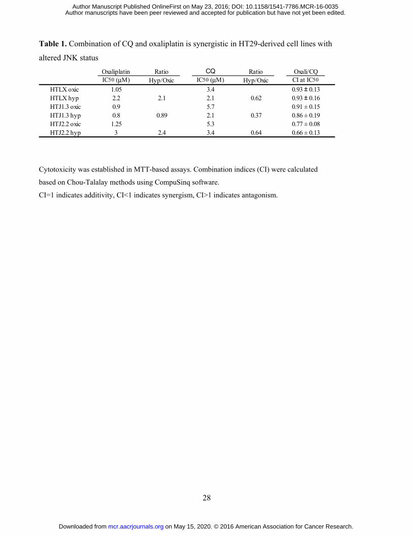
hypoxia in all cell lines, with highest ratio of sensitization in HTJ1.3 and lowest in HTJ2.2 cells (Table
on May 15, 2020. © 2016 American Association for Cancer Research. mcr.aacrjournals.org Downloaded from
Author manuscripts have been peer reviewed and accepted for publication but have not yet been edited. Author Manuscript Published OnlineFirst on May 23, 2016; DOI: 10.1158/1541-7786.MCR-16-0035

13
1). In the presence of CQ (5 μM, 24 hours) the most significant enhancement of apoptosis was detected
in hypoxic HTJ1.3 cells (Figure 3A), as judged by processing of caspase 7. Protein markers confirmed
inhibition of autophagy in all CQ-treated cell lines, both in normal and hypoxic conditions, whereas
only in JNK1-deficient cells autophagy was less pronounced in the absence of CQ (in accordance with
Figure 2A). More robust apoptosis in HTJ1.3 cells was also confirmed by fluorescent staining with
FITC-Annexin V (Supplemental Figure S4A and S4B). Use of CQ ( 2.5 μM, corresponding to IC30 of
oxic control) in combination with oxaliplatin sensitized all cell lines under both oxic and hypoxic
conditions, but the strongest effect was observed, again, in HTJ1.3 cell line (Figure 3B), in which
hypoxic resistance to oxaliplatin was completely abolished. Finally, when combining CQ and
oxaliplatin in MTT assays, we established synergism of the combination in all three cell lines, with
lowest CI50 in HTJ2.2 cells (Table 1), which suggests that inhibition of autophagy in its late stage can
partially reverse oxaliplatin resistance in JNK2-deficient cells under hypoxia. We also have noticed
that in CQ-treated cells hypoxia-induced expression and activation of c-Jun is diminished, which
suggests the inhibition of JNK signaling. It was shown, that CQ can cause ATM-dependent decrease
in JNK activity in macrophages (23), which, if true for colon cancer cell lines, could have additional
deleterious effect on cell survival in hypoxic conditions.
Sensitization to oxaliplatin in JNK1-deficient HT29 cells is associated with enhancement of intrinsic
apoptosis. To assess the cell death pathways in our panel, we evaluated induction of apoptosis and
necrosis in hypoxic cells by fluorescent staining (Figure 4A). Stronger and faster induction of
apoptosis was evident in HTJ1.3 cells. The percentage of apoptotic cells for each condition (n=3) from
three independent experiments was calculated and is shown next to the corresponding image. Western
blot analysis of protein extracts from cells treated with oxaliplatin under hypoxia (5 μM for 24 hours)
demonstrated inhibition of hypoxia-induced autophagy in the JNK1-deficient line, which was
on May 15, 2020. © 2016 American Association for Cancer Research. mcr.aacrjournals.org Downloaded from
Author manuscripts have been peer reviewed and accepted for publication but have not yet been edited. Author Manuscript Published OnlineFirst on May 23, 2016; DOI: 10.1158/1541-7786.MCR-16-0035
14
accompanied by enhanced apoptosis, as evident by processing of caspase 3 (Figure 4B). When hypoxia
was followed by reoxygenation for additional 24 hours, we could still detect differential activation of
autophagy, but apoptosis was activated equivalently in all three cell lines, suggesting that the severity
of the stress could not be counteracted by autophagy in HTLX and HTJ2.2 cells (Supplemental Figure
S5). Finally, caspase activation assays demonstrated activation of intrinsic apoptotic pathway in
HTJ1.3 cells (Figure 4C), especially under hypoxia, whereas in HTLX and HTJ2.2 cell lines it was
much less pronounced. These data is in accord with our previous findings, pointing to low level of
apoptotic cell death in hypoxic HT29 treated with oxaliplatin (24), as compared to necrosis.
Bcl-X knock-down enhances apoptosis in HT29 cell without affecting induction of autophagy.
Since for intrinsic apoptosis the interactions between Bcl2-family members are crucial in setting cell
death threshold, we wished to find out if down-regulation of JNK1 affects Bcl2 proteins in hypoxic
HT29 cells. Our data show, that expression of majority of proteins does not differ significantly
between HTLX, HTJ1.3 and HTJ2.2 cell lines under hypoxia. The only difference was the higher level
of Bcl-XL phosphorylation in JNK1-deficient cell line (Figure 5A). Also, levels of Bcl2 were barely
detectable in all cell lines. In immunoprecipitation experiments we identified Bcl-XL, but not Bcl2, as
the binding partner of Beclin1 in HTLX, HTJ1.3 and HTJ2.2 cells is (Figure 5B). Notably, despite the
differing levels of phosphorylation, Bcl-XL stayed bound to Beclin 1 in both HTJ1.3 and HTJ2.2 cells,
at least at six hours of hypoxic exposure, whereas effects on autophagy induction varied (Figure 2).
These conflicting data prompted us to investigate if down-regulation of Bcl-XL would affect autophagy
and oxaliplatin sensitivity in HT29 cells. To do so, we isolated several HT29-derived monoclonal sub-
lines, in which Bcl-XL expression was down-regulated by lentiviral delivery of shRNA constructs
(HTsBX4 and HTxBX5, with control HT-LVC line). Western blotting did not reveal significant
changes in autophagy induction under hypoxia in these cell lines (Figure 6A), whereas MTT analysis
clearly demonstrated abrogation of hypoxic resistance to the drug (Figure 6B). The acridine orange
on May 15, 2020. © 2016 American Association for Cancer Research. mcr.aacrjournals.org Downloaded from
Author manuscripts have been peer reviewed and accepted for publication but have not yet been edited. Author Manuscript Published OnlineFirst on May 23, 2016; DOI: 10.1158/1541-7786.MCR-16-0035

15
staining also demonstrated comparatively equal accumulation of acidic vesicles by 24 hours in hypoxic
control and HTsBX5 cells, with the latter showing slightly lesser staining at 6 hours of hypoxia
(Supplemental Figure S6). At the same time, HTsBX5 cells exhibit faster and stronger induction of
apoptosis under hypoxia, when compared to control, which is likely to be the basis of increased
cytotoxicity of oxaliplatin in this cell line (Figure 6C).
on May 15, 2020. © 2016 American Association for Cancer Research. mcr.aacrjournals.org Downloaded from
Author manuscripts have been peer reviewed and accepted for publication but have not yet been edited. Author Manuscript Published OnlineFirst on May 23, 2016; DOI: 10.1158/1541-7786.MCR-16-0035
16
DISCUSSION.
Involvement of JNK in the regulation of autophagy has been implicated in multiple studies, some of
which relied on pan-inhibition of JNK, whereas others employed genetic approaches to investigate
input of specific isoforms. In their pioneer paper (25) Wei and co-authors showed that in HeLa and
MCF7 cells, under starvation, Bcl2 undergoes multisite phosphorylation by JNK, leading to its
dissociation from Beclin1, thus blocking the anti-autophagy function of Bcl2. Further, using MEFs
with targeted disruption of jnk1 or jnk2, they demonstrated essential role of JNK1 in Bcl2
phosphorylation, and inability of JNK2 to compensate for it in these conditions. Finally, they also
proposed the model, linking the switch from autophagy to apoptosis in starved HeLa cells with the
duration of signal and the differing affinity of Bcl-2 to its binding partners, Beclin 1 and BAX,
dependent on the level of multi-site Bcl-2 phosphorylation (26). Although since then multiple studies
have implicated JNK in induction of protective autophagy in various models, data on its impact on
cytotoxicity of chemotherapeutic drugs in solid hypoxic tumors are scarce.
This project was initiated to broaden our study of the impact of hypoxia-induced signaling to AP-1
transcription factor on sensitivity of hypoxic colon cancer cell lines to oxaliplatin. After showing
synergism of JNK inhibition with oxaliplatin in a panel of cell lines, we demonstrated increased
sensitivity to the drug in HT29 cell line with impaired JNK1 function (21). Coupling that with data
showing synergism of CQ and oxaliplatin under hypoxia (22) led us focus on autophagy and its effects
on outcome of chemotherapeutic treatment in HT29-derived cell lines in which either JNK1 or JNK2
activities were down-regulated by overexpression of corresponding dominant-negative construct.
The majority of JNK isoform activities are considered redundant, but specific functions of different
isoforms in various cellular processes have been described (15, 16), and autophagy is not an exception:
several recent studies showed central role of JNK2 in autophagy induced by different stressors (27-29),
on May 15, 2020. © 2016 American Association for Cancer Research. mcr.aacrjournals.org Downloaded from
Author manuscripts have been peer reviewed and accepted for publication but have not yet been edited. Author Manuscript Published OnlineFirst on May 23, 2016; DOI: 10.1158/1541-7786.MCR-16-0035
17
including hypoxia-induced mitophagy (30). Our results confirm inhibition of autophagy by SP600125
in both hypoxia- and oxaliplatin-treated HT29 cells. We also show that JNK1 is essential for hypoxia-
induced autophagy, and cannot be substituted by JNK2 (Figure 2), suggesting that higher sensitivity of
HTJ1.3 cells to oxaliplatin could be, at least in part, based on autophagy down-regulation in this cell
line. We have observed increased levels of c-Jun and phospho-cJun in a JNK1-deficient cell line (21),
but our experiments with shJNK2 constructs show that it cannot be attributed solely to JNK2
overcompensation, suggesting either the activity of another mediator of hypoxia-induced signaling to
c-Jun or disruption of this protein’s turnover. The latter is suggested by highest c-Jun levels in HTJ1.3
cells, especially under hypoxia. Previous studies in JNK1-/- and JNK2-/- MEFs reported down-
regulation of c-Jun in the absence of JNK1 and its up-regulation in JNK2-deficient fibroblasts (31,32).
In our cells both endogenous JNK1 and dominant negative JNK1 are present, resulting in competitive
inhibition of signaling to c-Jun, which creates more complex environment. Since JNK1 was shown to
be most effective activating kinase of the E3 ligase Itch, which is responsible for c-Jun ubiquitination
and degradation (33), it is plausible to assume that JNK1 inhibition in HT29 cells could lead to
increase in c-Jun levels. And although this phenomenon could be investigated further, we did not
pursue it in this study, since it does not seem to affect autophagy inhibition in HTJ1.3 cells, as judged
by multiple autophagy markers.
Autophagy induction, as a cellular response to stress, is dependent on genetic background. Thus,
certain features of HT29 cells able to affect this process should be mentioned. It was shown that in
multiple cell lines hypoxia-induced autophagy is mediated through the HIF-1 transcriptional target
BH3-only protein BNIP3, which can displace Bcl2 from Beclin1, prompting autophagy activation (34,
35). However, in colon cancer cell lines, including HT29, expression of BNIP3 is extremely low, due
to epigenetic silencing of the promoter (36). On the other hand, we have shown earlier high
constitutive activity of ERK pathway in HT29 cells, both in normal conditions and upon hypoxia or
on May 15, 2020. © 2016 American Association for Cancer Research. mcr.aacrjournals.org Downloaded from
Author manuscripts have been peer reviewed and accepted for publication but have not yet been edited. Author Manuscript Published OnlineFirst on May 23, 2016; DOI: 10.1158/1541-7786.MCR-16-0035

18
oxaliplatin treatments (20, 37), which could enhance autophagy (38) and underlie low apoptotic cell
death in our model when treated with oxaliplatin, as compared to necrosis (24). Deregulation of
intrinsic apoptosis could also be a consequence of the p53 mutation in HT29, since majority of BCL2-
family members are p53 transcriptional targets (39). When Benard et al (40) investigated activity of
apoptotic genes in a panel of colon cancer cell lines, they designated the status of apoptotic pathways
in HT29 as “inconclusive”, with the cell line demonstrating very low induction of intrinsic apoptosis
upon cisplatin and radiation treatments. However, when autophagy was inhibited either by CQ or
JNK1 down regulation in our experiments, we observed significant enhancement of apoptosis (Figures
3A, S4 and 4C) under hypoxic conditions. Thus our data strongly suggest that hypoxia-induced
autophagy is a barrier to apoptosis in HT29 cells treated with oxaliplatin.
Bcl2-family proteins are indispensable for an intrinsic apoptosis induced by multiple stimuli (41),
DNA damage in particular. Reversible phosphorylation and dephosphorylation of both anti- and pro-
apoptotic members of the family plays central role in their regulation. Nevertheless, in general,
consequences of Bcl2 proteins’ phosphorylation in direct regulation of cell death are still somewhat
controversial, since phosphorylation of the same protein on different amino acid residues can cause
activation or inhibition of apoptosis (42). It is accepted that phosphorylation of Bcl-XL mostly leads to
inhibition of its anti-apoptotic function, with JNK1 being responsible in a majority of circumstances
(42). In contrast, study by Du et al (43) suggested that JNK does not phosphorylate Bcl-XL after
vinblastine treatment, and in this model apoptosis induction correlates with dephosphorylation of the
protein. Kinases other than JNK1 were reported to phosphorylate Bcl-XL, JNK2 among others (44-46).
Our data show that increased phosphorylation of Bcl-XL (with no change in the protein content) in
JNK1-deficient cells does not affect autophagy significantly, but is pro-apoptotic under these
conditions. We have not attempted to elucidate mechanism of hyper-phosphorylation of Bcl-XL in this
on May 15, 2020. © 2016 American Association for Cancer Research. mcr.aacrjournals.org Downloaded from
Author manuscripts have been peer reviewed and accepted for publication but have not yet been edited. Author Manuscript Published OnlineFirst on May 23, 2016; DOI: 10.1158/1541-7786.MCR-16-0035

19
study, but are planning to do so after expansion of our model to a panel of colon cancer cell lines
representative of human disease.
The complexity of cellular responses to stress, including hypoxia and DNA-damaging drugs, is
staggering. Engagement of the same signaling pathways and molecular mechanisms with differing
outcomes underlies the terminology often used when discussing the topic: double-edged sword,
balance, switch, crosstalk. Crosstalks between autophagy, apoptosis, necrosis and necroptosis were
described for various models (11), leading to the search for approaches to exploit these phenomena in
targeted treatment of cancer (47). Inhibitors of autophagy are currently tested in multiple clinical trials
(48), and combination of hydroxychloroquine with a standard chemotherapy was shown to be
beneficial for the treatment of solid tumors (49, 50). In our studies inhibiting autophagy with CQ (22)
or JNK activity (through pan-JNK pharmacological inhibition or molecular JNK1 down-regulation
(21)) lead to enhanced cytotoxicity of oxaliplatin and 5-FU in hypoxic colon cancer cell lines, which
strongly supports studies of the JNK inhibitors in clinical setting to improve chemotherapy efficacy
both alone and in combination with CQ.
on May 15, 2020. © 2016 American Association for Cancer Research. mcr.aacrjournals.org Downloaded from
Author manuscripts have been peer reviewed and accepted for publication but have not yet been edited. Author Manuscript Published OnlineFirst on May 23, 2016; DOI: 10.1158/1541-7786.MCR-16-0035

20
REFERENCES.
1. Vaupel P, Mayer A. Hypoxia in cancer: significance and impact on clinical outcome. Cancer
Metastasis Rev 2007, 26:225-39.
2. Span PN, Bussink J. Biology of hypoxia. Semin Nucl Med 2015, 45:101-9.
3. Rowher N, Cramer T. Hypoxia-mediated drug resistance: Novel insights on the functional
interaction of HIFs and cell death pathways. Drug Res Updates 2011, 14:191-201.
4. Karakashev SV, Reginato MJ. Progress toward overcoming hypoxia-induced resistance to solid
tumor therapy. Cancer Manag Res 2015,7:253-64.
5. Sun Q, Li X. Targeting cyclic hypoxia to prevent malignant progression and therapeutic resistance
of cancers. Histol Histopathol 2015, 30:51-60.
6. Semenza GL. HIF-1 mediates metabolic responses to intratumoral hypoxia and oncogenic
mutations. J Clin Invest 2013, 9:3664-71.
7. Cummins EP, Taylor CT. Hypoxia-responsive transcription factors. Pflugers Arch 2005, 450:363-
71.
8. Yang Z, Klionsky DJ. Mammalian autophagy: core molecular machinery and signaling regulation.
Curr Opin Cell Biol 2010, 22:124-31.
9. Galluzzi L, Pietrocola F, Bravo-San Pedro JM, Amaravadi RK, Baehrecke EH, Cecconi F et al.
Autophagy in malignant transformation and cancer progression. EMBO J 2015, 34:856-80.
10. He C, Levine B. The Beclin1 interactome. Curr Opin Cell Biol 2010, 22:140-9.
11. Mariño G, Niso-Santano M, Baehrecke EH, Kroemer G. Self-consumption: the interplay between
autophagy and apoptosis. Nat Rev Mol Cell Biol 2014, 15:81-94.
12. Rouschop KMA, Wouters BG. Regulating of autophagy through multiple independent hypoxic
signaling pathways. Curr Mol Med 2009, 9:417-24.
on May 15, 2020. © 2016 American Association for Cancer Research. mcr.aacrjournals.org Downloaded from
Author manuscripts have been peer reviewed and accepted for publication but have not yet been edited. Author Manuscript Published OnlineFirst on May 23, 2016; DOI: 10.1158/1541-7786.MCR-16-0035
21
13. Sui X, Kong N, Ye L, Han W, Zhou J, Zhang Q, He C, Pan H. p38 and JNK MAPK pathways
control the balance of apoptosis and autophagy in response to chemotherapeutic agents. Cancer Lett
2014, 344:174-9.
14. Davis RJ. Signal transduction by the JNK group of MAP kinases. Cell 2000; 103:239-52.
15. Bode AM, Dong Z. The functional contrariety of JNK. Mol Carcinogenesis 2007, 46:591-8.
16. Tournier C. The 2 Faces of JNK Signaling in Cancer. Genes &Cancer 2013, 4:397-400.
17. Pietrocola F, Izzo V, Niso-Santano M, Vacchelli E, Galluzzi L, Maiuri MC, Kroemer G.
Regulation of autophagy by stress-responsive transcription factors. Semin Cancer Biol 2013, 23:310-
22.
18. Vasilevskaya IA, O’Dwyer PJ. Role of Jun and Jun kinase in resistance of cancer cell to therapy.
Drug Res Updates 2003, 6:147-56.
19. Picco V, Pages G. Linking JNK activity to the DNA damage response. Genes & Cancer 2013,
4:360-8.
20. Vasilevskaya IA, Selvakumaran M, O'Dwyer PJ. Disruption of signaling through SEK1 and
MKK7 yields differential responses in hypoxic colon cancer cells treated with oxaliplatin. Mol
Pharmacol 2008, 74:246-54.
21. Vasilevskaya IA, Selvakumaran M, Cabal-Hierro L, Goldstein SR, Winkler JD, O'Dwyer PJ.
Inhibition of JNK Sensitizes Hypoxic Colon Cancer Cells to DNA-Damaging Agents. Clin Cancer Res
2015, 21:4143-52.
22. Selvakumaran M, Amaravadi RK, Vasilevskaya IA, O’Dwyer PJ. Autophagy inhibition sensitizes
colon cancer cells to antiangiogenic and cytotoxic therapy. Clin Cancer Res 2013, 19:2995-3007.
23. Schneider JG, Finck BN, Ren J, Standley KN, Takagi M, Maclean KH, Bernal-Mizrachi C, Muslin
AJ, Kastan MB, Semenkovich CF. ATM-dependent suppression of stress signaling reduces vascular
disease in metabolic syndrome. Cell Metab 2006, 4:377-89.
on May 15, 2020. © 2016 American Association for Cancer Research. mcr.aacrjournals.org Downloaded from
Author manuscripts have been peer reviewed and accepted for publication but have not yet been edited. Author Manuscript Published OnlineFirst on May 23, 2016; DOI: 10.1158/1541-7786.MCR-16-0035

22
24. Rakitina TV, Vasilevskaya IA, O'Dwyer PJ. Inhibition of G1/S transition potentiates oxaliplatin-
induced cell death in colon cancer cell lines. Biochem Pharmacol 2007, 73:1715-26.
25. Wei Y, Pattingre S, Sinha S, Bassik M, Levine B. JNK1-mediated phosphorylation of Bcl-2
regulates starvation-induced autophagy. Mol Cell 2008, 30:678-88.
26. Wei Y, Sinha S, Levine B. Dual role of JNK1-mediated phosphorylation of Bcl-2 in autophagy and
apoptosis regulation. Autophagy 2008, 4:949-51.
27. Raciti M, Lotti LV, Valia S, Pulcinelli FM, Di Renzo L. JNK2 is activated during ER stress and
promotes cell survival. Cell Death Dis 2012,3:e429.
28. Lin Z, Liu T, Kamp DW, Wang Y, He H, Zhou X, Li D, Yang L, Zhao B, Liu G. AKT/mTOR and
c-Jun N-terminal kinase signaling pathways are required for chrysotile asbestos-induced autophagy.
Free Radic Biol Med 2014, 72:296-307.
29. Tu QQ, Zheng RY, Li J, Hu L, Chang YX, Li L, Li MH, Wang RY, Huang DD, Wu MC, Hu HP,
Chen L, Wang HY. Palmitic acid induces autophagy in hepatocytes via JNK2 activation. Acta
Pharmacol Sin 2014, 35:504-12.
30. Zhang Q, Kuang H, Chen C, Yan J, Do-Umehara HC, Liu XY, Dada L, Ridge KM, Chandel NS,
Liu J. The kinase Jnk2 promotes stress-induced mitophagy by targeting the small mitochondrial form
of the tumor suppressor ARF for degradation. Nat Immunol 2015, 16:458-66.
31. Sabapathy K, Hochedlinger K, Nam SY, Bauer A, Karin M, Wagner EF. Distinct roles for JNK1
and JNK2 in regulating JNK activity and c-Jun-dependent cell proliferation. Mol Cell 2004, 15:713-
25.
32. Jaeschke A, Karasarides M, Ventura JJ, Ehrhardt A, Zhang C, Flavell RA, Shokat KM, Davis RJ.
JNK2 is a positive regulator of the cJun transcription factor. Mol Cell 2006, 23:899-911.
33. Gao M, Labuda T, Xia Y, Gallagher E, Fang D, Liu YC, Karin M. Jun turnover is controlled
through JNK-dependent phosphorylation of the E3 ligase Itch. Science 2004, 306:271-5.
on May 15, 2020. © 2016 American Association for Cancer Research. mcr.aacrjournals.org Downloaded from
Author manuscripts have been peer reviewed and accepted for publication but have not yet been edited. Author Manuscript Published OnlineFirst on May 23, 2016; DOI: 10.1158/1541-7786.MCR-16-0035
23
34. Azad MB, Chen Y, Henson ES, Cizeau J, McMillan-Ward E, Israels SJ, et al. Hypoxia induces
autophagic cell death in apoptosis-competent cells through a mechanism involving BNIP3. Autophagy
2008, 16:195–204.
35. Bellot G, Garcia-Medina R, Gounon P, Chiche J, Roux D, Pouyssegur J, et al. Hypoxia-induced
autophagy is mediated through hypoxia-inducible factor induction of BNIP3 and BNIP3L via their
BH3 domains. Mol Cell Biol 2009, 10:2570–81.
36. Bacon AL, Fox S, Turley H, Harris AL. Selective silencing of the hypoxia-inducible 1 target gene
BNIP3 by histone deacetylation and methylation in colorectal cancer. Oncogene 2007, 26:132–41.
37. Rakitina TV, Vasilevskaya IA, O'Dwyer PJ. Additive interaction of oxaliplatin and 17-allylamino-
17-demethoxygeldanamycin in colon cancer cell lines results from inhibition of nuclear factor kappaB
signaling. Cancer Res 2003, 63:8600-5.
38. Cagnol S, Chambard JC. ERK and cell death: mechanisms of ERK-induced cell death--apoptosis,
autophagy and senescence. FEBS J 2010, 277:2-21.
39. Galluzzi L, Morselli E, Kepp O, Vitale I, Pinti M, Kroemer G. Mitochondrial liaisons of p53.
Antioxid Redox Signal 2011, 15:1691-714.
40. Benard A, Janssen CM, van den Elsen PJ, van Eggermond MC, Hoon DS, van de Velde CJ,
Kuppen PJ. Chromatin status of apoptosis genes correlates with sensitivity to chemo-, immune- and
radiation therapy in colorectal cancer cell lines. Apoptosis 2014, 19:1769-78.
41. Czabotar PE, Lessene G, Strasser A, Adams JM. Control of apoptosis by the BCL-2 protein family:
implications for physiology and therapy. Nat Rev Mol Cell Biol 2014, 15:49-63.
42. Kutik O and Letai A. Regulation of Bcl-2 family proteins by posttranslational modifications. Cur
Mol Med 2008, 8: 102-18.
43. Du L, Lyle CS, Chambers TC. Characterization of vinblastine-induced Bcl-xL and Bcl-2
phosphorylation: evidence for a novel protein kinase and a coordinated
on May 15, 2020. © 2016 American Association for Cancer Research. mcr.aacrjournals.org Downloaded from
Author manuscripts have been peer reviewed and accepted for publication but have not yet been edited. Author Manuscript Published OnlineFirst on May 23, 2016; DOI: 10.1158/1541-7786.MCR-16-0035

24
phosphorylation/dephosphorylation cycle associated with apoptosis induction. Oncogene 2005;
24:107-17.
44. Arena G, Gelmetti V, Torosantucci L, Vignone D, Lamorte G, De Rosa P, Cilia E, Jonas EA,
Valente EM. PINK1 protects against cell death induced by mitochondrial depolarization, by
phosphorylating Bcl-xL and impairing its pro-apoptotic cleavage. Cell Death Differ 2013; 20:920-30.
45. Tamura Y, Simizu S, Muroi M, Takagi S, Kawatani M, Watanabe N, Osada H. Polo-like kinase 1
phosphorylates and regulates Bcl-x(L) during pironetin induced apoptosis. Oncogene 2009; 28:107-16.
46. Wang J, Beauchemin M, Bertrand R. Phospho-Bcl-xL(Ser62) plays a key role at DNA damage-
induced G2 checkpoint, Cell Cycle 2012, 11:2159-69.
47. Radogna F, Dicato M, Diederich M. Cancer-type-specific crosstalk between autophagy,
necroptosis and apoptosis as a pharmacological target. Biochem Pharmacol 2015, 94:1-11.
48. Duffy A1, Le J, Sausville E, Emadi A. Autophagy modulation: a target for cancer treatment
development. Cancer Chemother Pharmacol 2015, 75:439-47.
49. Poklepovic A, Gewirtz DA. Outcome of early clinical trials of the combination of
hydroxychloroquine with chemotherapy in cancer. Autophagy 2014, 10:1478-80.
50. Rebecca VW, Amaravadi RK. Emerging strategies to effectively target autophagy in cancer.
Oncogene 2016, 35:1-11.
on May 15, 2020. © 2016 American Association for Cancer Research. mcr.aacrjournals.org Downloaded from
Author manuscripts have been peer reviewed and accepted for publication but have not yet been edited. Author Manuscript Published OnlineFirst on May 23, 2016; DOI: 10.1158/1541-7786.MCR-16-0035

25
FIGURE LEGENDS.
Figure 1. JNK inhibition attenuates autophagy in HT29 cell line. A, Cells on glass slides were
subjected to hypoxia or oxaliplatin for 24 hours with or without SP600125 (10 μM ) and observed
under electron microscopy to assess autophagy by appearance of autophagosomes. B, Quantitative
analysis was carried out: cells with more than 5 autophagosomes were considered “positive” for
induction of autophagy. Graph represents average values from at least three slides for each condition,
bars represent standard deviation; **, P<0.01.
Figure 2. Functional JNK1 in necessary for induction of autophagy in HT29 cells. A, HT29-derived
cell lines were subjected to hypoxia, and the levels of autophagy markers assessed by Western blot
analysis. B, Lower GFP- LC3 puncta formation was observed in HTJ1.3 cells, as compared to control
and HTJ2.2 lines. Scale bar – 100μm. C, Autophagic cells were counted in multiple fields (n= 6-12)
for each condition, and their content (average percentage values of total cell count) is presented as a
graph. Cells which lost even cytoplasmic fluorescence and the ones containing more than 15 clearly
visible puncta were considered positive for autophagy induction. Bars represent standard deviation, *,
P < 0.05; **, P<0.01; ***, P<0.001. D, In HTJ1.3 and HTJ2.2 lines the counterpart JNK isoform was
down-regulated by control or shRNA-encoding retroviruses (sJ2 or sJ1, respectively), puromycin-
resistant cells were pooled, subjected to hypoxia and assessed for c-Jun and LC3B by western blot
analysis. E, Results of clonogenic assays in HT29-derived cell lines after down-regulation of each, or
both of the JNK. Graph shows average values from at least three independent experiments in
duplicates, bars represent standard deviation. *, P<0.05; **, P<0.01.
on May 15, 2020. © 2016 American Association for Cancer Research. mcr.aacrjournals.org Downloaded from
Author manuscripts have been peer reviewed and accepted for publication but have not yet been edited. Author Manuscript Published OnlineFirst on May 23, 2016; DOI: 10.1158/1541-7786.MCR-16-0035

26
Figure 3. Chloroquine enhances cell death in hypoxic HT29-derived cell lines. A, Western blot
analysis shows inhibition of JNK signaling, down-regulation of autophagy and enhancement of
apoptosis in the presence of CQ, especially in HTJ1.3 cell line. B, Results of MTT assays demonstrate
strongest enhancement of oxaliplatin cytotoxicity in hypoxic HTJ1.3 cells, when combined with 2.5
μM of CQ. Graph shows average values of IC50 derived from at least three independent experiments in
triplicate, bars represent standard deviation. *, P < 0.05; **, P<0.01; ***, P<0.001.
Figure 4. JNK1 deficiency results in enhancement of apoptosis in HT29 cell line. A, Cells were seeded
on glass slides, subjected to hypoxia for 6 or 24 hours, and stained using Apoptosis and Necrosis
Quantification kit. Representative images from three independent experiments are shown, column 1-
Hoechst 33342, column 2 – FITC-Annexin V+ Ethidium Homodimer III, column 3 - merged images;
scale bar – 100μm. Apoptotic cells were counted from 3 different fields in each experiment, average
values are shown. B, Cells were subjected to hypoxia with or without oxaliplatin (5 μM) for 24 hours
followed by Western blot analysis. C, Colorimetric caspase activation assays were carried out as
described in Material and Methods. Results are presented as folds increase in caspase activity when
compared to untreated control. Graph shows average values from 2 independent experiments in
duplicate, bars represent standard deviation.
Figure 5. Bcl-XL is a key mediator of hypoxia-induced autophagy in HT29 cells. A, Cells were
subjected to hypoxia, followed by Western blot analysis of Bcl2-family proteins, to assess possible
impact of JNK inhibition in this setting. B, Immunoprecipitation experiments with Beclin 1 antibody
revealed Bcl-XL as its binding partner in HT29-derived cell lines. In the first lane total protein extract
(TotPE) was loaded at the concentration of 10 μg; figure is a representative blot from two independent
experiments.
on May 15, 2020. © 2016 American Association for Cancer Research. mcr.aacrjournals.org Downloaded from
Author manuscripts have been peer reviewed and accepted for publication but have not yet been edited. Author Manuscript Published OnlineFirst on May 23, 2016; DOI: 10.1158/1541-7786.MCR-16-0035

27
Figure 6. Bcl-XL mediates hypoxic resistance to oxaliplatin in the HT29 colon adenocarcinoma model.
A, Bcl-XL was down-regulated in HT29 cells by lentiviral introduction of scrambled (HT-LVC) or Bcl-
X-targeting shRNA constructs, followed by isolation of monoclonal sub-lines. Cells were subjected to
hypoxia and assessed for expression of target protein and of autophagy markers. B, MTT assays
revealed abrogation of hypoxia-induced oxaliplatin resistance in HT29 cells lacking Bcl-XL. Graph
shows average values of IC50 in three monoclonal sub-lines derived from at least three independent
experiments in triplicates. Bars represent standard deviation; **, P<0.01. C, Staining for apoptosis and
necrosis demonstrate faster induction of apoptosis under hypoxia in HT29 cells lacking Bcl-XL.
on May 15, 2020. © 2016 American Association for Cancer Research. mcr.aacrjournals.org Downloaded from
Author manuscripts have been peer reviewed and accepted for publication but have not yet been edited. Author Manuscript Published OnlineFirst on May 23, 2016; DOI: 10.1158/1541-7786.MCR-16-0035
28
Table 1. Combination of CQ and oxaliplatin is synergistic in HT29-derived cell lines with
altered JNK status
Cytotoxicity was established in MTT-based assays. Combination indices (CI) were calculated
based on Chou-Talalay methods using CompuSinq software.
CI=1 indicates additivity, CI<1 indicates synergism, CI>1 indicates antagonism.
Oxaliplatin Ratio CQ Ratio Oxali/CQIC50 (μM) Hyp/Oxic IC50 (μM) Hyp/Oxic CI at IC50
HTLX oxic 1.05 3.4 0.93 ± 0.13HTLX hyp 2.2 2.1 2.1 0.62 0.93 ± 0.16HTJ1.3 oxic 0.9 5.7 0.91 ± 0.15HTJ1.3 hyp 0.8 0.89 2.1 0.37 0.86 ± 0.19HTJ2.2 oxic 1.25 5.3 0.77 ± 0.08HTJ2.2 hyp 3 2.4 3.4 0.64 0.66 ± 0.13
on May 15, 2020. © 2016 American Association for Cancer Research. mcr.aacrjournals.org Downloaded from
Author manuscripts have been peer reviewed and accepted for publication but have not yet been edited. Author Manuscript Published OnlineFirst on May 23, 2016; DOI: 10.1158/1541-7786.MCR-16-0035

INHIBITION OF JNK1 ATTENUATES HYPOXIA-
INDUCED AUTOPHAGY IN COLON CANCER CELL
LINES AND RESULTS IN HIGHER SENSITIVITY TO
CHEMOTHERAPY
Irina A Vasilevskaya
Muthu Selvakumaran
David Roberts
Peter J O’Dwyer
FIGURES (Revised)
on May 15, 2020. © 2016 American Association for Cancer Research. mcr.aacrjournals.org Downloaded from
Author manuscripts have been peer reviewed and accepted for publication but have not yet been edited. Author Manuscript Published OnlineFirst on May 23, 2016; DOI: 10.1158/1541-7786.MCR-16-0035

Figure 1
Control Oxaliplatin Hypoxia
S
P60
0125
C
on
tro
l
0
10
20
30
40
50
60
Control Oxaliplatin Hypoxia
Au
top
ha
go
so
me
po
sit
ive
ce
lls (
%)
** **
A B
on May 15, 2020. © 2016 American Association for Cancer Research. mcr.aacrjournals.org Downloaded from
Author manuscripts have been peer reviewed and accepted for publication but have not yet been edited. Author Manuscript Published OnlineFirst on May 23, 2016; DOI: 10.1158/1541-7786.MCR-16-0035

Figure 2
A B
C
D E
on May 15, 2020. © 2016 American Association for Cancer Research. mcr.aacrjournals.org Downloaded from
Author manuscripts have been peer reviewed and accepted for publication but have not yet been edited. Author Manuscript Published OnlineFirst on May 23, 2016; DOI: 10.1158/1541-7786.MCR-16-0035
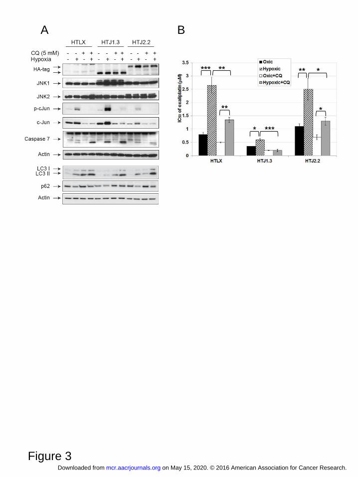
Figure 3
A B
on May 15, 2020. © 2016 American Association for Cancer Research. mcr.aacrjournals.org Downloaded from
Author manuscripts have been peer reviewed and accepted for publication but have not yet been edited. Author Manuscript Published OnlineFirst on May 23, 2016; DOI: 10.1158/1541-7786.MCR-16-0035

Figure 4.
A B
C
on May 15, 2020. © 2016 American Association for Cancer Research. mcr.aacrjournals.org Downloaded from
Author manuscripts have been peer reviewed and accepted for publication but have not yet been edited. Author Manuscript Published OnlineFirst on May 23, 2016; DOI: 10.1158/1541-7786.MCR-16-0035

Figure 5.
A B
on May 15, 2020. © 2016 American Association for Cancer Research. mcr.aacrjournals.org Downloaded from
Author manuscripts have been peer reviewed and accepted for publication but have not yet been edited. Author Manuscript Published OnlineFirst on May 23, 2016; DOI: 10.1158/1541-7786.MCR-16-0035
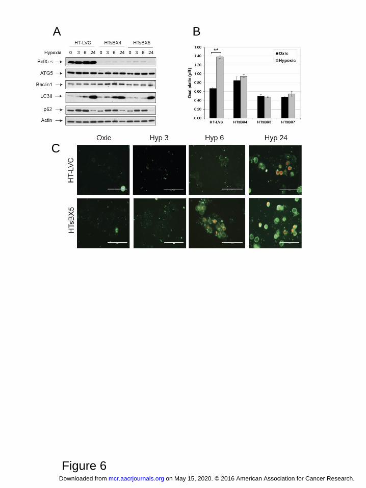
Figure 6
A B
C
on May 15, 2020. © 2016 American Association for Cancer Research. mcr.aacrjournals.org Downloaded from
Author manuscripts have been peer reviewed and accepted for publication but have not yet been edited. Author Manuscript Published OnlineFirst on May 23, 2016; DOI: 10.1158/1541-7786.MCR-16-0035

Published OnlineFirst May 23, 2016.Mol Cancer Res Irina A Vasilevskaya, Muthu Selvakumaran, David Roberts, et al. AUTOPHAGY AND SENSITIZES TO CHEMOTHERAPYJNK1 INHIBITION ATTENUATES HYPOXIA-INDUCED
Updated version
10.1158/1541-7786.MCR-16-0035doi:
Access the most recent version of this article at:
Material
Supplementary
http://mcr.aacrjournals.org/content/suppl/2016/06/25/1541-7786.MCR-16-0035.DC1
Access the most recent supplemental material at:
Manuscript
Authoredited. Author manuscripts have been peer reviewed and accepted for publication but have not yet been
E-mail alerts related to this article or journal.Sign up to receive free email-alerts
Subscriptions
Reprints and
To order reprints of this article or to subscribe to the journal, contact the AACR Publications
Permissions
Rightslink site. Click on "Request Permissions" which will take you to the Copyright Clearance Center's (CCC)
.http://mcr.aacrjournals.org/content/early/2016/05/21/1541-7786.MCR-16-0035To request permission to re-use all or part of this article, use this link
on May 15, 2020. © 2016 American Association for Cancer Research. mcr.aacrjournals.org Downloaded from
Author manuscripts have been peer reviewed and accepted for publication but have not yet been edited. Author Manuscript Published OnlineFirst on May 23, 2016; DOI: 10.1158/1541-7786.MCR-16-0035