jetter et al, j clin case rep 21, :5 o f : 141221210965 r ... · pdf filejetter et al, j clin...
TRANSCRIPT
Volume 7 • Issue 5 • 1000965J Clin Case Rep, an open access journalISSN: 2165-7920
OMICS InternationalCase Report
Jetter et al., J Clin Case Rep 2017, 7:5DOI: 10.4172/2165-7920.1000965
Journal of Clinical Case ReportsJour
nal o
f Clinical Case Reports
ISSN: 2165-7920
*Corresponding author: Nathan Jetter and Maria M Tsoukas, Department of Dermatology, University of Illinois, College of Medicine East Building, USA, Tel: (312)9966966; Fax: (312)9961188; E-mail: [email protected]; [email protected]
Received May 03, 2017; Accepted May 22, 2017; Published May 27, 2017
Citation: Jetter N, Juhl M, Tsoukas M (2017) Blastomycosis-Like Pyoderma in a Patient with Syphilis and HIV Resolving with Antibiotic Therapy. J Clin Case Rep 7: 965. doi: 10.4172/2165-7920.1000965
Copyright: © 2017 Jetter N, et al. This is an open-access article distributed under the terms of the Creative Commons Attribution License, which permits unrestricted use, distribution, and reproduction in any medium, provided the original author and source are credited.
Blastomycosis-Like Pyoderma in a Patient with Syphilis and HIV Resolving with Antibiotic TherapyNathan Jetter*, Mark Juhl and Maria Tsoukas*Department of Dermatology, University of Illinois, College of Medicine East Building, USA
AbstractBlastomycosis-like pyoderma (BLP) is a rare, exaggerated, vegetative tissue reaction that occurs in patients with
local or systemic immune dysregulation. Reported causes of immune compromise include human immunodeficiency virus, malnutrition, alcoholism, leukemia, immunosuppressant use, radiation therapy, and others. Nearly all cases involve an underlying, prolonged pyogenic bacterial infection. Historically monotherapy with systemic antibiotics requires long-term treatment and often fails. We describe a case of a male patient with BLP of the face and scalp, which cultured positive for methicillin-resistant Staphylococcus aureus (MRSA). Subsequent evaluation uncovered a concurrent Treponema pallidum and HIV infections. Treatment of the MRSA with a short course of doxycycline and the syphilis with penicillin, resulted in complete resolution of the skin lesions. Highly Active Anti-Retroviral Therapy for HIV was initiated after the skin lesions had cleared. Resolution of BLP by antibiotic therapy alone in the context of untreated HIV leads the authors to postulate that syphilis may have been the relevant factor contributing to his immune dysregulation.
Keywords: Blastomycosis-like pyoderma; BLP; Syphilis; HIV; Psudoepitheliomatous hyperplasia; Doxycycline
IntroductionBlastomycosis-like pyoderma (BLP) is a rare chronic form of
pyoderma that presents with one or more large, indurated, verrucous plaques studded with pustules and draining sinuses [1,2]. This slow-growing, vegetative infection typically presents similarly to the cutaneous manifestations of blastomycosis, a dimorphic fungi prevalent in the Ohio and Mississippi river valleys of North America. The fungus most commonly affects the lungs (91%), followed by the skin (18%) and bones (4%). Cutaneous lesions due to blastomycosis have been described variously as nodular, papular, and verrucous, grow slowly, and often ulcerate. While blastomycosis can infect both immunocompetent and immunocompromised patients, Blastomycosis-like pyoderma itself rarely occurs outside the setting of immune dysregulation [3]. It has been postulated previously that BLP’s pathogenesis includes an exaggerated tissue reaction due to the host’s inability to clear a bacterial colonization [3,4].
In 1979, various diagnostic criteria have been put forward with recent literature generally agreeing on: (1) large verrucous plaques with multiple pustules and an elevated border, (2) typical histologic findings of psudoepitheliomatous hyperplasia with abscesses on biopsy, (3) growth of one or more pathogenic bacteria, and (4) negative cultures for other infectious etiologies, particularly fungi or mycobacteria. In their original publication Su et al. included two additional criteria: (5) negative fungal serology, and (6) normal bromide and iodide blood levels [5]. However, modern case reports of BLP tend to make the diagnosis with fewer criteria. Of the fourteen case reports published in English-language journals since 1995, only one tested for fungal serology [6]. Similarly, only two reports tested for halogen toxicity with the majority of papers stating that halogenoderma could be satisfactorily ruled out by a lack of halogen medication or ingestion in the patient’s history [4,6].
Many systemic immune dysregulatory states have been described in patients diagnosed with BLP including leukemia, chemotherapy, malnourishment, and alcoholism [3,7]. There are cases of BLP arising at areas of local disturbance such as sun exposure, trauma and tattoo in immunocompetent persons [2,3,7].
Multiple treatments modalities with varying results have been reported in the literature, including: curettage, surgical excision, carbon
dioxide laser, potassium iodide, permanganate soaks, radiotherapy, disodium chromoglycate, and acitretin [4,8,9]. Effective treatment with systemic retinoids has also been reported [7].
We present a case of BLP with positive cultures for S. aureus and C. koseri in a patient with syphilis and HIV who responded to antibiotic therapy with doxycycline and penicillin G.
Case ReportA 35-year-old male presented for a facial rash of six months
duration. He described several small papules, initially on the left chin, which progressively enlarged. Subsequently, he developed similar boggy lesions on the scalp and cheek. The lesions were mildly pruritic but otherwise asymptomatic. He endorsed shaving, but denied other forms of trauma, sick contacts, or recent travel.
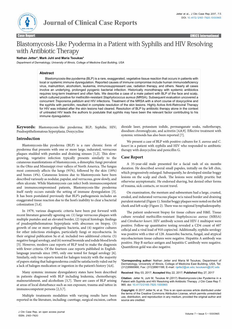
On examination, the mentum and submentum had a large, crusted, eroded, and indurated verrucous plaque w/a raised border and draining purulent material (Figure 1). Similar boggy plaques were noted on the left cheek and left scalp (Figure 2). There was no regional lymphadenopathy.
The patient underwent biopsy for tissue culture and H&E. Tissue culture revealed methicillin-resistant Staphylococcus aureus (MRSA) and Citrobacter koseri. HIV antibody screen and HIV-1 multispot were positive. Follow-up quantitative testing revealed a CD4 count of 607 cells/μl and a viral load of 910 copies/ml. Additionally, syphilis serology was positive with a titer of 128. Anaerobic bacteria, fungal, and atypical mycobacterium tissue cultures were negative. Hepatitis A antibody was positive. Hep B surface antigen and hepatitis C antibody were negative. Quantiferon gold was also negative.
Citation: Jetter N, Juhl M, Tsoukas M (2017) Blastomycosis-Like Pyoderma in a Patient with Syphilis and HIV Resolving with Antibiotic Therapy. J Clin Case Rep 7: 965. doi: 10.4172/2165-7920.1000965
Page 2 of 3
Volume 7 • Issue 5 • 1000965J Clin Case Rep, an open access journalISSN: 2165-7920
abscesses may also be present [10]. The differential diagnosis includes blastomycosis and other deep fungal infections, halogenoderma, pemphigus vegetans, tuberculosis verrucosa cutis, pyoderma gangrenosum, mycobacterial infections, giant keratoacanthoma, and squamous cell carcinoma [2,4,7-12]. Our patient had four of the six criteria for BLP established by Su et al. In accordance with the pertinent literature, we refrained from preforming fungal serology and blood halogen level testing.
Minor trauma, such as shaving, and chronic sun-damage are thought to increase the likelihood of BLP [8-12]. The most commonly reported pathogen is Staphylococcus aureus [2]. Additionally, β-hemolytic streptococci, Pseudomonas aeruginosa, and citrobacter species have been reported [1].
Monotherapy with systemic antibiotics usually requires long-term treatment and often fails [7,9]. Our patient’s immune status was compromised by an untreated HIV infection, as well as a concurrent syphilitic infection. Syphilis has been reported to cause immune dysregulation including altered cell surface markers and increased likelihood of HIV co-infection [13]. Further, the probability of HIV transmission is higher when syphilis is present in either individual in a sexual partnership involving discordant HIV infection status [13,14]. It has been shown that secondary syphilis markedly decreases total
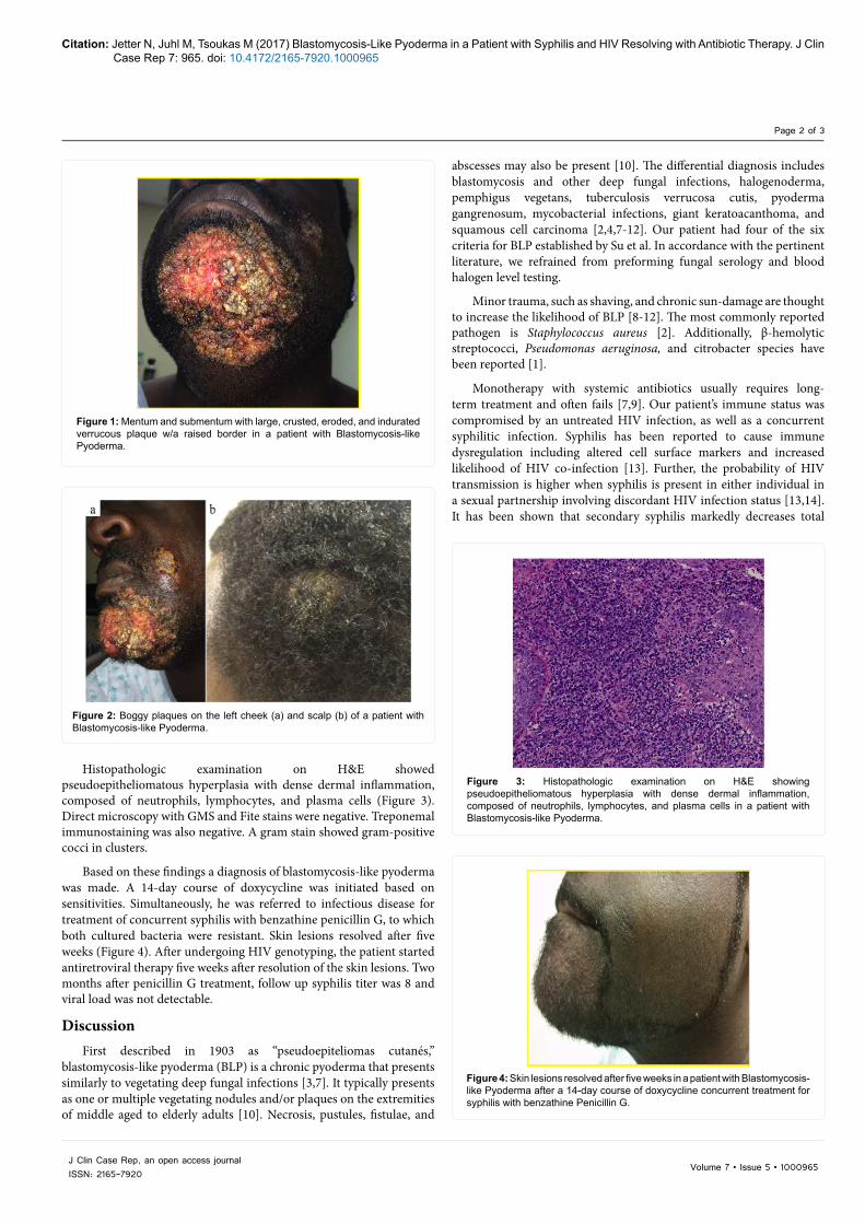
Histopathologic examination on H&E showed pseudoepitheliomatous hyperplasia with dense dermal inflammation, composed of neutrophils, lymphocytes, and plasma cells (Figure 3). Direct microscopy with GMS and Fite stains were negative. Treponemal immunostaining was also negative. A gram stain showed gram-positive cocci in clusters.
Based on these findings a diagnosis of blastomycosis-like pyoderma was made. A 14-day course of doxycycline was initiated based on sensitivities. Simultaneously, he was referred to infectious disease for treatment of concurrent syphilis with benzathine penicillin G, to which both cultured bacteria were resistant. Skin lesions resolved after five weeks (Figure 4). After undergoing HIV genotyping, the patient started antiretroviral therapy five weeks after resolution of the skin lesions. Two months after penicillin G treatment, follow up syphilis titer was 8 and viral load was not detectable.
DiscussionFirst described in 1903 as “pseudoepiteliomas cutanés,”
blastomycosis-like pyoderma (BLP) is a chronic pyoderma that presents similarly to vegetating deep fungal infections [3,7]. It typically presents as one or multiple vegetating nodules and/or plaques on the extremities of middle aged to elderly adults [10]. Necrosis, pustules, fistulae, and
Figure 1: Mentum and submentum with large, crusted, eroded, and indurated verrucous plaque w/a raised border in a patient with Blastomycosis-like Pyoderma.
Figure 2: Boggy plaques on the left cheek (a) and scalp (b) of a patient with Blastomycosis-like Pyoderma.
Figure 3: Histopathologic examination on H&E showing pseudoepitheliomatous hyperplasia with dense dermal inflammation, composed of neutrophils, lymphocytes, and plasma cells in a patient with Blastomycosis-like Pyoderma.
Figure 4: Skin lesions resolved after five weeks in a patient with Blastomycosis-like Pyoderma after a 14-day course of doxycycline concurrent treatment for syphilis with benzathine Penicillin G.

Citation: Jetter N, Juhl M, Tsoukas M (2017) Blastomycosis-Like Pyoderma in a Patient with Syphilis and HIV Resolving with Antibiotic Therapy. J Clin Case Rep 7: 965. doi: 10.4172/2165-7920.1000965
Page 3 of 3
Volume 7 • Issue 5 • 1000965J Clin Case Rep, an open access journalISSN: 2165-7920
circulating NK-cell numbers and is associated with the emergence of an atypical circulating CD56-negative CD16-positive NK-cell population which is poorly cytolytic and has impaired capacity to produce IFN gamma and other cytokines [15]. Interestingly, these NK-cell line changes are also observed in uncontrolled HIV patients. The mechanism for syphilis-driven immune dysregulation is not fully elucidated. The spread of syphilis into the bone marrow may alter the proper development of the myeloid and lymphoid progenitors of NK-cells along with macrophages and dendritic cells [15,16].
Our patient’s rapid and complete response to a short course of doxycycline is unusual for BLP. Resolution of the skin lesions in the context of untreated HIV raises the hypothesis that the concurrent syphilitic infection may have contributed to immune evasion by these bacteria.
Although our patient underwent treatment of syphilis with penicillin, both cultured bacteria were resistant. This leads the authors to postulate that treatment of syphilis may have hastened the resolution of his BLP lesions. Patients presenting with vegetating skin lesions should be evaluated for treatable causes of immunocompromise including syphilis and HIV. Our patient represents a unique case of MRSA-induced BLP in a patient with syphilis and untreated HIV that resolved with antibiotic therapy.
References
1. Crowley JJ, Kim YH (1997) Blastomycosis-like pyoderma in a man with AIDS. J Am Acad Dermatol 36: 633-634.
2. Scuderi S (2016) Heterogeneity of blastomycosis-like pyoderma: A selection of cases from the last 35 years. Australas J Dermatol.
3. Guidry JA (2015) Deep fungal infections, Blastomycosis-like Pyoderma, andgranulomatous sexually transmitted infections. Dermatol Clin 33: 595-607.
4. Lee YS (2011) Blastomycosis-like Pyoderma with good response to acitretin.Ann Dermatol 23: 365-368.
5. Su WP (1979) Blastomycosis-like pyoderma. Arch Dermatol 115: 170-173.
6. Cerullo L (2009) An unusual presentation of blastomycosislike pyoderma(Pyoderma vegetans) and a review of the literature. Cutis 84: 201-204.
7. Kobraei KB, Wesson SK (2010) Blastomycosis-like pyoderma: Response tosystemic retinoid therapy. Int J Dermatol 49: 1336-1338.
8. Su O (2004) Localized blastomycosis-like pyoderma with good response tocotrimoxazol and cryotherapy. Int J Dermatol 43: 388-390.
9. Rongioletti F (1996) Blastomycosis-like pyoderma (Pyoderma vegetans) responding to antibiotics and topical disodium chromoglycate. Int J Dermatol35: 828-830.
10. Cecchi R (2011) Blastomycosis-like pyoderma in association with recurrentvesicular hand eczema: good response to acitretin. Dermatol Online J 17: 9.
11. Sawalka SS (2007) Blastomycosis-like pyoderma. Indian J Dermatol VenereolLeprol 73: 117-119.
12. Nguyen RT, Beardmore GL (2005) Blastomycosis-like pyoderma: Successfultreatment with low-dose acitretin. Australas J Dermatol 46: 97-100.
13. Colmegna I (2006) Musculoskeletal and autoimmune manifestations of HIV,syphilis and tuberculosis. Curr Opin Rheumatol 18: 88-95.
14. Chessona H (1999) New HIV cases attributable to syphilis in the USA:Estimates from a simplified. AIDS 13: 1387-1396.
15. Cruz A (2012) Immune evasion and recognition of the syphilis spirochete inblood and skin of secondary syphilis patients: Two immunologically distinctcompartments. PLoS Negl Trop Dis 6: e1717.
16. Mavilio D, Lombardo G, Benjamin J, Kim D, Follman D, et al. (2005)Characterization of CD562/CD16+ natural killer (NK) cells: A highlydysfunctional NK subset expanded in HIV-infected viremic individuals. ProcNatl Acad Sci USA 102: 2886-2891.