jbc papers in press. published on june 13, 2012 as ... · intravascular hemolysis and liver damage...
TRANSCRIPT

Intravascular hemolysis and liver damage
1
Impact of Intravascular Hemolysis in Malaria on Liver Dysfunction Involvement of Hepatic Free Heme Over-load NFκB Activation and Neutrophil Infiltration
Sumanta Dey Samik Bindu Manish Goyal Chinmay Pal Athar Alam Mohd Shameel Iqbal Rahul Kumar Souvik Sarkar and Uday Bandyopadhyay
From Division of Infectious Diseases and Immunology CSIR -Indian Institute of Chemical Biology 4 Raja S C Mullick Road Kolkata -700032 West Bengal India
Running head Intravascular hemolysis and liver damage
Address correspondence to Uday Bandyopadhyay Division of Infectious Diseases and Immunology CSIR - Indian Institute of Chemical Biology 4 Raja S C Mullick Road Kolkata -700032 West Bengal India Tel 91-33-24733491 Fax 91-33-24730284 E -mail ubandyo_1964yahoocom
Keywords Hemolysis free heme iron oxidative stress NF-κB neutrophil malaria liver damage Background Multi-organ failure is evident in conditions of intravascular hemolysis Results Persistent intravascular hemolysis in malaria causes liver damage because of excess hepatic free heme accumulation TNFα release NF-κB activation and neutrophil infiltration Conclusion Intravascular hemolysis may result in hepatic failure as result of oxidative stress Significance Intravascular hemolysis in any condition may damage liver or other vascular organ SUMMARY We have investigated the impact of persistent intravascular hemolysis on liver dysfunction using mouse malaria model Intravascular hemolysis showed a positive correlation with liver damage along with the increased accumulation of free heme and reactive oxidants in liver Hepatocytes over-induced heme oxygenase-1 (HO-1) to catabolize free heme in building up defence against this pro-oxidant milieu However in a condition of persistent free heme overload in malaria the over-activity of HO-1 resulted in continuous transient generation of free iron to favour reactive oxidants production as evident from 27-dichlorofluorescein fluorescence studies Electrophoretic mobility shift assay documented the activation of NF-κB which in turn up-regulated intracellular adhesion molecule 1 as evident from chromatin
immunoprecipitation studies NF-κB activation also induced vascular cell adhesion molecule 1 keratinocyte chemoattractant and macrophage inflammatory protein 2 which favoured neutrophil extravasation and adhesion in liver The infiltration of neutrophils correlated positively with the severity of hemolysis and neutrophil depletion significantly prevented liver damage The data further documented the elevation of serum TNFα in infected mice and the treatment of anti-TNFα antibodies also significantly prevented neutrophil infiltration and liver injury Deferoxamine which chelates iron interacts with free heme and bears antioxidant properties prevented oxidative stress NF-κB activation neutrophil infiltration hepatocyte apoptosis and liver damage Furthermore the administration of N-acetyl cysteine also prevented NF-κB activation neutrophil infiltration hepatocyte apoptosis and liver damage Thus hepatic free heme accumulation TNFα release oxidative stress and NF-κB activation established a link to favour neutrophil infiltration in inducing liver damage during hemolytic condition in malaria Intravascular hemolysis is common pathological condition in a wide variety of diseases including malaria paroxysmal nocturnal hemoglobinuria thalassemia and sickle cell anaemia (12) Irrespective of the cases severe hemolysis leads
httpwwwjbcorgcgidoi101074jbcM112341255The latest version is at JBC Papers in Press Published on June 13 2012 as Manuscript M112341255
Copyright 2012 by The American Society for Biochemistry and Molecular Biology Inc
by guest on June 2 2018httpw
ww
jbcorgD
ownloaded from
Intravascular hemolysis and liver damage
2
to several systemic complications and severe organ damage (13-5) Renal failure liver damage spleen enlargement lung injury vascular injury and stroke are some of the major complications due to hemolysis (13-5) Hemolysis leads to the release of free hemoglobin in plasma Once outside the red blood cells hemoglobin dissociates to αβ dimers which are bound by haptoglobulin and removed from the circulation (6) But once hemolysis becomes severe haptoglobulin fails to scavenge all the extracellular hemoglobin (7) Free ferrous hemoglobin in the plasma gets oxidized to ferrihemoglobin which dissociates to free heme and globin (7) Free heme is removed from the blood by the protein hemopexin (89) which is taken up by the hepatocytes through receptor mediated endocytosis But hemopexin similarly fails to counter the condition when hemolysis is severe and persistent Under such conditions free heme concentration is increased in serum Besides hemopexin mediated heme transport to liver there can be other ways of heme entry inside the liver when hemopexin is saturated Extracellular free hemoglobin and free heme is very toxic due to their pro-oxidant nature (10-16) The heme iron reacts with endogenous H2O2 via the Fenton reaction and Haber-Weiss reaction to produce oxygen free radicals The hydrophobic nature of heme allows it to intercalate into cell membranes causing the oxidant mediated killing of cells (1517) The damage to cells and tissue is further aggravated in presence of H2O2 which causes the release of iron from heme (18) Once inside the tissue toxicity of heme is counteracted by the combination of heme oxygenase-1 (HO-1) and ferritin The action of HO-1 on heme results in the generation of equimolar concentration of iron biliverdin and CO (15) Once released this iron is generally chelated by ferritin (19) although the limits of ferritin are not known in pathological conditions However in conditions of persistent hemolysis and continuous supply of heme HO-1 is over-induced and generated huge quantities of free iron which may cross the limit of its ferritin sequestration This chaos or failure of the cytoprotective system leads to severe oxidative stress mediated cell death through necrosis and apoptosis (152021) Malaria one of the most devastating infectious diseases is endemic in 91 countries Around 40 of the worlds population is at risk of acquiring the dreadful infection Each year there are 300ndash500 million cases of malaria and 15ndash27 million
deaths due to malaria (22) Intravascular hemolysis is highly accelerated due to malaria infection (23-25) In the erythrocytic stage the malaria parasites modify the erythrocytes (26) and degrade approximately 60 to 80 of the total RBC hemoglobin (2728) Severe organ damage has been reported due to malaria infection (1528-34) However the mechanism of organ damage under hemolytic condition is not yet clear Here we provide evidence that intravascular hemolysis in malaria damages liver as a result of increased accumulation of free heme and reactive oxidants in liver which favour neutrophil infiltration We demonstrated that heme overload and oxidative stress activated the transcriptional factor NF-κB which in turn enhanced neutrophil infiltration and extravasation through the up-regulation of intracellular adhesion molecules and chemokines Further the chelation of free iron scavenging of reactive oxidants and depletion of neutrophil significantly prevented liver damage during malaria in mice EXPERIMENTAL PROCEDURES In vivo growth of Plasmodium yoelii- P yoelii (MDR strain) is grown in vivo in male BALBc mice (20-25 g) by inoculation of infected blood as described (35-36) Parasite burden in blood ( parasitemia) was monitored by preparing thin smear of blood and subsequent Giemsa staining All animals are maintained in the animal house and procedures were conducted in accordance with the guidelines of Institutional Animal Ethics Committee and Committee for the Purpose of Control and Supervision of Experiments on Animals (CPCSEA) Soret spectroscopy to detect released hemoglobinheme in serum due to hemolysis and assay of liver enzymes in serum- Blood was collected by puncture of heart from different groups of mice and put into 15 ml microcentrifuge tube Serum was separated by centrifugation at 600 x g for 5 min and kept at -20 ordmC Serum of different groups of mice was diluted (1100) in distilled water and was analysed in a spectrophotometer to determine the release of hemoglobin or heme from the erythrocytes in the serum Although most of the heme in serum probably comes from hemolysis but could also come from other sources such as muscle cells and hepatocytes Activity of liver enzymes and bilirubin in serum of mice were
by guest on June 2 2018httpw
ww
jbcorgD
ownloaded from
Intravascular hemolysis and liver damage
3
measured to assess liver function Enzyme activities of alanine transaminase (ALT) aspartate transaminase (AST) and alkaline phosphatase (ALP) were measured We also measured the total amount of bilirubin (TBIL) and conjugated or direct bilirubin (DBIL) in serum All assays were performed by using kits purchased from Randox Laboratories Ltd (Ardmore Diamond Road Crumlin Co Antrim United Kingdom) Manufacturerrsquos instructions were strictly followed These assays served as parameters to evaluate the extent of hemolysis and liver damage in mice Quantitation of heme-Total heme or free heme was quantified in serum and liver homogenate by using Quantichrome heme assay kit (Bioassay Systems CA USA) according to the manufacturerrsquos instructions Assay of HO-1 activity-HO-1 activity in liver homogenate was measured based on the amount of bilirubin formed in an assay system as described (3738) Liver excised from mouse was homogenized in a homogenization buffer [Tris-HCl (pH 74) 5 mll Triton X-100 and protease inhibitor cocktail] Liver homogenate of different groups of mice were used for the assay The assay mixture consisted of a 11 mixture of liver homogenate (200 microg of protein) and assay buffer (08 mM NADPH 2 mM glucose-6-phosphate 02 U of glucose 6-phosphate dehydrogenase 1 mM MgCl2 100 mM potassium phosphate buffer 20 microM hemin and 2 mg of mouse liver cystosol as a source of biliverdin reductase) The final volume was made up to 1 ml The mixture was incubated at 37ordmC for 1 h in the dark Reactions were terminated by keeping the samples on ice for 5 min The bilirubin formed was extracted in chloroform and A464-530 was measured The amount of bilirubin formed was calculated from its extinction coefficient (40 mMLcm)(37) The HO-1 activity was expressed as nmol of bilirubin formedmg proteinh RNA isolation and RT-PCR-TRIZOL reagent was used to isolate RNA from the perfused tissue according to the manufacturerrsquos instructions (Invitrogen Carlsbad CA) Total RNA (2 microg) was then reverse transcribed using the RevertAid First Strand cDNA synthesis kit (Fermentas USA) according to the manufacturerrsquos instructions The cDNA obtained was diluted for PCR reactions The primers were purchased from Integrated DNA Technologies Inc (San Diego California USA) The primers are given along with their product size in Table
1 The cycling conditions for PCR amplification were initial denaturation at 95 degC for 30 s and 40 cycles of denaturation at 95 degC for 30 s annealing at 55 degC for 30 s and extension at 72 degC for 60 s respectively The PCR products were detected with 15 agarose gel electrophoresis and densitometric analysis of the bands allowed semi-quantification The images were captured using Quantity onereg software (Bio Rad Hercules CA USA) Western immunoblotting- Liver was perfused for a long time to remove all contaminations of blood cells after which the homogenates were prepared The perfused liver from different groups of mice were homogenized in a glass homogenizer in buffer (140 mM NaCl 10 mM EDTA 10 glycerol 20 mM Tris pH 80 supplemented with 1 NP40 as detergent and 20000 Uml aprotinin 200 mM PMSF) The samples were then subjected to centrifugation for 20 min at 13000 x g at 4 degC The supernatant (liver tissue lysate 70 μg) was mixed with protein loading buffer (Fermentas Maryland USA) and boiled for 4 min The proteins were separated in a 12 polyacrylamide-SDS gel at constant voltage (100 V) Proteins were then transferred to a nitrocellulose membrane in a transfer apparatus with a current intensity of 400 mA for 120 min in a 190 mM glycine 20 mM Tris base buffer (pH 83) The membrane was incubated for 3 h in the blocking buffer TBS (25 mM Tris 150 mM NaCl 2 mM KCl pH 74) to which 5 non fat dry milk had been added The mixture was then quickly washed with the same buffer without milk The membrane was incubated overnight in the last buffer with 02 bovine serum albumin (BSA) solution containing 11000 anti-HO-1(Abcam) ferritin heavy chain (US Biological) anti-NF-κB p65 IKK β IκB-α and β-Actin (Santa Cruz) for separate experiments The membrane was then washed well with TBS containing 01 Tween 20 The membrane was incubated for 2 h in the same buffer containing secondary antibody (11000 HRP-labeled anti-rabbit or goat IgG) The membrane was washed well with the incubation buffer The protein detection was performed with a standard Western blot detection system Pre-stained standards were used as molecular weight markers and were run in parallel Isolation of hepatocytes from liver and preparation of lysates- Hepatocytes were isolated from liver after perfusion by a two-step collagenase digestion method and purified by
by guest on June 2 2018httpw
ww
jbcorgD
ownloaded from
Intravascular hemolysis and liver damage
4
Percoll gradient (39) Hepatocytes were then counted and used for further experiments For the preparation of hepatocyte lysates cells were lysed in 100 μl of buffer (140 mM NaCl 10 mM EDTA 10 glycerol 20 mM Tris pH 80 supplemented with 1 NP-40 as detergent and 20000 Uml aprotinin 200 mM PMSF) by three freeze-thaw cycles and sonication and the cytosolic fraction was prepared by centrifugation at 13000 times g for 20 min at 4degC Protein content in supernatant was determined by the method of Lowry (40) Equal amount of protein was used for western immunoblotting (41) Measurement of free iron mediated changes in redox reactions - Hepatocytes isolated from different groups of mice were loaded with DCFDA (10 microM) (Molecular Probes Eugene OR USA) for 30 min in dark Images were captured in fluorescence microscope using green filter Upon oxidation DCFDA exhibited bright green fluorescence (excitationemission maxima ~490515 nm) which was viewed under Leica DM 2500 Fluorescence microscope (Leica Microsystems GmbH Wetzlar Germany) (4243) Preparation of nuclear extract-Nuclear extract from liver tissue was prepared with the help of two nuclear extraction buffers Buffer I (10mM HEPES 15 mM MgCl2 10 mMKCl and 05 Triton X) and Buffer II (1M NaCl 02 M EDTA 20 glycerol and 05 mM DTT) Liver tissue from different groups of mice were minced in the presence of buffer I and then homogenized The sample was then centrifuged and the cytosol was separated The pellet was resuspended in a mixture of buffer I and II (11)The samples were vigorously vortexed for 30 s kept on ice for the next 30 min and centrifuged at high speed at 4ordmC The supernatants obtained were nuclear extracts and were stored at -80ordmC until use Electrophoretic mobility shift assay (EMSA)-NF-кB double-stranded consensus oligonucleotide 5rsquo-AGTTGAGGGGACTTTCCCAGGC-3rsquo and 3rsquo-TCAACTCCCCTGAAAGGGTCCG-5rsquo(44-45) were used for EMSA The probes were labeled at the 5rsquo end with T4 Polynucleotide kinase (Fermentas Maryland USA) and [γ-32P] ATP (Perkin Elmer MA USA) Excess unreacted ATP was removed from the labeled probe by ethanol precipitation at -20degC (overnight) and washing with 70 ethanol Binding reaction was performed with 10 microg nuclear protein in 10 microl binding buffer (10 mM HEPES buffer pH 76 50 mM NaCl 1 mM EDTA 5 mM MgCl2 01
mM dithiothreitol 1mgml BSA and 005 Triton X-100) Samples were incubated for 30 min on ice Complexes were separated by 5 native polyacrylamide gel electrophoresis Gels were fixed in a solution containing 30 methanol and 10 glacial acetic acid and dried in a gel drying equipment Dried gels were visualized by autoradiography Antibody super-shift assay was performed by incubating the sample with polyclonal antibodies against p65 subunit of NF-кB (Santa Cruz Biotechnology Inc Santa Cruz CA) for 30 min on ice before 32P-labelled probe addition A competitive assay was also done by incubating the sample with cold probe (100- fold excess) for 30 min on ice before adding 32P-labelled probe In order to analyze the results of EMSA a densitometric quantification of each spot of film was performed Chromatin immunoprecipitation (ChIP)-Liver from different groups of mice were slightly minced and washed in cold PBS After removal of PBS the tissues were fixed in 1 formaldehyde in PBS Nucleus was isolated as mentioned previously Chromatin was sheared by sonication (10 times for 10 sec at one-fifth of maximum power) centrifuged and diluted in dilution buffer [50 mM Tris (pH 80) 5 mM EDTA 02 M NaCl and 05 Nonidet P-40] Extracts were pre-cleared for 2 h with a non-specific antibody bound to Protein A Sepharose For immunoprecipitation 5 microl of anti-NF-κB p65 (Santa Cruz) was added to Protein Andashsepharose and incubated for 3 hrs The unbound antibodies were removed by washing The blocking step was performed with herring sperm DNA which was added to the anti-NF-κB-protein A sepharose complex A brief washing was done after blocking The pre-cleared liver lysates were added and left overnight at 4degC Immune complexes were collected with Protein A sepharose after a series of washing once in washing buffer (20 mM Tris [pH 80] 01 SDS 05 M NaCl 2 mM EDTA and 1 Nonidet P-40) and once in 05 M LiCl followed by three washes with TE buffer Immune complexes were extracted 3 times with 100 microl of extraction buffer (TE buffer containing 2 SDS) DNA cross-links were reverted by heating for 8 h at 65degC After proteinase K (100 microg for 2 h) digestion DNA was extracted with phenolchloroform and precipitated in ethanol PCR was done both with DNA isolated from an aliquot of the total nuclear extract (input) and DNA isolated from the immunoprecipitated
by guest on June 2 2018httpw
ww
jbcorgD
ownloaded from
Intravascular hemolysis and liver damage
5
complex Mouse ICAM-1 promoter-specific primers (given in Table 1) were used for PCR at optimal PCR conditions and annealing at 59degC for 30 s The PCR products were then analyzed in a 1 agarose gel Quantitation of precipitated DNA relative to input DNA was undertaken using Quantity onereg software (Bio Rad Hercules CA USA) Isolation of neutrophil from mice blood-Neutrophils were isolated from mouse blood as described (46) Neutrophil was isolated from mouse blood using the Histopaque gradient technique with slight modifications 5 ml of mouse blood was slowly layered onto equal volume of Histopaque (1077 mgml) in a 15 ml tube This results in the formation of a two step gradient Then the tube was centrifuged at 400 x g for 45 min in a swinging rotor The upper phase was discarded and 2 ml of 6 Dextran solution (MW 250000-500000) was added The volume was increased to 7 ml with phosphate buffered solution and homogenized The tube was then incubated at 37ordmC for 20 min The supernatant was then centrifuged at 270 g for 10 min To remove erythrocytes the pellet was then suspended in 083 (wv) NH4Cl and kept for 5 min after which it was again centrifuged at 480 g for 10 min Finally the cells were washed with HBSS by centrifugation at 270xg for 8 min and suspended in 1ml HBSS with 01 gelatin Neutrophil adhesion assay-Neutrophil adhesion assay was performed as described by Wiemer et al (47) This gives a quantitative measurement of the number of neutrophils that adhere to hepatocytes isolated from different groups of mice under similar conditions First neutrophils were labeled with calcein-AM (Invitrogen) in a medium containing 193 microM of the cell-permeable fluorescent indicator calcein-AM (Invitrogen) for 15 minutes at 37degC and 75 CO2 The excess fluorescent dye was removed by washing the neutrophils with PBS without Ca2+Mg2+ Neutrophils were then diluted to a concentration of 1 times 106 cellsml in Dulbeccos phosphate buffered saline (D-PBS) without Ca2+Mg2+ for the standard curve or in a medium for further experiments Prior to this experiment hepatocytes were isolated from different groups of mice The live cells were counted after isolation and washed well Then the plates were coated with equal number of live cells Neutrophils were then allowed to adhere for 30 minutes on hepatocyte coated black medium-binding 96-well plates After 30 min of incubation of the hepatocytes with the
neutrophils unbound neutrophils were removed by washing Measurement of fluorescence was done at an excitationemission of 485535 nm using a Hitachi F-2500 Fluorescence Spectrophotometer All samples were run in triplicates The number of neutrophils adhered were counted with the help of a standard curve Neutrophil migration assay-Neutrophil migration or chemotaxis towards homogenates of liver from different groups of mice were studied in a Transwell system (Corning NY USA) using 5-microm polycarbonate membrane (34) Liver from different groups of mice were weighed and homogenized in equal volume of PBS (Ca2+ Mg2+ free) and centrifuged to collect the cytosol Equal volume of all samples was then loaded in the bottom wells Neutrophils present in RPMI 1640 (5 x 10⁴ 50 microl ) were added to the upper wells and incubated for 2 h at 37 degC under 5 CO2 atmosphere The number of migrated neutrophils was counted on Neubauer chambers As a positive control 1 nM leukotriene B4 was taken in the bottom wells whereas for negative control only PBS was used The ratio of the migrated neutrophils in the presence of samples or leukotriene B4 and the number of neutrophils migrated only in presence of PBS (Ca2+ Mg2+ free) is referred to as chemotactic indices Detection of neutrophils Immunohistochemistry for neutrophil infiltration and myeloperoxidase (MPO) assay-Neutrophil infiltration in liver was detected with the help of immunohistological study using primary neutrophil marker antibody (NIMP-R14) (Santa Cruz CA) and measuring myeloperoxidase (MPO) chlorination activity For immunohistological characterization specimens of liver were fixed in neutral buffered formalin embedded in paraffin cut in a microtome to 3ndash5 microm thickness and affixed onto the slide This was followed by deparaffinization in xylene rehydration in graded ethanol proteolytic antigen retrieval washing in TBS plus Triton X-100 (0025) blocking with 1 BSA in TBS treatment with primary neutrophil marker antibody (NIMP-R14) treatment with FITC conjugated secondary anti-rat antibody washed well and counter-stained with the fluorescent nucleic acid stain DAPI The slides were viewed under 20x objective of a Leica DM 2500 Fluorescence microscope (Leica Microsystems GmbH Wetzlar Germany) The measurement of MPO-chlorinating activity was based on the chlorination of taurine with the
by guest on June 2 2018httpw
ww
jbcorgD
ownloaded from
Intravascular hemolysis and liver damage
6
MPOH2O2Cl- system (48) Prior to the start preparation of TNB from DTNB is done The pH of a 2 mM DTNB solution was raised to 12 to prepare TNB TNB concentration was detected spectrophotometrically at 412 nm (Extinction coefficient is 14100 M-1cm-1) (48) Liver excised from different groups of mice were homogenized in PBS (pH 74) and were centrifuged at 10000 x g for 10 min and the supernatant was discarded The pellet was then dissolved in ice-cold solubilisation buffer containing 05 hexadecyltrimethyl ammonium bromide in PBS (pH 74) The samples were then sonicated and freeze thawed three times to ensure lyses Samples were then centrifuged at 12000 g for 30 min at 4 ordmC and the supernatant was used for this assay The temperature of water bath was fixed at 25ordmC and the tubes containing 880 microl PBS (pH 74) and 80 microl tissue samples were incubated for 5 min in the water bath Then 40 microl of 25 mM H2O2 was added and mixed well The reaction was terminated after 30 min by adding 40 microl of catalase and 100 microl TNB was added to each well Hypochlorous acid reacts with taurine to produce taurine chloramine which then reacts with yellow 5-thio-2-nitrobenzoic acid (TNB) to produce colorless 55rsquo-dithiobis-2-nitro benzoic acid (DTNB) The absorbance was measured at 412 nm after 20 min The decrease in TNB concentration is proportional to the amount of MPO in the sample and MPO activity was expressed as units gm wet liver One unit is the amount of MPO that can produce 10 nmole of taurine chloramine which in turn can oxidize 20 nmoles of TNB to DTNB under the given assay conditions Depletion of neutrophils - Neutrophil depletion was performed as described (49) in mice by intraperitoneal administration of 100 microl of anti-PMN antibody (host species rabbit) (Accurate Chemicals Westbury NY USA) diluted 10 times in normal saline to make a total volume of 1 ml from Day 3 to Day 7 post infection Equal amount of normal serum of rabbit was administered in separate groups of control and malaria infected mice Estimation of TNFα in serum and TNFα neutralization‐ TNF α was measured from the serum of mice using an ELISA assay kit (RayBiotech Inc Norcross GA USA) TNF α neutralization in mice was done by intraperitoneal administration of 200 microg of anti-TNFα antibody (host species hamster) (e-biosciences CA USA) from Day 3 to Day 7
post infection (23) Neutralization of TNFα was confirmed by ELISA of serum samples Equal amount of normal serum of hamster was administered in separate groups of control and malaria infected mice Animal treatment with deferoxamine mesylate (DFO) and N-acetyl cysteine (NAC)-Male BALBc mice (20-25 g) with infection on lsquoDay 0rsquowere treated as lsquocontrolrsquo group Infected mice attained a parasitemia of 30-35 on Day 4 and a peak parasitemia of 65-70 on Day 8 Infected mice were treated with DFO and NAC purchased from Sigma (St Louis MO USA) from the 3rd day of infection till the 7th day of infection and experiments were performed on Day 8 These treated groups of mice are referred to as lsquoDay 8+ DFOrsquo and lsquoDay 8 + NACrsquo treated groups There were six animals in each group The dose of DFO and NAC was selected after a series of studies and the optimal doses were reported DFO was administered two times per day at 300 mg Kg and NAC was administered once a day at 250 mg Kg for 5 consecutive days All the drugs were administered intraperitoneally in water Measurement of oxidative stress in liver-Lipid peroxidation was monitored in tissue by measuring the formation of thiobarbituric acid-reactive substances (TBARS)The TBARS were measured as described before (5051) A 5 homogenate of liver of different groups of mice were prepared in ice-cold 09 saline Then 1 ml of each homogenate was mixed with a 2 ml solution containing trichloroacetic acid-thiobarbituric acid (0375wv 15 wv respectively) in 025 N HCl and 001 butylatedhydroxytoluene This was subjected to heating in a boiling water bath for 15 min and cooled at room temperature The supernatant obtained after centrifugation of these samples were used for the spectrophotometric measurement of TBARS at 535 nm Tetraethoxypropane was taken as standard Protein carbonyl a marker for oxidative stress was measured as previously described (52) The liver from different groups of mice was homogenized in 50 mM sodium phosphate buffer pH 74 to obtain a 10 homogenate The samples were then centrifuged at 600 x g for 10min and the proteins in the supernatants were precipitated with 5 tricholoroacetic acid To this precipitated proteins 05 ml of 10 mM 2 4 dinitrophenylhydrazine was added and kept for 1 hour Next the proteins were precipitated with 10 trichloroacetic acid and washed three times
by guest on June 2 2018httpw
ww
jbcorgD
ownloaded from
Intravascular hemolysis and liver damage
7
with a solution of ethanol ethylacetate (11) The samples were then dissolved in 06 ml of a solution containing 6 M guanidine-HCl in 20 mM potassium phosphate (pH 23 with trifluoroacetic acid) The samples were subjected to centrifugation and the supernatant was used for spectrophotometric measurement of carbonyl content at 362 nm Detection of apoptosis Caspase-3 activity and Terminal Deoxynucleotidyl Transferase dUTP Nick End Labeling (TUNEL) assay- Caspase-3 activity assay in liver homogenate was determined with a commercially available kit (Sigma St Louis MO USA) (3653) The absorbance of pNA released from caspase-3 specific substrate (Ac-DEVD-pNA) was measured at 405 nm in a microtiter plate reader Results of caspase-3 activity are expressed as pmoles mg protein min Mean values of triplicate measurements were presented TUNEL assay was performed to detect hepatic DNA fragmentation characteristic of apoptosis in liver sections with the help of DeadEndtrade Colorimetric TUNEL System (Promega Madison USA) (3554) Briefly paraffin embedded liver sections from different groups of mice livers were deparaffinized and then washed with PBS The instructions given in the technical bulletin were strictly followed Finally the tissue sections were mounted in 80glycerol and the slides were observed in a light microscope Data analysis-Data of all experiments were expressed as mean plusmn SEM Calculations of the levels of significance were based on unpaired Studentrsquos t test and one way ANOVA as applied P value less than 005 (Plt005) was taken as statistically significant
RESULTS Association of hemolysis with liver damage in malaria-To measure the impact of hemolysis on liver damage in malaria we inoculated parasite (P yoelii) infected RBC (pRBC) to develop infection in mice and in this process parasite will not infect liver The extent of hemolysis was analysed through Soret spectroscopy by measuring extracellular hemoglobin or free heme in serum on Day 0 (lt1 parasitemia) Day 4 (30-35 parasitemia) and Day 8 (65-70 parasitemia) of post infection (Fig 1A) The level of serum hemoglobin (absorbance at 412 nm) increased with the increase of parasite burden with time indicating a positive correlation between parasite burden and
hemolysis Oxidation of free hemoglobin in serum leads to the formation of free heme in serum (7) Therefore we measured the level of free heme a major hemoglobin degradation product in serum (Fig 1B) Results indicated a steady increase of free heme in serum with the increase of parasite burden with time Free heme concentration was ~16 microM in serum collected from malaria infected mice on Day 8 The presence of both free heme and hemoglobin in serum is an indication of severe hemolysis in malaria infected mice We also observed increased levels of alanine transaminase (ALT) aspartate transaminase (AST) and alkaline phosphatase (ALP) in serum which are cellular markers for hemolysis A significant increase of the level of these enzymes was correlated well with the increase of parasitemia with time after infection (Fig 1C) The levels of ALT AST and ALP increased by ~6 folds ~4 folds and ~3 folds in malaria infected mice on Day 8 The increased level of these marker enzymes in serum not only indicates hemolysis but also indicates liver injury Therefore we were interested to evaluate the status of liver function under this hemolytic condition To resolve this issue we measured the direct and total bilirubin a better indicator of liver injury under hemolytic conditions We observed a significant increase in conjugated bilirubin (Fig 1D) in serum of infected mice with the progress of infection indicating liver damage It appeared that the damage of liver was positively associated with the severity of intravascular hemolysis as evident from the level of hemoglobin and free heme in serum in infected mice (Fig1) Evidence for free heme over-load and reactive oxidants in liver of infected mice-Hemolysis results in high amount of free heme in blood which is carried to liver by hemopexin where the heme-hemopexin receptor on the hepatocyte membrane allows its entry inside the cells (855)Under high hemolytic condition there are also other non-specific ways of heme entry in the liver (56) Therefore we quantified the amount of the total and free heme present in liver in infected mice (Fig 2A) The data indicated an increase of both total and free heme by 3 folds in liver of mice on Day 8 post infection Total heme takes into account both the heme present in all hemoproteins and the free heme in the liver Over-load of heme in liver stimulated HO-1 activity significantly in malaria infected mice with the increase of parasite burden (Fig 2B) We also measured the expression of HO-1 and
by guest on June 2 2018httpw
ww
jbcorgD
ownloaded from
Intravascular hemolysis and liver damage
8
observed that both its transcript and protein levels were significantly elevated in infected mice as revealed by RT-PCR (Fig 2C) and western immunoblot (Fig 2D) with the progress of infection But in spite of over-induction of HO-1 free heme accumulated in the liver indicating that HO-1 could not act on all the heme molecules under acute hemolytic conditions Heme when degraded by HO-1 results in the formation of Fe (II) Free iron may also be produced due to the reaction of free heme with H2O2 Free iron produced is removed from the tissue by ferritin to neutralize its pro-oxidant effect The expression of ferritin was also found to be augmented in the liver of infected mice as revealed by RT-PCR (Fig 2C) and western immunoblot (Fig 2D) It is proposed that the continuous supply of free heme to liver in malaria induced HO-1 which consequently produced huge quantity of free iron by degrading free heme Ferritin although was induced under these circumstances in liver to sequester free iron but may have failed to prevent the immediate oxidative stress under severe conditions of infection Increased hepatic free heme may favour the generation of reactive oxidants because of their pro-oxidant nature Heme may be degraded either by HO-1 or H2O2 This will result in the generation of free iron (Fe2+) and subsequently free iron may lead to the generation of oxidants 27-dichlorodihydrofluorescein diacetate (DCFDA) (Molecular Probes Invitrogen) a fluorescent redox indicator probe was used to see the alterations in free iron signaling in hepatocyte isolated from liver of infected mice (42)The data indicated a significant alteration in redox reactions mediated by free iron or the presence of one-electron-oxidizing species (green fluorescence) and it was found to be increased again with the progress of parasitemia with time (Fig 3) NF-κB over-expression and activation in liver during malaria infection-Excessive alteration of redox reactions may be a cause of oxidative stress that can stimulate NF-κB in the liver of infected mice NF-κB is a major transcription factor controlling a wide array of genes (5758) We were interested to find out the response of NF-κB in liver in hemolytic conditions Activation of NF-κB under conditions of oxidative stress is dependent on its upstream proteins IKK β and IκB IKK β stimulation or reactive oxidants stimulates IκB degradation which activates NF-κB and allows to translocate
from cytosol to nucleus (59) We performed western immunoblotting to determine the levels of IKKβ IκB and NF-κB in liver of infected mice The data indicated that the expression of IKKβ was induced with the increase of parasite burden with time (Fig 4A B) Densitometric analyses revealed ~3 folds increase in the expression of IKKβ in liver of malaria infected mice on Day 3 and 5-6 folds increase in liver of malaria infected mice on Day 8 (Fig 4B) We found an increase of IκB degradation in liver of infected mice in parallel to the increase of IKK β expression (Fig 4A B) IκB degradation can be mediated by IKK or by direct effect of increasing reactive oxidants NF-κB expression was also found to be enhanced in parallel with the increase of post infection period (Fig 4A B) IκB degradation led to activation of NF-κB and its transport inside the nucleus as revealed by electrophoretic mobility shift assay (EMSA) (Fig 4C) No detectable complex was formed when protein of the nuclear extract obtained from mice liver on Day 0 post infection was subjected to EMSA with an oligonucleotide probe containing the NF-κB binding sequence (Fig 4C lane 1) In contrast a clear shift of band was observed using nuclear extracts from the liver of infected mice on Day4 post infection which increased significantly with time till Day 8 (Fig 4C lane 2-3) indicating the formation of NF-κB-DNA complex NF-κB-DNA complex formation was completely inhibited in presence of excess of unlabelled oligonucleotide probes indicating the specificity of binding (Fig 4C lane 4) No band was detected when a free labelled probe was run in absence of nuclear extract (Fig 4C lane 5) A distinct supershifted band was detected when the nuclear extracts of liver of severely infected mice was incubated with the NF-κB antibody (Fig 4C lane 6) Densitometric analyses of the NF-κB-DNA complex revealed ~17 and ~25 fold increase in Day 4 and Day 8 post infected liver nuclear extracts (Fig 4D) Increase in NF-κB level in nuclear extract was confirmed through immunoblotting and subsequent densitometric analysis The data were normalised with the band obtained by using a control antibody against histone H3 (Fig 4E) We purified hepatocytes by percoll gradient to demonstrate NF-κB activation in hepatocyte Cell lysates and nuclear extracts were prepared from these purified hepatocytes for western immunoblotting Data indicated that there was significant increase in NF-κB level both in lysate
by guest on June 2 2018httpw
ww
jbcorgD
ownloaded from
Intravascular hemolysis and liver damage
9
and in the nuclear extracts indicating translocation of NF-κB in the nucleus (Fig 4 FG) Expression of adhesion molecules neutrophil adhesion and chemotaxis in liver of infected mice- NF-κB activation leads to the induction of endothelial cell adhesion molecules and chemokines which promote neutrophil infiltration and adhesion (58) RT-PCR data documented that adhesion molecules and chemokines were significantly induced in liver with the increase of parasitemia (Fig 5A) The expressions of ICAM-1 VCAM-1 KC and MIP-2 were significantly high with the progress of infection with time (Fig 5A) The fold increment of the expression was analysed by densitometric analysis of the transcript of these genes with respect to the control (Fig 5B) All the above mentioned genes have κB binding sequence in their promoters and κB binding results in the induction of expression of these genes We performed a ChIP assay to check the binding of NF-κB to the promoter We chose to study the binding of NF-κB to ICAM-1 promoter a representative of the above mentioned genes Isolated chromatin from Days 0 4 and 8 post-infected mice were immunoprecipitated by anti-NF-κB p65 antibody or rabbit IgG (as negative control) PCR analysis showed that NF-κB p65 antibody precipitated the ICAM-1 promoter region from infected mice (Fig 5C top panel) whereas the same from control mice (Day 0 infected) did not demonstrate any significant DNA binding (Fig 5C top panel) Chromatin samples immunoprecipitated by rabbit IgG did not exhibit any DNA binding as revealed by PCR analysis (Fig 5C second panel) Input samples confirmed equal loading of samples Therefore data revealed a steady increase in the binding of NF-κB to ICAM-1 with increase in time after infection (Fig 5C) Further to confirm the increase in endothelial cell adhesion molecules on the surface of hepatocyte cells were isolated from liver of infected mice Incubation of calcein labelled neutrophils with hepatocytes clearly indicated that adhesion of neutrophils to hepatocyte from infected mice significantly increased with time after infection as evident from fluorescence intensity This increase in adhesion of neutrophil was positively correlated with parasite burden (Fig 5D) Furthermore we performed neutrophil chemotaxis assay to document neutrophil attraction towards liver homogenate of mice which was prepared after
extensive perfusion of the tissue The chemotaxis of neutrophil was represented as chemotactic index and it was positively correlated with the increase of parasite burden and hemolysis (Fig 5E) Evidence for neutrophil infiltration in liver and its effect on liver damage during malaria- The data indicated a significant infiltration of neutrophils in liver sinusoids and extravasation inside the liver parenchyma cells during malaria in mice (Fig 6) The presence of neutrophils in liver was assessed by immunofluorescence studies with neutrophil marker antibody (Fig 6A B) In liver of infected mice on Day 0 insignificant infiltration of neutrophils was evident (Fig6A B) But neutrophil infiltration was found to be prominent and increased gradually with the severity of parasitemia In liver of infected mice on Day 4 post infection the number of neutrophils [as indicated by green fluorescence of neutrophil bound neutrophil marker (NIMP-R14) antibodies] in the sinusoids and parenchyma were comparatively lesser than in the liver of mice on Day 8 post infection (Fig 6A B) MPO activity in liver of infected mice was measured for further confirmation of neutrophil infiltration MPO chlorination activity was found to be increased with the increase of parasite burden and hemolysis (Fig 6C) We used an anti-PMN (polymorphonuclear) antibody to deplete neutrophils in mice to confirm the role of neutrophils in liver damage in malaria Depletion of neutrophils was carried out by administration of anti-PMN antibody daily from Day 3 to Day 7 Neutrophil depletion prevented oxidative stress as evident from reduced lipid peroxide and protein carbonyl formation (Fig 6D) Neutrophil depletion also inhibited the induction of apoptosis as evident from caspase-3 activity assay (Fig 6E) Subsequently neutrophil depletion in malaria infected mice significantly reduced the activity of serum marker enzymes for liver function like ALT ALP and AST (Table 2) and levels of conjugated and unconjugated bilirubin in serum (Fig 6F) No significant change was noticed in malaria infected mice which received normal rabbit serum with respect to malaria infected mice which did not receive any serum or antibody (Table 2) Therefore we conclude that neutrophil depletion significantly prevented liver injury in malaria infected mice The role of TNFα in mediating liver injury-Accumulation of free heme in liver may also mediate its cytotoxic effects by sensitizing the
by guest on June 2 2018httpw
ww
jbcorgD
ownloaded from
Intravascular hemolysis and liver damage
10
hepatocytes to TNFα mediated apoptosis (23) TNFα also plays a major role in neutrophil infiltration in liver and other organs (60-62) We were interested to find out the possible role of TNFα in liver injury in malaria We found a significant increase in TNFα level in serum of infected mice which positively correlated with the degree of infection and hemolysis (Fig 7A) To understand the role of TNFα in liver injury we neutralized TNFα by injecting anti-TNFα antibody daily from Day 3 to Day 7 post infection We found that TNFα neutralization resulted in significant decrease in MPO in liver as evident from MPO chlorination assay in liver homogenates indicating significant inhibition of neutrophil infiltration in liver (Fig 7B) Subsequently we measured caspase-3 activity to determine whether or not TNFα neutralization inhibited hepatocyte apoptosis Data indicated hepatocyte apoptosis was reduced as evident from caspase-3 activity (Fig 7C) Finally we assayed AST ALP ALT and bilirubin in serum of malaria infected mice after TNFα neutralization to understand the effect of TNFα neutralization on liver injury in malaria Data indicated significant reduction of the bilirubin level (Fig 7D) and liver marker enzymes activity in malaria infected mice after TNFα neutralization (Table 2) Administration of hamster serum in malaria infected mice (negative control) did not show any significant alteration with respect to malaria infected mice which did not receive any serum or antibody (Table 2) Therefore we conclude that TNFα neutralization prevented neutrophil infiltration in liver of malaria infected mice and significantly prevented malaria induced liver damage Chelation of iron and scavenging of reactive oxidants inhibit NFκB activation expression of endothelial cell adhesion molecules neutrophil infiltration and liver injury- Both TNFα and free heme can induce oxidative stress and neutrophil infiltration in liver during malaria To assess the role of free iron derived from heme and reactive oxidants derived from both heme and TNFα in liver injury we administered DFO which is an iron chelator (63) scavenger of reactive oxidants (64) and interacts with heme (65) Further we also used N-acetyl cysteine (NAC) which is a scavenger of reactive oxidants and is devoid of iron chelating or heme-interacting properties DFO and NAC inhibited NF-κB activation in liver of infected mice as documented by EMSA (Fig 8A) The NF-κB-DNA complex formation was significantly reduced in DFO and NAC
treated mice liver as revealed by EMSA and densitometric analyses (Fig 8A B) Immunoblot of nuclear extracts and densitometric analysis indicated significant decrease of NF-κB inside nucleus (Fig 8C D) in DFO and NAC treated infected mice Thus data supports that free iron and reactive oxidants are responsible for NF-κB activation in liver under severe hemolytic conditions in malaria ChIP assay was performed to check the binding of NF-κB to the ICAM-1 promoter one of the downstream genes of NF-κB Chromatin isolated from liver of DFO and NAC treated malaria infected mice 8 days after infection were immunoprecipitated by anti-NF-κB p65 antibody or rabbit IgG (as negative control) PCR and densitometric analyses showed that the ICAM-1 promoter region precipitated by NF-κB p65 antibody was significantly reduced in DFO and NAC treated mice Thus data revealed that DFO and NAC significantly inhibited the binding of NF-κB to ICAM-1 in liver of malaria infected mice (Fig 8E F) Further we performed PCR and densitometric analyses of some of the genes downstream to NF-κB activation like the endothelial cell adhesion molecules ICAM-1 and VCAM-1 and the chemokines KC and MIP-2 Data revealed that the expression of these genes was also significantly reduced by DFO and NAC (Fig 9A) Results of immunofluorescence studies confirmed that administration of DFO and NAC in infected mice significantly inhibited neutrophil extravasation in liver as evident from neutrophil count (Fig 9B) To confirm our results we performed MPO chlorination assay of the liver samples obtained after treatment with DFO and NAC and found that there was a significant inhibition in MPO activity (Fig 9C) Therefore we conclude that free iron and reactive oxidants produced due to hemolysis in malaria are responsible for neutrophil infiltration and extravasation in liver Finally we were interested to see the effect of DFO and NAC on liver dysfunction under hemolytic conditions in malaria Both DFO and NAC reduced the generation of reactive oxidants in liver as revealed by decrease in DCFDA positive cells We found that DFO and NAC also significantly prevented oxidative stress as measured by lipid peroxidation and protein carbonylation in liver of infected mice (Fig 10A) Reactive oxidants have been reported to be the cause of apoptosis to damage liver (35-36) DFO and NAC significantly prevented apoptosis in infected mice as evident from the
by guest on June 2 2018httpw
ww
jbcorgD
ownloaded from
Intravascular hemolysis and liver damage
11
inhibition of caspase-3 activity (Fig 10B) and decrease in number of TUNEL positive cells (Fig 10C) Finally to investigate whether DFO and NAC could protect liver from damage in malaria we measured bilirubin levels in serum after treatment with DFO and NAC Data indicated that both DFO and NAC significantly prevented liver injury as evident from reduced levels of conjugated bilirubin (Fig 10D) The activity of ALT AST and ALP levels in serum were also significantly reduced in DFO and NAC treated mice (Table 2) Therefore data confirmed that both DFO and NAC protected liver under conditions of persistent hemolysis DISCUSSION We have presented evidence to support a positive correlation between intravascular hemolysis and liver damage using malaria as a model where hemolysis is prevalent The data indicate that under hemolytic condition the overload of heme in liver leads to oxidative stress which promotes neutrophil infiltration and extravasation in liver through activation of NF-κB which further aggravates liver damage through the generation of exogenous reactive oxidants Thus a vicious reactive oxidants generating cycle is created to damage liver Rodent malaria is an excellent model to explore the impact of persistent hemolysis on organ dysfunction particularly highly vascular liver (23) Malaria infection-induced liver injury has been reported by many workers (233566-69) but the reason is not clear It is clear that the erythrocytic stage of the life cycle is mainly responsible for hepatopathy or host pathology (233566-69) We have inoculated mice with malaria parasite (P yoelii)-infected mouse RBCs (erythrocytic phase) to develop infection Because the erythrocytic stage of malaria parasite cannot infect the hepatocytes it is reasonable to assume that the effect if any on liver is not due to parasite invasion to hepatocyte but due to the toxic products or factors released in blood as result of parasite infection (303135) Earlier reports suggest that liver damage occurs in malaria infected mice due to oxidative stress-mediated apoptosis (233536) However the correlation between hemolysis and liver damage if any is not yet established Our work was targeted to explore the link between hemolysis and liver damage using hemolytic malaria as a model We found severe hemolysis in our model of malaria infected mice which subsequently led to
heme overload in liver and induced HO-1 and ferritin over-expression The data indicate that under severe and persistent hemolysis HO-1 failed to act on all the free heme causing heme overload in liver HO-1 acts on heme to produce free iron [Fe (II)] which is also a pro-oxidant as it can act as a source of hydroxyl radical (OH) Although iron may be sequestered by ferritin but ferritin may not be able to counter the immediate oxidative stress Malaria infection also causes an increase in TNF α in serum Heme also sensitizes cells to undergo TNF α mediated cell death through oxidative stress (23) Therefore we hypothesize that heme overload in liver is the cause of tremendous reactive oxidants generation in liver under conditions of severe and persistent hemolysis through Fe(II) mediated reactions and through synergistic action with TNF α Reactive oxidants can regulate NF-κB in a number of ways (70) and reactive oxidants-induced NF-κB activation is well-documented (71-73) The mechanism of reactive oxidants induced NF-κB is also elusive (7074) Although NF-κB has been shown to have an anti-apoptotic effect it can also be pro-apoptotic under certain conditions (75) TNF α can also stimulate NF-κB activation (76) NF-κB regulates the expression of a wide array of genes required for various cellular processes such as inflammation immunity cell proliferation and apoptosis (57-58) Inappropriate regulation of immune and inflammation cascades causes severe liver injury (7778) Therefore we were interested to find out the response of NF-κB in liver under conditions of acute hemolysis We found a signification increase in NF-κB activation in malaria which is increased with hemolysis NF-κB signalling is very much related to neutrophil infiltration and inflammation in many diseases (79-81) We further explored whether activation of NF-κB can cause neutrophil activation in liver under hemolytic conditions Activation of NF-κB enhances the expression of CXC chemokines in the hepatocytes like KC and MIP-2 These together with other inflammatory mediators can increase the expression of β2integrins like CD11bCD18 and many receptors on the surface of the neutrophils (82) Further they also activate the machinery for reactive oxidants generation in the neutrophils (82) But this is not enough to cause damage to liver (82) Neutrophil extravasation in the liver parenchyma from the hepatic
by guest on June 2 2018httpw
ww
jbcorgD
ownloaded from
Intravascular hemolysis and liver damage
12
microvasculature is essential for liver injury (82) We observed extravasation of neutrophils in liver of malaria infected mice This can be accredited to both enhanced reactive oxidants generation and production of chemokines in apoptotic and necrotic liver cells Data from neutrophil migration assay suggested that the ability to attract neutrophils by malaria infected liver increases with time of infection The observed chemotactic changes in the liver homogenate obtained after an extensive perfusion may be due to many factors The increased levels of chemokines due to oxidative stress mediated NF- κB activation in the liver are one of the reasons Heme has been shown to induce neutrophil chemotaxis (34) Hemozoin is also pro-inflammatory in nature (8384) We propose that heme and chemokines in the liver homogenate of malaria infected mice act as major chemoattractants for neutrophil However we cannot ignore the possibility of proteins of parasite origin (antigens) to act as chemoattractants Although intra-erythrocytic stages of parasites do not infect the hepatocytes they may exist in the blood vessels of liver We have perfused the liver and therefore the parasites in the blood were completely removed But there may be a possibility of the presence of proteins which are secreted by the parasite that may enter the liver and act as chemoattractants Once the neutrophils enter liver parenchyma β2integrins interact with adhesion molecules like ICAM-1 and VCAM-1 on the hepatocytes resulting in adhesion of neutrophils to the hepatocytes The expression of ICAM-1 and VCAM-1 are also induced by activated NF-κB by binding to the promoter of these genes Adhesion of neutrophils to hepatocytes triggers the generation of tremendous amount of reactive oxidants Neutrophil adhesion assay confirmed that hepatocytes isolated from malaria infected liver cells on Day 8 can allow adherence of higher number of neutrophils compared to the infected liver cells on Day 0 NADPH oxidase in activated neutrophils generates superoxide which is dismutated to oxygen and hydrogen peroxide Myeloperoxidase (MPO) released from the azurophilic granules of neutrophils generates hypochlorous acid from H2O2 and chloride ions (Cl-) during respiratory burst of neutrophils The subsequent products formed are chlorine chloramines hydroxyl radicals singlet oxygen and ozone that can damage cellular
targets in vivo (8586) Other than these oxidants neutrophils also produce serine proteases like neutrophil proteinase-3 and elastase which further aggravate hepatocyte damage (87) Our results suggested an association of NF-κB oxidative stress and neutrophil infiltration in liver in the form of a vicious cycle under hemolytic conditions Thus it is clear that persistent hemolysis leads to continuous supply of free heme which is very toxic to major organs Liver consists of 60 ndash 65 hepatocytes (parenchymal cells) by number Hepatocytes make up to 70 - 80 of total cytoplasmic mass of liver (88) The rest are non-parenchymal cells The changes in NF-κB signalling certainly occur in hepatocytes of malaria infected mice but may also take place in non-parenchymal cells or infiltrating phagocytes To confirm that free iron and reactive oxidants derived from free heme plays a major role in liver damage during malaria we used a heme interacting iron chelator and a reactive oxidants scavenger If free iron (derived from heme) and reactive oxidants are the main causes of oxidative stress NF-κB activation and neutrophil infiltration in liver then administration of a heme interacting iron chelator and reactive oxidants scavenger would reduce these events and protect the liver under conditions of severe hemolysis The data suggested that reactive oxidants scavenger and iron chelator (DFO) and the antioxidant (NAC) reduced NF-κB activation in liver expression of chemokines like KC and MIP2 and expression of endothelial cell adhesion molecules like ICAM-1 and VCAM-1 Further scavenging of reactive oxidants and chelation of free iron prevented not only oxidative stress but also neutrophil infiltration into liver and therefore effectively prevented liver injury under hemolytic conditions DFO has heme interacting iron chelating and radical scavenging properties (63-65) NAC is not an iron chelator rather it is an effective free radical scavenger Although we did not find increase in iron per milligram liver of infected mice on Day 8 with respect to infected mice on Day 0 we presume that the immediate participation of Fe (II) in Fentonrsquos reaction leads to the generation of free radicals Free iron is then sequestered by ferritin and redistributed by hepcidin from the hepatocytes to the liver macrophages and spleen (89) The iron chelator would therefore counter the immediate oxidative stress DFO has been used to protect patients from toxic effects of iron overload especially in cases
by guest on June 2 2018httpw
ww
jbcorgD
ownloaded from
Intravascular hemolysis and liver damage
13
of thalassemia and transfusion-dependent anemias (90) NAC was chosen over other reactive oxidants scavengers because of its strong efficiency to enter inside cells and less toxicity in comparison to other reactive oxidants scavengers NAC has been used as reactive oxidants scavenger in vivo (9192) Neither DFO nor NAC reduced the parasite burden at the dose used indicating that the hepatoprotective effect of DFO and NAC was not due to any antiparasitic effect The dose of DFO and NAC was decided on the basis of their efficiencies Since DFO and NAC significantly prevented inflammation induced changes in liver of malaria infected mice we can conclude that reactive oxidants play a major role in eliciting inflammatory changes in liver during malaria However such inflammatory changes may also be induced simply by parasite infection and other factors This led us to propose that both free iron and reactive oxidants derived from heme act as
triggers for oxidative stress and neutrophil infiltration in liver Oxidative stress and neutrophil infiltration are linked with each other through NF-κB activation Other than the use of iron chelator and reactive oxidants scavenger the use of anti-neutrophil antibody and anti- TNFα antibody can be other options to diminish this cycle of reactive oxidants generation Anti-neutrophil antibody may prevent neutrophil infiltration but it would not deter the generation of oxidative stress in liver due to the accumulation of free heme and iron To protect liver or any major organ damage due to accumulation of free heme and subsequent neutrophil infiltration in malaria a combination therapy of antimalarial and antioxidant-iron chelator is highly recommended We therefore conclude that under conditions of acute hemolysis liver or any other major vascular organ can be protected by an efficient reactive oxidants scavenger and an iron chelator
REFERENCES
1 Belcher J D Beckman J D Balla G Balla J and Vercellotti G (2010) Heme degradation and vascular injury Antioxid Redox Signal 12 233-248
2 Kato G J and Taylor J G 6th (2010) Pleiotropic effects of intravascular haemolysis on vascular homeostasis Br J Haematol 148 690-701
3 Woollard K J Sturgeon S Chin-Dusting J P Salem H H and Jackson S P (2009) Erythrocyte hemolysis and hemoglobin oxidation promote ferric chloride-induced vascular injury J Biol Chem 284 13110-13118
4 Qian Q Nath K A Wu Y Daoud T M and Sethi S (2010) Hemolysis and acute kidney failure Am J Kidney Dis 56 780-784
5 Yang F Haile D J Berger F G Herbert D C Van Beveren E and Ghio A J (2003) Haptoglobin reduces lung injury associated with exposure to blood Am J Physiol Lung Cell Mol Physiol 284 L402-409
6 Kato G J (2009) Haptoglobin halts hemoglobins havoc J Clin Invest 119 2140-2142 7 Tolosano E Fagoonee S Hirsch E Berger F G Baumann H Silengo L and Altruda
F (2002) Enhanced splenomegaly and severe liver inflammation in haptoglobinhemopexin double-null mice after acute hemolysis Blood 100 4201-4208
8 Smith A and Morgan W T (1981) Hemopexin-mediated transport of heme into isolated rat hepatocytes J Biol Chem 256 10902-10909
9 Smith A and Morgan W T (1984) Hemopexin-mediated heme uptake by liver Characterization of the interaction of heme-hemopexin with isolated rabbit liver plasma membranes J Biol Chem 259 12049-12053
10 Pal C Kundu M K Bandyopadhyay U and Adhikari S (2011) Synthesis of novel heme-interacting acridone derivatives to prevent free heme-mediated protein oxidation and degradation Bioorg Med Chem Lett 21 3563-3567
11 Pal C and Bandyopadhyay U (2011) Redox-active antiparasitic drugs Antioxid Redox Signal 101089ars20114436
by guest on June 2 2018httpw
ww
jbcorgD
ownloaded from
Intravascular hemolysis and liver damage
14
12 Buehler P W and DAgnillo F (2010) Toxicological consequences of extracellular hemoglobin biochemical and physiological perspectives Antioxid Redox Signal 12 275-291
13 Reeder B J Svistunenko D A Cooper C E and Wilson M T (2004) The radical and redox chemistry of myoglobin and hemoglobin from in vitro studies to human pathology Antioxid Redox Signal 6 954-966
14 Kapralov A Vlasova II Feng W Maeda A Walson K Tyurin V A Huang Z Aneja R K Carcillo J Bayir H and Kagan V E (2009) Peroxidase activity of hemoglobin-haptoglobin complexes covalent aggregation and oxidative stress in plasma and macrophages J Biol Chem 284 30395-30407
15 Kumar S and Bandyopadhyay U (2005) Free heme toxicity and its detoxification systems in human Toxicol Lett 157 175-188
16 Rother R P Bell L Hillmen P and Gladwin M T (2005) The clinical sequelae of intravascular hemolysis and extracellular plasma hemoglobin a novel mechanism of human disease JAMA 293 1653-1662
17 Jeney V Balla J Yachie A Varga Z Vercellotti G M Eaton J W and Balla G (2002) Pro-oxidant and cytotoxic effects of circulating heme Blood 100 879-887
18 Nagababu E and Rifkind J M (2004) Heme degradation by reactive oxygen species Antioxid Redox Signal 6 967-978
19 Orino K Lehman L Tsuji Y Ayaki H Torti S V and Torti F M (2001) Ferritin and the response to oxidative stress Biochem J 357 241-247
20 Ryter S W and Tyrrell R M (2000) The heme synthesis and degradation pathways role in oxidant sensitivity Heme oxygenase has both pro- and antioxidant properties Free Radic Biol Med 28 289-309
21 Balla J Vercellotti G M Jeney V Yachie A Varga Z Jacob H S Eaton J W and Balla G (2007) Heme heme oxygenase and ferritin how the vascular endothelium survives (and dies) in an iron-rich environment Antioxid Redox Signal 9 2119-2137
22 Breman J G (2009) Eradicating malaria Sci Prog 92 1-38 23 Seixas E Gozzelino R Chora A Ferreira A Silva G Larsen R Rebelo S Penido C
Smith N R Coutinho A and Soares M P (2009) Heme oxygenase-1 affords protection against noncerebral forms of severe malaria Proc Natl Acad Sci U S A 106 15837-15842
24 Larkin D de Laat B Jenkins P V Bunn J Craig A G Terraube V Preston R J S Donkor C Grau G E van Mourik J A and ODonnell J S (2009) Severe Plasmodium falciparum malaria is associated with circulating ultra-large von Willebrand multimers and ADAMTS13 inhibition PLoS Pathog 5 e1000349
25 Fendel R Brandts C Rudat A Kreidenweiss A Steur C Appelmann I Ruehe B Schroumlder P Berdel W E Kremsner P G and Mordmuumlller B (2010) Hemolysis is associated with low reticulocyte production index and predicts blood transfusion in severe malarial anemia PLoS ONE 5 e10038
26 Haldar K Hiller N L van Ooij C and Bhattacharjee S (2005) Plasmodium parasite proteins and the infected erythrocyte Trends Parasitol 21 402-403
27 Francis S E Sullivan D J Jr and Goldberg D E (1997) Hemoglobin metabolism in the malaria parasite Plasmodium falciparum Annu Rev Microbiol 51 97-123
28 Bandyopadhyay U and Dey S (2011) Antimalarial Drugs and Molecules Inhibiting Hemozoin Formation in Apicomplexan Parasites Wiley-VCH Verlag GmbH amp Co KGaA 205-234
29 Pamplona A Ferreira A Balla J Jeney V Balla G Epiphanio S Chora A Rodrigues C D Gregoire I P Cunha-Rodrigues M Portugal S Soares M P and Mota M M (2007) Heme oxygenase-1 and carbon monoxide suppress the pathogenesis of experimental cerebral malaria Nat Med 13 703-710
30 Buffet P A Safeukui I Deplaine G Brousse V Prendki V Thellier M Turner G D and Mercereau-Puijalon O (2011) The pathogenesis of Plasmodium falciparum malaria in humans insights from splenic physiology Blood 117 381-392
31 Haldar K Murphy S C Milner D A and Taylor T E (2007) Malaria mechanisms of erythrocytic infection and pathological correlates of severe disease Annu Rev Pathol 2 217-249
by guest on June 2 2018httpw
ww
jbcorgD
ownloaded from
Intravascular hemolysis and liver damage
15
32 Postma N S Mommers E C Eling W M and Zuidema J (1996) Oxidative stress in malaria implications for prevention and therapy Pharm World Sci 18 121-129
33 Orjih A U Banyal H S Chevli R and Fitch C D (1981) Hemin lyses malaria parasites Science 214 667-669
34 Porto B N Alves L S Fernandez P L Dutra T P Figueiredo R T Graca-Souza A V and Bozza M T (2007) Heme induces neutrophil migration and reactive oxygen species generation through signaling pathways characteristic of chemotactic receptors J Biol Chem 282 24430-24436
35 Guha M Kumar S Choubey V Maity P and Bandyopadhyay U (2006) Apoptosis in liver during malaria role of oxidative stress and implication of mitochondrial pathway FASEB J 20 1224-1226
36 Dey S Guha M Alam A Goyal M Bindu S Pal C Maity P Mitra K and Bandyopadhyay U (2009) Malarial infection develops mitochondrial pathology and mitochondrial oxidative stress to promote hepatocyte apoptosis Free Radic BiolMed 46 271-281
37 Farombi E O Shrotriya S Na H K Kim S H and Surh Y J (2008) Curcumin attenuates dimethylnitrosamine-induced liver injury in rats through Nrf2-mediated induction of heme oxygenase-1 Food Chem Toxicol 46 1279-1287
38 Bindu S Pal C Dey S Goyal M Alam A Iqbal M S Dutta S Sarkar S Kumar R Maity P and Bandyopadhyay U (2011) Translocation of heme oxygenase-1 to mitochondria is a novel cytoprotective mechanism against non-steroidal anti-inflammatory drug-induced mitochondrial oxidative stress apoptosis and gastric mucosal injury J Biol Chem
39 Goncalves L A Vigario A M and Penha-Goncalves C (2007) Improved isolation of murine hepatocytes for in vitro malaria liver stage studies Malar J 6 169
40 Lowry O H Rosebrough N J Farr A L and Randall R J (1951) Protein measurement with the Folin phenol reagent J Biol Chem 193 265-275
41 Kim Y M de Vera M E Watkins S C and Billiar T R (1997) Nitric oxide protects cultured rat hepatocytes from tumor necrosis factor-alpha-induced apoptosis by inducing heat shock protein 70 expression J Biol Chem 272 1402-1411
42 Kalyanaraman B Darley-Usmar V Davies K J Dennery P A Forman H J Grisham M B Mann G E Moore K Roberts L J 2nd and Ischiropoulos H (2012) Measuring reactive oxygen and nitrogen species with fluorescent probes challenges and limitations Free Radic Biol Med 52 1-6
43 Maity P Bindu S Dey S Goyal M Alam A Pal C Mitra K and Bandyopadhyay U (2009) Indomethacin a non-steroidal anti-inflammatory drug develops gastropathy by inducing reactive oxygen species-mediated mitochondrial pathology and associated apoptosis in gastric mucosa a novel role of mitochondrial aconitase oxidation J Biol Chem 284 3058-3068
44 Sajan M P Standaert M L Nimal S Varanasi U Pastoor T Mastorides S Braun U Leitges M and Farese R V (2009) The critical role of atypical protein kinase C in activating hepatic SREBP-1c and NFkappaB in obesity J Lipid Res 50 1133-1145
45 Fouad D Siendones E Costan G and Muntane J (2004) Role of NF-kappaB activation and nitric oxide expression during PGE protection against d-galactosamine-induced cell death in cultured rat hepatocytes Liver Int 24 227-236
46 Russo-Carbolante E M S Azzolini A E C S Polizello A C M and Lucisano-Valim Y M (2002) Comparative Study of Four Isolation Procedures to Obtain Rat Neutrophils Comp Clin Path 11 71-76
47 Wiemer A J Lokuta M A Surfus J C Wernimont S A and Huttenlocher A (2010) Calpain inhibition impairs TNF-alpha-mediated neutrophil adhesion arrest and oxidative burst Mol Immunol 47 894-902
48 Weiss S J Klein R Slivka A and Wei M (1982) Chlorination of taurine by human neutrophils Evidence for hypochlorous acid generation J Clin Invest 70 598-607
49 Hao Q Chen Y Zhu Y Fan Y Palmer D Su H Young W L and Yang G Y (2007) Neutrophil depletion decreases VEGF-induced focal angiogenesis in the mature mouse brain J Cereb Blood Flow Metab 27 1853-1860
by guest on June 2 2018httpw
ww
jbcorgD
ownloaded from
Intravascular hemolysis and liver damage
16
50 Pal C Bindu S Dey S Alam A Goyal M Iqbal M S Sarkar S Kumar R Halder K K Debnath M C Adhikari S and Bandyopadhyay U (2011) Tryptamine-gallic acid hybrid prevents non-steroidal anti-inflammatory drug-induced gastropathy Correction of mitochondrial dysfunction and inhibition of apoptosis in gastric mucosal cells J Biol Chem
51 Guha M Maity P Choubey V Mitra K Reiter R J and Bandyopadhyay U (2007) Melatonin inhibits free radical-mediated mitochondrial-dependent hepatocyte apoptosis and liver damage induced during malarial infection J Pineal Res 43 372-381
52 Biswas K Bandyopadhyay U Chattopadhyay I Varadaraj A Ali E and Banerjee R K (2003) A novel antioxidant and antiapoptotic role of omeprazole to block gastric ulcer through scavenging of hydroxyl radical J Biol Chem 278 10993-11001
53 Pal C Bindu S Dey S Alam A Goyal M Iqbal M S Maity P Adhikari S S and Bandyopadhyay U (2010) Gallic acid prevents nonsteroidal anti-inflammatory drug-induced gastropathy in rat by blocking oxidative stress and apoptosis Free Radic Biol Med 49 258-267
54 Maity P Bindu S Dey S Goyal M Alam A Pal C Reiter R and Bandyopadhyay U (2009) Melatonin reduces indomethacin-induced gastric mucosal cell apoptosis by preventing mitochondrial oxidative stress and the activation of mitochondrial pathway of apoptosis J Pineal Res 46 314-323
55 Smith A and Morgan W T (1985) Hemopexin-mediated heme transport to the liver Evidence for a heme-binding protein in liver plasma membranes J Biol Chem 260 8325-8329
56 Eskew J D Vanacore R M Sung L Morales P J and Smith A (1999) Cellular protection mechanisms against extracellular heme heme-hemopexin but not free heme activates the N-terminal c-jun kinase J Biol Chem 274 638-648
57 Li Q and Verma I M (2002) NF-kappaB regulation in the immune system Nat Rev Immunol 2 725-734
58 Liang Y Zhou Y and Shen P (2004) NF-kappaB and its regulation on the immune system Cell Mol Immunol 1 343-350
59 Kabe Y Ando K Hirao S Yoshida M and Handa H (2005) Redox regulation of NF-kappaB activation distinct redox regulation between the cytoplasm and the nucleus Antioxid Redox Signal 7 395-403
60 Bombini G Canetti C Rocha F A and Cunha F Q (2004) Tumour necrosis factor-alpha mediates neutrophil migration to the knee synovial cavity during immune inflammation Eur J Pharmacol 496 197-204
61 Lokuta M A and Huttenlocher A (2005) TNF-alpha promotes a stop signal that inhibits neutrophil polarization and migration via a p38 MAPK pathway J Leukoc Biol 78 210-219
62 Perry B C Soltys D Toledo A H and Toledo-Pereyra L H (2011) Tumor necrosis factor-alpha in liver ischemiareperfusion injury J Invest Surg 24 178-188
63 Kalinowski D S and Richardson D R (2005) The evolution of iron chelators for the treatment of iron overload disease and cancer Pharmacol Rev 57 547-583
64 Caraceni P Van Thiel D H and Borle A B (1995) Dual effect of deferoxamine on free radical formation and reoxygenation injury in isolated hepatocytes Am J Physiol 269 G132-137
65 Sullivan S G Baysal E and Stern A (1992) Inhibition of hemin-induced hemolysis by desferrioxamine binding of hemin to red cell membranes and the effects of alteration of membrane sulfhydryl groups Biochim Biophys Acta 1104 38-44
66 Santos L C Abreu C F Xerinda S M Tavares M Lucas R and Sarmento A C (2012) Severe imported malaria in an intensive care unit a review of 59 cases Malar J 11 96
67 Haque A Best S E Amante F H Ammerdorffer A de Labastida F Pereira T Ramm G A and Engwerda C R (2011) High parasite burdens cause liver damage in mice following Plasmodium berghei ANKA infection independently of CD8(+) T cell-mediated immune pathology Infect Immun 79 1882-1888
68 Yoshimoto T Takahama Y Wang C R Yoneto T Waki S and Nariuchi H (1998) A pathogenic role of IL-12 in blood-stage murine malaria lethal strain Plasmodium berghei NK65 infection J Immunol 160 5500-5505
by guest on June 2 2018httpw
ww
jbcorgD
ownloaded from
Intravascular hemolysis and liver damage
17
69 Seixas E Oliveira P Moura Nunes J F and Coutinho A (2008) An experimental model for fatal malaria due to TNF-alpha-dependent hepatic damage Parasitology 135 683-690
70 Bubici C Papa S Dean K and Franzoso G (2006) Mutual cross-talk between reactive oxygen species and nuclear factor-kappa B molecular basis and biological significance Oncogene 25 6731-6748
71 Schieven G L Kirihara J M Myers D E Ledbetter J A and Uckun F M (1993) Reactive oxygen intermediates activate NF-kappa B in a tyrosine kinase-dependent mechanism and in combination with vanadate activate the p56lck and p59fyn tyrosine kinases in human lymphocytes Blood 82 1212-1220
72 Schoonbroodt S Ferreira V Best-Belpomme M Boelaert J R Legrand-Poels S Korner M and Piette J (2000) Crucial role of the amino-terminal tyrosine residue 42 and the carboxyl-terminal PEST domain of I kappa B alpha in NF-kappa B activation by an oxidative stress J Immunol 164 4292-4300
73 Takada Y Mukhopadhyay A Kundu G C Mahabeleshwar G H Singh S and Aggarwal B B (2003) Hydrogen peroxide activates NF-kappa B through tyrosine phosphorylation of I kappa B alpha and serine phosphorylation of p65 evidence for the involvement of I kappa B alpha kinase and Syk protein-tyrosine kinase J Biol Chem 278 24233-24241
74 Morgan M J and Liu Z G (2011) Crosstalk of reactive oxygen species and NF-kappaB signaling Cell Res 21 103-115
75 Perkins N D and Gilmore T D (2006) Good cop bad cop the different faces of NF-kappaB Cell Death Differ 13 759-772
76 Baldwin A S Jr (1996) The NF-kappa B and I kappa B proteins new discoveries and insights Annu Rev Immunol 14 649-683
77 Adams D H Ju C Ramaiah S K Uetrecht J and Jaeschke H (2010) Mechanisms of immune-mediated liver injury Toxicol Sci 115 307-321
78 Dienes H P and Drebber U (2010) Pathology of immune-mediated liver injury Dig Dis 28 57-62
79 Tak P P and Firestein G S (2001) NF-kappaB a key role in inflammatory diseases J Clin Invest 107 7-11
80 Nanji A A Jokelainen K Rahemtulla A Miao L Fogt F Matsumoto H Tahan S R and Su G L (1999) Activation of nuclear factor kappa B and cytokine imbalance in experimental alcoholic liver disease in the rat Hepatology 30 934-943
81 McDonald B and Kubes P (2012) Neutrophils and intravascular immunity in the liver during infection and sterile inflammation Toxicol Pathol 40 157-165
82 Jaeschke H (2006) Mechanisms of Liver Injury II Mechanisms of neutrophil-induced liver cell injury during hepatic ischemia-reperfusion and other acute inflammatory conditions Am J Physiol Gastrointest Liver Physiol 290 G1083-1088
83 Jaramillo M Plante I Ouellet N Vandal K Tessier P A and Olivier M (2004) Hemozoin-inducible proinflammatory events in vivo potential role in malaria infection J Immunol 172 3101-3110
84 Griffith J W Sun T McIntosh M T and Bucala R (2009) Pure Hemozoin is inflammatory in vivo and activates the NALP3 inflammasome via release of uric acid J Immunol 183 5208-5220
85 Klebanoff S J (2005) Myeloperoxidase friend and foe J Leukoc Biol 77 598-625 86 Vasilyev N Williams T Brennan M L Unzek S Zhou X Heinecke J W Spitz D R
Topol E J Hazen S L and Penn M S (2005) Myeloperoxidase-generated oxidants modulate left ventricular remodeling but not infarct size after myocardial infarction Circulation 112 2812-2820
87 Shimakura A Kamanaka Y Ikeda Y Kondo K Suzuki Y and Umemura K (2000) Neutrophil elastase inhibition reduces cerebral ischemic damage in the middle cerebral artery occlusion Brain Res 858 55-60
88 Ramadori G Moriconi F Malik I and Dudas J (2008) Physiology and pathophysiology of liver inflammation damage and repair J Physiol Pharmacol 59 Suppl 1 107-117
by guest on June 2 2018httpw
ww
jbcorgD
ownloaded from
Intravascular hemolysis and liver damage
18
89 Portugal S Carret C Recker M Armitage A E Goncalves L A Epiphanio S Sullivan D Roy C Newbold C I Drakesmith H and Mota M M (2011) Host-mediated regulation of superinfection in malaria Nat Med 17 732-737
90 Cianciulli P (2009) Iron chelation therapy in thalassemia syndromes Mediterr J Hematol Infect Dis 1 e2009034
91 Sathish P Paramasivan V Palani V and Sivanesan K (2011) N-acetylcysteine attenuates dimethylnitrosamine induced oxidative stress in rats Eur J Pharmacol 654 181-186
92 Baniasadi S Eftekhari P Tabarsi P Fahimi F Raoufy M R Masjedi M R and Velayati A A (2010) Protective effect of N-acetylcysteine on antituberculosis drug-induced hepatotoxicity Eur J Gastroenterol Hepatol 22 1235-1238
FOOTNOTES Sumanta Dey gratefully acknowledges Council of Scientific and Industrial Research (CSIR) New Delhi for providing Senior Research fellowship to carry out this work We thank Council of Scientific and Industrial Research (CSIR) New Delhi for providing grants through Suprainstitutional Project Address correspondence to Uday Bandyopadhyay Division of Infectious Diseases and Immunology CSIR - Indian Institute of Chemical Biology 4 Raja S C Mullick Road Kolkata -700032 West Bengal India Tel 91-33-24733491 Fax 91-33-24730284E -mail ubandyo_1964yahoocom
The abbreviations used are HO-1 Heme oxygenase-1 TUNEL terminal deoxynucleotidyltransferased UTP nick end labeling DFO deferoxamine mesylate NAC N-acetyl cysteine ICAM-1 Intracellular Adhesion molecule-1 VCAM-1 Vascular cell Adhesion molecule KC Keratinocyte chemoattract MIP-2 macrophage inflammatory protein 2 TNF Tumor Necrosis Factor
FIGURE LEGENDS
FIGURE 1 Hemolysis and liver dysfunction in malaria infected mice (A) Soret spectroscopy of serum of malaria infected mice on (a) Day 0 (b) Day 4 and (c) Day 8 post infection (B) Measurement of heme in serum of malaria infected mice on Day 0 Day 4 and Day 8 post infection (C) Measurement of the activities of alanine transaminase (ALT) aspartate transaminase (AST) and alkaline phosphatase (ALP) in serum of malaria infected mice on Day 0 Day 4 and Day 8 post infection (D) Measurement of total unconjugated and conjugated bilirubin in serum of malaria infected mice on Day 0 Day 4 and Day 8 post infectionMalaria infected mice on Day 0 is considered as control The details of the methodology are described under lsquoEXPERIMENTAL PROCEDURESrsquo Data are presented as mean plusmn SEM ( = Plt001 vs control = Plt0001 vs control n = 6) FIGURE 2 The status of heme heme-oxygenase-1 and ferritin in liver of infected mice (A) Measurement of heme in liver of malaria infected mice on Day 0 Day 4 and Day 8 post infection (B) Heme oxygenase-1 activity in liver of malaria infected mice on Day 0 Day 4 and Day 8 post infection (C) RT PCR analysis of heme oxygenase-1 and ferritin (heavy chain) in liver of malaria infected mice on Day 0 Day 4 and Day 8 post infection (D) Western immunoblot analysis of heme oxygenase-1 and ferritin (heavy chain) in liver of malaria infected mice on Day 0 Day 4 and Day 8 post infection Malaria infected mice on Day 0 is considered as control The details of the methodology are described under lsquoEXPERIMENTAL PROCEDURESrsquo Data are presented as mean plusmn SEM ( = Plt01 vs control = Plt001 vs control n = 6)
by guest on June 2 2018httpw
ww
jbcorgD
ownloaded from
Intravascular hemolysis and liver damage
19
FIGURE 3 Induction of iron mediated reactive oxidants generation Iron mediated changes in redox reactions was measured in liver of malaria infected mice on Day 0 Day 4 and Day 8 post infection Detection of reactive oxidants by DCFDA (green fluorescence) in hepatocyte isolated from the liver of infected mice The details of the methodology are described under lsquoEXPERIMENTAL PROCEDURESrsquo FIGURE 4 Activation of NF-κB in the liver of malaria-infected mice NF-κB activation was monitored in three separate groups of mice at Day 0 Day 4 and Day 8 post infection (A) Western immunoblot of IKK β IκB and NF-κB in liver homogenate of infected mice on different days after infection (B) Assessment of the expression of IKK β IκB and NF-κB by densitometric analyses of the western immunoblot data (C) Translocation of NF-κB to nucleus as documented by EMSA Lane 1 nuclear extract of liver from mice on Day 0 post infection with labelled probe lane 2 nuclear extract of liver from infected mice at day 4 post infection with labelled probe lane 3 nuclear extract of liver from infected mice at day 8 post infection with labelled probe lane 4 nuclear extract of liver from severely infected mice with labelled probe and competition with 100-fold excess of cold probe lane 5 only labelled probe and no nuclear extract lane 6 super shift with an antibody specific for NF-κB and nuclear extract of liver from infected mice on Day 8 post infection (D) Densitometric analysis of EMSA (E) Detection of NF-κB in nuclear extract from liver of different groups of infected mice by western immunoblot and its densitometric analysis (lower panel) (F) Western immunoblot of NF-κB in hepatocyte lysates and nuclear extracts isolated from hepatocytes of infected mice on different days post infection β ndashactin and Histone H3 served as loading controls for hepatocyte lysates and nuclear extracts respectively (G) Densitometric analyses of western immunoblot Malaria infected mice on Day 0 is considered as control The details of the methodology are described under lsquoEXPERIMENTAL PROCEDURESrsquo Data are presented as mean plusmn SEM ( = Plt001 vs control n = 6)
FIGURE 5 Expression of endothelial cell adhesion molecules and neutrophil chemoattractants (CXC chemokines) in the liver of malaria infected mice and measurement of neutrophil adhesion The expression of adhesion molecules and the neutrophil adhesion were checked in liver of three separate groups of mice on Day 0 Day 4 and Day 8 post infection (A) RT-PCR analysis of ICAM-1 VCAM-1 KC MIP-2 and β-actin (B) Densitometric analyses of the RT-PCR data (C) ChIP analysis to follow the recruitment of NF-κB on ICAM-1 gene promoter Lane 1-3 Loaded with samples of infected mice on Day 0 Day 4 and Day 8 post infection Upper panel ChIP assay PCR products from immunoprecipitates obtained using antibodies against NF-κB with nuclear extracts of liver Second panel Input denotes amplification of the DNA in soluble chromatin before immunoprecipitation Bottom panel Chromatin immunoprecipitated with normal rabbit IgG (D) Neutrophil adhesion assay with liver cells isolated from infected mice on Day 0 Day 4 and Day 8 post infection (E) Chemotactic index of neutrophils indicating neutrophil migration in liver of infected mice on Day 0 Day 4 and Day 8 post infection Malaria infected mice on Day 0 is considered as control The details of the methodology are described under lsquoEXPERIMENTAL PROCEDURESrsquo Data are presented as mean plusmn SEM ( = Plt001 vs control n = 6)
FIGURE 6 Infiltration of neutrophil in liver during malaria (A) Histology of liver showing neutrophil migration Neutrophils (as indicated by green fluorescence) were detected in infected mice on Day 0 Day 4 and Day 8 post infection (B) Counting of neutrophils in liver sections from different groups of mice (C) Assay of myeloperoxidase (MPO) activity (a marker enzyme for neutrophil) in liver of infected mice on Day 0 Day 4 and Day 8 post infection (D) Fold increase of lipid peroxidation and protein carbonylation in malaria infected mice on Day 8 which received either only rabbit serum or anti-PMN antibody from Day 3 to Day 7 post-infection (E) Measurement of caspase-3 activity in malaria infected mice on Day 0 and Day 8 after infection treated with either normal rabbit serum or anti-PMN antibody (F) Measurement of bilirubin in malaria infected mice treated with normal rabbit serum or anti-PMN antibody Malaria infected mice on Day 0 is considered as control The details of the methodology are described under lsquoEXPERIMENTAL PROCEDURESrsquo Data are presented as mean plusmn SEM ( = Plt001 vs control = Plt001 vs infected mice on Day 0 treated with normal rabbit serum = Plt001 vs infected mice on Day 8 treated with normal rabbit serum n = 6)
by guest on June 2 2018httpw
ww
jbcorgD
ownloaded from
Intravascular hemolysis and liver damage
20
FIGURE 7 Role of TNFα in liver injury in malaria infected mice (A) Measurement of TNFα in serum of malaria infected mice on Day 0 Day 4 and Day 8 post infection (B) Assay of myeloperoxidase (MPO) activity in liver of infected mice treated with normal hamster serum or anti-TNFα antibody from Day 3 to Day 7 post-infection (C) Measurement of caspase-3 activity in malaria infected mice treated with normal hamster serum or anti-TNFα antibody (D) Measurement of bilirubin in malaria infected mice treated with normal hamster serum or anti-TNFα antibody Malaria infected mice on Day 0 is considered as control The details of the methodology are described under lsquoEXPERIMENTAL PROCEDURESrsquo Data are presented as mean plusmn SEM ( = Plt001 vs control = Plt001 vs infected mice on Day 0 treated with normal hamster serum = Plt001 vs infected mice on Day 8 treated with normal hamster serum n = 6) FIGURE 8 Effect of DFO and NAC on the activation of NF-κB in liver of malaria-infected mice (A) Translocation of NF-κB to nucleus as documented by EMSA Lane 1only labelled probe and no nuclear extract lane 2 nuclear extract of liver from mice at day 0 post infection with labelled probe lane 3 nuclear extract of liver from infected mice on Day 8 post infection with labelled probe lane 4 nuclear extract of liver from infected mice on Day 8 post infection and treated with DFO with labelled probe lane 5 nuclear extract of liver from infected mice on Day 8 post infection and treated with NAC with labelled probe lane 6 nuclear extract of liver from infected mice on Day 8 post infection with labelled probe and competition with 100-fold excess of cold probe lane 7 super shift reaction with an antibody specific for NF-κB and nuclear extract of liver from infected mice on Day 8 post infection (B) Densitometric analysis of EMSA (C) Detection of NF-κB in nuclear extract from liver of different groups of infected and treated mice by western immunoblot (D) Densitometric analysis of western immunoblot after normalization with the levels of Histone H3 (lower panel) (E) ChIP analysis to follow the recruitment of NF-κB at ICAM-1 promoter after treatment with DFO and NAC (F) Densitometric analysis of ChIP Malaria infected mice on Day 0 is considered as control The details of the methodology are described under lsquoEXPERIMENTAL PROCEDURESrsquo Data are presented as mean plusmn SEM ( = Plt01 vs control = Plt001 vs control= Plt01 vs infected at Day 8= Plt001 vs infected at Day 8 n = 6) FIGURE 9 Effect of DFO and NAC on the expression of endothelial cell adhesion molecules chemokines and neutrophil infiltration during the infection (A) Reduction in expression of ICAM-1 VCAM-1 KC and MIP-2 in liver of infected mice on Day 8 after treatment with DFO and NAC calculated from densitometric analyses of PCR products of the genes (B) Inhibition of neutrophil infiltration in liver of infected mice on Day 8 after treatment with DFO and NAC as analysed through immunofluorescence studies by neutrophil count (C) Inhibition of MPO activity as determined through MPO chlorination assay of infected mice on Day 8 after treatment with DFO and NACThe details of the methodology are described under lsquoEXPERIMENTAL PROCEDURESrsquo Data are presented as mean plusmn SEM ( = Plt01 vs infected at Day 8 = Plt001 vs infected at Day 8 n = 6) FIGURE 10 Effect of DFO and NAC on oxidative stress apoptosis and liver injury in malaria infected mice (A) Inhibition of protein carbonylation and lipid peroxidation in liver and reactive oxidants generation in hepatocyte by DFO and NAC (B) Caspase-3 activity assay in liver homogenate of infected mice on Day 8 day after treatment with DFO and NAC (C) TUNEL analysis in liver of infected mice on Day 8 after treatment with DFO and NAC (D) Assay of total unconjugated and conjugated bilirubin in serum of infected mice on Day 8 after treatment with DFO and NACMalaria infected mice on Day 0 is considered as control The details of the methodology are described under lsquoEXPERIMENTAL PROCEDURESrsquo Data are presented as mean plusmn SEM ( = Plt0001 vs control = Plt001 vs infected at Day 8 n = 6)
by guest on June 2 2018httpw
ww
jbcorgD
ownloaded from
Intravascular hemolysis and liver damage
21
Table 1 Primers for RT-PCR Primers used in the study are tabulatedwith the product size Details of the PCR conditions are described under lsquoEXPERIMENTAL PROCEDURESrsquo
Genes
Forward Primer (5rsquo-3rsquo) Reverse Primer (5rsquo-3rsquo) Product size (bp)
Icam1
taagaggactcggtggatgg tttccccagactctcacagc
347
Vcam1
ctccacaaggcttcaagagg acgtcagaacaaccgaatcc 308
KCCxcl1
tgagctgcgctgtcagtgcct agaagccagcgttcaccaga 260
Mip2Cxcl2 gctggccaccaaccaccagg
agcgaggcacatcaggtacg 357
Hmox1
gcactatgtaaagcgtctcc gactctggtctttgtgttcc 353
Fth1
ctttgccaaatactttctcc aaagagatattctgccatgc 361
Actin
gtcagaaggactcctatgtg gctcgttgccaatagtgatg 618
Icam1 promoter gagagatgagagggggaagg gatccgctgtgagaaagtcc 363
by guest on June 2 2018httpw
ww
jbcorgD
ownloaded from
Intravascular hemolysis and liver damage
22
Table 2 Biochemical analysis of Liver function after treatment Liver function tests in malaria infected mice on Day 8 post infection (pi) with or without administration of DFO (300 mg Kg) x 2 and NAC (250 mg Kg) mice for 5 consecutive days (3rd day-7th day) The activity of the liver enzymes in serum is expressed in Unitsl (Ul) Malaria infected mice on Day 0 is considered as control The details of the methodology are described under lsquoEXPERIMENTAL PROCEDURESrsquo
Data were presented as mean plusmn SEM ( = Plt001 vs control = Plt001 vs infected on Day 8 = Plt0001 vs infected on Day 8 n = 6)
Experimental groups Biochemical analysis Liver function
ALT (Ul)
AST (Ul)
ALP (Ul)
Infected mice on Day 0 pi 1716 plusmn 172 448 plusmn 45
10543 plusmn 1134
Infected mice on Day 8 pi 1424 plusmn 1421 21082 plusmn205
27497 plusmn 3056
Infected on Day 8 pi+ DFO 6954 plusmn 696 10865 plusmn 956 15778 plusmn 1612
Infected on Day 8 pi+ NAC 6151 plusmn 678 9545 plusmn 966 1461 plusmn 1389
Infected mice on Day 8 pi + rabbit serum 1396 plusmn 1388 21435 plusmn205
27266 plusmn 2889
Infected mice on Day 8 pi + Anti-PMN
antibody 7776 plusmn 808 12194 plusmn1445 17793 plusmn 2017
Infected mice on day 8 pi + hamster IgG 14476 plusmn 408 20796 plusmn2035
27121 plusmn 2725
Infected mice on day 8 pi + Anti-TNF α antibody 5941 plusmn 678 9132 plusmn 948 13781 plusmn 1578
by guest on June 2 2018httpw
ww
jbcorgD
ownloaded from
Intravascular hemolysis and liver damage
23
by guest on June 2 2018httpw
ww
jbcorgD
ownloaded from
Intrava
24
ascular hemoolysis and livver damage
by guest on June 2 2018httpw
ww
jbcorgD
ownloaded from
Intravascular hemolysis and liver damage
25
by guest on June 2 2018httpw
ww
jbcorgD
ownloaded from
Intravascular hemolysis and liver damage
26
by guest on June 2 2018httpw
ww
jbcorgD
ownloaded from
Intravascular hemolysis and liver damage
27
by guest on June 2 2018httpw
ww
jbcorgD
ownloaded from
Intravascular hemolysis and liver damage
28
by guest on June 2 2018httpw
ww
jbcorgD
ownloaded from
Intravascular hemolysis and liver damage
29
by guest on June 2 2018httpw
ww
jbcorgD
ownloaded from
Intravascular hemolysis and liver damage
30
by guest on June 2 2018httpw
ww
jbcorgD
ownloaded from
Intrava
31
ascular hemoolysis and livver damage
by guest on June 2 2018httpw
ww
jbcorgD
ownloaded from
Intrava
32
ascular hemoolysis and livver damage
by guest on June 2 2018httpw
ww
jbcorgD
ownloaded from
Intravascular hemolysis and liver damage
33
by guest on June 2 2018httpw
ww
jbcorgD
ownloaded from
Iqbal Rahul Kumar Souvik Sarkar and Uday BandyopadhyaySumanta Dey Samik Bindu Manish Goyal Chinmay Pal Athar Alam Mohd Shameel
B Activation and Neutrophil InfiltrationκHepatic Free Heme Over-load NFImpact of Intravascular Hemolysis in Malaria on Liver Dysfunction Involvement of
published online June 13 2012J Biol Chem
101074jbcM112341255Access the most updated version of this article at doi
Alerts
When a correction for this article is postedbull
When this article is citedbull
to choose from all of JBCs e-mail alertsClick here
by guest on June 2 2018httpw
ww
jbcorgD
ownloaded from

Intravascular hemolysis and liver damage
23
by guest on June 2 2018httpw
ww
jbcorgD
ownloaded from
Intrava
24
ascular hemoolysis and livver damage
by guest on June 2 2018httpw
ww
jbcorgD
ownloaded from
Intravascular hemolysis and liver damage
25
by guest on June 2 2018httpw
ww
jbcorgD
ownloaded from
Intravascular hemolysis and liver damage
26
by guest on June 2 2018httpw
ww
jbcorgD
ownloaded from
Intravascular hemolysis and liver damage
27
by guest on June 2 2018httpw
ww
jbcorgD
ownloaded from
Intravascular hemolysis and liver damage
28
by guest on June 2 2018httpw
ww
jbcorgD
ownloaded from
Intravascular hemolysis and liver damage
29
by guest on June 2 2018httpw
ww
jbcorgD
ownloaded from
Intravascular hemolysis and liver damage
30
by guest on June 2 2018httpw
ww
jbcorgD
ownloaded from
Intrava
31
ascular hemoolysis and livver damage
by guest on June 2 2018httpw
ww
jbcorgD
ownloaded from
Intrava
32
ascular hemoolysis and livver damage
by guest on June 2 2018httpw
ww
jbcorgD
ownloaded from
Intravascular hemolysis and liver damage
33
by guest on June 2 2018httpw
ww
jbcorgD
ownloaded from
Iqbal Rahul Kumar Souvik Sarkar and Uday BandyopadhyaySumanta Dey Samik Bindu Manish Goyal Chinmay Pal Athar Alam Mohd Shameel
B Activation and Neutrophil InfiltrationκHepatic Free Heme Over-load NFImpact of Intravascular Hemolysis in Malaria on Liver Dysfunction Involvement of
published online June 13 2012J Biol Chem
101074jbcM112341255Access the most updated version of this article at doi
Alerts
When a correction for this article is postedbull
When this article is citedbull
to choose from all of JBCs e-mail alertsClick here
by guest on June 2 2018httpw
ww
jbcorgD
ownloaded from

Intrava
24
ascular hemoolysis and livver damage
by guest on June 2 2018httpw
ww
jbcorgD
ownloaded from
Intravascular hemolysis and liver damage
25
by guest on June 2 2018httpw
ww
jbcorgD
ownloaded from
Intravascular hemolysis and liver damage
26
by guest on June 2 2018httpw
ww
jbcorgD
ownloaded from
Intravascular hemolysis and liver damage
27
by guest on June 2 2018httpw
ww
jbcorgD
ownloaded from
Intravascular hemolysis and liver damage
28
by guest on June 2 2018httpw
ww
jbcorgD
ownloaded from
Intravascular hemolysis and liver damage
29
by guest on June 2 2018httpw
ww
jbcorgD
ownloaded from
Intravascular hemolysis and liver damage
30
by guest on June 2 2018httpw
ww
jbcorgD
ownloaded from
Intrava
31
ascular hemoolysis and livver damage
by guest on June 2 2018httpw
ww
jbcorgD
ownloaded from
Intrava
32
ascular hemoolysis and livver damage
by guest on June 2 2018httpw
ww
jbcorgD
ownloaded from
Intravascular hemolysis and liver damage
33
by guest on June 2 2018httpw
ww
jbcorgD
ownloaded from
Iqbal Rahul Kumar Souvik Sarkar and Uday BandyopadhyaySumanta Dey Samik Bindu Manish Goyal Chinmay Pal Athar Alam Mohd Shameel
B Activation and Neutrophil InfiltrationκHepatic Free Heme Over-load NFImpact of Intravascular Hemolysis in Malaria on Liver Dysfunction Involvement of
published online June 13 2012J Biol Chem
101074jbcM112341255Access the most updated version of this article at doi
Alerts
When a correction for this article is postedbull
When this article is citedbull
to choose from all of JBCs e-mail alertsClick here
by guest on June 2 2018httpw
ww
jbcorgD
ownloaded from

Intravascular hemolysis and liver damage
26
by guest on June 2 2018httpw
ww
jbcorgD
ownloaded from
Intravascular hemolysis and liver damage
27
by guest on June 2 2018httpw
ww
jbcorgD
ownloaded from
Intravascular hemolysis and liver damage
28
by guest on June 2 2018httpw
ww
jbcorgD
ownloaded from
Intravascular hemolysis and liver damage
29
by guest on June 2 2018httpw
ww
jbcorgD
ownloaded from
Intravascular hemolysis and liver damage
30
by guest on June 2 2018httpw
ww
jbcorgD
ownloaded from
Intrava
31
ascular hemoolysis and livver damage
by guest on June 2 2018httpw
ww
jbcorgD
ownloaded from
Intrava
32
ascular hemoolysis and livver damage
by guest on June 2 2018httpw
ww
jbcorgD
ownloaded from
Intravascular hemolysis and liver damage
33
by guest on June 2 2018httpw
ww
jbcorgD
ownloaded from
Iqbal Rahul Kumar Souvik Sarkar and Uday BandyopadhyaySumanta Dey Samik Bindu Manish Goyal Chinmay Pal Athar Alam Mohd Shameel
B Activation and Neutrophil InfiltrationκHepatic Free Heme Over-load NFImpact of Intravascular Hemolysis in Malaria on Liver Dysfunction Involvement of
published online June 13 2012J Biol Chem
101074jbcM112341255Access the most updated version of this article at doi
Alerts
When a correction for this article is postedbull
When this article is citedbull
to choose from all of JBCs e-mail alertsClick here
by guest on June 2 2018httpw
ww
jbcorgD
ownloaded from
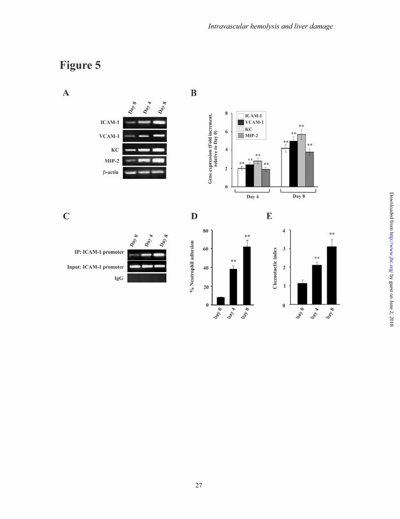
Intravascular hemolysis and liver damage
30
by guest on June 2 2018httpw
ww
jbcorgD
ownloaded from
Intrava
31
ascular hemoolysis and livver damage
by guest on June 2 2018httpw
ww
jbcorgD
ownloaded from
Intrava
32
ascular hemoolysis and livver damage
by guest on June 2 2018httpw
ww
jbcorgD
ownloaded from
Intravascular hemolysis and liver damage
33
by guest on June 2 2018httpw
ww
jbcorgD
ownloaded from
Iqbal Rahul Kumar Souvik Sarkar and Uday BandyopadhyaySumanta Dey Samik Bindu Manish Goyal Chinmay Pal Athar Alam Mohd Shameel
B Activation and Neutrophil InfiltrationκHepatic Free Heme Over-load NFImpact of Intravascular Hemolysis in Malaria on Liver Dysfunction Involvement of
published online June 13 2012J Biol Chem
101074jbcM112341255Access the most updated version of this article at doi
Alerts
When a correction for this article is postedbull
When this article is citedbull
to choose from all of JBCs e-mail alertsClick here
by guest on June 2 2018httpw
ww
jbcorgD
ownloaded from

Intrava
32
ascular hemoolysis and livver damage
by guest on June 2 2018httpw
ww
jbcorgD
ownloaded from
Intravascular hemolysis and liver damage
33
by guest on June 2 2018httpw
ww
jbcorgD
ownloaded from
Iqbal Rahul Kumar Souvik Sarkar and Uday BandyopadhyaySumanta Dey Samik Bindu Manish Goyal Chinmay Pal Athar Alam Mohd Shameel
B Activation and Neutrophil InfiltrationκHepatic Free Heme Over-load NFImpact of Intravascular Hemolysis in Malaria on Liver Dysfunction Involvement of
published online June 13 2012J Biol Chem
101074jbcM112341255Access the most updated version of this article at doi
Alerts
When a correction for this article is postedbull
When this article is citedbull
to choose from all of JBCs e-mail alertsClick here
by guest on June 2 2018httpw
ww
jbcorgD
ownloaded from