january/february - west virginia state medical association
TRANSCRIPT

Chronic or severe headaches are among the most commonreasons patients visit a physician.
Many headaches can be treated adequately with medicationsor changes in lifestyle. But for some patients, their headachesprove to be intractable and sometimes debilitating.
The newWVU Headache Center in Morgantown welcomesreferrals of such patients.
Neurologist DavidWatson, MD, medical director of the center,is the only physician inWest Virginia fellowship trained andcertified as a headache specialist.
When scans or other diagnostic measures are needed,WVUhas the most sophisticated imaging equipment and diagnosticlabs in the region.
For information, contact Dr.Watson through the MARS line(1-800-WVA-MARS).
®WVU Headache Center
To schedule an appointment for your patient:
304-598-6127
Intractable
David Watson, MD
wvuhealth.com
Headaches
A better way to care

Continuing Medical Education Opportunities at CAMC Health Education and Research Institute
The CAMC Institute is dedicated to improving health through research, education and community health development. The institute’s education division offers live conferences, seminars, workshops, teleconferences and on-site programs to health care professionals. The institute’s CME program is accredited by the Accreditation Council for Continuing Medical Education to sponsor continuing medical education programs for physicians. The CAMC Institute designates this educational activity for a maximum of 1 AMA PRA Category 1 Credit(s)™. Physicians should only claim credit commensurate with the extent of their participation in the activity. For more information about these and future programs provided by the CAMC Institute, call (304) 388-9960, fax (304) 388-9966.
SEMInARS28th Cardiovascular ConferenceSunday through Wednesday, Feb. 1 - 4Mountain Lodge Conference CenterSnowshoe, WV
2009 West Virginia Trauma SymposiumWednesday through Friday, Feb. 11-13Canaan Valley Resort & Conference CenterDavis, WV
LIfE SuppORt tRAInIng Log-on to our web site to register at www.camcinstitute.org
Basic Life Support – Health Care ProviderJan. 13, 27Feb. 10, 24
Advanced Cardiac Life Support – RenewalJan. 13Feb. 5, 17
Advanced Cardiovascular Life Support (ACLS) – ProviderJan. 14Feb. 5, 17
Pediatric Advanced Cardiac Life Support (PALS) - RenewalJan. 27Feb. 10
Pediatric Advanced Cardiac Life Support - ProviderFeb. 1
Advanced Trauma Life Support (ATLS)Jan. 29
CME OnLInE pROgRAMS/ARCHIvEd guESt LECtuREpROgRAMSLog on to our web site at www.camcinstitute.orgSystem requirementsEnvironment: Windows 98, SE, NT, 2000 or XPResolution: 800 x 600Web Browser: Microsoft’s Internet Explorer 5.0 or above or Netscape Navigator 4.7x. (Do not use Netscape 7.1)Video Player: Windows Media Player 6.4 or better. Dial-up or broadband connection. Minimum speed, 56k (broadband is recommended)
WV Mutual Insurance Company: Enhancing the Disclosure of Unanticipated Outcomes to PatientsThomas H. Gallagher, MD
Osteoporosis Prevention Education ProgramGayle Manchin and Jessica Wright, RN, MPH, CHES
Diabetes Education for the Primary Care ProviderDaniel J. Dickman, MD; Kristy Lucas, PharmD; Barbara D. Smith, RPh, CDE; Sara O’Conner, MD; Asif Rahman, MD; Harry L. Reahl, MD; Lori Tucker, DO; Arthur B. Rubin, DO, FACOP; Robin Bowyer, RN, BSN, CDE; Marie Gravely, RD, LD, CDE; Cassandra B. Ford, RPh, CDE
Diabetes Education 2 – Recertification for Primary Care ProvidersPalliative Care, Pain Management, Decision Making and Use of the Post FormKim Ashcraft, MS, MSN, RN, C-FNP
Asthma Education for Primary Care ProvidersRobert J. Crisalli, MD, FCCP; Robert A. Kaslovsky, MD; Michael J. Romano MD, MBA; Michael J. Smith PhD, RPh; Sandra E. Swisher RN, MSN, C-FNP
Other archived CME opportunities:Geriatric Series
Ethics Series
Research Series
20534-L08

contents
Certified Partner

contents
EditorF. Thomas Sporck, M.D., F.A.C.S.Charleston
Managing Editor/Director of CommunicationsAngela L. Lanham, Charleston
Executive DirectorEvan Jenkins, Huntington
January/February 2009, Volume 105, No. 1
In this issue…Scientific Articles
10 The Use of MR-Myelography Combining Flexion and Extension Imaging in the Diagnosis of Cervical Myelopathy: A Case Report
15 Melkersson-Rosenthal Syndrome with Migraine-Like Headaches Treated With Minocycline: A Case Report and Review of the Literature
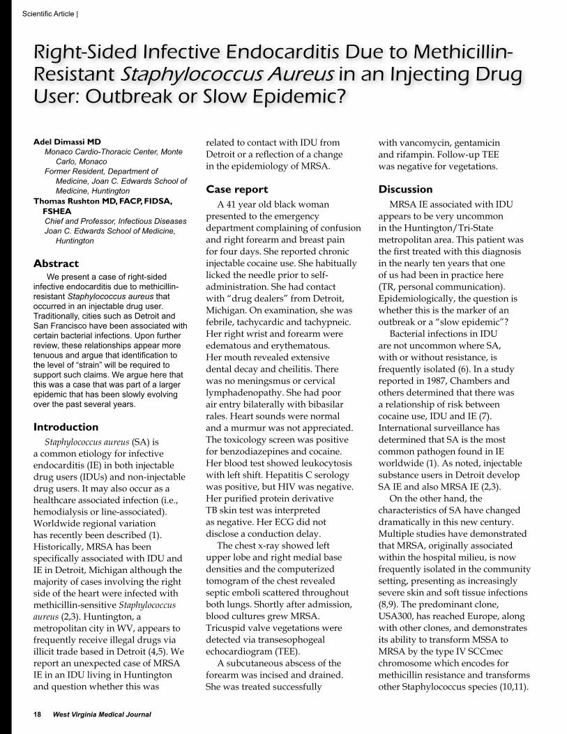
18 Right-Sided Infective Endocarditis Due to Methicillin-Resistant Staphylococcus Aureus in an Injecting Drug User: Outbreak or Slow Epidemic?
20 Selective Renal Artery Embolization Following Blunt Renal Trauma: Case Report and Current Treatment Recommendations for Renal Trauma
Call for Papers39 Dedicated Issue—Breast Cancer
UPComing EvEntS27 Annual Meeting & Mid-Winter
Conference
The West Virginia Medical Journal is published bimonthly by the West Virginia State Medical Association, 4307 MacCorkle Ave., SE, Charleston, WV 25304, under the direction of the Publication Committee. The views expressed in the Journal are those of the individual authors and do not necessarily reflect the policies or opinions of the Journal’s editor, associate editors, the WVSMA and affiliate organizations and their staff.
WVSMA Info: PO Box 4106, Charleston, WV 25364 1-800-257-4747 or 304-925-0342
features 4 President’s Message 6 Our Editor Speaks 7 Letter to the Editor 8 Medical Journal Questionnaire24 General News26 MPLA Suit Statistics28 Court Watch 29 Insurance Commissioner Report 30 Obituaries 32 West Virginia School of Osteopathic
Medicine News33 Marshall University Joan C. Edwards School
of Medicine News34 Robert C. Byrd Health Sciences Center of
West Virginia University News36 Bureau for Public Health News38 Physician Practice Advocate News40 WV Medical Insurance Agency News42 WESPAC Contributors 44 AMA News46 New Members47 Classified Advertising48 Manuscript Guidelines/Advertisers
Cover photo courtesy of Dan Shirley.
Robert J. Marshall, M.D., HuntingtonDavid Z. Morgan, M.D., MorgantownMartha D. Mullett, M.D., MorgantownLouis C. Palmer, M.D., Clarksburg
Associate EditorsJames D. Felsen, M.D., M.P.H.James D. Helsley, M.D., MorgantownDouglas L. Jones, M.D., White Sulphur SpringsSteven J. Jubelirer, M.D., Charleston

� West Virginia Medical Journal
Throughout West Virginia and the United States everyone is talking about healthcare reform. It seems everyone has a healthcare “reform plan” and it is definitely going to be on our state’s legislative agenda. We need to crystallize our own thoughts on what we as physicians know is needed to promote access, quality, and cost efficient healthcare. The following is my opinion based on information gleaned from numerous sources and my thirty years as a family physician caring for West Virginia patients.
These are basic principles:The concept of having a medical home.
I have always tried to advocate for my patients. I provide the services that I feel are appropriate and I try to make sure they are referred to a specialist when necessary and appropriate. I have always felt that it is best for the patient when their physician fully discusses their course of treatment and comes to an agreement as to what tests and procedures should be done, and what referrals should be made. The patient should assume some responsibility in this. When the physician acts as an advisor to the patient, care is likely to be appropriate and the patient is likely to be compliant. When patients have chronic diseases, the treatment team expands and much of the coordination and management is not done at face-to-face visits. The present system does not pay the physician for this service and for many ancillary services such as diabetic education. This needs to change. There
•
should be widespread agreement that everyone should have a primary care physician as his or her medical home.
The availability of insurance must be addressed in any plan for healthcare reform.
Cost shifting has caused tremendous problems. One of the first steps in universal coverage is to develop risk pools for patients with devastating illnesses, which cause them to be uninsurable. Everyone must have insurance and insurance carriers must pay the actual cost of medical care. There are various proposals as to how to do this. See the AMA’s proposal for expanding health insurance coverage and choice at www.voicefortheuninsured.org. But the end result will have to be that insurance is provided to all with no screens. Physical, mental, long-term and dental coverage must be provided to everyone. This insurance must also pay for the implementation of Health Information Technology.
We have to separate medical care from healthcare.
We have to stop blaming our medical care system for problems such as infant mortality and premature death, which are due to social problems such as substance abuse and unhealthy lifestyles. Society as a whole must take responsibility for encouraging proper diet, exercise and a safe environment. Prevention is a part of medical care but it must be a part of all aspects of our lives.
We have to be very careful about making specific recommendations
•
•
•
such as mandating specific services that must be provided.
The problem is we lack in many areas, evidence-based information on what works and what doesn’t work. We need to have a system which allows continued innovation and monitoring of data.
We must encourage the use of information technology.
This will help to eliminate errors and allow the gathering of information and measurement of outcomes. Best practices will continue to evolve, but we are not yet in a position for a specific mandate.
Physicians try to do what is in the best interest for their patients. When the system tries to impose regulations, money is spent on administration instead of patient care. If costs, payments and outcomes are widely known, market forces will address quality, access and price.
The Healthcare Planning Authority needs to stop interfering with the capitalistic market and simply supply information about charges, payments and outcomes. No one would order a meal or buy a car without knowing the price. Why then do we purchase medical care without knowing the cost?
If we can provide universal health insurance and universal use of information technology and transparency in terms of outcomes and of payments, healthcare may truly be reformed.
Stephen L. Sebert, MDWVSMA President
•
President’s Message
Healthcare Reform?

Charleston | Morgantown | Wheeling
www.fsblaw.com | (304) 345-0200 | (800) 416-3225
Edward C. Martin, Responsible [email protected]
health care practice groupStephen R. Brooks
Ryan A. BrownRobert L. CoffieldAlaina N. Crislip
Peter T. DeMastersJ. Dustin Dillard
Sam Fox
David S. GivensPhillip T. Glyptis
Michele GrinbergJohn D. HoffmanStacie D. Honaker
Amy R. HumphreysRobert C. James
Richard D. JonesEdward C. MartinMark A. RobinsonAmy L. Rothman
Don R. Sensabaugh, Jr. Salem C. Smith
take the right path……in professional liability defense, litigation, privacy and security compliance, licensure and professional disciplinary matters, health care fraud and abuse, Stark law analysis, reimbursement issues, employment issues, certificate of need, contractual matters and business transactions.
Helping West Virginia pHysicians

� West Virginia Medical Journal
There is little question that the early days of the new federal administration and Congress will be consumed with the attempted rescue of the economy. It is just a matter of time however, that they will under the direction of former Senator Tom Daschle begin yet another attempt to restructure the healthcare delivery “system” of this republic.
As that debate begins we will frequently hear of the number of uninsured in this country. When we debated “Hillary-Care” 15 years ago the magic number was about 37 million. Today it is somewhere in the high 40 million. Interestingly, as a percentage of the total population that number remains stable at about 15%.
This number is an annualized figure which means that at some point during a twelve month period each of these individuals is without insurance even for a
few days. The number that would be meaningful is the “any given day figure” which would be the number without insurance on any one day. I have been told that this number would be between half and two-thirds of the 45 million annualized. That would equate to between 22 and 30 million or at most 10% of the total population.
A few years ago I read an article comparing “Universal Care Systems” in a number of countries including Canada, Great Britain and many European countries. One interesting factor stuck out—in any of these systems even though “free,” about 5% of the population refused to enroll.
The October 2008 issue of The American Legion Magazine published a very interesting piece dealing with this issue. They took the Census Bureau’s numbers for 2004 and made the following computation:
with private health insurance 198,262,000with government health insurance 79,086,000not covered during the year 45,820,000totAl 323,168,000minus the reported total population 2004 291,155,000disparity 32,013,000 surplus
As you can see we obviously can’t count on the Census Bureau for accurate numbers.
Many of you have been and will be involved in this debate at the state level and may be at the federal level as well. Before we throw the baby out with the bath water lets be sure we know what the real demographics are and not be consumed by the romantic emotion buoyed by these very large numbers.
Tread carefully as we move forward.
F. Thomas Sporck, MDWV Medical Journal Editor
Just How Many Uninsured?
Our Editor Speaks

January/February, 2009, Vol. 105 �
The tobacco industry has a long history of marketing products that purport to be “less harmful” than other tobacco products. This began with filter cigarettes, then with “light” cigarettes. More recently, the industry introduced cigarette-like products having less combustion and emissions than conventional cigarettes. None of these products has succeeded in the marketplace.
In 2006, snus, an oral smokeless tobacco, was introduced to the United States. A teabag-like pouch of snus is placed underneath the upper lip (reducing salivation and thus the need to expectorate) to give the user a boost in blood nicotine concentration. Snus is common in Sweden and Norway, but virtually nowhere else.
Given our high adult tobacco use data (27% smoking; 16% males using smokeless tobacco), West Virginia is a logical state to introduce these products. Our principal concerns are that smokers could opt for snus when they cannot smoke. The tobacco industry makes this “advantage” very clear in its marketing. Should this occur, this could lead to fewer people quitting. Secondly, we are concerned that snus could be an entry tobacco product for minors. Thirdly, there are two Group A carcinogens in these products.
Sales of Camel Snus began in a chain of gasoline/convenience stores in West Virginia in the summer of 2007. In May of 2008,
Tourney Snus was introduced in a different chain in another part of West Virginia. The snus producers seem to believe that the public has no right to have any information about these products—and there are no federal or state statutes requiring disclosure. For the benefit of the consumer and the public health community, we decided to find out about the nicotine and nitrosamine content of these products.
We purchased the products in stores the same way the consumer does, and sent them to an independent laboratory in Canada. They analyzed the three flavors of Camel Snus (Original, Frost, and Spice), and the three flavors of Tourney Snus (Original, Spearmint, and Wintergreen).
Laboratory analyses determined the levels of nicotine, free nicotine, moisture, dry matter, pH, and tobacco-specific nitrosamines (TSNAs): nitrosonornicotine (NNN), nitrosoanatabine (NAT), nitrosoanabasine (NAB), and 4-(N-nitrosomethylamino)-1 (3-pyridyl)-1-butanone (NNK). NNN and NNK are Group A carcinogens. (Free nicotine, i.e., “unbound” or “un-ionized” nicotine needs to be calculated since not all of the nicotine in tobacco is absorbed by the user. Only the free nicotine in a tobacco product can cross biological membranes.)
Camel Snus was found to have a high level of nicotine. On a per gram
basis, it is comparable to a popular snuff product, Copenhagen. From a practical standpoint, the dosage must be taken into account. The Camel brands tested sold in 8 g tins, with 20 pouches to a tin—or 0.4 g/pouch. The Tourney brands tested sold in 12 g tins, with 20 pouches to a tin—or 0.6 g/pouch.
The Camel Snus Original provided 2.03 mg of nicotine in each pouch; the Tourney Snus Original yielded 1.30 mg of nicotine in each pouch. Following a November 2008 press release of our findings, a Camel Snus spokesperson revealed that each pouch of Camel Snus (currently being sold in 0.6 g pouches instead of the 0.4 g pouches that we had analyzed) contained 8 g of nicotine.
That amount of nicotine will surely provide the “satisfaction”—the industry codeword for nicotine—that the user desires. With new forms of oral tobacco slated for an aggressive marketing campaign in 2009—which will include dissolvable tobacco products—physicians are well advised to counsel their smoking patients to avoid all tobacco use.
Letter to the Editor
Bruce Adkins, PADivision of Tobacco Prevention
WV Bureau for Public Health
Robert H. Anderson, MA, CHESPrevention Research Center
Translational Tobacco Reduction Research Program
West Virginia University

� West Virginia Medical Journal
Questionnaire
Dear Member/Subscriber,
The West Virginia State Medical Association (WVSMA), publisher of the West Virginia Medical Journal would like to ask for your feedback concerning the content, design, and focus of the Journal. Please take a moment to answer the questions below. Feel free to add any other comments, criticisms and/or suggestions you may have. Your opinions and perceptions mean a great deal to the WVSMA.
We are here to serve our membership, and the Journal is one of the many valuable services you receive as a member of our state’s largest physician advocacy organization.
If you have other suggestions concerning value-added services the WVSMA could adopt to further enhance membership, these are also welcome. We want to know how we can best serve you and your West Virginia physician colleagues. The WVSMA is proud to be the voice of medicine in West Virginia.
Warmest regards,
Angie LanhamManaging EditorWV Medical Journal
WEST VIRGINIA MEDICAL JOURNAL QUESTIONNAIRE
1. Are the scientific articles of interest to you and relevant to your practice?
2. Are there enough scientific articles or too many in each issue?
3. Is the layout and design of the Journal easy to read?
4. What elements of the current design do you like?
Dislike?
5. Has the addition of new sections (i.e., WVMPHP News, WVMIA News, Court Watch, Physician Practice Advocate News, MPLA Suit Statistics and AMA News) been helpful?
Which section(s) are you most likely to read?
6. What would you add to the Journal?
What would you remove?
7. Other comments and suggestions are welcome and appreciated!
Please copy, then fax the completed questionnaire to Angie Lanham at 304.925.0345

January/February, 2009, Vol. 105 �
Scientific Articles
The Use of MR-Myelography Combining Flexion and Extension Imaging in the Diagnosis of CervicalMyelopathy: A Case Report p. 10
Melkersson-Rosenthal Syndrome with Migraine-LikeHeadaches Treated With Minocycline: A Case Reportand Review of the Literature p. 15
Right-Sided Infective Endocarditis Due toMethicillin-Resistant Staphylococcus Aureus in anInjecting Drug User: Outbreak or Slow Epidemic? p. 18
Selective Renal Artery Embolization Following BluntRenal Trauma: Case Report and Current TreatmentRecommendations for Renal Trauma p. 20

10 West Virginia Medical Journal
Scientific Article |
Charles L. Rosen MD, PhDDepartment of Neurosurgery
John R. Orphanos MDDepartment of Neurosurgery
Robert G. Nugent MDDepartment of Neurosurgery
Gary Marano MDDepartment of Radiology
West Virginia University School of Medicine, Morgantown
AbstractThis report describes the use of a
non-invasive MR-Myelogram combining flexion and extension views to demonstrate causative factors in cervical myelopathy. Utilizing a new approach to MRI, mimicking an older technique of flexion/extension cervical myelography, we were able to identify the pathology of a patient suffering from progressive cervical myelopathy.
We report a patient suffering from progressive multilevel cervical myelopathy due to posterior compression of the spinal cord by an inbuckling ligamentum flavum. Neutral position radiologic assessment failed to reveal any significant spinal cord compression.
In this patient, the dynamic MRI and MR-Myelography was critical for demonstrating the posterior compression of the spinal cord by the ligamentum flavum. Flexion and extension MRI images of the cervical spine complemented by non-invasive MR-myelography were obtained. Surgical decompression was indicated based on these additional studies.
The use of invasive myelography and in particular, flexion/extension myelography is in decline. MR-myelography combining flexion and extension views can be a useful non-invasive means of studying patients with possible ligamentous hypertrophy and dynamic cord compression.
IntroductionImportant pathophysiologic factors
that play a role in the development
of cervical myelopathy include: osteophyte formation, inbuckling of the ligamentum flavum, spinal cord damage resulting from normal flexion and extension of the congenitally narrow cervical spinal canal, and ischemia due to the attenuation of blood vessels supplying the spinal cord by the aforementioned compressive forces (4,5,10,12,13). When the canal diameter is < 10-12mm as a result of these pathological processes, the patient is at an increased risk of suffering cord contusion and subsequent cervical myelopathy (3,11). Clinical presentation, physical examination, and radiologic evaluation establish a diagnosis of cervical myelopathy.
Cervical myelopathy requires evaluation of the anatomical structures involved. The test of choice is MRI of the cervical spine. Many imaging parameters are available, but a T1 weighted signal to look at anatomic structures, and some T2 or similar signal sequence to evaluate for spinal cord edema and anatomy of the CSF spaces is usually of most value. Typical findings include narrowing of the spinal canal, effacement of the spinal cord, and “high signal” within the spinal cord (4,9).
Historically, a myelogram with flexion-extension views was performed to confirm posterior compression of the thecal sac and cord by an inbuckling ligamentum flavum. Today, with the use of MR-Myelography, this same approach, using flexion-extension views can be used in a non-invasive manner providing information confirming the etiology of cervical myelopathy and provide valuable information as to the etiology of cervical myelopathy.
Case ReportPresentation:
The patient is a 59-year-old male with a history of previous C5-6 anterior cervical discectomy and fusion performed in 1996 for C6 radiculopathy and progressive signs of myelopathy including difficulty with ambulation, positive Lhermitte’s sign, and bilateral Hoffman’s reflex. Following surgery, the patient had resolution of his symptoms and no other complaints. The patient returned to West Virginia University in 2002 complaining of recurrent aching neck pain. He had been in his usual state of health until one year ago. He presented with a six-month worsening of his neck pain along with developing shaking, jerking, and difficulty in moving his legs. These symptoms were worse with extension of the head and neck. The patient also described decreased strength and numbness in his hands. The patient’s symptoms were symmetric, and he denied any symptoms consistent with radiculopathy. The patient has had no recent trauma and denied any bowel, bladder, or sexual dysfunction.
Examination:The cranial nerve examination
(II-XII) was grossly intact. His gait was steady and he was able to heel, toe, and tandem walk. There was slightly increased tone in the lower extremities and motor examination was 5/5 for all muscle groups. There was some decrease in light touch and pinprick sensation bilaterally in the lower extremities. Stretch reflexes were all brisk and there was an extensor plantar response on the left. In the upper extremities there was an asymmetry of his stretch reflexes, with the right radial and biceps jerks
The Use of MR-Myelography Combining Flexion and Extension Imaging in the Diagnosis of Cervical Myelopathy: A Case Report

January/February, 2009, Vol. 105 11
| Scientific Article
being clearly brisker than on the left. There was a positive Hoffman’s sign bilaterally and the patient had a strongly positive Lhermitte’s sign.
Radiological Assessment:The patient’s initial radiographic
evaluation included a series of cervical spine radiographs and an MRI. A neutral position T2-weighted image (Figure 1a,b) depicts the previous fusion at the C5-C6 levels. The spinal canal shows some narrowing at the fusion site but no compression of the cervical cord. A CSF space persists around the cord at all levels on axial cuts,
though it is narrowed in the region of the previous fusion. The spinal cord has an area of increased signal posterior to the fusion site (Figure 1c). It is not associated with post-contrast enhancement and was thought to possibly be related to the previous injury at this site. There are disk herniations at the C3-C4 and C4-C5 levels of the protrusion type. The disk herniation at C4-C5 causes moderate-to-severe left neural foraminal stenosis, but as
mentioned before, the patient had no findings of a radiculopathy.
Careful review of the MRI failed to demonstrate definite spinal cord compression. As no recent films were available for comparison, we could not determine whether the area of increased signal was an area of chronic or more current injury.
Since the patient’s symptoms were more prominent when he extended his cervical spine, we repeated the MRI in flexion and extension and performed MR-myelography in flexion and extension to visualize CSF spaces in a manner similar to invasive myelography.
Figures 2a and b are T2-weighted sagittal views of the cervical spine in flexion and extension, respectively. The sagittal flexion view (Figure 2a) shows some mild indentation of the ventral thecal sac at the level of the superior endplate of C5 and effacement of the dorsal thecal sac posteriorly at the C5-C6 level. The sagittal extension view (Figure 2b) shows marked effacement of the ventral and dorsal thecal sac at the C4-C5 level, causing severe central canal stenosis. There is also effacement of the dorsal and ventral
Figure 1aNeutral position sagittal T2 MRI depicts the previous fusion at C5-C6 with spinal canal narrowing at this site but no frank compression of the spinal cord.
Figure 1bT2 MRI axial cut through the level of C4-5 depicting preserved CSF signal around spinal cord.
Figure 2a, 2bSagittal flexion and extension T2 MRI
Figure 1cSagittal T2 extension MRI showing areas of increased canal stenosis above and below the level of prior fusion.

12 West Virginia Medical Journal
Scientific Article |
thecal sacs at the C6-C7 level, causing moderate central canal stenosis. Figure 1d shows an axial view through the level of C4-5 in extension and demonstrates moderate to severe canal compromise.
Figures 3a-d are examples of non-invasive MR-myelography combining flexion and extension views. Flexion
films, seen in lateral (Figure 3a) and antero-posterior views (Figure 3b), show minimal amounts of spinal cord effacement. The extension films, also seen in lateral (Figure 3c) and antero-posterior (Figure 3d) views, more dramatically show the effacement (areas of hypointensity) of the spinal cord at the levels of C4-C5 (severe stenosis) and C6-C7 (moderate stenosis).
Treatment:We discussed with the patient
his pathology and surgical options. The issues of posterior decompression with laminectomy versus multi-level discectomy and fusion are beyond the scope of this discussion. The patient elected for posterior cervical decompression. A laminectomy from C3 to C6 was performed. No instrumentation or fusion technique was utilized. At last follow-up, the vast majority of his symptoms had abated, and he was back to normal levels of activity.
DiscussionPosterior compression of
the cervical spinal cord by the ligamentum flavum along with multiple level disc herniations, in association with a narrow cervical canal, was the cause of progressive myelopathy in this patient.
A recent study by Chen et al was conducted to determine if there were any neutral-position imaging criteria that would predict functional cord impingement at flexion-extension cervical magnetic resonance (MR) imaging. They used five criteria of neutral position MRI to predict functional cord impingement including: cervical curvature, spinal canal space, cervical degeneration, resting instability, and intramedullary high intensity signal on T2 MRI 1. They found that none of their criteria had the ability to predict impingement by flexion MRI; however there was a probable increase of cord impingement to 79% with extension MRI if spinal canal diameter and cervical degenerative
Figure 1dT2 extension MRI axial cut through the level of C4-5 depicting moderate to severe canal compromise.
Figure 3a, 3bLateral and anterior-posterior views of MR-Myelogram in flexion.

January/February, 2009, Vol. 105 13
| Scientific Article
change were used as criteria 1. Using these same criteria in our case we found that they predicted spinal cord impingement using MR-myelography with extension views.
Inufusa et al. examined the relationship between cervical flexion and extension and spinal canal diameter. The study found that cervical extension significantly decreased spinal canal area, midsagittal diameter, and subarticular sagittal diameter (5). This correlation of critical stenosis with spinal canal diameter (<12mm) was also evident in our case. On neutral position MRI, spinal canal diameter was found to be 11.9mm, 10.2mm, and 11.1mm at the levels of C4-C5, C5-C6, and C6-C7, respectively. Applying the criteria of neutral positioning MRI proposed by Chen et al., significant stenosis could be predicted to occur on extension films as shown in Figure 2b. Stenosis was also seen on the extension MR-
Myelogram. As a result, it would appear that neutral position criteria for the prediction of critical stenosis on dynamic MRI can also be applied to a dynamic MR-Myelogram.
In years past, to evaluate a patient with cervical myelopathy, many physicians obtained invasive cervical myelograms, often with flexion-extension views, to diagnose the presence and source of spinal cord compression. With the advent of high resolution CT and MRI, the need for invasive flexion/extension myelography has abated. This report highlights the use of a non-invasive MR-Myelogram combining flexion and extension views that can be used to complement MRI assessment of the patient suffering from myelopathic symptoms. This method is especially useful because it avoids the nephrotoxic complications associated with dye loads as well as the radiation
exposure that can be particularly harmful in certain populations.
ConclusionIn this particular patient with
a myelopathy, identification of posterior ligamentous compression, secondary to inbuckling of the liagmentum flavum, was identified with a “dynamic” MRI. The extension views revealed the presence of spinal cord compression not present in the neutral or flexion views and indicated that this process could be treated surgically. This evaluation was performed in a minimally invasive manner and avoided many of the complications that can be associated with conventional myelographic techniques. In cases of “questionable” spinal cord compression we recommend that consideration be given to the use of non-invasive “dynamic MRI” studies including extension views.
Figure 3c, 3dLateral and anterior-posterior views of MR-Myelogram in extension. Hypodense areas represent areas of compression secondary to loss of signal.

1� West Virginia Medical Journal
Scientific Article |
References 1. Chen CJ, Hsu HL, Niu CC et al: Cervical
Degenerative Disease at Flexion-Extension MR Imaging: Prediction Criteria. Radiology 227:136-142, 2003.
2. Dvorak J: Epidemiology, Physical Examination, and Neurodiagnostics. Spine 23(24):2663-2673, 1998.
3. Edwards CC, Heller JG, Murakami H: Corpectomy versus Laminoplasty for Multilevel Cervical Myelopathy: an Independent Matched-Cohort Analysis. Spine 27(11):1168-1175, 2002.
4. Emery SE: Cervical Spondylotic Myelopathy: Diagnosis and Treatment. J Am Acad Orthop Surg 9(6):376-388, 2001.
5. Fehlings MG, Skaf G: A Review of the Pathophysiology of Cervical Spondylotic Myelopathy with Insights for Potential Novel Mechanisms Drawn From Traumatic Spinal Cord Injury. Spine 23(24):2730-2736, 1998.
6. Fouyas IP, Statham PF, Sandercock PA: Cochrane Review on the Role of Surgery in Cervical Spondylotic Radiculomyelopathy. Spine 27(7):736-747, 2002.
7. Inufusa A, An HS, Lim TH, Hasegawa T, Haughton VM, Nowicki BH: Anatomic Changes of the Spinal Canal and Intervertebral Foramen Associated With Flexion-Extension Movement. Spine 21(21):2412-2420, 1996.
8. Muhle C, Metzner J, Weinert D et al: Classification System Based on Kinematic MR Imaging in Cervical Spondylitic Myelopathy. American Journal of Neuroradiology 19:1763-1771, 1998.
9. Muhle C, Wiskirchen J, Weinert D et al: Biomechanical Aspects of the Subarachnoid Space and Cervical Cord in Healthy Individuals Examined with Kinematic Magnetic Resonance Imaging. Spine 23:556-567, 1998.
10. Nugent GR: Clinicopathologic Correlations in Cervical Spondylosis. Neurology 9:273-279, 1959.
11. Okada Y, Ikata T, Katoh S, Yamada H: Morphologic Analysis of the Cervical Spinal Cord, Dural Tube, and Spinal Canal by Magnetic Resonance Imaging in Normal Adults and Patients with Cervical Spondylotic Myelopathy. Spine 19:2331-2335, 1994.
12. Sampath P, Bendebba M, Davis JD, Ducker TB: Outcome of Patients Treated for Cervical Myelopathy: A Prospective, Multicenter Study with Independent Clinical Review. Spine 25(6):670-676, 2000.
13. Young WF: Cervical Spondylotic Myelopathy: A Common Cause of Spinal Cord Dysfunction in Older Persons. Amer Fam Phys 62(5):1064-1070, 2000.
Drug or Alcohol Problem? Mental Illness?If you have a drug or alcohol problem, or are suffering from a mental illness you can get help by
contacting the West Virginia Medical Professionals Health Program. Information about a practitioner’s participation in the program is confidential. Practitioners entering the program as self-referrals without a complaint filed against them are not reported to their licensing board.
ALL CALLS ARE CONFIDENTIAL(304) 414-0400
West Virginia Medical Professionals Health ProgramPO Box 40027
Charleston, WV 25364

January/February, 2009, Vol. 105 15
| Scientific Article
Matthew A. Hazey, BSMedical Student
Anthony J. Van Norman, MDResident, Section of Dermatology
Drury L. Armistead, MDAssistant Professor, Section of
DermatologyWest Virginia University School of Medicine, Morgantown
AbstractCheilitis granulomatosa (CG), which
presents clinically as persistent lip swelling, is characterized histologically by noncaseating granulomatous inflammation of unknown origin. CG may also be part of the classic triad of the Melkersson-Rosenthal Syndrome (MRS) and alone is considered by some to be an oligosymptomatic form of MRS. We report a case of CG associated with migraine-like headaches in a 44-year-old woman. The clinical presentation, histologic findings, and subsequent treatment are described. A brief review of the literature also is provided.
Case ReportA 44-year-old white female
presented with persistent, painless, non-pruritic swelling of the upper lip for 8 months. The swelling, which was associated with an overlying erythema, was more severe on the
left side (Figure 1). The patient’s symptoms were initially intermittent but became persistent. She gave a history of severe headaches that were temporally related to increased swelling of her upper lip. She was otherwise well and denied symptoms in any other organ system. Further, the patient denied a history of any drug ingestion, nor could she recall any provocative factors such as foods, contactants, or activities. Physical examination revealed soft, non-pitting edema of the upper lip, left greater than right, with overlying erythema. The tongue was normal in appearance, and there were no findings consistent with facial nerve paralysis. Serologic evaluation including complete blood count,
C1 esterase inhibitor, C3, C4, serum angiotensin-converting enzyme level, and antinuclear antibody were within normal limits. Chest x-ray and magnetic resonance imaging of the brain and sinuses were performed and found to be within normal limits. A 4-mm punch biopsy specimen from the cutaneous upper lip showed scattered noncaseating granulomas, mild edema, lymphectasia, as well as superficial and deep perivascular lymphocytic inflammation (Figures 2 and 3). Special stains were negative for evidence of fungi or acid-fast bacilli. No foreign bodies were seen on examination of the specimen under polarized light.
Treatment was initiated with minocycline hydrochloride,100mg,
Melkersson-Rosenthal Syndrome with Migraine-Like Headaches Treated With Minocycline: A Case Report and Review of the Literature
Figure 1.Before treatment. Upper lip with firm edema accompanied by erythema.
Figure 2.Noncaseating granulomas, mild edema, lymphectasia, as well as superficial and deep perivascular lymphocytic inflammation (H&E, original magnification x 20).

1� West Virginia Medical Journal
Scientific Article |
twice daily. This regimen resulted in improvement of the upper lip swelling and erythema (Figure 4). Of note, the patient also noted concordant improvement of her severe headaches. The patient was quite satisfied with this result and the response to therapy has persisted 6 months.
DiscussionThe symptomatic relationship
between recurrent orofacial edema and recurrent facial nerve palsy was first recognized by Melkersson in 1928. Rosenthal later associated lingua plicata with this symptomatology in 1931 (1). While this triad of symptoms known as Melkersson-Rosenthal syndrome (MRS) only presents in a mere 25% of cases, oligosymptomatic and monosymptomatic cases present more frequently as modified forms of the syndrome (2). MRS is uncommon with an estimated incidence reported at 0.08% (3). The
literature reports an age range at symptom onset spanning from 2 to 81 years, however, a majority of cases report onset during the second decade of life (4). Most authors state an equal gender distribution (1,5), while a few report a slight female predilection (4). No apparent racial predilection exists, as cases have been reported from across the globe (5).
Although many theories have been proposed, the etiology of Melkersson-Rosenthal Syndrome is still unknown. Cases have been reported suggesting delayed-type hypersensitivity to food and cosmetic additives, such as cinnamaldehyde and gallates, as a possible cause (6,7). There has even been report of an association between MRS and a delayed-type hypersensitivity to protein in cow’s milk (8). Other theories propose that MRS signifies the body’s reaction to specific infection, however, studies suggest that Borrelia burgdorferi, Toxoplasma gondii, Treponema pallidum, and herpes simplex virus are not
involved pathogenically (9). Reports exist describing the presence of Mycobacterium tuberculosis rRNA in the biopsy specimens of some patients with MRS, but no clear correlation with disease etiology has been elucidated (10). Many case reports in the literature describe an association between Crohn’s disease and orofacial edema, suggesting that this monosymptomatic form of MRS represents a rare extraintestinal sign of Crohn’s disease (11). Although it is uncertain whether or not a definitive genetic predisposition for MRS exists, an implicated gene has been mapped to 9p11 with inheritance thought to occur in an autosomal dominant fashion with variable expression (12).
The clinical presentation of MRS is widely variable. As stated earlier, the complete symptom triad of orofacial edema, facial nerve palsy, and lingua plicata is somewhat uncommon, manifesting in only 25% of cases (2). Orofacial edema has been reported as the presenting symptom in 69% of cases, while Bell’s palsy is described as the initial symptom 19% of the time (4). Recurrent orofacial edema is regarded as the most prominent manifestation of MRS, with labial swelling in 75% of cases and facial swelling in 50% of cases (4). In regard to the specific sites of swelling, the upper lip is reported as being involved most often, followed by the lower lip, buccal mucosa, and palate in decreasing frequency (13).
Figure 3.High-power magnification of noncaseating granulomas with Langhans’ giant cells (H&E, original magnification x 40).
Figure 4.After treatment. Upper lip with reduction in lip swelling and erythema 3 months after initiating minocycline.

| Scientific Article
Moreover, periorbital and upper eyelid edema have been reported in multiple cases (14-18). Recurrent facial nerve palsy is reported in 47% of patients, while lingua plicata is reported in up to 50% (5). Furthermore, in many cases various additional neurological symptoms have been reported, including ear pain, diplopia, vertigo, tinnitus, and decreased saliva (4). In one study 11% of patients reported associated migraines, which our patient also experienced (5).
Histologically, the early stage is often represented by edema, lymphangiectasia, and a predominately perivascular lymphoplasmacytic infiltrate (5,19). In the later stages, after weeks of persistent edema, the classic histopathologic picture emerges. The classic histopathologic findings are characterized by small and scattered noncaseating epithelioid granulomas with lymphangitis and perilymphangitis (5,19). Granulomatous inflammation is not present in all biopsies, and its absence does not exclude the diagnosis of MRS.
The differential diagnosis of persistent swelling of the lip is extensive (Table 1), but a thorough history and careful clinical
examination will usually eliminate many diagnostic possibilities. Although many of the causes of recurrent lip swelling can be excluded on the basis of history and physical examination, a full-thickness mucosal biopsy is needed in order to make a definitive diagnosis. Special stains of the specimen are used in an effort to rule out a mycobacterial or deep fungal infection; polarization can help identify a foreign material. Correlation of historical and clinical data with the histologic findings ultimately culminates in the diagnosis.
MRS is often refractory to treatment, with no individual or combination therapy resulting in complete remission. Topical, intralesional and systemic corticosteroids have been used with some success. Other reported therapies, including prednisone, methotrexate, hydroxychloroquine, dapsone, thalidomide, ranitidine, diphenhydramine, metronidazole, clofazimine, sulfasalazine, penicillin, erythromycin, clindamycin, tetracycline, and minocycline as single therapy or as a combination regimen, have been used with some efficacy (2,15,20). Recently, Barry et al. reported a case of granulomatous cheilits refractory to minocycline, erythromycin, topical tacrolimus, and clofazimine and was successfully treated with infliximab (21). Radiotherapy, facial nerve decompression for the facial palsy, and surgical debulking for cosmesis have also been used (15,22,23).
References1. Rogers RS 3rd. Granulomatous cheilitis,
Melkersson-Rosenthal syndrome, and orofacial granulomatosis. Arch Dermatol. 2000;136:1557-1558.
2. Khachemoune A, Papadopoulos A, Ehrsam E, Kauffmannn L. Self-Assessment examination of the American Academy of Dermatology: Woman with swelling of the lower lip. J Am Acad Dermatol. 2004;50:815-817.
3. Ziem PE, Pfrommer C, Goerdt S, Orfanos CE, Blume-Peytavi U. Melkersson-Rosenthal syndrome in childhood: a challenge in differential diagnosis and treatment. Br J Dermatol. 2000;143:860-863.
4. Zimmer WM, Rogers RS 3rd, Reeve CM, Sheridan PJ. Orofacial manifestations of
Melkersson-Rosenthal syndrome: A study of 42 patients and review of 220 cases from the literature. Oral Surg Oral Med Oral Pathol. 1992;74:610-610.
5. Greene RM, Rogers RS 3rd. Melkersson-Rosenthal syndrome: a review of 36 patients. J Am Acad Dermatol. 1989;21:1263-1270.
6. McKenna KE, Walsh MY, Burrows D. The Melkersson-Rosenthal syndrome and food additive hypersensitivity. Br J Dermatol. 1994;131:921-922.
7. Wong GA, Shear NH. Melkersson-Rosenthal syndrome associated with allergic contact dermatitis from octyl and dodecyl gallates. Contact Dermatitis. 2003;49:266-270.
8. Levy FS, Bircher AJ, Buchner SA. Delayed-type hypersensitivity to cow’s milk protein in Melkersson-Rosenthal syndrome: coincidence or pathogenic role? Dermatology. 1996;192:99-102.
9. Muellegger RR, Weger W, Zoechling N, Kaddu S, Soyer HP, El Shabrawi-Caelen L, Kerl H. Granulomatous cheilitis and Borrelia burgdorferi: polymerase chain reaction and serologic studies in a retrospective case series of 12 patients. Arch Dermatol. 2000;136:1502-1506.
10. Apaydin R, Bahadir S, Kaklikkaya N, Bilen N, Bayramgurler D. Possible role of Mycobacterium tuberculosis complex in Melkersson-Rosenthal syndrome demonstrated with Gen-Probe amplified Mycobacterium tuberculosis direct test. Australas J Dermatol. 2004;45:94-99.
11. van de Scheur MR, van der Waal RI, Volker-Dieben HJ, Klinkenberg-Knol EC, Starink TM, van der Waal I. Orofacial granulomatosis in a patient with Crohn’s disease. J Am Acad Dermatol. 2003;49:952-954.
12. Smeets E, Fryns JP, Van den Berghe H. Melkersson-Rosenthal syndrome and de novo autosomal t(9;21)(p11:p11) translocation. Clin Genet. 1994;45:323-324.
13. Ang KL, Jones NS. Melkersson-Rosenthal syndrome. J Laryngol Otol. 2002;116:386-388.
14. Pierre-Filho Pde T, Rocha EM, Natalino R, Cintra ML, CaldatoR. Upper eyelid oedema in Melkersson-Rosenthal syndrome. Clin Experiment Ophthalmol. 2004;32:439-440.
15. Chen C, Selva D, James C, Huilgol SC. Chronic periorbital swelling in an elderly man. Arch Dermatol. 2003;139:1075-1080.
16. Akarsu C, Atasoy P, Erdgan S, Kocak M. Bilateral upper eyelid edema in Melkersson-Rosenthal syndrome. Ophthal Plast Reconstr Surg. 2005;21:243-245.
17. Cocuroccia B, Gubinelli E, Annessi G, Zambruno G, Girolomoni G. Persistent unilateral orbital and eyelid oedema as a manifestation of Melkersson-Rosenthal syndrome. J Eur Acad Dermatol Venereol. 2005;19:107-111.
Allergic contact dermatitisAscher syndromeCheilitis glandularisCheilitis granulomatosa (Melkersson-Rosenthal syndrome)Crohn’s diseaseFacial edema with eosinophiliaGranulomatous infectionHemangiomaHereditary angioedemaLymphangiomaNeoplasticOdontogenic infectionRecurrent erysipelasSarcoidosis
Table 1. Differential diagnosis of persisent swelling of the lip (5,11).
Please contact the authors for additional references.
January/February, 2009, Vol. 105 1�
1� West Virginia Medical Journal
Scientific Article |
Adel Dimassi MDMonaco Cardio-Thoracic Center, Monte
Carlo, MonacoFormer Resident, Department of
Medicine, Joan C. Edwards School of Medicine, Huntington
Thomas Rushton MD, FACP, FIDSA, FSHEAChief and Professor, Infectious DiseasesJoan C. Edwards School of Medicine,
Huntington
AbstractWe present a case of right-sided
infective endocarditis due to methicillin-resistant Staphylococcus aureus that occurred in an injectable drug user. Traditionally, cities such as Detroit and San Francisco have been associated with certain bacterial infections. Upon further review, these relationships appear more tenuous and argue that identification to the level of “strain” will be required to support such claims. We argue here that this was a case that was part of a larger epidemic that has been slowly evolving over the past several years.
IntroductionStaphylococcus aureus (SA) is
a common etiology for infective endocarditis (IE) in both injectable drug users (IDUs) and non-injectable drug users. It may also occur as a healthcare associated infection (i.e., hemodialysis or line-associated). Worldwide regional variation has recently been described (1). Historically, MRSA has been specifically associated with IDU and IE in Detroit, Michigan although the majority of cases involving the right side of the heart were infected with methicillin-sensitive Staphylococcus aureus (2,3). Huntington, a metropolitan city in WV, appears to frequently receive illegal drugs via illicit trade based in Detroit (4,5). We report an unexpected case of MRSA IE in an IDU living in Huntington and question whether this was
related to contact with IDU from Detroit or a reflection of a change in the epidemiology of MRSA.
Case reportA 41 year old black woman
presented to the emergency department complaining of confusion and right forearm and breast pain for four days. She reported chronic injectable cocaine use. She habitually licked the needle prior to self-administration. She had contact with “drug dealers” from Detroit, Michigan. On examination, she was febrile, tachycardic and tachypneic. Her right wrist and forearm were edematous and erythematous. Her mouth revealed extensive dental decay and cheilitis. There was no meningsmus or cervical lymphadenopathy. She had poor air entry bilaterally with bibasilar rales. Heart sounds were normal and a murmur was not appreciated. The toxicology screen was positive for benzodiazepines and cocaine. Her blood test showed leukocytosis with left shift. Hepatitis C serology was positive, but HIV was negative. Her purified protein derivative TB skin test was interpreted as negative. Her ECG did not disclose a conduction delay.
The chest x-ray showed left upper lobe and right medial base densities and the computerized tomogram of the chest revealed septic emboli scattered throughout both lungs. Shortly after admission, blood cultures grew MRSA. Tricuspid valve vegetations were detected via transesophogeal echocardiogram (TEE).
A subcutaneous abscess of the forearm was incised and drained. She was treated successfully
with vancomycin, gentamicin and rifampin. Follow-up TEE was negative for vegetations.
DiscussionMRSA IE associated with IDU
appears to be very uncommon in the Huntington/Tri-State metropolitan area. This patient was the first treated with this diagnosis in the nearly ten years that one of us had been in practice here (TR, personal communication). Epidemiologically, the question is whether this is the marker of an outbreak or a “slow epidemic”?
Bacterial infections in IDU are not uncommon where SA, with or without resistance, is frequently isolated (6). In a study reported in 1987, Chambers and others determined that there was a relationship of risk between cocaine use, IDU and IE (7). International surveillance has determined that SA is the most common pathogen found in IE worldwide (1). As noted, injectable substance users in Detroit develop SA IE and also MRSA IE (2,3).
On the other hand, the characteristics of SA have changed dramatically in this new century. Multiple studies have demonstrated that MRSA, originally associated within the hospital milieu, is now frequently isolated in the community setting, presenting as increasingly severe skin and soft tissue infections (8,9). The predominant clone, USA300, has reached Europe, along with other clones, and demonstrates its ability to transform MSSA to MRSA by the type IV SCCmec chromosome which encodes for methicillin resistance and transforms other Staphylococcus species (10,11).
Right-Sided Infective Endocarditis Due to Methicillin-Resistant Staphylococcus Aureus in an Injecting Drug User: Outbreak or Slow Epidemic?

| Scientific Article
This patient might have experienced an infection with an USA300 clone of MRSA which created her soft tissue infection. Once she became bacteremic, right-sided IE developed. If so, her connection with Detroit is less remarkable.
It is useful to consider the history and epidemiology of Serratia marcescens in San Francisco, California: IDU-associated IE has been caused by Serratia marcescens (12,13). It has been suggested that this organism became part of the endogenous microbial flora due to biological warfare experiments conducted by the US military after World War II. Culture media were placed at various geographic points to determine the effectiveness of off-shore disbursement methods. The wild strain of this bacterium grows as red-pigmented colonies which allowed for rapid identification in environmental cultures. While it was hypothesized that the experiments created environmental contamination that led to infections associated with IDU in San Francisco, later analysis proved that military strain was different from that found in such patients a quarter of a century later (12).
More recently, Spaargaren and colleagues describe an outbreak of lymphogranuloma venereum (LGV) L2b occurring in Amsterdam and San Francisco, which is equally illustrative (14). Researchers in Amsterdam detected an ongoing outbreak of LGV proctitis. From 2002-2005, cases were analyzed using the polymerase chain reaction assay. These were compared to samples collected in San Francisco in the 1980’s. What appeared at first to be an outbreak associated only with Amsterdam is now correctly identified as an ongoing “slowly evolving epidemic” that involves both Europe and the United States, with the L2b strain implicated in patients infected over almost a quarter of a century (14).
While a city or defined geographic boundary may become associated
with a particular infection or strain, it would be incorrect to conclude that this represents a localized, isolated event. A community based study showed that individuals in non-healthcare locations had an overall Staphylococcus aureus colonization rate of almost 27% and an MRSA colonization rate of 1% (15).
ConclusionThis patient received her cocaine
through contacts originating in Detroit, a city associated with MRSA IE. Concomitantly, the epidemiology of MRSA was shifting from a nosocomial, hospital-based infection, to a community –acquired epidemic. It now appears that these new, more virulent strains spread quite rapidly by skin and soft-tissue infections. The most common type isolated is the USA300 strain. This patient might have been colonized by contact with her Detroit-based dealers or she might have been infected with the USA300 strain.
At first, it was an attractive hypothesis that the Huntington-Detroit relationship connected both the illicit drug trade and MRSA IE associated with IDU. A review of other geographically identified infections, and the dramatic shift in the epidemiology of MRSA that was just coming to light when this patient was diagnosed, argues that she was part of a larger epidemic.
Merely knowing the genus and species of a bacterial infection now seems woefully inadequate. It would appear that precise identification, at least to the level of “strain”, should be employed to establish the exact nature of the infection present (16). The threat to public welfare produced by infections such as MRSA is critical enough to warrant this analysis and hopefully lead to effective control measures.
References1. Fowler VG, Miro JM, Hoen V, Cabell CH,
Abrutyn GR, et al. Staphylococcus aureus endocarditis: a consequence of medical progress. JAMA. 2005; 293: 3012-3021.
2. Crane LR, Levine DP,. Levine, Zervos MJ, Cummings G. Bacteremia in narcotic addicts at the Detroit Medical Center. I. Microbiology, epidemiology, risk factors, and empiric therapy. Rev Infect Dis. 1986; 8: 364-373.
3. Levine DP, Crane LR, Zervos MJ. Bacteremia in narcotic addicts at the Detroit Medical Center.II. Infectious endocarditis: a prospective comparative study. Rev Infect Dis. 1986; 8: 374-396.
4. Manolatos T. and Shepardson D. Detroit’s drug trade linked to 4 West Virginia slayings: Huntington police blame city for a ‘substantial amount of our violent crime.’ The Detroit News, May 25, 2005. www.detnews.com, accessed July 21, 2007.
5. U.S. Census Bureau. Census 2000 PHC T-29. Ranking tables for population of metropolitan statistical areas, micropolitan statistical areas, combined statistical areas New England city and town areas, and combined New England city and town areas: 1990-2000; table 1a. www.census.gov/population/cen2000/phct29/tab01a.pdf. Internet release date: December 30, 2003; accessed May 4, 2007.
6. Gordon RJ and Lowy FD. Bacterial infections in drug users. N Engl J Med. 2005; 353: 1945-54.
7. Chambers HF, Morris DL, Tauber MG, et al. Cocaine use and the risk for endocarditis in intravenous drug users. Ann Intern Med. 1987; 106: 833-36.
8. King MD, Humphrey BJ, Wang YF, Kourbatova EV, et al. Emergence of community-acquired methicillin-resistant Staphylococcus aureus USA 300 clone as the predominant cause of skin and soft-tissue infections. Ann Intern Med. 2006; 309-17.
9. Johnson JK, Khoie T, Shurland S, Kreisel K, et al. Skin and soft tissue infections caused by methicillin-resistant Staphylococcus aureus USA300 clone. Emerg Infect Dis. 2007; 13: 1195-1200.
10. Tristan A, Bes M, Meugnier H, Lina G, et al. Global distribution of Panton-Valentine leukocidin-positive methicillin-resistant Staphylococcus aureus, 2006. Emerg Infect Dis. 2007 ; 13 : 594-600.
11. Hiramatsu K, Kuroda M, Baba T, Ito T, and Okuma, K. Application of genomic information to diagnosis, management, and control of bacterial infections: the Staphylococcus aureus model. In: Persing DH, Tenover FC, Versalovic J, Tang Y-W, et al, (eds). Molecular microbiology: diagnostic principles and practice. Washington D.C.: ASM Press, 2004; pp.:407-18.
12. Yu VL. Serratia marcescens: historical perspective and clinical review. N Engl J Med. 1979; 300: 887-893.
13. Mills J, Drew D. Serratia marcescens endocarditis. Ann Intern Med. 1976; 85: 397.
14. Spaargaren J, Schachter J, Moncada J, de Vries HJC, Fennema HSA, et al. Slow epidemic of lymphogranuloma venereum L2b strain. Emerg Infect Dis. 2005 ; 11 : 1787-88.
January/February, 2009, Vol. 105 1�
Please contact authors for additional references.

20 West Virginia Medical Journal
Scientific Article |
Brian P. DeFade, DOChief Urology Resident, Charleston Area
Medical CenterJames P. Tierney, DO
Program Director, Chair of Urology, Charleston Area Medical Center
Patrick A. Stone, MDAssistant Professor of Surgery, WVU
School of Medicine, Charleston Division
Department of Surgery: Division of Vascular and Endovascular Surgery
Richard R. TruxilloMedical Student, West Virginia School of
Osteopathic Medicine
AbstractManagement of renal trauma has
become more conservative as newer techniques evolve. In 2004, the Renal Trauma Subcommittee modified their algorithms for the management of renal injuries to include selective angiography and embolization for grade III and IV lacerations for both blunt and penetrating renal lacerations (1). These algorithms are based on the renal organ injury scale defined by The American Association for the Surgery of Trauma (AAST) and
whether the patient is hemodynamically stable or unstable (Table 1) (1). Historically, grade III and IV renal injuries would have been managed by renal exploration.
The goals of treating patients with severe renal injuries are to prevent significant hemorrhage and retain sufficient functional nephrons to prevent end-stage kidney failure (1). Selective arterial embolization provides a minimally invasive treatment option for renovascular injuries and potentially obviates the need for surgical exploration with its higher incidence of nephrectomy (2). We present a case of grade IV renal laceration following blunt renal trauma, which was successfully treated with selective renal arterial embolization.
Case ReportA 21-year-old white male
presented to the Charleston Area Medical Center (CAMC) Trauma unit with an isolated renal injury sustained during a snowboarding accident. His Glasgow coma score (GCS) was 15 on arrival. He denied
any loss of consciousness. The patient complained of bilateral flank pain and right hip pain. Physical exam revealed bilateral flank and hip abrasions with no other injuries identified. A Foley catheter was inserted and revealed gross hematuria. The patient was hemodynamically stable and underwent computed tomography (CT) of the abdomen and pelvis with intravenous contrast. Findings on CT scan demonstrated a grade IV renal laceration of the right middle and upper pole with extension to the hilum and associated perinephric hematoma (Fig. 1). Caliceal system involvement was suspected, but there was no evidence of contrast extravasation on initial CT evaluation. The patient was admitted to the intensive care unit (ICU) for observation.
The patient remained hemodynamically stable until hospital day four when his
Selective Renal Artery Embolization Following Blunt Renal Trauma: Case Report and Current Treatment Recommendations for Renal Trauma
Table 1. Renal organ injury scale as defined by the American Association for the Surgery of Trauma (AAST).
AAST Renal Injury Classification Scheme
Grade I Renal Contusion: Microscopic or gross hematuria, urologic studies normal. Subcapsular Hematoma: Non-expanding without parenchymal lacerationGrade II Cortical Laceration: <1 cm parenchymal depth of renal cortex Perirenal Hematoma: Non-expanding, confined to renal capsule.Grade III Parenchymal Laceration: Deep, >1 cm parenchymal depth of renal cortex without collecting system rupture
or urinary extravasation.Grade IV Parenchymal Laceration: Involving the collecting system, with or without a devascularized segment. Vascular: Main Renal Artery or Vein Injury with contained hemorrhage.Grade V Laceration: Completely Shattered Kidney Vascular: Renal Artery Thrombosis, avulsion of the renal pedicle.
*For bilateral injuries, upstate the injury by one grade up to grade III.
Figure 1. Adapted from Wessells, H., et al.: Urologic Emergencies: A Practical Guide. 2005. (From Figure 2, page 5).

| Scientific Article
hematocrit dropped from 38.5% to 21.3%. Two units of packed red blood cells were transfused and a second CT scan was performed. This study showed extension of the perinephric hematoma with trace contrast extravasation (Fig. 2).
An endovascular consult was requested to consider selective arterial embolization. Selective right renal artery angiography followed soon thereafter (Fig. 3). Three branches of the right renal artery, found to be actively bleeding, were coil embolized with 2 mm platinum coils—in the anterior superior segmental artery, anterior inferior segmental artery, and a small branch of the posterior
segmental artery (Fig. 4). Following selective embolization the vital signs and hematocrit stabilized.
On hospital day nine the patient was ambulating, tolerating a regular diet, his fever had subsided, and his urine had cleared of hematuria. The patient was discharged home after a repeat non-contrasted CT scan of the abdomen and pelvis was found to be grossly unaltered from his previous contrasted studies.
Two months following successful embolization of the renal artery, CT scan of his abdomen and pelvis with IV contrast demonstrated only a small low-attenuation density posterior to the right kidney, which represented a resolving hematoma. Six-month follow-up revealed that the patient had returned to normal daily activities, was pain free, had no hypertension. His urinalysis was free of protein and blood.
DiscussionThe current indications for
nephrectomy with renal trauma have narrowed to persistent, life threatening hemorrhage, an expanding or pulsatile retroperitoneal hematoma, and uncontrolled retroperitoneal bleeding (4,5). Other factors must be considered, such as the presence of concomitant injuries and their management,
hemodynamic instability after initial resuscitation, and the grade and mechanism of injury (3). Despite a more conservative trend, nephrectomy remains the most commonly performed renal surgery for renal injuries and is seen at much higher rates in institutions without specialization in renal trauma (4). A recent review of the National Trauma Data Bank by Michael Metro found that on multivariate analysis of these data that renal injury severity was the strongest predictor of nephrectomy (4).
Currently conservative treatment for Grade I to Grade III renal lacerations with either penetrating or blunt renal trauma is considered standard of care. There is, however, some disagreement over the management of Grade IV and Grade V renal lacerations. Buckley et al. reviewed 153 Grade IV renal traumas over the past 25 years and subdivided these patients into isolated or non-isolated renal injuries. Forty-three (28%) of the 153 patients were found to have isolated renal injuries and of these 43 patients 18 (42%) underwent renal exploration with the majority having penetrating injuries(6). The nephrectomy rate for those 18 patients was 11%. The remaining 25 isolated renal injuries were managed non-operatively with a renal salvage
January/February, 2009, Vol. 105 21
Figure 1.Anterior/posterior view of the patient’s IV contrasted CT scan of abdomen and pelvis showing the Grade IV renal injury.
Figure 2.Transverse view of the patient’s IV contrasted CT scan of the abdomen and pelvis showing perirenal hematoma and Grade IV injury.
Figure 3.Pre-embolization angiogram showing segmental renal artery bleeding.
Figure 4.Post-embolization angiogram showing segmental renal artery embolization with coils, and resolution of active bleeding.

22 West Virginia Medical Journal
Scientific Article |
rate of 88% and a lower transfusion rate as compared to operative management—2.5 units of packed red blood cells versus 8.5 units of packed red blood cells (6). Conversely, the remaining 110 patients were found to have non-isolated renal injuries. Of these 110 cases, (of non-isolated renal injuries) 103 (94%) were operated (6). The operative rate for Grade IV renal injuries in this group was 77% with a 15% nephrectomy rate. The overall renal salvage rate was 84% (6). This review provides evidence that Grade IV isolated and non-isolated renal injuries may be managed conservatively with good outcomes, yet penetrating and non-isolated renal injuries were more likely to be explored.
Alsikafi and Rosenstein reviewed 2900 patients from San Francisco General Hospital database who had blunt renal trauma. Of these patients, only 2.6% were managed operatively with a 0.7% nephrectomy rate (3). They concluded that most blunt renal injuries could be managed conservatively, even those with urinary extravasation and nonviable renal tissue (3). Similarly, Altman et al. showed that Grade V shattered but perfused kidneys that were hemodynamically stable could be treated conservatively without surgery (3). 66% of these kidneys functioned at discharge from the hospital. None developed subsequent hypertension (3).
In spite of the increasing trend towards more conservative treatment of both blunt and penetrating injuries, open surgical exploration still occurs at a higher rate in cases of penetrating and non-isolated renal injuries. Superselective embolization is becoming a tool that may help decrease the incidence of surgical management for both blunt and penetrating renal injuries; thereby decreasing the morbidity and higher nephrectomy rate associated with open surgical management. Superselective coil embolization has been reported to be effective in the treatment of iatrogenic and
penetrating renal injuries. Dinkel et al showed that even grade IV and grade V renal injuries, which were unstable could be treated with selective coil embolization and surgery averted (7). Heyns et al reviewed 93 patients with stab wounds involving the upper urinary tract. Of these patients, 53 (67%) were managed non-operatively and 11 underwent selective coil embolization of segmental branches of the renal artery because they were found to be actively bleeding (8). They suggested that renal angiography and superselective coil embolization should be performed in patients with renal stab wounds before surgical exploration for suspected injury to adjacent organs (8). Even with many recent studies supporting selective coil embolization for the management of blunt and penetrating renal injuries, there are many institutions not utilizing this technique.
ConclusionAlthough conservative
management of grade I-III blunt and penetrating renal trauma is accepted, the current literature supports selective renal artery embolization in hemodynamically stable patients for grade IV and grade V blunt and penetrating renal trauma before surgical management is attempted. Further recommendations from the Jackson Hole Urology Meeting in 2000 suggested that the majority of renal traumas should be managed non-operatively. Current guidelines suggest that only 1.7% of blunt injuries and 57% of penetrating injuries require surgical exploration (9). The mechanism of injury is less important than the extent of damage. Operative decisions should be based on acute or delayed complications (9). A CT scan should be the imaging modality of choice for renal injury staging. Delayed images should be included to identify injuries to the collecting system (9).
Recommendations based on renal injury grade were as follows: grade I and grade II injuries usually have no
indication for surgery; grade III and grade IV injuries should be managed non-operatively as long as the patient remains hemodynamically stable. Superselective coil embolization may be considered for these stage injuries as well. Grade IV vascular injuries and grade V injuries warrant surgical exploration, but may be managed with selective coil embolization in select cases (9). The endovascular approach to renal injuries using selective coil embolization has shown an ever increasing role in the management of both blunt and penetrating renal trauma. There are few drawbacks to endovascular management. The most common post-operative complication is postembolization syndrome subsequent to large volume parenchymal infarction (10). These reports support the increasing role of selective coil embolization in the treatment of bleeding renal injuries. Further randomized studies may help further support these reports, yet it appears that the current literature supports the increasing role of selective coil embolization as the primary management strategy for renal trauma in order to preserve renal function, and decrease morbidity and mortality from open operative techniques.
References 1. Masters, VA, McAninch, JW. Operative
Management of Renal Injuries: Parenchymal and Vascular. Urol Clin N Am. 2006;33:21-31.
2. Beaujeux, R, Saussine, C, Al-Fakir, A, et al. Superselective Endo-Vascular Treatment of Renal Vascular Lesions. The Journal of Urology. 1995;153:14-17.
3. Alsikafi, NF, Rosenstein, DI. Staging, Evaluation, and Nonoperative Management of Renal Injuries. Urol Clin N Am. 2006;33:13-19.
4. Metro, M. Renal and Extrarenal Predictors of Nephrectomy from the National Trauma Data Bank Reviewed. The Journal of Urology. 2006;175:970-975.
5. Carroll, PR, Klosterman, PW, McAninch, JW. Surgical Management of Renal Trauma: Analysis of Risk Factors, Techniques, and Outcomes. The Journal of Urology. 1998;28:1071-1077.
Please contact authors for additional references.


2� West Virginia Medical Journal
Scientific Article |
The West Virginia Healthy Kids and Families Coalition is pleased to announce that Dr. William Neal, founder and director of the Coronary Artery Risk Detection in Appalachian Communities (CARDIAC) Project, is the recipient of the 2008 “No Greater Legacy Award.” The “No Greater Legacy Award” is given to those who have contributed to the quality of child health in West Virginia. The 2008 award is given to Dr. Neal because of his accomplishments in the area of childhood obesity in West Virginia.
West Virginia has one of the highest rates of childhood obesity in the nation. Dr. Neal’s work on
childhood obesity has enabled West Virginia to lower its childhood obesity rates. Of the children screened during 2006 to 2008 school years, a decline in obesity was noted among kindergarten students (36.2 vs. 33.2%), second grade students (42.1 vs. 37.5%), and fifth grade students (46 vs. 45.5%).
Dr. Neal is a Professor and the James H. Walker, MD Chair of Pediatric Cardiology in the Department of Pediatrics at West Virginia University. He is a member of a number of medical associations including, the American Pediatric Society, Society for Pediatric Research, and the American Academy of
Pediatrics. Dr. Neal is the recipient of numerous awards and his scholarly work is contained in publications, books, and abstracts. He has served in the Navy as a flight surgeon aboard the USS Constellation.
He is married to Dr. Martha Mullett, a neonatalogist, also practicing at the West Virginia University School of Medicine. They have six children and 13 grandchildren.
To keep up to date on the West Virginia Healthy Kids and Families Coalition conference developments, including speakers and registration, sign up for e-mail updates at www.wvhealthykids.org.
Dr. William Neal is Honored with the“No Greater Legacy Award”
General | NEws

January/February, 2009, Vol. 105 25
The West Virginia State Medical Association (WVSMA) hosted the Physician Practice Management Conference, Wednesday, November 19, 2008. Despite treacherous weather conditions, a large crowd braved the elements to attend this informative conference.
Following a welcome by WVSMA’s Executive Director, Evan Jenkins, The conference kicked off with a presentation by Bethany Chiaramonte of the American Medical Association. Bethany traveled from Chicago to give our participants an update on the “Heal That Claim” campaign.
Following the AMA presentation, participants enjoyed visiting exhibitor booths including Sage Software Healthcare, Inc., the West Virginia Medical Insurance Agency, West Virginia Health Information Network, Standard Register, West
Virginia Medical Foundation and the CPR Solutions Group, Inc.
Pamela Harvit, protocol consultant, led the mid-morning presentation with “Office Protocol and Etiquette.” Her unique, interactive presentation style had participants up on their feet!
During a special luncheon, Bob Lane of Standard Register enlightened participants about prescription fraud and prescription pad security. A special presentation was given on the WVSMA’s Tamper-Resistant Prescription Pad Grant Program.
Following lunch, Dr. Barbara Connors, chief medical officer for CMS Region III, gave an excellent presentation on E-prescribing and PQRI.
The last session of the day, “2009 Medicare Update” was led by Tim Allman of Palmetto GBA. Tim kept the crowd alert and focused. The audience was full of questions and
fortunately, Tim had the answers!The overall feedback from the
physicians, office managers, and office staff who attended was excellent. Physicians were able to receive 6 CME credits for the daylong conference.
“I’m pleased that that our participants felt the sessions provided at the conference were relative and valuable. Many expressed appreciation for practical information that could be used everyday in their medical practices, with the ultimate goal being better patient care,” said Barbara Good, physician practice advocate for the WVSMA and organizer of the Physician Practice Management Conference.
The large attendance at the conference demonstrated the need for educational events of this caliber. The WVSMA plans to hold a similar conference in the spring of 2009 so watch for information and plan to attend!
General | NEws (Continued)
November 19, 2008, Physician Practice Management Conference a Great Success

COUNTY
BarbourBerkeleyBooneBraxtonBrookeCabellCalhounClayDoddridgeFayetteGilmerGrantGreenbrierHampshireHancockHardyHarrisonJacksonJeffersonKanawhaLewisLincolnLoganMarionMarshallMasonMcDowellMercerMineralMingoMonongaliaMonroeMorganNicholasOhioPendeltonPleasantsPocahontasPrestonPutnamRaleighRandolphRitchieRoaneSummersTaylorTuckerTylerUpshurWayneWebsterWetzelWirtWoodWyoming
TOTALS(BY INDIVIDUAL YEAR)
2002
0600529010114500021170201504838041700015000241320200011003090
239
2003
19107280005027111141266201026231704310142000012021300011121020140
315
2004
02111150000003121611204040211902701170000106300130010010110
130
2005
140147000100410081137109223043510102100010
1261040100001002060
273
2006
0311014000501301053047112121180315001500024720002003001152
154
2007
03102
140013015100942
461040221903
15102600005
1451110101000061
174
TOTAL2002-September 2008
328342611501116193044248129
317111468182011603231062211660015
1738319152822101091593
1405
Each month, the WVSMA tracks the number of MPLA suits filed in each county throughout West Virginia. Below is a chart summa-rizing the case filings from 2002 to September 2008. Please note the annual total for 2005 was significantly impacted by the large number of suits brought in Putnam County that year, most of which related to Dr. King. Excluding the 2005 filings in Putnam County, year-end total filings 2004-2007 were 130, 147, 154 and 174 respectively.
Thru9/08
1100790001013000412
310023032502
11001300004
1200102001000080
86
2� West Virginia Medical Journal

January/February, 2009, Vol. 105 2�
Participate in charting the direction of healthcare reform in West Virginia. Attend the WVSMA Mid-Winter Business Meeting.
2009 Mid-wiNTERbUSiNESS MEETiNG
February 6-8Waterfront Place Hotel, Morgantown, WV

Court Watch is funded through physicians’ membership investment in the WVSMA and prepared bythe law firm Flaherty, Sensabaugh & Bonasso PLLC.
Short answer: Yes.
In Cartwright v. McComas, et al., Plaintiff’s then four-year-old daughter was admitted to Cabell Huntington Hospital (“CHH”) from October 9, 1999 through October 16, 1999. During this time period the defendant physician did not order an MRI. On November 8, 1999, during an office visit, the defendant physi-cian ordered that the child have an MRI of the spine. The MRI was completed in December 1999, and revealed a vascular abnormality which was compressing the child’s spinal cord and causing paralysis. The child was referred out and underwent a surgical procedure elsewhere to resect a hemorrhagic mass near her spinal cord. After the surgery the child continues to be paralyzed and incontinent. Plaintiff’s expert opined that the delay reduced the child’s likelihood of making a recovery.
Plaintiff filed a medical malpractice action against the defendant physician on April 23, 2003, asserting that he devi-ated from the standard of care by failing to order an MRI of the child’s spine while she was hospitalized at CHH. On June 15, 2005, Plaintiff was granted leave to file an amended complaint to add CHH as a defendant. CHH moved for summary judgment claiming that the 2003 amendments to the MPLA, which took effect on July 1, 2003, precluded the Plaintiff from pursuing a claim against CHH for the acts of the defendant physician (ostensible agency claim). On July 3, 2007, the circuit court entered an order granting summary judgment in favor of CHH. Plaintiff appealed.
On appeal the Court determined that the summary judgment ruling of the lower court which threw out the claim against CCH represents plain error1, and the Supreme Court reinstated the case in the circuit court..
First, the Court determined that reversible error exists because the amendment to the complaint adding the defen-dant hospital should relate back to the date that the original complaint was filed in 2003, and not to the 2005 date when the amendment was filed. If the 2003 date is used, then the ostensible agency prohibition was not yet part of the MPLA. Second, the Court stated that the Plaintiff child has a substantial right in pursuing her claim against CHH, and failure to preserve the child’s right would call into question the fairness and integ-rity of the judicial process. The Court noted that the Legislature expressly permits an extended time to minors to file malpractice claims. Minors under ten may bring an action until their twelfth birthday. In this case the minor’s twelfth birthday was in 2007, well after the plaintiff sought to add the hospital.
1The necessary elements of plain error are present in this case. The Court pointed out in syllabus point twelve of Keesee v. General Refuse Service, Inc., 216 W. Va. 199, 604 S.E.2d 449 (2004), in order “[t]o trigger an application of the “plain error” doctrine there must be (1) an error; (2) that is plain; (3) that affects substantial rights; and (4) seriously affects the fairness, integrity, or public reputation of the judicial proceedings…”
C o u r tA special WVSMA
membership update
on the actions
and decisions of
the West Virginia
Supreme Court of
Appeals regarding
key issues impacting
West Virginia
physicians. The
WVSMA, West
Virginia’s largest
physician advocacy
organization, is
dedicated to a fair
and equitable civil
justice system and is
fighting to preserve
our hard-fought
medical liability
reforms. Monitor
our website at
www.wvsma.com
for the latest
updated
information.
Cartwright, as guardian and parent of Cartwright, a minor childv. McComas, and Cabell Huntington Hospital/Opinion No. 33868
Appeal from the Circuit Court of Cabell County to the West Virginia Supreme Court of Appeals
ISSUE: Does the MPLA provision providing the right of a minor under ten years of age to file suit prior to a minor’s twelfth birthday permit a hospital to be added as a defendant based on alleged malpractice of a physician, when the original case was filed in April 2003, and when as of July 1, 2003, a hospital may no longer be sued under a theory of “ostensible agency?”
Forshey v. Jackson/Opinion No. 33834Appeal from the Circuit Court of Kanawha County to the West Virginia Supreme Court of Appeals
ISSUE: When does the continuous medical treatment doctrine apply to a medical malpractice case?
In Forshey v. Jackson, the defendant physician performed carpel tunnel surgery on the patient on July 6, 1995. After surgery, the patient continued to experience pain, swelling and tenderness in his left hand. Nevertheless, the physician failed to order any follow-up x-rays on the patient’s hand. The patient’s last visit with the physician was on January 31, 1997.
During the summer of 2005, the patient suffered an unre-lated injury to his left hand. An x-ray of the hand revealed a metallic foreign body in the patient’s hand, which was ultimately
determined to be a piece of a knife blade. In September 2006, the patient filed a medical malpractice claim against the physi-cian, claiming that the only explanation for the foreign body was the carpal tunnel surgery performed by the physician in 1995. The trial court dismissed the patient’s suit, for failing to file his claim within the ten year statute of repose found in West Virginia’s Medical Professional Liability Act, W. Va. Code § 55-7B-4.1 The patient appealed.
2� West Virginia Medical Journal
January/February, 2009, Vol. 105 2�
W A t C HC o u r tOn appeal, the patient argued that the continuous medical
treatment doctrine should be applied to his case, because the physician committed malpractice each time he saw the patient by not ordering x-rays and diagnosing the foreign body in his hand. Under the continuous medical treatment doctrine, the running of the statute of limitations is tolled when a course of treatment that includes wrongful acts has run continuously and is related to the original complaint. In its opinion, the West Virginia Supreme Court of Appeals officially adopted the continuous medical treatment doctrine, holding that:
[U]nder the continuous medical treatment doctrine, when a patient is injured due to negligence that occurred during a continuous course of medical treatment, and due to the continuous nature of the treatment is unable to ascertain the precise date of the injury, the statute of limitations will begin to run on the last date of treatment.
Despite adopting the continuous medical treatment doctrine, the Court found that the doctrine did not apply to the patient’s case because the patient’s injury occurred on a certain date – the date of the carpal tunnel surgery – and did not result from a continuing course of treatment. The Court further found that “where the patient suffers an identifiable injury through some affirmative act of negligence on part of the practitioner, the fact that thereafter the practitioner continues to care for
and treat the patient does not postpone the commencement of the limitation period.” The Court found that the patient’s claim arose on the date of surgery, July 6, 1995. Under the statute of repose, the patient’s claim had to be filed no later than July 6, 2005, ten years after the date of injury.
The patient also argued that his case should not have been dismissed because it constituted a continuing tort. The West Virginia Supreme Court of Appeals previously recognized that a continuing tort “involves a continuing or repeated injury” and found that “the statute of limitations begins to run from the date of the last injury.” The Court noted that the concept of a continuing tort requires a showing of repetitious wrongful conduct. The Court further stated that a wrongful act with continuing damages is not a continuing tort. Thus, the Court held that “in the context of a medical malpractice action, in order to establish a continuing tort theory a plaintiff must show repetitious wrongful conduct. Merely establishing the continuation of the ill effects of an original wrongful act will not suffice.” Because the patient could not show repetitious wrongful conduct, the West Virginia Supreme Court of Appeals affirmed the dismissal of the case by the trial court.
1Section 55-7B-4 provides that no action shall be filed more than ten years after the date of injury.
♦ West Virginia’s overall medical malpractice experience in 2007 declined from the exceptional results realized during 2006. It is notable however, that the written premium volume for 2007 ($60,322,954) was at its lowest overall level since 1999 ($44,387,157).
♦ Countrywide net operating results for all lines of business in 2007 remained favorable (88.4%), and overall medical malpractice results improved to 67.6% (from 72.3% in 2006.)
♦ The key volume writer in the state, West Virginia Mutual Insurance Company which holds 82.2% of the admitted market and 59.9% of the entire West Virginia market, again experienced a favorable year in 2007, posting a pure direct loss ratio of only 40.37%. However, only 2 companies were found to meet the 5% or more market share threshold for inclusion in the 2007 report. There were 3 companies which met this criteria in 2006.
♦ During 2007 Medical Malpractice rates continued to decline in West Virginia.
♦ The small size of our medical malpractice marketplace in West Virginia, with only 3,837 active physicians practicing in our State, inherently lends itself toward volatility, as can be demonstrated by simply comparing the 2006 results to the 2007 results alone.
♦ An analysis of West Virginia Board of Medicine data revealed the following: • The number of paid claims continues to
generally decline in West Virginia. • Roughly 31% of claims filed are still being dismissed • The total number of claims in 2007 (164) is less than half of
that experienced in 2004 (375).This appears to suggest that the “certificate of merit”, required by H. B. 601, continues to have a diminishing impact on medical mal-practice claims. o Approximately 10% of claims filed actually go to court. o 60% of malpractice claims are settled outside of court. o There does not appear to be any clear and credible pattern
of escalating jury awards, as the small number of awards yields little in the way of credible data upon which to draw sound conclusions.
♦ A review of Medical Liability fund data indicates that the number of filed actions in West Virginia continued to decline in 2007. This appears to be generally consistent with other similar measurements contained within this report.
Insurance Commissioner Issues ReportIn late November, the offices of the West Virgina Insurance Commissioner issued the annual report for West Virginia’s medical liability
insurance market. This report is a comprehensive analysis of many facts and figures that impact the cost of medical liability insurance for physicians in West Virginia. Below is a summary of key findings of this report. (Note: The data compiled in the November, 2008 report is as of 12/31/07). The full report is available on the WVSMA website at www.wvsma.com.

30 West Virginia Medical Journal
ObituariesThe WVSMA remembers our esteemed colleagues…
Jack H. Baur MD, FACPDr. Jack H. Baur, age 84, of Huntington, WV, died Monday, Dec. 1, 2008 at his residence. He was born May 31, 1924, in Cincinnati, Ohio, a son of the late Samuel and
Emma Homberg Baur. In addition to his parents, he was preceded in death by a son, Donald A. Baur.
He was a graduate of the University of Cincinnati Medical School and came to Huntington in 1955, and practiced at the C&O Hospital then joined HIMG in 1970.
Dr. Baur was a former Associate Dean at the Marshall University School of Medicine. He then went to Cabell Huntington Hospital as Vice President of Medical Affairs, where he retired in 1993. He also served as the Governor of the A.C.P. in 1975 and 1976. He was the first hematologist in Huntington. He served in the U.S. Navy as a medical officer from 1949 to 1954.
He is survived by his wife, Maxine Plumley Baur; his daughter, Jane; two sons, Jay and Tom; a stepdaughter, Angela Fain; grandchildren Marc and Michael Baur, Jill Bunn, Steve and Dan Baur, Lindsay and Sarah Roberts and Eric, Erin and Elizabeth Fain; three great-grandchildren; a brother, Robert Baur, and a niece and nephew. The family would like to express a heartfelt thank you to his caregivers and special friends, Deborah Branham, Judy Dallis,
Mabel Arthur, Cheryl Ruley and Brandy Ward. Those who wish, may make memorial contributions to the Donald A. Baur Scholarship Fund c/o Wake Forest University, P.O. Box 7227, Winston-Salem, NC 27109 or to the charity of one’s choice.
Kirk Jamieson David, MDKirk Jamieson David, MD, 89, of Huntington, WV, died Thursday, July 31, 2008, at his home in the Woodlands Retirement Community
Dr. David was born in Moscow, Idaho. He was a graduate of the University of Idaho and of Columbia University College of Physicians and Surgeons. He served in the United States Army Medical Corps in Korea and practiced surgery in Huntington from 1952 to 1991.
Dr. David was a Fellow of the American College of Surgeons and was a member of Our Lady of Fatima Catholic Church, the Cabell County Medical Society, the Knights of Columbus, the Huntington Museum of Art and the Marshall Artist Series.
Survivors include his wife of 62 years, Rosemary Jordan David, and five children, Kathleen David, Marguerite David, Joseph J. David, Kirk J. David Jr., and Maureen David; one grandson, Kirk J. David III.
Memorial contributions may be made to the Palotti Fund of St. Mary’s Medical Center or the charity of one’s choice. Family guestbook at www.klingelcarpenter.com.
Jeffrey George, MDJeffrey Edward
George, M.D., age 49, of Roanoke, Virginia, formerly of Huntington, West Virginia, went to be with the Lord on December 3, 2008 near Roanoke. Born December
20, 1958, son of the late Edward and Frances Shaw George, Dr. George is survived by his loving wife of 20 years, Teresa Kimbler George. He was graduated from Duke University in 1980 and West Virginia University Medical School in 1984. He completed his general surgery residency at Roanoke Memorial Hospital in 1989 and his cardiothoracic surgery residency at the University of Louisville in 1991. His honors include valedictorian of Huntington High School and, later, recipient of the Lange Outstanding Senior Medical Student Award at WVU. He received the Baird Memorial Surgical Research award many times during his general surgery training. Furthermore, he was designated as one of the “Best Doctors in America” six separate times. He was also chairman of the Hospitality House (now the Jeffrey E. George Comfort House); director of Cardiothoracic Surgery, Chief of Surgery and Chief of Staff at St. Mary’s Medical Center; Chairman of St. Mary’s Medical Center Foundation; Professor and Chief, Division of Cardiothoracic Surgery, Joan C. Edwards School of Medicine. Dr. George and his family returned to Roanoke, where he joined Carilion Cardiothoracic Surgical Associates. His tireless

January/February, 2009, Vol. 105 31
efforts on behalf of all patients continued through his work on the safety and quality improvement team for improving patient outcomes.
Survivors include two aunts, Linda George Sakakini and Rosalie George. Surviving cousins include: Joy and Jim Booten, Steven Sakakini, Lynette Sakakini Bunyan, Julia Denise George, Fredric George, Phillip Charles George, Alan George, Barbara George Ricks, Gregory George, Mark George, Chris George, Joy George Biddar, Roseanne George Giles, John George, the late Deidra George and their respective families.
The family guest book may be signed at www.klingel-carpenter.com. In lieu of flowers, expressions of sympathy may be made to either Jeffrey E. George Comfort House or Fifth Avenue Baptist Church.
Edward Lewis, MDEdward Lewis, M.D., 90, born in Charleston in March of 1918, passed away in Heartland Nursing Home on November 29, 2008 after a short illness.
Edward attended and graduated from Charleston High School where he was a basketball All-Star. He graduated from the University of West Virginia in 1949 and from the University of Virginia Medical School, Charlottesville, as a medical doctor.
He was a veteran of World War II, serving in the Army 261st Infantry Company C. He served in the European Theatre under General George Patton and during the Battle of the Bulge. Edward
was awarded the Bronze Star.He interned at Charleston General
Hospital. He had a general practice in Charleston until he retired.
He was preceded in death by his parents, George and Nazza Lewis; and brother, Fred Lewis, in 1996. He is survived by brother, Richard A. Lewis, M.D.; and sisters, Rosemary Bsharsh and Nazza Skaff, and several nieces and nephews.
In lieu of flowers, donations may be made to St. George Orthodox Cathedral, Lee and Court Streets, Charleston, WV 25301.You may send condolences at www.barlowbonsall.com.
entchas.com
500 Donnally StreetCharleston, WV
Physician’s Office:Suite 200
Hearing Aid Center:Suite 102
304.340.2200for appointments
Hearing aid Center:304.340.2222
Complete comprehensive services including:
Ears, nose and throat medical and surgical care
Audiological testing
Inhalant allergy testing and treatment
Hearing aid evaluation and placement services
Computed Tomography (CT) for sinuses and ears
Board Certified Specialists:
F. Thomas Sporck, MD, FACS D. Richard Lough, MD P. Todd Nichols, MD G. Stephen Dawson, MD
Michael R. Goins, MD
Obituaries | (Continued)

32 West Virginia Medical Journal
West Virginia School of Osteopathic Medicine | NEws
The Board of Governors of the West Virginia School of Osteopathic Medicine has announced that Dr. Richard Rafes will succeed Dr. Olen Jones as President.
Following an extensive national search, the Board made their final decision today. The Higher Education Policy Commission will act on the appointment Friday, November 21 during their regular Commission meeting.
“We welcome Dr. Rafes to WVSOM”, said Sharon Rowe, Board Chair. “We are indeed fortunate to have an individual with his credentials and experience to assume the presidency of WVSOM.”
Dr. Rafes is currently serving as President of East Central University (ECU) in Ada, Oklahoma. ECU is a comprehensive student-centered regional public university with
69 academic program offerings and about 4500 students.
Prior to his presidency at ECU, Dr. Rafes served in various capacities for 26 years at the University of North Texas (UNT) and the University of North Texas System (University of North Texas, University of North Texas Health Science Center at Fort Worth and University of North Texas at Dallas). His most recent position at UNT was Senior Vice President for Administration. UNT is the third largest university in Texas.
The University of North Texas Health Science Center at Fort Worth developed from its first college, Texas College of Osteopathic Medicine (TCOM), into a comprehensive health science center. Dr. Rafes was intricately involved as general counsel in the development of the osteopathic medical school and the health science center. He served as a faculty member in the College of Education at UNT for 25 years as well as the College of Business Administration. At TCOM he taught
osteopathic medical students medical jurisprudence and medical ethics.
Dr. Rafes holds a B.A. in government from Lamar University and two doctoral degrees, a J.D. from the University of Houston Law Center and a Ph.D. in higher education from the University of North Texas.
In accepting the appointment, Dr. Rafes said, “I am thrilled to join the outstanding team of faculty and staff at the West Virginia School of Osteopathic Medicine and work to further the institution’s mission of educating primary care osteopathic physicians for rural communities. My wife, Tommye, and I look forward to becoming active members of the Lewisburg community and are most appreciative to the Board of Governors for affording us this wonderful opportunity.”
Dr. Rafes will become President February 1, 2009 and will begin the transition January 10.
Dr. Richard Rafes Named President at WVSOM
Longtime WVSOM President Honoredby Robert C. Byrd Clinic
The Robert C. Byrd Clinic honored Olen E. Jones, Jr., Ph.D., recently by naming a portion of its facility after the long time WVSOM president.
During a ceremony on November 6, the Robert C. Byrd Clinic dedicated a meeting room in its new East Wing addition to Dr. Jones, renaming it the “Olen E. Jones, Jr., Ph.D. Resource Room.” The room will be adorned with a large portrait of Dr. Jones, as well as a bronze plaque.
“Dr. Jones has been a visionary leader and proactive supporter for medical education in southern West Virginia,” said Mike Painter,
Executive Director for the clinic. “Throughout his 21-year career, he has supported the clinic in all aspects and embraced our duel role of treating patients and educating medical students.”
“This tribute is a small token of appreciation to say ‘thank you’ to Dr. Jones for his commitment to WVSOM and the Robert C. Byrd Clinic. Your legacy will live on. Congratulations and we appreciate everything you have done,” said Painter.
In accepting the honor, Jones responded, “I’m humbled. Many
people deserve credit for the success at the clinic. You have done an outstanding job training our students and serving the people of the Greenbrier Valley. You have affected the quality of life in this area in a positive way.”
Rafes
on hand for the dedication ceremony (l to r): Dr. Jane Johnson, chairperson of the RBC Clinic Board of Directors; mike Painter, Executive Director of the RCB Clinic; Dr. olen Jones; Patty Jones, wife; and Delegate tom Campbell of the 28th District.

| Scientific ArticleMarshall University Joan C. Edwards School of Medicine | NEws
Marshall University Welcomes New Specialty Faculty
New Marshall faculty physicians offering subspecialty medical services include the region’s first orthopedic oncologist and physicians in several medical and surgical specialties. New faculty members include:
EndocrinologyTipu Saleem, MD,
trained at Penn State and Cornell; he has special interest in new protocols for weight reduction and glucose control in diabetes,
as well as thyroid ultrasound and ultrasound-guided biopsy for early detection of thyroid cancer.
Ayhman Elkadry, MD, trained at Marshall and has special interests in tight glucose control with diabetes and in thyroid diseases,
including the management and early detection of thyroid cancer.
GastroenterologyJoe Gerges el Khouri
trained at Yale and the University of Florida and has special interests in treating gastroesophageal reflux
disease, liver diseases and inflammatory bowel diseases.
Vikram Tarugu, MD, trained at the University of Mississippi and specializes in treating pancreatobiliary diseases and inflammatory bowel diseases.
Infectious DiseaseJose-Mario Fontanilla,
MD, MPH, trained at Dartmouth-Hitchcock Medical Center and the University of Connecticut. He has special interests
in outpatient parenteral antibiotic therapy, treatment of bone and joint infections, and treatment of prosthesis-related infections, prevention of healthcare-associated infections and hospital epidemiology.
NeurologyMark Stecker, MD,
PhD, who trained at the University of Pennsylvania, has expertise in treatment for seizures and epilepsy. He
also is director of the expanded neurodiagnostics laboratory at Cabell Huntington Hospital.
Hasan Ercan, MD, trained at the University of Chicago and the University of South Carolina and has a special interest in the treatment
of sleep disorders.
OphthalmologyParveen Nagra, MD,
trained at Albert Einstein Medical Center and Wills Eye Hospital, with a fellowship in cornea and external diseases.
OrthopedicsFelix Cheung, MD,
who trained at West Virginia University and Harvard Medical School, performs specialized orthopedic treatments
for bone and soft-tissue cancers and the latest techniques in limb-sparing reconstruction.
Douglas S. Tice, MD, trained at West Virginia University and the Mercy Hospital of Pittsburgh. He specializes in orthopedic trauma.
Plastic and Reconstructive Surgery
Farid Mozzafari, MD, trained at Marshall University and the Medical College of Georgia; his surgical interests include pediatric and craniofacial
surgery, post-bariatric surgery, body contouring and aesthetic surgery.
Keith Pitzer, MD, trained at Scott and White Hospital at Texas A&M and has special interest in post-bariatric contouring, cancer reconstructive
surgery, breast surgery, aesthetic surgery, skin cancer surgery and facial rejuvenation.
Pulmonary MedicineFadi Alkhankan, MD,
who trained at Marshall University, has interest in the full spectrum of pulmonary medicine.
Alejandro Lorenzana, MD, who trained at the University of Massachusetts and Sinai Hospital in Detroit, has special interest
in the treatment of asthma and COPD, as well as early detection and treatment of lung cancer.
UrologyLawrence Wyner,
MD, received his fellowship training in renovascular surgery and transplantation from the Department
of Urology at the Cleveland Clinic Foundation, and remains a member of its associate staff. He also trained at the University of Pittsburgh.
January/February, 2009, Vol. 105 33

3� West Virginia Medical Journal
Bonnie’s Bus Ready to RollWVU OFFERing digiTAl MAMMOgRAPHy in RURAl AREAS
West Virginia University officials rolled out Bonnie’s Bus Nov. 6, complete with equipment and staff, poised to begin the trek throughout the state to reach women who lack easy access to mammograms.
Bonnie’s Bus will offer life-saving early detection for breast cancer in a state with the fifth highest mortality rates in the nation.
The bus and mammograms are made possible through a $2.5 million gift of Jo and Ben Statler to the Mary Babb Randolph Cancer Center, part of last year’s historic $25 million donation to WVU. WVU Hospitals owns and operates the bus.
Named after Mrs. Statler’s late mother, Bonnie Wells Wilson, Bonnie’s Bus and the Cancer Center also received $300,000 from Susan G. Komen for the Cure to support outreach to women and follow-up tracking studies and a two-year $400,000 grant from the Claude Worthington Benedum Foundation to establish a clinical trials network.
“Bonnie’s Bus is far more than a mobile digital mammography unit,” said Scot Remick, M.D., director of WVU’s Mary Babb Randolph Cancer Center. “Not only are we building a tremendous research database, but also we are creating networks with doctors and hospitals throughout West Virginia to deliver the care women need. Through Bonnie’s Bus, we will reduce deaths from breast cancer in our state.”
Dr. Remick added: “Together, Bonnie’s Bus and the Cancer Center at WVU are devoted to improving the cancer care of West Virginians. Collaborative relationships in Martinsburg, Wheeling, Clarksburg and Charleston are well beyond the formative stages, and we plan to engage other new partners.”
Women whose mammograms suggest a need for further diagnosis and treatment will be guided to doctors in their home communities or to hospitals and, most important, to state-of-the-art clinical trials for
patients who want access to them. The network will make it easier for women who live far from hospitals or an academic medical center to gain access to investigational drug trials.
The mammograms are not free. But billing to third-parties will be provided, and women who lack insurance will be matched to government or nonprofit charities.
For information on Bonnie’s Bus see www.wvucancer.org/bonnie.
WVU Announces Creation of Multimillion Dollar Pediatric Research InstituteREzUlin SETTlEMEnT, WAlkER BEqUEST PROVidE $12 MilliOn FOR CHildREn’S HEAlTH
West Virginia University has announced the creation of the multimillion dollar WVU Pediatric Research Institute. The institute builds on a strong history of pediatric research at WVU.
“We are exhilarated to be able to take our research to the next
level to serve the needs of children throughout West Virginia,” said Giovanni Piedimonte, M.D., chair of the Department of Pediatrics and physician-in-chief at WVU Children’s Hospital. “Whenever we invest in children, we invest in our future.”
Funding for the WVU Pediatric Research Institute comes from two sources: a generous bequest from a former WVU physician, and unclaimed settlement money from a class action lawsuit.
Through a bequest from the late James H. Walker, M.D., former
Robert C. Byrd Health Sciences Center of West Virginia University | NEws
Ribbon cutting for Bonnie’s Bus. l-R: James Brick, m.D.; Fred Butcher, Ph.D.; Jo Statler; C. Peter magrath, Ph.D.; gary marano, m.D.; Ben Statler; Scot Remick, m.D.

January/February, 2009, Vol. 105 35
WVU’s Dr. Bill Neal is First Walker Chair in Pediatric CardiologyJAMES H. WAlkER BEqUEST TOTAlS MORE THAn $6 MilliOn
Robert C. Byrd Health Sciences Center of West Virginia University | NEws Continued
WVU physician and professor on the Charleston campus, $6.378 million will go toward the institute and the creation of the James H. Walker Chair in Pediatric Cardiology.
WVU pediatric cardiologist William A. Neal, M.D. is the inaugural recipient of the Walker Chair.
In addition, the institute will receive $5.67 million from unclaimed settlement money in a class action lawsuit involving Warner-Lambert Co. and Parke-Davis, creators of the diabetes drug Rezulin. In September, Raleigh County
Circuit Judge John Hutchison approved the payment to WVU.
Dr. Piedimonte said the money would be used primarily to research a link between obesity, diabetes and asthma in West Virginia’s children. The Pediatric Research Institute will hire research scholars, who along with the current team of researchers, will continue to work to solve these problems.
Currently, about 60 percent of faculty members in the Department of Pediatrics are actively engaged in research, with more than 16,500 square feet of
space within the Department of Pediatrics dedicated to research.
Dollars invested in research on pediatric issues at WVU has doubled in the past two years, jumping from almost $3 million in 2006 to more than $6 million in 2008.
The institute will occupy two floors of an existing classroom and laboratory building on the north side of the Health Sciences Campus, beginning in 2009.
For more information about pediatric research at WVU see www.hsc.wvu.edu/som/pediatrics/research.asp.
William A. Neal, M.D., professor of pediatric cardiology at the West Virginia University School of Medicine, has been named the inaugural recipient of the James H. Walker, M.D., Chair in Pediatric Cardiology.
The chair is funded through a gift from the estate of the late James H. Walker, M.D., a clinical professor of surgery at the WVU School of Medicine in Charleston from 1980 until his death in 2006. His gift also establishes a permanent endowment for support of the chair. The total bequest will be $6.378 million.
“The reason we have a top-notch pediatric cardiology program here at WVU Children’s Hospital is in no small means due to the efforts of Dr. Walker,” Neal said.
Walker recognized the growing needs of children in West Virginia for high quality pediatric cardiology services, Dr. Neal said. Together with Neal, Walker established WVU’s program, which today performs 250 cardiothoracic procedures, 100 cardiac catheterizations and 3,000 ultrasound examinations annually.
A native of Huntington, Neal graduated from Xavier University
in Cincinnati, then earned his medical degree at WVU, where he was the first recipient of the Edward J. Van Liere Award for medical student research. He continued his training at Milwaukee County General Hospital and studied pediatric cardiology at the University of Minnesota.
In 1998, Neal established the CARDIAC (Coronary Artery Risk Detection in Appalachian Communities) Project to address the high illness and death rates from cardiovascular disease in West Virginia. In 2007, the U.S. Department of Health and Human Services gave the project a national Innovation in Prevention Award for its success in addressing chronic diseases and promoting healthier lifestyles.
Neal also staffs the Pediatric Lipid Clinic in Morgantown and outreach clinics in Beckley, Huntington, Lewisburg, Princeton and Wheeling.
William neal, mD, first James H. Walker Chair in Pediatric Cardiology

3� West Virginia Medical Journal
Bureau for Public Health | NEws
Methamphetamine has been a problem in West Virginia for over a decade. Eighty to ninety percent of methamphetamine found in West Virginia originates from other states and Mexico. Mexican drug cartels are the leading producer and seller of methamphetamine nationwide. It is the other 10-20% that affects citizens of the state. It is West Virginia’s rural environment that poses the greatest threat when fighting the war against methamphetamine. It can be made anywhere now, and often escapes notice by law enforcement.
Methamphetamine is manufactured by using toxic chemicals and over-the-counter cold medication. In the early 1990s, labs were found in locations such as warehouses with technical-grade lab equipment and chemicals. The recipes were lengthy, and it took two days to cook methamphetamine. Then, the rise of the Internet led to faster and easier lab recipes. The time it took to cook methamphetamine was about eight to 12 hours, and the average size of a lab was that of a kitchen. These labs were known as mom-and-pop or “Beavis and Butthead” labs. Mainly, these names were adopted by law enforcement and the forensic community due to the fact that the labs were anything but technical in nature. Meth cookers are now using household kitchen equipment and chemicals to produce the drug.
Today, methamphetamine is produced in 2-liter bottles using very little, if any, equipment. The time has been reduced to approximately 45 minutes. But no matter the size of the lab, the toxic waste and methamphetamine residue still exist. The lab leaves behind a hazardous environment any unknowing individual could walk into, including innocent children.
Several years ago, laws were passed to limit the sale of pseudoephedrine-containing medications. The laws have extended their reach to include the sale of other meth-making ingredients. There have even been laws passed to give law enforcement the authority to arrest individuals who possess the ingredients to produce methamphetamine. But it wasn’t until 2007 that the West Virginia Legislature passed a law that changed the future for once-known meth houses.
On April 2, 2008, Governor Joe Manchin, signed into law the Clandestine Drug Laboratory Remediation Act. The new rule provides the guidance and authority the state needs to combat the public health hazards of former meth labs. The rule includes the responsibilities placed on law enforcement, property owners, and the contractors performing the remediation. It also gives the West Virginia Department of Health and Human
Resources (DHHR) the authority to write regulations concerning
• Property clean up• Licensing of contractors
and technicians• Accreditation of
training providersThe maximum contamination
level of 0.1 ug/100 cm2 for methamphetamine residue was adopted and is used by most states. Law enforcement has the new role of notifying the Clandestine Drug Laboratory Remediation Program, as well as the property owner, within 24 hours of lab bust. Property owners are now responsible for securing and maintaining the property as vacant, hiring a licensed contractor to perform testing and remediation. They must also disclose this information to potential buyers or renters.
The property must remain secure and unoccupied until DHHR issues a Certificate of Remediation Completion. Properties cannot be sold or rented until a Certificate is issued by the Clandestine Drug Laboratory Remediation Program. The option of demolition is permissible if the owner does not feel the need to remediate.
For more information, please contact Mr. Lewis at 304-558-6782 or by e-mail at [email protected].
Brandon LewisEnvironmental Resources Specialist
Methamphetamine Lab Properties Get New Life
Visit us on our website for more information or contact: Toni Charlton – President at 304-670-7197 or Donna Lee - State VP Membership at 276-322-5732.
OFFICE MANAGERS ASSOCIATIONOF HEALTHCARE PROVIDERS, INC.
www.officemanagersassociation.comWe invite you to join our organization which consists of members
who manage the daily business of healthcare providers.Our objectives are to promote educational opportunities, professional knowledge
and to provide channels of communication to officemanagers in all areas of healthcare. We currently have
eleven chapters in West Virginia.
Visit us on our website for more information or contactDonna Zahn (President) at 740-283-4770 ext. 105 or
Tammy Mitchell (Membership) at 304-324-2703.
We invite you to join our organization which consists of members who
manage the daily business of healthcare providers. Our objectives are
to promote educational opportunities, professional knowledge and to
provide channels of communication to office managers in all areas of healthcare.
We currently have eleven chapters in West Virginia.

3� West Virginia Medical Journal
Medicare has announced a number of changes for 2009. The Centers for Medicare and Medicaid Services (CMS) is encouraging physicians to adopt electronic prescribing for their practices as soon as possible. According to CMS, one of the major advantages of adopting e-prescribing is the elimination of medication errors which may result from misreading handwritten prescriptions. CMS states that Medicare beneficiaries’ out-of-pocket costs should be less as e-prescribing will improve communication between physicians and pharmacies regarding lower-cost generic alternatives.
In the 2009 Physician Fee Schedule Final Rule, CMS has created incentives for physicians in order to persuade them to become early adopters of e-prescribing. Beginning January 1, 2009, eligible professionals may participate in the E-prescribing Incentive Program by submitting information required by the E-prescribing measure on their Medicare Part B claims.
CMS is providing a 2% payment incentive for successful use of e-prescribing for the years 2009 and 2010. In 2011 and 2012, the payment incentive decreases to 1% of covered Medicare Part B charges. In addition, the rule states that in 2012, eligible physicians who are not successfully using electronic prescribing will be penalized 1% of their covered Medicare Part B charges. This means that physicians would be paid at 99% of Medicare reimbursement. Penalties would increase as physicians would
receive 98.5% of their allowed charges in 2013, and only 98% in 2014.
CMS Secretary Leavitt has the ability and authority to change the requirements for successful e-prescribing in the future. Also, the Medicare Improvements for Patients and Providers Act of 2008 (MIPPA) allows for the use of Part D data instead of claims based reporting for eligible physicians.
In the Final Rule, CMS stated what physicians were required to do in order to earn their e-prescribing incentive payments. Physicians will need to have a qualified e-prescribing system which must be able to communicate with the patient’s pharmacy and help the physician identify appropriate drugs, as well as to provide information on generic substitutes for brand-name drugs. The system must be able to provide information on formulary and tiered formulary medications. It must also be able to forewarn physicians about possible adverse events, such as improper dosing, drug-to-drug interactions, or allergy concerns.
CMS has also adopted PQRI improvements in the Final Rule. Physicians and other professionals who participate in the PQRI initiative will now be eligible to receive a 2% bonus of allowable charges which are submitted no later than two months after the end of the reporting period for 2009 quality measures.
In addition to removing quality measure 125 that was used to report on the use of e-prescribing, CMS added nine new measure groups, including CABG surgery,
rheumatoid arthritis, back pain, coronary artery disease, and HIV/AIDS. This brings the total number of reportable measures to 153 for 2009.
In order to earn the incentive payment, physicians will need to report on every patient visit, using one of three “G” codes for the e-prescribing measure. When submitting claims for specified types of medical visits to earn the incentive payment, physicians may indicate that they didn’t prescribe any medications during the visit (G8445) or that they used e-prescribing for any medications prescribed during the visit (G8443). They may also report (G 8446) that some or all the prescriptions generated during the visit were printed or phoned in as required by state or federal law or regulations (i.e. for controlled substances); patient request; or the pharmacy system being unable to receive electronic transactions.
Both of the 2% e-prescribing and the 2% PQRI incentive payments are in addition to the 1.1 percent fee schedule update physicians will receive for services provided under the 2009 Physician Fee Schedule Final Rule. That means that a physician could earn up to 5.1 percent more in 2009 for successful reporting under both the e-prescribing and PQRI initiatives.
It is important to note that a physician or other eligible professional does not have to enroll in order to participate in the E-prescribing Incentive Program. Furthermore, participation in PQRI is not required in order to participate in this incentive program.
CMS Changes for 2009
Physician Practice Advocate | NEws
January is here, bringing with it many changes, particularly in the world of insurance. As a reminder, some of your patients may also have changed insurance carriers, effective January 1, 2009. In order to ensure that you do not delay your practice’s reimbursement by billing the wrong carrier, it is critical that you require your patients to show their insurance cards when they come for the first appointment in 2009. That will ensure that your practice has the current information on file for each patient.
January/February, 2009, Vol. 105 3�
tHEmE: Breast Cancer
toPiCS: Detection • Historical Perspectives • incidence Statistics • Biology • Screening • Prevention • treatment • Statewide Programs and Partnerships
DEADlinE: may 1, 2009
Please send your manuscripts, double-spaced to Angie Lanham, Managing Editor, WV Medical Journal, PO Box 4106, Charleston, WV 25364 or email to: [email protected]. Please keep in mind that all figures, photos, tables, etc. need to be separate .jpg, .tif or .pdf files or presented camera-ready for scanning and placement.
All scientific articles should be prepared in accordance with the “Uniform Requirements for Submission of Manuscripts to Biomedical Journals.” Please go to www.icmje.org for complete details.
For additional requirements, please refer to page 48, Manuscript Guidelines.
| Call for Papers
C A L L F O R P A P E R S
3211 Dudley Avenue, Parkersburg, WV 26104Call Nicole Needs (304) 834-4977 or Jeff Matheny (304) 422-0578
web: physiciansbusinessoffice.com • e-mail: [email protected]
Refer your business operations to a specialist.
Just as physicians have specific areas ofexpertise, developed through educationand experience, Physician’s Business Office specializes in the business operations of a medical practice. We can be an extension of your office staff, with the training, experience and cutting-edge tools to make smooth sailing of your practice. Call today to see how our specialists can work with you.
�0 West Virginia Medical Journal
Medical Professional Liability Insurance Frequently Asked Questions/Points of Concern
WV Medical Insurance Agency | NEws
This article is a mixture of information about medical professional liability insurance dealing with frequently asked questions and issues we have observed over the past months. The information provided herein is an overview of these issues and not intended to bind any insurance company to the views expressed by the West Virginia Medical Insurance Agency. Should you have a question with a specific set of facts that applies to your practice, we suggest you submit that information to us for presentation to your insurance carrier to receive a specific response.
(1) Tail insurance Extended Reporting Endorsement
coverage, more commonly referred to as tail insurance, is a frequent topic of discussion. Most carriers have “55 and 5” requirements for free tail at retirement. This means three important criteria must be met to receive free tail at retirement. The individual retiring (a) must be at least 55 years of age, (b) must be insured by the carrier providing the free tail for the most recent 5 consecutive year period and (c) must retire from the practice of medicine. While the first two points are very easily understood, the third presents some issues. Doctors frequently ask “if I change practices and my new role (generally as an employee) comes with my insurance coverage being provided and I meet the age and length of coverage requirement, why can I not get free tail coverage from the carrier I am
leaving since my old carrier has no exposure for my new position?”
The key point of the third requirement is that the individual must retire or cease the practice of medicine to receive free tail coverage.
The following was written by David Rader, the Mutual’s President and CEO, and will hopefully provide you additional information needed to answer questions regarding the Mutual’s DDR policy.
“The way the ‘free’ tail works is: the Mutual, and most other carriers (but not all), set up a special fund to meet the needs of certain policyholders who might have special issues. We call this the DDR fund, named for the fact that the Mutual uses these monies to assist physicians (or their estates) who have died, become disabled or chose to fully retire (DDR – Death, Disability & Retirement). To set up this fund, the Mutual’s actuaries determine how much will be needed each year to meet these special needs. The Mutual then places monies into this fund and we draw out of that fund whenever a policyholder dies, becomes disabled or retires under certain specific definitions. If the Mutual did not have these stringent retirement restrictions, we would not be able to properly determine the amount to be set aside each year. All the policyholders pay a bit of their premium into this DDR Fund so that a very small number of policyholders can be assisted when they have a special situation.”
“We only set aside funds for those who fully retire – not those who
merely wish to move to another carrier and continue working. If we handled the DDR in that manner, we would need to have a much larger fund and that would drive everyone’s premium up substantially.”
(2) Part-Time CoverageWhat is considered a part-time
practice? In most circumstances, when a practice is limited to 20 hours or less per week (as an average week), then insurers will offer a part-time discount which allows a premium to be discounted.
All factors related to the practice should be considered when trying to determine how many hours are worked, such as, but not limited to, time spent conducting: office hours, hospital visitations, and medical records charting. A second concern is: why is a greater discount not provided if you work only 10 hours per week? While there may not be a specific answer to this, it could be asked why insurers do not charge for hours worked in excess of 40 hours per week (we don’t get this question much!), when most full-time physicians do work in excess of 40 hours per week. It seems to be an underwriting judgment to not increase premium in excess of 40 hours as well as to not provide a larger discount or credit for hours worked less than 20. Further, we believe it would lead to an additional administrative expense to try to monitor hours worked by doctors which would create unnecessary barriers between insurers and their clients.

January/February, 2009, Vol. 105 �1
(3) locum Tenens
The key element here is utilization of locum tenens. An individual physician is generally allowed to utilize up to 90 days of locum tenen coverage each policy year. But note this factor: locum tenen physicians are replacements for the existing insured physician, while he/she is attending CME, on vacation, or away from the practice temporarily for illness or injury. Locum tenen coverage is not to be utilized to allow the physician being replaced to practice medicine elsewhere. This could have the effect of potentially providing coverage for multiple physicians at the same time with the same limit at one premium; this is not the intent of your policy.
One of the biggest problems in granting locum tenen coverage is making sure the carrier has adequate time to evaluate the replacement physician. The underwriting process
is sacred ground for insurers; they need time to be able to evaluate the risk they will insure. Locum tenen coverage is a feature offered by carriers, which is frequently utilized by physicians, but working with carriers when it is needed is essential. We recommend a minimum of 2 – 3 weeks advance notice (submission of an application) be given to insurers.
(4) Collaborative AgreementsWhile state law allows
practitioners to enter into collaborative agreements (i.e. nurse practitioners being supervised by physicians), insurance carriers do not generally like to insure these arrangements. Why you ask, well the answer is directly related to the arrangement. State law allows for autonomy in medical practice to the benefit of nurse practitioners. Insurance carriers providing medical professional liability insurance to doctors are
concerned about the practice of their insured physicians; therefore, if the supervisory responsibility of physicians is to supervise a nurse practitioner who has autonomy to make his/her own medical decisions, how can the insurer adequately protect this layer of medical practice which is not controlled by its insured and why would it want to provide such protection when it has not underwritten the risk.
Insurance carriers writing physicians do insure nurse practitioners, but almost always as employees of their insured. With this arrangement they believe the adequate level of control is provided by their insured for the duties performed by the nurse practitioner and they will receive an adequate premium for the exposure, as well as being better able to successfully defend the practice against claims.
WV Medical Insurance Agency | NEws Continued
A specialized insurance agency dedicated to meeting the medical professional liability
insurance needs of physicians.
Formed by physicians for physicians
We will evaluate your professional liability insurance needs and provide a specialized source of information and data for your insurance buying decisions.
West Virginia Medical Insurance Agency: Steve Brown, Agency Manager; Megan Hughes, Account Executive; Robin Saddoris, Account Manager and Evan Jenkins, Executive Director, West Virginia State Medical Association.
West Virginia Medical insurance Agency 4307 MacCorkle Avenue, SE, Charleston, WV 25304
1-800-257-4747 w 304-925-0342 w 304-925-3166 (fax)
Extensions: Steve – 22; Megan – 29; Robin – 17
This article is the first in a series of articles designed to answer the most frequently asked insurance questions.

�2 West Virginia Medical Journal
| WESPAC Contributors
2008 WESPAC Contributors
Chairman’s Club ($1000)Frederick D. Gillespie, MDDavid W. Avery, MDCindy Dugan, William D. Given, MDThomas M. Jung, MDWilliam C. Morgan Jr, MDDana Olson, MDStephen L. Sebert, MDFriday G. Simpson, MDPhillip R. Stevens, MDRon D. Stollings, MDDavid L. Waxman, MDCharles F. Whitaker, III, MD
Extra Miler Plus (> $500)Michael A. Morehead, MDGreenbrier D. Almond, MDConstantino Y. Amores, MDEdward F. Arnett, MDGary S. DeGuzman, MDMarion H. Drews, MD
Extra Miler ($500)Frank W. Alderman, MDDavid A. Bowman, MDJames L. Comerci, MDGeneroso D. Duremdes, MDAhmed D. Faheem, MDPhillip Bradley Hall, MDR. Mark Hatfield, MDBarry J. Lifson, MDJuddson Lindley, MDAhmet H. Ozturk, MDAlbert J. Paine, Jr, MDFrank A. Scattaregia, MDJoseph B. Selby, MDMark F. Sheridan, MDElizabeth L. Spangler, MDMichael A .Stewart, MDL. Blair Thrush, MDRichard E. Topping, MDMark D. White, MD
Dollar-A-Day Plus (> $365)Ujjal S. Sandhu, MDRichard M. Fulks, MDMuthusami Kuppusami, MDDomingo T. Chua, MD
Dollar-A-Day ($365)Derek H. Andreini, MDCharles D. Bess, MDJames L. Bryant II, MDHoyt J. Burdick, MDGina R. Busch, MDMaryAnn Nicolas Cater, DOSamuel R. Davis, MDJohn A. Draper, Jr MDEdgar C. Gamponia, MDKathy D. Harvey, DOJohn D. Holloway, MDLisa D. Hrutkay, DOCarlos C. Jimenez, MDRoger E. King, MDD. Richard Lough, MDSushil K. Mehrotra, MDPrasadarao Mukkamala, MDDennis R. Niess, MDLucas J. Pavlovich Jr, MDWilliam R. Post, MDBradley J. Richardson, MDNeil R. Strobl, MDW. Parke Thrush, MDRobert L. Vawter, MDJohn A. Wade, Jr., MDJames D. Walker, MDR. Austin Wallace, MDStephen J. Wetmore, MD
Campaigner Plus (> $100)Terry Waxman, Allan B. Kunkel, MDDavid A. Mosman, MDDiane E. Shafer, MDDavid F. Colvin, MDKelby L. Frame, MD
Zane P. Lazer, MDSimon McClure, MDFinbar G. Powderly, MDP. Alex Skaff II, MDKenneth J. Allen, MDJames M. Carrier, MDRobert A. Caveney, MDThomas R. Douglass, MDJames D. Felsen, MDManuel A. Gomez, MDNimish K. Mehta, MDRichard C. Rashid, MDGurijala N. Reddy, MDWayne Spiggle, MDSadtha Surattanont, MDOphas Vongxaiburana, MDBetty Kuppusami, W. Alva Deardorff, MDSatyanarayana M. Mamidi, MDPeter W. Strobl, MD
Campaigner ($100)George F. Adam Jr., MDMarsha S. Anderson, MDLoretto R. Auvil, MDRano S. Bofill, MDJames M. Carter, MDDeborah R. Cintron, MDPatsy P. Cipoletti, MDLynn Comerci, Nestor F. Dans, MDMichael S. Dewitt, DOJohn E. Dudich, MDSanford E. Emery, MDPaul F. Francke III, MDVivian Ghiz Scott R. Gibbs, MDBruce F. Haupt, MDTheodore A. Jackson, MDDavid M. Keadle, MDJudith Kemp, MDCarl A. Liebig Sr, MDTony C. Majestro, MD
Harry A. Marinakis, MDKenneth F. McNeil, MDSteven C. Miller, MDStephen K. Milroy, MDScott A. Naegele, MDDonna Niess, Michael R. Panger, MDVijaykumar R. Phade, MDCharles D. Pruett, MDFred T. Pulido, MDDavid S. Ratliff, MDJoseph B. Reed, MDRomeo C. Reyes, MDMohammad Roidad, MDArturo B. Sabio, MDScott H. Strickler, MDPhilip H. Strobl, MDGanpat G. Thakker, MDLinda Turner, Byron L. Van Pelt, MDHaven N. Wall Jr, MDSyed A .Zahir, MD
Resident/Student ($20)Alberto G. Capinpin, MDShawn Reesman, MDDavid C. Tingler, MD
DonorTodd Goldberg, MDEllen L. Kitts, MDPedro F. Lo, MDNancy N. Lohuis, MDBabulal M. Pragani, MDBrian L. Wood, MDLuis A. Almase, MDKrista L. Hopkins, MD
The WVSMA would like to thank the following physicians, residents, medical students and Alliance members for their recent contributions to WESPAC. These contributions were received as of December 12, 2008:

January/February, 2009, Vol. 105 �3
2008 WESPAC Contributors
WESPAC Board Members2008-2009
| WESPAC Contributors
2009 WESPAC Contributors
Extra Miler ($500)Hoyt J. Burdick, MDJames P. Clark II, MDGeneroso D. Duremdes, MDDavid A. Gnegy, MDPhillip Bradley Hall, MDDavid E. Hess, MDLucas J. Pavlovich Jr, MDFrank A .Scattaregia, MD
Dollar-A-Day Plus (> $365)Mark D. White, MD
Dollar-A-Day ($365)Derek H. Andreini, MDJoseph P. Assaley, MDStephen P. Cassis, MDSamuel R. Davis, MDGary S. DeGuzman, MDMichael A. Istfan, MDMichael A. Kelly, MDTeodoro G. Medina, MDSushil K. Mehrotra, MDJohn A. Wade, Jr, MDJames D. Walker, MD
Campaigner Plus (> $100)Kamalesh Patel, MDRichard M. Fulks, MDDiane E. Shafer, MD
Campaigner ($100)James D. Felsen, MDStephen K. Milroy, MDMichael C. Shockley, MDStephen M. Smith, MDSasidharan Taravath, MDGanpat G. Thakker, MDOphas Vongxaiburana, MD
Resident/Student ($20)Kyle T. Kutrovac, RES
DonorRoger A. Abrahams, MDPatsy P. Cipoletti, MDJoseph B. Reed, MD
STATE AT-LARGE - 2 SEATSPhillip R. Stevens, MD, Chairman
M. Tony Kelly, MD
WVSMA COUNCIL REPRESENTATIVE - 1 SEATF. Tom Sporck, MD, Secretary
FIRST CONGRESSIONAL DISTRICT - 2 SEATSKen Nanners, MD
David W. Avery, MD
SECOND CONGRESSIONAL DISTRICT - 2 SEATSJohn Wade, MD
Other seat vacant
THIRD CONGRESSIONAL DISTRICT - 2 SEATSAhmed D. Faheem, MD
Ron Stollings, MD
ALLIANCE REPRESENTATIVE - 1 SEATTerry Waxman
DIRECTORAmy N. Tolliver, MS, Treasurer

�� West Virginia Medical Journal
q: What is AMA Therapeutic Insights?
A: AMA Therapeutic Insights is an online program that offers disease-specific newsletters with unique prescribing information and evidence-based treatment guidelines. The newsletters are written in case study format, and continuing medical education (CME) credit is available for each issue. Some physicians will also have access to their own personal prescribing profile showing their actual prescribing patterns for the featured diseases. This aspect of the program is being rolled out on a state by state basis. Intended for primary care physicians, a new therapeutic topic is offered every quarter.
q: What makes AMA Therapeutic Insights unique?
A: AMA Therapeutic Insights takes traditional treatment-orientated CME programs that focus on disease management one step further by providing prescribing data for the conditions. With these data, physicians have an unprecedented capability to see exactly which medications are being prescribed for the selected diseases on a state, national, specialty and individual level.
The prescribing data are provided by IMS Health. The AMA does not compile or have access to individual physician prescribing data in any form.
q: How does AMA Therapeutic Insights help physicians help patients?
A: By providing physicians the most current treatment recommendations, up-to-date overviews of the classes of drugs used, current prescription data and the treatment approaches of their peers, AMA Therapeutic Insights helps physicians to effectively evaluate their therapeutic approach to each disease condition.
q: What is included in a physician’s personal prescribing profile?
A: Through AMA Therapeutic Insights, physicians can gain secure and confidential access to their own personal prescribing profiles for each of the featured diseases. The profile shows the physician’s prescribing activity alongside state and national prescribing patters as well as the top three specialties treating the condition. These statistics are updated on a monthly basis, so physicians are encouraged to check back periodically to see if treatment trends have shifted.
Q & A with the American Medical Association’s Director of Educational Products R. Mark Evans, PhD
AMA | NEws
Physician Prescribing Information Adds Context to AMA CME Program

January/February, 2009, Vol. 105 �5
AMA Therapeutic Insights delivers. The new online continuing medical education (CME) newsletter from the American Medical Association offers:
Visit www.ama-assn.org/go/therapeuticinsights
West Virginia physicians can now access personal prescribing reports for all four newsletter topics.
Want a little insight into therapeuticprescribing?
As an additional benefit, since these data are based on filled prescriptions, they also serve as an overall marker for compliance in the physician’s patient population.
q: Where does the prescribing data for AMA Therapeutic Insights come from?
A: The prescribing data are provided by IMS Health, a healthcare information organization (HIO). The HIOs routinely obtain prescribing data from pharmacies, claims processors, and Pharmacy Benefit Managers (PBMs). Prescribing data purchased by HIOs, which are subject to HIPAA privacy requirements, do not contain patient identifiable information.
q: Who develops the content for the newsletters?
A: The AMA’s extensive editorial board of top experts permits the AMA to collaborate with leading disease specialists in the development of AMA Therapeutic Insights newsletters. In many cases the physician author of a newsletter was also involved in the development of the treatment guidelines for that disease topic.
q: How can CME credit be obtained?
A: Each program is certified for AMA PRA Category 1 creditTM. The CME self-assessment may be taken online or the completed answer sheet provided with the newsletter may be faxed or mailed to the AMA.
q: What are the current newsletter topics and what topics are coming up?
A: The four most recent issues are available at: www.ama-assn.org/go/therapeuticinsights• Medical treatment of lower
urinary tract symptoms secondary to BPH
• Gaining control of asthma in children
• Management of osteoporosis in primary care
• Management of depression in primary careEach newsletter is available
online for one year, with a new Therapeutic Insights topic added each quarter
q: How do I obtain more information on AMA Therapeutic Insights?
A: The program can be accessed online at www.ama-assn.org/go/therapeuticinsights.
AMA | NEws Continued

�� West Virginia Medical Journal
We would like to welcome the following physicians and medical students to the WVSMA:
Cabell County medical SocietyCassandra M Addis, FYMSRezwan Ahmed, FYMSKrista M Allen, FYMSAdam R Alley, FYMSGargi Bajpayee, FYMSLisa Bajpayee, FYMSCrystal M Bennett, FYMSLauren E Bevins, FYMSJacquelyn H Bowen, FYMSJared T Brownfield, FYMSKarah R Cloxton, FYMSMelissa L Compton, FYMSRichard Coulon, MDLarry Dial, MDRotem Elitsur, FYMSSaba Faiz, MDJoshua C Ferrell, FYMSElizabeth A Freeman, FYMSZachary T Grambos, FYMSAndres T Guidry, FYMSBeth A Gustke, FYMSMargaret S Guy, FYMSJenelle B Hao, FYMSErin M Hare, FYMSBrian M Hensley, FYMSJonathan A Hess, FYMSAdam D Hill, FYMSJoshuaL Houser, FYMSKassie R Ice, FYMSKevin A Johnson, FYMSRahal Y Kahonda, FYMSBrian A Kilgore, FYMSAlan Koester, MDAlyssa M Kraynie, FYMSToussaint Leclercq, MDMonica R Lee, FYMSKristen A McClung, FYMSMark E Minor, FYMSFarid Mozaffari, MDReema S Patel, FYMSYesha H Patel, FYMSLauren E PattonJodi M Pitsenbarger, FYMSKeith Pitzer, MDDaniel J Poole, FYMSJennifer E Rinehart, FYMSCraig A Robinson, FYMSSarah A Samuel, FYMSEmily A seidler, FYMSKrish D Sekar, FYMSShirali C Shah, FYMSJeffrey Shaw, MD
Kara E Siford, FYMSRyan K Skeens, FYMSBrooke D Smith, FYMSMichael A Staton, FYMSMark Stecker, MDLeonard Treanor, MDElmo Tucker, MDHaresh K Visweshwar, FYMSKrista T Wagoner, FYMSJohnson B Walker, FYMSPrice S Ward, FYMSIan N Wilhelm, FYMSJuliet E Wolford, FYMS
Central West virginia medical SocietyAhmed Farooq, MD
Eastern Panhandle medical SocietyJames Henick, MDAllen Meske, MDWanda Raczkowski, MDJeffrey Skiles, MDKaroly Varga DO
Harrison County medical SocietyPaul Pickholtz, MD
Kanawha County medical SocietyChristopher Dewese, MDPickens A. Gantt, MDJ. Michelle Jackson, MDNicole Morrison, DOQuentin Tanko, MDWilliam Wood, MD
marion County medical SocietyMervin Manuel, MD
monongalia County medical SocietyMatthew B Anastasi, FYMSJeffrey Brejwo, FYMSZachary S. Brewer, FYMSJoshua Briscoe, FYMSStephanie Busby, FYMSTimothy A. Carpenter, FYMSAudrey M. Chase, FYMSKara M. Clark, FYMSScott Daffner, MDJason W. DeBerry, FYMSMatthew Paul Elliott, FYMSJohn M. Guido, FYMSJohn Guilfoose, MDBrittany Z. Gusic, FYMS
Donald J Hahn, FYMSMary Hall, MDBryce Harvey, FYMSLucas M Heller, FYMSKelly D Jones, FYMSMiranda King, FYMSChristine Lansdale, FYMSNathaniel A Lee, FYMSJason W Likens, FYMSMorgan P McBee, FYMSKevin Mcginnis, MDTara M Melgary, FYMSMatthew J. Miller, FYMSDiana Minatsakanova, FYMSUnbar Moghal, MDGrant A Morris, FYMSAdam K Morrison, FYMSAshley Neal, FYMSRebecca L Neusch, FYMSAllison G Norrod, FYMSJustine A Pagenhardt, FYMSHolly K Payne, FYMSDino R Phillippi, FYMSCatherine A Polak, FYMSJill C Rotruck, FYMSShon Rowan DOZachary B Royce, FYMSDanielle N. Sauro, FYMSLeah K Schrier, FYMSMonica J Shah, FYMSNicholas W Sheets, FYMSSamantha Stamper, FYMSMark E Tarakji, FYMSPatrick J Tate, FYMSZachariah M Thomas, FYMSRyan C Turner, FYMSScott Michael Vascik, FYMSCory T Walsh, FYMSDevin M Weber, FYMSAndrew T Wilcox, FYMSHarold Williams, MD
ohio County medical Society Leah Jones, MDBradley Schmitt, MD
Parkersburg Academy of medicine Leah Hopkins, MDCatherine Adkins, MD
Raleigh County medical Society Michele Staton, MD
New Members |
Membership for the period from October 2008 to December 15, 2008 has increased by 36 new members. In addition, 48 first year medical students from WVU and 54 first year medical students from MU have joined the WVSMA and AMA.
The voice of medicine grows stronger with each new member.

January/February, 2009, Vol. 105 ��
| Classifieds
MEDICAL EQuIPMENt& SuPPLIES
Since 1858Equipment Leasing
Also Available(New & Used)
McLAIN SurGICAL SuPPLY
205 Leon Sullivan WayCharleston, WV 25301-2408
Phone: 304-343-4384 800-729-3195
FAX: 304-343-4385
A West Virginia Company
3000 Washington St. West
the magazinephysicians turn to.
A Unique OpportunityPatient Choice, Inc., an innovative Internal Medicine practice in beau‑tiful Lexington, VA is seekinga physician to enjoy attractive compensation and flexible schedul‑ing. We serve patients in the office, hospital, nursing home, assisted living facility and at home. Our new state-of-the art office features a full laboratory, electronic health records, e‑prescribing and a stable, caring staff.
Please ContactWalter C. KersChl, MD, CMDor JOaNNa WeIr, CPC, CPC-h
The quality of life here is exceptional!

�� West Virginia Medical Journal
The WVSMA reserves the right to deny advertising space to any individual, company, group or association whose products or services interfere with the mission, objectives, endorsement agreement(s) and/or any contractual obligations of the WVSMA. The WVSMA, in its sole discretion, retains the right to decline any submitted advertisement or to discontinue publishing any advertisement previously accepted. The Journal does not accept paid political advertisements. The fact that an advertisement for a product, service, or company appears in the Journal is not a guarantee by the WVSMA of the product, service or company or the claims made for the product in such advertising. The WVSMA reserves the right to enter into endorsements, sponsorship and/or marketing agreements that may limit the placement of advertisements for certain products or services.
Subscription Rates: $60 a year in the United States$100 a year in foreign countries$10 per single copy
POSTMASTER: Send address changes to the West Virginia Medical Journal, P.O. Box 4106, Charleston, WV 25364. Periodical postage paid at Charleston, WV.
USPS 676 740 ISSN 0043 - 3284 Claims for back issues should be made within six months after publication. Microfilm editions beginning with the 1972 volume are available from University Microfilms International, 300 N. Zeeb Rd., Ann Arbor, MI 48106.
©2007, West Virginia State Medical Association
Originality: All scientific and special topic manuscripts for the West Virginia Medical Journal will not be considered for publication if they have already been published or are described in a manuscript submitted or accepted for publication elsewhere. All scientific articles should be prepared in accordance with the “Uniform Requirements for Submission of Manuscripts to Biomedical Journals.” Please go to www.icmje.org for complete details.
Authors: A cover letter from the corresponding author should be submitted with the manuscript. All persons listed as authors should have participated sufficiently in the work to take public responsibility for the concept.
Format: All articles may be submitted by email or on CD. Microsoft Word is preferred, but other programs are acceptable. All tables or figures should be created separately from the body of the manuscript as .tif, .jpg or .pdf files in a high resolution format with corresponding file names such as,Table 1, Figure 1, etc. Legends should be included for all tables and figures.
References: References should be prepared in accordance to the “American Medical Association Manual of Style.” These instructions for authors are available online at www.jama.com.
Photographs: All photos are printed in black and white. Please submit digital files either from a digital camera or scan at 300 dpi at 100%. All original photos should have a label on the back indicating the number of the photo, the author’s name and an indication of “top.” Do not write on the back of photos or scratch them with paper clips.
Please address articles to the editor at this address only: F. Thomas Sporck, M.D., F.A.C.S. West Virginia Medical Journal P.O. Box 4106 Charleston, WV 25364
If you need more information or wish to correspond via email, contact Angela L. Lanham, Managing Editor, at (304) 925-0342, Ext. 20; email: [email protected]
Thanks To Our Advertisers!
Blackbox Networking Services, Inc. ...........................27
CAMC Health Ed. and Research Institute ....................1
Chapman Printing Co. .................Inside Back Cover, 47
CPR Solutions Group, Inc. ...........................................2
Ear, Nose & Throat Assoc. of Charleston, Inc. ...........31
Flaherty Sensabaugh & Bonasso PLLC ......................5
McLain Surgical Supply .............................................47
Office Managers Association......................................36
Patient Choice, Inc. ....................................................47
Physician’s Business Office .......................................39
Stationers, Inc. ...........................................................47
Suttle & Stalnaker ......................................................24
West Virginia Medical Insurance Agency ...................41
West Virginia Mutual Insurance Co. ............ Back Cover
West Virginia University ................... Inside Front Cover
Manuscript Guidelines
Advertising Policy

HAVING AN INCOMPLETEMARKETING STRATEGYDOESN’T MAKE MUCH SENSE EITHER.
HAVING AN INCOMPLETEMARKETING STRATEGYDOESN’T MAKE MUCH SENSE EITHER.
CHAMPION INDUSTRIES, INC.
Your Complete Marketing Fulfillment Solution
NORTH CAROLINA TENNESSEE WEST VIRGINIA KENTUCKY PENNSYLVANIA LOUISIANA OHIO INDIANA
PRINTING, MAIL SERVICE, OFFICE FURNITURE, OFFICE SUPPLIES, and PROMOTIONAL PRODUCTS
We have you fully covered. From conception to the mailstream for your customer. Call a representative today!
304.528.2791 Huntington 800.624.3431 Toll Free

West Virginia Medical JournalP.O. Box 4106
Charleston, WV 25364www.wvsma.com
BA
CK
CO
VER
what you need to knowabout medical professional
liability rates
wV mutual professional liability premium trend mature policyclass 3 family practice
$24,856
2005
$17,552
2008 2008 with 10% Risk Management
Premium Credit
$19,502
Class 3 Family Practice Class 10 Surgeon Class 13 Obstetrician
2005 WVMIC $24,856 $82,821 $117,599
2008 WVMIC * $19,502 $55,308 $76,792
2008 Premium with 10% Risk Management $17,552 $49,777 $69,113Credit Applied
reduction $7,304 $33,044 $48,486since 2005 29% 40% 41%
* includes 5% renewal credit
(304) 343-3000 • (888) 998-7642
www.wvmic.com