jaco - academy of chiropractic orthopedists · pdf filejournal of the academy of chiropractic...
TRANSCRIPT

JACO Journal of the Academy of
Chiropractic Orthopedists
2016
Volume 13
Issue 1
September, 2016

JACO Journal of the Academy of
Chiropractic Orthopedists
The Open Access, Peer-Reviewed and Indexed Publication of
the Academy of Chiropractic Orthopedists
September 2016 – Volume 13, Issue 1
Editorial Board Editor-In-Chief
Shawn M. Neff, DC, MAS, FACO
Editor Stanley N. Bacso, DC, FACO, FCCO (Can)
Associate Editors James Demetrious, DC, FACO
David Swensen, DC, FACO
Alicia Marie Yochum, R.N, D.C.
Current Events Editor James R. Brandt, DC, MPS, FACO
Editorial Advisory Board James R. Brandt, DC, MPS, FACO
Ronald C Evans, DC, FACO
James Demetrious, DC, FACO
Michael Henrie, DO
Reed Phillips, DC, PhD
Robert Morrow, MD
Bruce Gundersen, DC, FACO
Editorial Review Board Scott D. Banks, DC MS Ward Beecher, D.C., FACO Thomas F. Bergmann, DC Gary Carver, DC, FACO Jeffrey R. Cates, DC, FACO Rick Corbett, DC, DACBR, FCCO(C) Donald S. Corenman, MD, DC, FACO Anthony Vincent D'Antoni, MS, DC, PhD James Demetrious, DC, FACO Daniel P. Dock, DC, FACO Neil L. Erickson, DC, DABCO, CCSP Simon John Forster, DC, DABCO Jaroslaw P. Grod, DC, FCCS(C) Evan M. Gwilliam, DC, MBA Tony Hamm, DC, FACO Dale Huntington, DC, FACO Keith R. Kamrath, DC, FACO Charmaine Korporaal, M.Tech: Chiropractic Ralph Kruse, DC, FACO Clark Labrum, DC, FACO Thomas Mack, DC, FACO Joyce Miller, DC, FACO Loren C. Miller DC, FACO William E. Morgan, DC, DAAPM Raymond S Nanko, DC, MD, DAAPM, FACO Deanna O'Dwyer, DC, FACO Casey Okamoto, DC Joni Owen, DC, FACO Gregory C. Priest, DC, FACO J Chris Romney, DC, FACO Roger Russell, DC, MS, FACO Stephen M. Savoie, DC, FACO Brandon Steele, DC Larry L. Swank, DC, FACO David Swensen, DC, FACO Cliff Tao, DC, DACBR John M. Ventura, DC, FACO Michelle A Wessely BSc, DC, DACBR Michael R. Wiles, DC, MEd, MS James A. Wyllie, DC DABCO Steve Yeomans, DC, FACO Alicia Marie Yochum, R.N, DC, DACBR
Articles, abstracts, opinions and comments appearing in this journal are the work of submitting authors, have been reviewed by
members of the editorial board and do not reflect the positions, opinions, endorsements or consensus of the Academy in any connotation.
Journal of the Academy of Chiropractic Orthopedists September 2016- Volume 13, Issue 1

Journal of the Academy of Chiropractic Orthopedists
September 2016 – Volume 13, Issue 1
Original Articles
Neff SM, Schielke AL: Chiropractic Management of a Patient with Chronic
Post-surgical Neck Pain: A Case Report: JACO 2016, 13(1):2-7
Hinkeldey NA, Morgan WE: Leg Length Inequality as a Cause of Functional
Scoliosis in a Patient Following a Total Hip Replacement JACO 2016,
13(1):8-13
Abstracts and Literature Review
van Alfen N, van Eijk JJ, et.al:Incidence of Neuralgic Amyotrophy (Parsonage
Turner Syndrome) In a Primary Case Setting – A Prospective Cohort Study;
Reviewed by Yeomans SG. JACO 2016, 13(1):14-16
Huisstede BM, Fride´n J, et al: Carpal Tunnel Syndrome: Hand Surgeons,
Hand Thereapists, and Physical Medicine and Rehabilitations Physicians
Agree on a Multidisciplinary Treatment Guideline; Reviewed by Huntington
DG JACO 2016, 13(1):17-21
Moradi A, Ebrahimzadeh MH, Jupiter JB: Radial Tunnel Syndrome, Diagnostic
and Treatment Dilemma; Reviewed by Romney JC JACO 2016, 13(1):22-23
Radiology Corner
Yochum AM: Case Presentation- 64 year old female with right shoulder pain.
JACO 2016, 13(1):24-27
Ortho Quiz
Kleinfield SL: Ortho Quiz. JACO 2016, 13(1):27-28
Current Events
Examination and Symposium
Answers to Ortho Quiz
Check your knowledge on page 29

Journal of the Academy of Chiropractic Orthopedists
Volume 13, Issue 1
2
Original Article
Chiropractic Management of a Patient with Chronic Post-surgical Neck Pain: A Case Report
Shawn M. Neff, DC, MAS, FACO1, 2, Alec L. Schielke3
1 Staff Chiropractor, Martinsburg Veterans Affairs Medical Center, Martinsburg, WV
2Adjunct Faculty, Palmer College of Chiropractic
3 Student, Palmer College of Chiropractic
Published: September 2016 Journal of the Academy of Chiropractic Orthopedists
September 2016, Volume 13, Issue 1
This is an Open Access article which permits unrestricted use, distribution, and reproduction in any medium, provided the original work is properly cited. The article copyright belongs to the author and the Academy of Chiropractic Orthopedists and is available at:
http://www.dcorthoacademy.com. © 2016 Neff/Schielke and the Academy of Chiropractic Orthopedists.
Abstract Introduction: This paper presents a case report of chiropractic management of a patient with
myofascial cervicalgia as a sequela to carotid endarterectomy.
Case Presentation: An 83 year old male presented with a chief complaint of chronic right
anterior neck pain and an associated tender mass. His surgical history includes a right carotid
endarterectomy performed 32 months prior to initial chiropractic consult. Management and Outcome: The patient was treated weekly with Instrument Assisted Soft
Tissue Mobilization (IASTM) of the right cervical spine musculature and instrument assisted
manipulation of the right first costovertebral joint. The patient’s pain resolved after five visits
without sustained soreness or other side effects following treatment. Discussion: Post-surgical pain is a common problem with a high degree of morbidity and a
high overall cost effect. Myofascial adhesions are a well-documented cause of postsurgical pain.
IASTM was effective and safe in treatment of pain and adhesion in a patient with chronic pain
following carotid endarterectomy. Keywords: Fascia; Therapy, Soft Tissue; Chiropractic; Endarterectomy, Carotid; Chronic Pain

3
Background
More than 140,000 individuals die from stroke every year, making it the third leading
cause of death in the United States, behind heart disease and cancer [1]. Patients with carotid
artery disease may undergo a preventative surgery known as carotid endarterectomy (CEA)
during which a surgeon will make a 5-10 cm incision on the carotid artery and remove
accumulations of atherosclerotic plaque [Figures A-C]. Another common alternative treatment
known as carotid angioplasty stenting (CAS) utilizes an inflatable balloon to displace the plaque
and a stent to lower the risk of future blockage [2]. It is estimated that 100,000 CAE procedures
occurred in the United States in 2010 [3].
The illustration shows the process of carotid endarterectomy. Figure A shows a carotid artery with plaque buildup. The inset
image shows a cross-section of the narrowed carotid artery. Figure B shows how the carotid artery is cut and how the plaque is
removed. Figure C shows the artery stitched up and normal blood flow restored. The inset image shows a cross-section of the
artery with plaque removed and normal blood flow restored.

Journal of the Academy of Chiropractic Orthopedists
Volume 13, Issue 1
4
The utilization of CEA declined between 1997 to 2010 as the use of CAS increased [3]
despite studies suggesting that CEA is the preferred procedure in symptomatic patients due to
decreased postsurgical risk of stroke when compared to CAS [4]. CEA has its own risks,
specifically with regard to health related quality of life measures such as difficulty swallowing
and persistent neck pain [5, 6]. The more invasive nature of the CEA increases the risk for and
intensity of postsurgical pain. As with other open surgeries, myofascial pain with adhesion is a
common postsurgical complication [7, 8, 9].
Instrument assisted soft tissue mobilization (IASTM) is an emerging treatment for
conditions with myofascial components. Evidence indicates that soft tissue mobilization
promotes healing by increasing fibroblast production [10] and instrument assisted cross fiber
massage has been shown to accelerate early tissue level healing as well as the orientation and
formation of collagen fibers in ligament injuries [11].
Case Presentation
The patient is an 83 year old caucasian male with right anterior neck pain and an
associated soft, tender, mobile mass measuring approximately 2 cm by 3 cm in size. His surgical
history includes a right carotid endarterectomy 32 months prior to initial chiropractic consult. He
reported that the pain began a year and a half ago without inciting event or injury. He described
the quality of pain as a soreness which he rated at a 2-3 out of 10 on the Numeric Pain Rating
Scale (NPRS) where 0 equals no pain and 10 equals the most intense pain imaginable. The
patient reported some pain relief from prescription acetaminophen (325 mg tablet, two per os
every 6 hours as needed for pain). The patient’s medical history includes right carpal tunnel
release, right hip replacement, left quadriceps tendon repair, left forearm osteomyelitis surgery,
and a bone marrow surgery. Two months prior, a computed tomography study showed signs of
cervical degeneration. The patient also reports a history of transient ischemic attacks prior to, as
well as several following the carotid endarterectomy. Patient consult was sent from an
otolaryngologist who was seeing the patient for the neck discomfort and dysphagia as well as
otitis externa and impacted cerumen.
Physical examination yielded mild to moderate palpatory tenderness at the right anterior
cervical region as well as the right supraclavicular fossa. Palpation also revealed moderate to
severe hypertonicity of the anterior cervical musculature including the right sternocleidomastoid
(SCM) and anterior and middle scalenes. Cervical range of motion was normal in flexion, but
decreased in extension, left rotation, right rotation, left lateral flexion, and right lateral flexion.
Active cervical right rotation, left lateral flexion, and right lateral flexion were provocative for
right anterior neck pain. Static and motion palpation revealed a tender superior prominence of
the right first rib as well as a decrease in superior to inferior fluid motion of the right first
costovertebral joint.
Management and Outcome
Following the consult evaluation, the patient’s costovertebral joint was adjusted by a
chiropractor with an Impulse® Adjusting Instrument (NeuroMechanical Innovations, Chandler,
AZ) with a superior to inferior and lateral to medial line of correction. Additionally, IASTM was
performed utilizing a FAKTR® Instrument F4 and emollient (FAKTR-PM, Inc., Asheville, NC)

5
on the right, hypertonic anterior cervical musculature. Following initial treatment the patient
rated his pain at 0 out of 10 on the NPRS and noted on a subsequent visit that he remained
without pain for three days.
The patient completed a short course of conservative care at a frequency of 1 visit per
week for 4 weeks, during which he underwent the same intervention of instrument adjusting and
IASTM as needed. Adjustment of the right first costovertebral joint was accomplished on visits
1-4 only. IASTM treatment progressed from visit to visit as follows: neutral cervical position;
passive right and left rotation; active patient cervical motion of combined flexion and right
lateral flexion and right rotation into combined extension left lateral flexion and left rotation;
resisted cervical motion from combined flexion and right lateral flexion and right rotation into
combined extension, left lateral flexion and left rotation. Patient pain presentation on the third
visit was decreased to 1-2 out of 10 on the NPRS and 0 out of 10 on the penultimate visit. Patient
reported feeling better following the first and second visit, and the patient stated that he had no
pain or soreness following the care of the 3rd
, 4th
, and 5th
visits.
During of the fifth and final visit of initial course of care, the patient presented with a
pain intensity of 0 out of 10 on the NPRS and reported only three exacerbations since the fourth
visit which he rated at 2 out of 10 in terms of pain intensity. Following active treatment on the
fifth visit, a home exercise program was described, demonstrated, and observed with instructions
regarding frequency provided to the patient for stretching of the anterior neck musculature,
specifically the SCM. The patient was asymptomatic at the conclusion of the fifth visit and was
discharged with self-care recommendations and the option to return to clinic on a symptomatic
basis.
Discussion
Fascia becomes important clinically when it loses stiffness, becomes too stiff or has
decreased shearing ability [12]. A surgical operation can cause adhesions that, regardless of the
surgical procedure adopted, are traceable in the underlying layers, with fascial tissue failing to
differentiate the adjacent structures effectively leading to entrapments [13, 14]. Fibrous
adhesions are known to be painful, prevent normal muscle mechanics and decrease soft-tissue
extensibility [15]. This case presented the use of a minimally invasive treatment for chronic
post-CEA neck pain of myofascial etiology and yielded a good outcome over 5 weeks, without
negative side effect.
The mean follow-up cost for CAE for one year is over $1,000 [16] and in 2012, the
United States alone had an estimated 100,000 inpatient CEA procedures [3]. This creates a cost
burden that is substantial and post-operative adhesions are associated with substantial morbidity
and present a risk over time that can run into decades [17]. Due to the nature of many operations,
fascial layers are often disturbed during surgery, but more often than not, little to no attention is
given to the remodeling to this anatomy.
When providing care directly to an area of increased and delicate vasculature, especially
with complications such as stroke, precautions such as a thorough history and physical
examination are of a paramount importance. Although the results from one case cannot be
generalized, further study is indicated to determine if this procedure could benefit other patients
for whom chronic postsurgical pain is a problem.
Journal of the Academy of Chiropractic Orthopedists
Volume 13, Issue 1
6
Limitations The authors recognize limitations of this case study. Generalization of the diagnostic
findings and outcomes represented in this case may not necessarily apply to other patients. Consent
Written consent for publication was obtained from the patient.
Acknowledgements
Supported by the Department of Veteran Affairs. The contents of this paper do not
represent the view of the Department of Veterans Affairs or the United States Government.
Public domain image(s) provided courtesy of The National Heart, Lung, and Blood Institute
(NHLBI) https://www.nhlbi.nih.gov/health/health-topics/topics/carend part of the National
Institutes of Health and the U.S. Department of Health and Human Services.
Competing Interests
The authors declare that they have no competing interests.
References
1. The Internet Stroke Center. [http://www.strokecenter.org/patients/about-stroke/stroke-statistics]
2. What Is Carotid Endarterectomy? (2010, Dec 1) [https://www.nhlbi.nih.gov/health/health-
topics/topics/carend]
3. Mozaffarian D, et al: American Heart Association Statistical Update. Heart Disease and Stroke
Statistics—2015 Update. Chapter 14 e179; 2014
4. International carotid stenting study investigators. Ederle J, Dobson J, Featherstone RL, Bonati LH, van
der Worp HB, de Borst GJ, Lo TH, Gaines P, Dorman PJ, Macdonald S, Lyrer PA, Hendriks JM,
McCollum C, Nederkoorn PJ, Brown MM: Carotid artery stenting compared with endarterectomy in
patients with symptomatic carotid stenosis (International Carotid Stenting Study): an interim
analysis of a randomised controlled trial. Lancet 2010, 375(9719):985–997.
5. Stolker JM, Mahoney EM, Safley DM, et al: SAPPHIRE Investigators Health-related quality of life
following carotid stenting versus endarterectomy: results from the SAPPHIRE (Stenting and
Angioplasty with Protection in Patients at High Risk for Endarterectomy) trial. JACC:
Cardiovascular Interventions 2010, 3(5):515-523.
6. Cohen DJ, Stolker JM, Wang K, Magnuson EA, Clark WM, Demaerschalk BM, et al: Health-Related
Quality of Life after Carotid Stenting versus Carotid Endarterectomy: Results from CREST
(Carotid Revascularization Endarterectomy Versus Stenting Trial). Journal of the American College
of Cardiology 2011, 58(15):1557–1565.

7
7. Hetmann F, Kongsgaard UE, Sandvik L, Schou-Bredal I: Prevalence and predictors of persistent
post-surgical pain 12 months after thoracotomy. Acta Anaesthesiologica Scandinavica 2015,
59(6):740-8.
8. Luleci N, et al: Myofascial pain at post-sternotomy patients after cardiac surgery: A clinical study
in 1226 patients. Journal of Back and Musculoskeletal Rehabilitation 2008, 21(4):239–243-239–243.
9. Ferguson, L: Idiopathic scoliosis: The tethered spine II: Post-surgical pain. Journal of Bodywork
and Movement Therapies 2014, 18(4):501-513.
10. Gehlsen, GM, Ganion LR, et al: Fibroblast responses to variation in soft tissue mobilization
pressure. Medicine & Science in Sports & Exercise 1999, 31(4):531-5.
11. Loghmani, M, Warden, S: Instrument-Assisted Cross-Fiber Massage Accelerates Knee Ligament
Healing. J Orthop Sports Phys Ther Journal of Orthopaedic & Sports Physical Therapy 2009, 39(7):506-
514.
12. Klingler W, Velders M, Hoppe K, et al: Clinical relevance of fascial tissue and dysfunctions. Curr
Pain Headache Rep. 2014, 18:439.
13. Bordoni, B, & Zanier, E: Skin, fascias, and scars: symptoms and systemic connections. Journal of
Multidisciplinary Healthcare 2014, 7:11–24.
14. Barnes, MF: The basic science of myofascial release: Morphologic change in connective tissue. J
Bodywork Move Ther 1997, 1:231–238.
15. MacDonald GZ Penney MD Mullaley ME, et al: An acute bout of self‐myofascial release increases
range of motion without a subsequent decrease in muscle activation or force. J Strength Cond Res.
2013, 27(3):812‐821.
16. Vilain KR, Magnuson EA, Li H, Clark WM, Begg RJ, Sam AD, 2nd, et al: Costs and cost-
effectiveness of carotid stenting versus endarterectomy for patients at standard surgical risk:
results from the Carotid Revascularization Endarterectomy Versus Stenting Trial (CREST) Stroke.
2012, 43:2408–2416.
17. H Ellis, BJ Moran, JN Thompson, MC Parker, MS Wilson, D Menzies, A McGuire, AM Lower, RJ
Hawthom, F O'Brien, S Buchan, AM Crowe: Adhesion-related hospital readmissions after abdominal
and pelvic surgery: A retrospective cohort study. Lancet 1999, 353:1476.
Public domain image(s) provided courtesy of The National Heart, Lung, and Blood Institute
(NHLBI) https://www.nhlbi.nih.gov/health/health-topics/topics/carend part of the National
Institutes of Health and the U.S. Department of Health and Human Services.

Journal of the Academy of Chiropractic Orthopedists
Volume 13, Issue 1
8
Original Article
Leg Length Inequality as a Cause of Functional Scoliosis in a Patient
Following a Total Hip Replacement
Nathan A. Hinkeldey, DC1, 2, William E. Morgan, DC, DAAPM3
1 Staff Chiropractor, VA Central Iowa Healthcare System, Des Moines, IA
2 Adjunct Faculty, Palmer College of Chiropractic
3President, Parker University
Published: September 2016 Journal of the Academy of Chiropractic Orthopedists
September 2016, Volume 13, Issue 1
This is an Open Access article which permits unrestricted use, distribution, and reproduction in any medium, provided the original work is properly cited. The article copyright belongs to the author and the Academy of Chiropractic Orthopedists and is available at:
http://www.dcorthoacademy.com. © 2016 Neff/Schielke and the Academy of Chiropractic Orthopedists.
ABSTRACT
Introduction: Leg Length Inequality (LLI) has been identified as a mechanism that can lead to
scoliosis. LLI is a relatively common outcome of total hip replacement (THR) surgery; however, few
studies illustrate the potential for THR to result in scoliosis. The current report discusses a scoliosis that
may have resulted from LLI following a THR.
Clinical Features: A 65-year-old woman sought care at a hospital based chiropractic clinic for an
acquired scoliosis following a THR surgery. This was not present in plain film imaging taken prior to her
THR surgery. Post-surgical leg length analysis revealed a significant LLI and subsequently, the rapid
progression of a scoliosis.
Interventions and Outcomes: Post THR surgery radiographs of the hips illustrated significant
asymmetrical leg lengths and progressing scoliosis.
Conclusion: A significant number of patients develop LLI following THR surgery. Non-surgical LLI
have been found to cause or contribute to scoliosis; this case identifies a potential link between an
iatrogenic LLI and the onset of scoliosis. The presenting case illustrates a patient that may present with
back pain to the general chiropractic office.
Introduction

9
Acquired scoliosis following total hip replacement (THR) surgery is not well documented within the
literature. Our paper describes a mature patient who presented post-THR who subsequently developed a
40-degree scoliosis. Total hip replacement surgery is an effective intervention for reducing hip
dysfunction and decreasing pain; however, a substantial number of patients report poor outcomes related
to Leg Length Inequality (LLI), nerve palsies, low back pain, and gait abnormalities (1, 2). Over 70% of
post-THR patients are left with some LLI (3), and 23-56% of THR surgeries result in LLI of greater than
1 cm (4, 5, 6); subsequently, the Joint Commission has listed LLI as one of the 19 major events deserving
additional focus and attention in healthcare safety (7). A search was conducted through Pubmed, Google
Scholar, and 17 other databases within OVID using the terms leg length inequality, scoliosis, iatrogenic
scoliosis, total hip replacement, and total hip arthroplasty. Scoliosis is defined as a lateral spinal deformity
in a skeletally mature adult with a Cobb angle greater than 10 degrees. There are four type of scoliosis.
Type 1 is termed primary degenerative scoliosis and is a result of disc degeneration and/or facet joint
arthritis. Type 2 is known as idiopathic adolescent scoliosis, which progresses into adult life. Type 3a is
termed functional scoliosis and results from pelvic obliquity due to LLI, hip pathology, or as secondary
curves. Type 3b is development of the lateral curvature as a result of metabolic pathology (8).
Functional scoliosis is related to LLI (8), but only one article has illustrated a possible connection
between THR and progression of lateral spinal curvature. Moreno et al evaluated the effects of leg length
discrepancy following THR using a 3D motion capture system in standing subjects and found a 1 cm
difference caused significant alterations in posture and gait. They also noted that limb length inequality
can result in pelvic obliquity, asymmetric loading patterns, and progressive dysfunction; therefore, the
intent of their study was to analyze the effects of heel lifts on LLI. Group I was fitted with a heel lift equal
to the LLI, group II was fitted with a heel lift not equal to LLI, and group III was not fitted with a heel
lift. After 3 months, the groups were re-evaluated. Group III had progressive pelvic obliquity, more back
pain, and noted spinal deformity, while group II had some who improved and others whose obliquity and
spinal deformity increased. Group I had full resolution of the symptoms and a stable pelvic posture as
defined by this study’s authors (9). Leg length inequality can lead to scoliosis (8), and total hip
replacement surgery can create leg length inequality (1, 2). However, little information is available
regarding LLI secondary to THR and resulting in scoliosis. There are those who believe that LLI post-
THR does not result in pain or alteration in gait(10); however, in addition to Moreno et all (8), Betsch et
al illustrated that LLI greater than 20mm resulted in significant biomechanical alterations related to pelvic
rotation and lateral deviation (11). Others have noted that LLI less than 2 mm is a static disorder and
correction of the LLI will eliminate the scoliosis (12). We identified a patient who, after undergoing a
THR, developed a LLI of 27mm and subsequent scoliosis of 40 degrees over 4 years.
Case Report
A 65-year old female patient presented initially to the hospital-
based chiropractic clinic on 4/18/2008 with a mild backache for
one visit before having a left THR on 4/29/2008. Plain film
radiographs illustrated severe degenerative joint disease in the
Figure 1

Journal of the Academy of Chiropractic Orthopedists
Volume 13, Issue 1
10
left hip that had resulted in prolonged pain and dysfunction. Her personal history was negative for
inflammatory arthropathies, obesity, surgeries, or other systemic diseases; however, lumbar spine
osteopenia was noted from a bone density examination. Following the surgery, the patient’s back pain
increased. During a post-operative follow-up visit on 9/19/2008 with the orthopedic surgeon, a CT
scanogram was taken and revealed a LLI of 27mm (figure 1).
A lumbar spine radiograph was also
taken on this date and revealed a 15
degree dextroscoliosis using a Cobb
angle (figure 2). The patient did have a
previous lumbar spine radiograph dated
3/10/2005, which was used for
comparison, and that radiograph
illustrated no significant lateral
curvature (figure 3). The patient
returned to the hospital based
chiropractic clinic for two additional
visits on 11/21/2008 and 12/10/2008
with subjective complaints of severe low
back pain. The patient’s treatment plan
consisted of continuous passive motion using a flexion distraction table
with light posterior-anterior joint manipulation in addition to
instruction for core bracing. The patient’s pain persisted, prompting
the writer to obtain a right hip radiograph which revealed degenerative
joint disease. The combination of degeneration and LLI resulted in the
patient and the orthopedic surgeon choosing to proceed with a right
THR on 7/17/2009. Three months following the right hip replacement,
another lumbar radiograph was taken that demonstrated a progression
of her scoliosis to 33 degrees (figure 4). The extent of the rotatory
progression was especially noteworthy. Symptoms persisted and a
scoliogram was performed on 1/29/2010 and revealed progression of
the lateral curvature to 35 degrees.
Figure 2 Figure 3
Figure 4
11
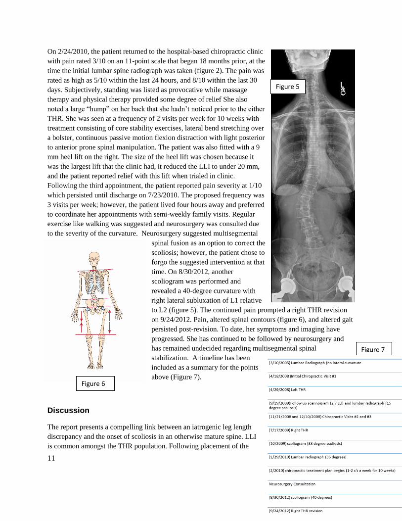
On 2/24/2010, the patient returned to the hospital-based chiropractic clinic
with pain rated 3/10 on an 11-point scale that began 18 months prior, at the
time the initial lumbar spine radiograph was taken (figure 2). The pain was
rated as high as 5/10 within the last 24 hours, and 8/10 within the last 30
days. Subjectively, standing was listed as provocative while massage
therapy and physical therapy provided some degree of relief She also
noted a large “hump” on her back that she hadn’t noticed prior to the either
THR. She was seen at a frequency of 2 visits per week for 10 weeks with
treatment consisting of core stability exercises, lateral bend stretching over
a bolster, continuous passive motion flexion distraction with light posterior
to anterior prone spinal manipulation. The patient was also fitted with a 9
mm heel lift on the right. The size of the heel lift was chosen because it
was the largest lift that the clinic had, it reduced the LLI to under 20 mm,
and the patient reported relief with this lift when trialed in clinic.
Following the third appointment, the patient reported pain severity at 1/10
which persisted until discharge on 7/23/2010. The proposed frequency was
3 visits per week; however, the patient lived four hours away and preferred
to coordinate her appointments with semi-weekly family visits. Regular
exercise like walking was suggested and neurosurgery was consulted due
to the severity of the curvature. Neurosurgery suggested multisegmental
spinal fusion as an option to correct the
scoliosis; however, the patient chose to
forgo the suggested intervention at that
time. On 8/30/2012, another
scoliogram was performed and
revealed a 40-degree curvature with
right lateral subluxation of L1 relative
to L2 (figure 5). The continued pain prompted a right THR revision
on 9/24/2012. Pain, altered spinal contours (figure 6), and altered gait
persisted post-revision. To date, her symptoms and imaging have
progressed. She has continued to be followed by neurosurgery and
has remained undecided regarding multisegmental spinal
stabilization. A timeline has been
included as a summary for the points
above (Figure 7).
Discussion
The report presents a compelling link between an iatrogenic leg length
discrepancy and the onset of scoliosis in an otherwise mature spine. LLI
is common amongst the THR population. Following placement of the
Figure 5
Figure 6
Figure 7

Journal of the Academy of Chiropractic Orthopedists
Volume 13, Issue 1
12
implant, if the surgeon feels that the implant is too loose, a larger implant will be placed to prevent
dislocation, which adds length to that lower extremity (1). While no literature exists to date correlating
post-THR LLI to scoliosis, the European Spine Journal released a review of adult scoliosis, which
identified LLI and pelvic obliquity as one of the 4 major possible causes of scoliosis (8). In addition,
Giles et al have provided evidence that LLI and pelvic torsion result in asymmetry of lumbosacral facet
joint angles, postural scoliosis, concavities in the vertebral body end-plates, wedging of the fifth lumbar
vertebra, and traction spurs (13,14). One reason this correlation may not be apparent in the literature is the
inherent difficulty in quantifying LLI. Full spine standing A-P radiographs are commonly used to initially
assess LLI; however, the CT scanogram is the most reliable and accurate technique because of its ability
to take into account soft tissue contractures (15). If physicians suspect that their patient has a LLI,
especially post-surgically, the CT scanogram would be the most appropriate method for quantification.
Historically, most of the studies examining this topic were performed before the CT scanogram was
identified as the method of choice for evaluating LLI; therefore, the amount of LLI may be over or
underestimated. Within the timeline, the patient had only reported to the chiropractic clinic for one visit
prior to the initial lumbar radiograph and lateral curvature measurement of 15-degrees. In addition, the
patient was only seen two more times before another radiograph illustrated a 35-degree lateral curvature.
The lateral curvature is much more likely to have been a result of LLI (8) or degenerative changes (8) as
there is no evidence to suggest that manipulation is a cause of scoliosis. As for other treatments, the
progression was rapid and earlier post-surgical follow up with the orthopedist may have resulted in
prompter recognition of the LLI. Early recognition could have resulted in early intervention including
corrective orthoses. It is worth noting that the literature does support initial treatment with a heel lift half
of the size of the measured LLI (16). It is possible that the combination of early detection and correction
may have slowed progression of the scoliosis.
Our case demonstrates a relationship between LLI following THR surgery and progression of scoliosis
using acceptable standards of measurement (11, 13). We acknowledge that causes other than surgery may
explain this particular acquired scoliosis. It is our opinion that the pain and dysfunction of degenerative
hips, psoas inhibition or hypertonicity, unrecognized organic disease, or several other causes could
potentially cause or contribute to this condition. It should be noted that the limitations related to this case
include a lack of scoliogram or lumbar radiograph immediately prior to the initial THR, so it is unclear
whether the LLI existed and, if so, to what degree, prior to the THR.
Conclusion
Leg length inequality can contribute to scoliosis (8) and total hip replacement frequently results in leg
length inequality (1). It is our opinion that this case demonstrates the potential for hip replacement
surgery to inadvertently cause or worsen a functional scoliosis. This case contributes to the body of
evidence regarding post-surgical structural changes and is directed toward healthcare professionals who
may treat musculoskeletal spinal disorders post-THR.
Funding Source and Potential Conflicts of Interest: No funding sources or conflicts of interest were
reported for this case report.
References

13
(1) Clark CR, Huddleston HD, Schoch III EP, Thomas BJ. Leg length discrepancy after total hip
arthroplasty. J Am Acad Orthop Sur. 2006;14(1):38-45. 3 4
(2) Ulf L. The Danish hip arthroplasty register. Acta Orthop Scan. 2000;71:433-9.
(3) Wylde V, Whitehouse SL. Prevalence and functional impact of patient-perceived leg length
discrepancy after hip replacement. Int Orthop (SICOT) 8 2009;33:905-9. 9
(4) Maloney WJ, Keeney JA. Leg length discrepancy after total hip arthroplasty. J Arthroplasty
2004;19:108-10.
(5) Ahmad R, Sharma V, Sandu H, Bishay M. Leg length discrepancy in total hip arthroplasty with the
use of cemented and non-cemented femoral stems. A prospective radiological study. Hip Int
2009;19:264-7.
(6) Ranawat CS, Rodriguez JA. Functional leg-length inequality following total hip arthroplasty. J
Arthroplasty 1997; 12: 359-64.
(7) Herndon JH. One more turn of the wrench. J Bone Joint Surg Am 21 2003;85:2036-8.
(8) Aebi M. The adult scoliosis. Eur Spine J 2005;14:925-948.
(9) D’amico M, Ciarrocca F, Liscio G, Seranfini P, Tommasini M, Vallasciani M. Balance lower limb
loads and 3D spine modifications after total hip joint replacement: effects of leg length discrepancy
correction. Stud Health Technol Inform 2006;123:409-14.
(10) White TO, Dougall TW. Arthroplasty of the hip. Leg length is not important. J Bone Joint Surg Br.
2002 Apr;84(3):335-8.
(11) Betsch M, Rapp W, Przibylla A, et al. Determination of the amount of leg length inequality that
alters spinal posture in healthy subjects using rasterstereography. Eur Spine J. 2013 Mar 13. [Epub ahead
of print]
(12) Raczkowski, J. W., Daniszewska, B., & Zolynski, K. Functional scoliosis caused by leg length
discrepancy. Archives of Medical Science 2010; 6(3), 2 393-398)
(13) Giles LGF. Lumbosacral facetal 'joint angles' associated with leg length inequality. Rheumatology
and Rehabilitation. 1981;20:233–238. 6
(14) Giles LGF, Taylor JR. Lumbar spine structural changes associated with leg length inequality. Spine.
1982;7:159–162.
(15) Sabharwal S, Kumar A. Methods for assessing leg length discrepancy. Clin Orthop Relat Res 2008
Oct;466:2910-22
(16) Baylis W.J., Rzonca E.C. Functional and structural limb length discrepancies: evaluation and
treatment. Clin Podiatr Med Surg. 1988;5(3):509–520.
Journal of the Academy of Chiropractic Orthopedists
Volume 13, Issue 1
14
Abstracts & Literature Review
Incidence of Neuralgic Amyotrophy
(Parsonage Turner Syndrome) in a Primary Care Setting - A Prospective Cohort Study
Nens van Alfen, Jeroen J. J. van Eijk, Tessa Ennik, Sean O. Flynn, Inge E.
G. Nobacht, Jan T. Groothuis, Sigrid Pillen, Floris A. van de Laar PLoS ONE. 2015;10(5):
e0128361. doi:10.1371/journal.pone.0128361; May 27. 2015
JACO Editorial Reviewer: Steven G. Yeomans, DC, FACO, DABCC
Published: September 2016
Journal of the Academy of Chiropractic Orthopedists
September 2016, Volume 13, Issue 1
The original article copyright belongs to the original publisher. This review is available from: http://www.dcorthoacademy.com ©
2015 Yeomans and the Academy of Chiropractic Orthopedists. This is an Open Access article which permits unrestricted use,
distribution, and reproduction in any medium, provided the original work is properly cited.
Authors’ Abstract: Objective: Neuralgic amyotrophy is considered a rare peripheral nervous system disorder but in
practice seems grossly under recognized, which negatively affects care for these patients. In this
study we prospectively counted the one-year incidence rate of classic neuralgic amyotrophy in a
primary care setting.
Methods: In a prospective cohort study during the year 2012 we registered all new cases of
neck, shoulder, or arm complaints from two large primary care centers serving a population of 14,118. Prior to study, general practitioners received a short training on how to diagnose classic
neuralgic amyotrophy. Neuralgic amyotrophy was defined according to published criteria
irrespective of family history. Only patients with a classic phenotype were counted as definite
cases. After inclusion, patients with suspected neuralgic amyotrophy who had not yet seen a
neurologist were offered neurologic evaluation for diagnostic confirmation.
Results: Of the 492 patients identified with new onset neck, shoulder or arm complaints, 34
were suspected of having neuralgic amyotrophy. After neurologic evaluation the diagnosis was
confirmed in 14 patients. This amounts to a one-year incidence rate for classic neuralgic
amyotrophy of 1 per 1000.
Conclusions: Our findings suggest that neuralgic amyotrophy is 30-50 times more common
than previously thought. Unawareness of the disorder and its clinical presentation seems the most

15
likely explanation for this difference. An incidence rate of 1 per 1000 and the long-term sequelae
many patients suffer warrant more vigilance in diagnosing the disorder, to pave the way for
timely treatment and prevent complications. Clinical Relevance This study suggests that neuralgic amyotrophy is much more common than that previously
thought/reported and should be considered when patients present with radiating neck pain to the
upper extremity.
JACO Editorial Summary
This article was written by authors primarily from the Netherlands (where the study took
place) with one author from Ireland.
Neuralgic amyotrophy or NA (a.k.a. Parsonage Turner syndrome or brachial plexus
neuritis) is a distinct type of peripheral neuropathy, with one or more episodes of acute,
severe pain in the upper extremity which is quickly followed by multifocal paresis with a
slow 1-2 year recovery time of which a large subset become disabled (estimated at 25%
of NA cases in this study).
Diagnostic difficulty is common (about 30% of the cases) due to the high variance in
clinical presentations such as painless episodes and lower brachial plexus, or other
peripheral nerve involvement.
In about 70%, the “classic presentation” is: acute shoulder region pain followed by
scapular winging, weakness in shoulder abduction, external rotation, grip/pinch strength,
and forearm pronation.
The cause is thought to be autoimmune but difficulty proving this has occurred most
commonly due to delay in diagnosis (median time: 11 weeks) and hence, difficulty
randomizing subjects to corticosteroids or IV gammaglobuline groups.
o A reference listed in the study identifies “idiopathic” and a “hereditary form.”
(https://www.radboudumc.nl/Informatiefolders/7130-
Neuralgic_Amyotrophy__id-i.pdf)
This study changes the prior incidence rate estimate from 1-3/100,000 per year to 1 in
1000 (30-50 times more frequent for the “classic presentation.”
History criteria: 1) New / subacute onset of uni- or bilateral shoulder pain; 2) NRS pain
score of ≥7/10; 3) History of weakness with abnormal shoulder ROM (abduction, ER,
FFL); 4) When ≥3 weeks post-onset: Paresis of the long thoracic, suprascapular, and
anterior interosseous nerves; 5) Slow recovery.
Physical exam criteria: 1) Scapular winging/dyskinesia; 2) Signs of muscle atrophy; 3)
Weakness of shoulder abduction, external rotation, serratus anterior, forearm pronation
and/or pinch strength.
Probable reasons for “missing” this diagnosis: 1) Diagnostic unfamiliarity (many
physicians simply do not know about it); 2) Given it’s rarity, it’s simply not considered as
a differential diagnosis; 3) Many physicians don’t look at scapular stability, don’t

Journal of the Academy of Chiropractic Orthopedists
Volume 13, Issue 1
16
perform a detailed neurological exam of all the upper extremity muscles, and/or don’t
“tie” the findings of NA together.
The ratio of classic NA to that of other disorders in this study was 3% or, 1 in every 33
patients with a new onset of neck, shoulder, or arm complaints in a primary care setting
will have NA (this compares to 47% or 1 in 2 patients that were diagnosed with shoulder
pathology; 15x more common).
An incidence rate of 1 per 1000 per year suggests NA is NOT “rare” which is defined as
1 per 2000 per year. This equates to 17,000 new cases per year in the Netherlands.
Table 4: Points that lead to a correct diagnosis
o Any patient with 1) acute onset, 2) severe pain (≥7/10), 3) Analgesic resistant
shoulder &/or upper arm pain
o Pain worse at night and still severe at rest
o Multifocal motor/sensory losses that can be bilateral but asymmetrical
o PE: Remove clothing – inspect/palpate shoulder/arms for scapular asymmetry and
muscle atrophy
o Look for scapular motion dyskinesia during slow abduction, FFL (video clips
available)
o Test/compare bilaterally the strength of the serratus anterior, shoulder external
rotation, long thumb and index finger flexors (pinch), forearm pronation: Any
weakness found in combination is suspect for NA and rare in other disorders with
similar presentations
Periscapular weakness and a pain score of ≥7/10 are needed as “minimums.”
Training of primary care physicians (PCP’s) in two, 1-hour teaching sessions followed by
a simple diagnostic protocol led to a 3-fold increase of identifying NA (confirmed by an
experienced neurologist).
Limitations in the study include: 1) All PCP’s involved were aware of the study goal
(which can lead to bias); 2) They did not systematically refer every patient with suspected
NA for neurological examination &/or ancillary tests (EMG, cervical MRI)
o There is no “gold standard” test to confirm NA; the diagnosis is made by adhering
to clinical criteria (history/examination).
Recommendation: Every PCP be given a short educational program on NA, confirm
suspected cases with a neurologist, in order to pave the way for acute phase
immunomodulating therapy trials to prevent long-term complications
Summary
The results of this investigation should raise awareness of all health care providers that
treat neuromusculoskeletal disorders that: 1) NA is not rare; 2) Diagnosis is clinical, NOT reliant
on expensive diagnostic tests; 3) A prompt diagnosis can help to avoid the common (25%
incidence) long-term disability associated with NA, i.e., to improve clinical outcomes.
17
Abstracts & Literature Review
Carpal Tunnel Syndrome: Hand Surgeons, Hand Therapists, and
Physical Medicine and Rehabilitation Physicians Agree on a
Multidisciplinary Treatment Guideline – Results: From the European
HANDGUIDE Study
Bionka M. Huisstede, PhD, Jan Fride´n, MD, PhD, J. Henk Coert, MD, PhD, Peter Hoogvliet, MD, PhD,
European HANDGUIDE Group
JACO Editorial Reviewer Dale G. Huntington, D.C., F.A.C.O.
Published: September 2016
Journal of the Academy of Chiropractic Orthopedists
September 2016, Volume 13, Issue 1
The original article copyright belongs to the original publisher. This review is available from: http://www.dcorthoacademy.com ©
2015 Huntington and the Academy of Chiropractic Orthopedists. This is an Open Access article which permits unrestricted use,
distribution, and reproduction in any medium, provided the original work is properly cited.
Author’s Abstract
Objective: To achieve consensus on a multidisciplinary treatment guideline for carpal tunnel
syndrome (CTS).
Design: Delphi consensus strategy.
Setting: Systematic reviews reporting on the effectiveness of surgical and nonsurgical
interventions were conducted and used as an evidence based starting point for a European Delphi
consensus strategy.
Participants: In total, 35 experts (hand surgeons selected from the Federation of European
Societies for Surgery of the Hand, hand therapists selected from the European Federation of
Societies for Hand Therapy, physical medicine and rehabilitation physicians) participated in the
Delphi consensus strategy.

Journal of the Academy of Chiropractic Orthopedists
Volume 13, Issue 1
18
Interventions: Not applicable.
Main Outcome Measures: Each Delphi round consisted of a questionnaire, analysis, and
feedback report.
Results: After 3 Delphi rounds, consensus was achieved on the description, symptoms, and
diagnosis of CTS. The experts agreed that patients with CTS should always be instructed, and
instructions combined with splinting, corticosteroid injection, corticosteroid injections plus
splinting, and surgery are suitable treatments for CTS. Relevant details for the use of
instructions, splinting, corticosteroid injections, and surgery were described. Main factors for
selecting one of the aforementioned treatment options were identified as follows: severity and
duration of the disorder and previous treatments received. A relation between the
severity/duration and choice of therapy was found by the experts and reported in the guideline.
Conclusions: This multidisciplinary treatment guideline may help physicians and allied health
care professionals to provide patients with CTS with the most effective and efficient treatment
available.
Comment
The complex movements and tactile sensation of the hand are essential for completing everyday
tasks. Consequently, hand disorders affecting these qualities have a significant impact on
activities of daily living. Of those with chronic non-traumatic complaints of the arm, neck and/or
shoulder, 29% reported complaints in the wrist/hand area. The most prevalent non-traumatic
hand disorder is carpal tunnel syndrome (CTS).
Although the exact causative mechanism of CTS is unknown, it is safe to state that CTS is
related to an increased pressure within the carpal tunnel, resulting in mechanical compression
and local ischemia – mediated damage to the median nerve. The occurrence of CTS can be
associated with work-related factors. The prevalence of CTS is reported to be 0.6% in men and
5.8% in women in the general population and 1 in 5 in symptomatic subjects.
Interventions used to treat CTS vary from splinting to exercise therapy and from ultrasound to all
kinds of surgical interventions. Ideally, a treatment guideline for CTS is based on systematic
reviews describing the long-term effects of all aspects relevant for the diagnosis and treatment of
the disorder. However, systematic reviews on the treatment of CTS mainly describe short-term
and mid-term effects and focus on the global picture of a treatment (eg, splinting, corticosteroid
injections, open surgery), without taking into account relevant details (eg, type of splint; when to
wear it; type of corticosteroid; number of injections; types of anesthesia, incision, and stitches).
Because such details can have significant consequences, a Delphi consensus strategy was
conducted to develop a treatment guideline for CTS. Development of evidence-based protocols
and treatment guidelines can aid in optimizing care for hand disorders. Therefore, in Europe, the
HANDGUIDE study was initiated with the goal to create multidisciplinary consensus on

19
treatment guidelines for non-traumatic hand disorders: trigger finger, De Quervain disease,
Dupuytren disease, CTS, and Guyon canal syndrome. In a Delphi consensus strategy, a series of
sequential questionnaires (or rounds) are presented to a panel of experts, interspersed with
controlled feedback, with the aim to achieve consensus of opinion among these experts. This
article reports on the results for CTS.
Methods:
Steering committee and advisory team: A steering committee consisting of a hand surgeon (with
a PhD), physical medicine and rehabilitation (PM&R) physician (with a PhD), and
physiotherapist (with a PhD) was composed to initiate and guide the HANDGUIDE Study. All 3
members of the steering committee have a clinical and scientific and/or epidemiologic
background. They designed the questionnaires, analyzed the responses, and formulated the
feedback reports. Further, an advisory team (consisting of 2 professors of hand surgery, 1
professor of PM&R, and 1 hand therapist with a PhD) was formed that received regular updates
on the progress of the HANDGUIDE Study. This team could be consulted by the steering
committee if necessary and could give the steering committee their opinions and advice as they
saw fit.
Preparation of the study:
Evidence for effectiveness of interventions of CTS to establish an evidence-based starting point
for this study, systematic reviews were conducted reporting on the evidence for the effectiveness
of nonsurgical, surgical, and postsurgical interventions to treat CTS.
Selection of experts:
The Federation of European Societies for Surgery of the Hand (FESSH) and European
Federation of Societies for Hand Therapy (EFSHT) endorsed this study. The national member
associations of the FESSH and EFSHT selected the experts in their respective field. Each
national member association was invited to select a maximum of 3 representative experts per
Delphi consensus strategy. In addition, some European PM&R physicians who specialize in hand
rehabilitation were invited to participate in this study.
Delphi consensus strategy on CTS:
Description, symptoms, and diagnosis of CTS First-round questionnaire: In the first round,
literature-based concepts for a short description of CTS, its symptoms, its diagnosis, and its
nonsurgical and surgical treatment were presented to the experts. Subsequently, the experts were
asked whether they agreed (yes/no/no opinion) with the aforementioned concepts followed by
the request to explain their answer (please explain your answer). This allowed the experts at any

Journal of the Academy of Chiropractic Orthopedists
Volume 13, Issue 1
20
time to object or suggest alterations to any of this steering committee’s suggestions regarding the
aforementioned items.
Results
Expert panel: A total of 112 experts (52 hand surgeons, 47 hand therapists, 13 PM&R
physicians) from 17 European countries were selected to participate in 1 of the 3 Delphi
consensus strategies of the HANDGUIDE Study, which was performed between June 2009 and
December 2012.
For the Delphi consensus strategy on CTS, 36 experts were selected (18 hand surgeons, 13 hand
therapists, 5 PM&R physicians). Of these, 1 expert did not finish any of the questionnaires.
Response rates of the remaining 35 experts for rounds 1 to 3 were 89%, 94%, and 89%,
respectively.
Conclusions
European experts (hand surgeons, hand therapists, PM&R physicians) achieved multidisciplinary
consensus on a treatment guideline for CTS. This guideline may help and guide physicians and
allied health care professionals to provide patients suffering from CTS with the most effective
and efficient treatment available.
JACO Editorial Summary:
This clinical research article was written by the authors; Bionka M. Huisstede, PhD, Jan
Friden, MD, PhD, J. Henk Coert, MD, PhD, Peter Hoogvliet, MD PhD, of the “European
Handguide Study”.
The purpose of this 2.5 years, 17 country study was to determine the most effective ways
of treating CTS via nonsurgical, surgical, and postsurgical interventions which is an
evidence based starting point for the first European Delphi consensus strategy.
This was a comprehensive study that was closely monitored to obtain the most reliable
results by the hand experts to assimilate the documentation from all the Delphi groups to
determine multidisciplinary results in the 3 separate interventions.
The diagnosis of CTS is primarily based on the clinical picture. The sensitivity of special
tests such as, (Phalen test and Tinel sign) are reported to be 34% to 59% and 51% to 93%
and 25% to 41% and 66% to 91% respectively. These tests are of limited value which
brings to point the importance of electro - diagnostic testing. There are reports of results
that do not clearly demonstrate whether the origin is at the wrist, cervical spine
radiculopathy or a mixed result.
Of those who reported non-traumatic neck and upper extremity pain 29% of the
complaints were in the wrist/hand area. The most prevalent non-traumatic hand disorder
was carpal tunnel. In addition the study revealed the prevalence of CTS reported to be
0.6% in men and 5.8% in women. This mere fact could be a study of its own.

21
The use of the cold laser has been utilized with some degree of effectiveness in North
America over the past 10 plus years as a primary or adjunctive therapy procedure in the
fields of the interventions that have been discussed in this clinical research model. To my
knowledge there has not been a controlled clinical study performed or an article written
in an indexed medical journal on that topic.
Summary:
The authors of this study are to be commended for their efforts in this comprehensive clinical
investigation that spanned over a period of two and one half years, seventeen countries, and 112
experts guiding this multidisciplinary study to a consensus of opinion. It is hoped that this will
launch other studies of its kind in the future.
Journal of the Academy of Chiropractic Orthopedists
Volume 13, Issue 1
22
Abstracts & Literature Review
Radial Tunnel Syndrome, Diagnostic and Treatment Dilemma
Ali Moradi, MD; Mohammad H Ebrahimzadeh, MD; Jess B Jupiter, MD Research performed at Hand and Upper Extremity Service, Massachusetts General Hospital, Harvard
Medical School, Boston, MA, USA
JACO Editorial Reviewer J. Chris Romney DC FACO
Published: September 2016
Journal of the Academy of Chiropractic Orthopedists
September 2016, Volume 13, Issue 1
The original article copyright belongs to the original publisher. This review is available from: http://www.dcorthoacademy.com ©
2015 Romney and the Academy of Chiropractic Orthopedists. This is an Open Access article which permits unrestricted use,
distribution, and reproduction in any medium, provided the original work is properly cited.
Author’s Abstract
Background: It is evident that radial tunnel syndrome should be considered in the diagnosis of
lateral elbow and dorsal forearm pain that may radiate to the wrist and dorsum of fingers.
Clinical examination of the elbow has been found to be the best method for the diagnosis of
radial tunnel syndrome. The paraclincal study options such as, electrodiagnostics, imaging
studies, or diagnostic ultrasound are utilized to rule out other pathologies to differentiate the
several conditions of lateral elbow pain. The most valuable examination is at the direct site of
pain and can be best identified by rule of nine test and detecting weakness of the third finger and
wrist extension. Treatment of radial tunnel syndrome should utilize various conservative non-
surgical efforts before undergoing surgical intervention. Methods: The study identifies the prevalence of compression of the superficial radial nerve is
at 0.003%, in comparison to carpal tunnel syndrome which has an annual incidence between
0.1% and 0.35%. The entrapment of the radial nerve and its deep branch has been found to occur
at five different sites within the radial tunnel. The study by Bolster reported 5 out of 12 patients
with the diagnosis of RTS had previous surgical intervention on the ipsilateral upper extremity
such as, trigger finger, CTS, and shoulder instability. Studies also indicate that patients with RTS
disease is more prevalent in women age 30-50 years, significantly right hand dominant with
bilateral involvement being rare.
Results: RTS is relatively uncommon but has distinct signs and symptoms that include
localized tenderness over the radial nerve 5cm distal to the lateral epicondyle. Patients report

23
aggravated pain at night that disturbs sleep and can become more severe when increased traction
is applied to the nerve by extending, flexing, or pronating the elbow. There are two accepted
clinical tests to confirm the diagnosis. although X-ray, MRI, and EMG have not been found to
play a key role in RTS diagnosis.
Conclusion: Because of the limited number of confirmatory diagnostic tests, RTS is diagnosed
by exclusion and is dependent on clinical signs and symptoms. Common non surgical methods
are recommended, however, the success rate is in doubt. Steven at al report shows only 4 out of
15 patients with the diagnosis of RTS had improvement with conservative treatment. Surgical
treatment was found to result in 93% success rate. RTS is a disease that should be consider in the
differential diagnosis of lateral elbow pain. JACO Editorial Summary
Clinical Relevance: Lateral elbow pain is commonly diagnosed as lateral epicondylitis, when
in some cases a closer look can identify the evidence of radial tunnel syndrome. This study
includes an allopathic conservative care regimen with little success. The chiropractic physician
can differentiate the diagnosis of lateral elbow pain by utilizing the proper historical and
diagnostic techniques. Once the doctor has arrived at the diagnosis of RTS, the treatment plan
might include the following: manipulation of the spinal segments of the cervical and thoracic
spine, deep tissue massage , ultrasound modality, exercise rehabilitation, and manipulation of
the radial head. If these efforts do not accomplish the preferred outcome then prolozone therapy
or steroid injection may be a viable option prior to referral for surgical intervention.

Journal of the Academy of Chiropractic Orthopedists
Volume 13, Issue 1
24
Radiology Corner
Case Presentation: 64 year old female with right shoulder pain
Alicia M. Yochum RN, DC, DACBR
Chesterfield, MO [email protected]
Published: September 2016
Journal of the Academy of Chiropractic Orthopedists
September 2016, Volume 13, Issue 1
This is an Open Access article which permits unrestricted use, distribution, and reproduction in any medium, provided the original
work is properly cited. The article copyright belongs to the author and the Academy of Chiropractic Orthopedists and is available at:
http://www.dcorthoacademy.com. © 2015 Yochum and the Academy of Chiropractic Orthopedists.
A 64 year old female presented for chiropractic care with anterior right shoulder pain.
She is retired but continues to participate in shot put, javelin, and the hammer throw which
exacerbates her pain. She had decreased range of motion with pain in abduction, internal
rotation, and flexion. Orthopedic testing revealed pain with supraspinatus press test.
Neurological and physical examination was negative.
Fig 1: Normal diagnostic ultrasonography of the rotator cuff. A- Longitudinal view which compares to the coronal magnetic
resonance imaging (MRI) perspective. B- Transverse view which compares to the sagittal MRI perspective.

25
Fig 2: A- Coronal T2 fat saturated MRI depicting a full thickness tear of the supraspinatus (arrows). There is also evidence of a
joint effusion (arrowhead). B- Longitudinal diagnostic ultrasound in the same patient showing the focal tear of the supraspinatus.
Fig 3: A- Sagittal T2 fat saturated MRI demonstrating a full thickness tear of the supraspinatus tendon with partial thickness
tearing as well. B- Transverse diagnostic ultrasound in the same patient showing the focal tearing of the supraspinatus.
Legend: Arrows all point to the area of tearing in the supraspinatus tendon
* - Biceps tendon (long head)
GT- Greater tuberosity
SST- Supraspinatus tendon
IST- Infraspinatus tendon
The shoulder is the most movable joint in the body and prone to dysfunction and injury.
Rotator cuff tears are the most common cause of shoulder pain and dysfunction in adults. They
are commonly related to overuse and degeneration within the tendon. In patients over 66 years

Journal of the Academy of Chiropractic Orthopedists
Volume 13, Issue 1
26
old who present with a symptomatic rotator cuff tear, 50% of those patients will have an
asymptomatic tear on the contralateral side as well.
Rotator cuff tears can be categorized into partial or complete (full thickness). A complete
tear extends through the entire thickness of the tendon and creates a communication between the
subacromial space and the glenohumeral joint. This will allow fluid to be visualized superficial
to the cuff tendons at the greater tuberosity and can be helpful when determining if a tear is
partial or full thickness. A complete tear may allow retraction of the tendon and could create a
wavy appearance to the tendon called the “cuff wave sign.” If this is present, it is associated with
easier reattachment of the tissue on surgical repair [1].
Partial thickness tears are divided into articular sided, bursal sided or intrasubstance.
They are twice as common as complete tears. Articular sided are adjacent to the humeral
cartilage while bursal sides are adjacent to the subacromial bursa. Intrasubstance tears are within
the center of the tendon. Bursal sided and intrasubstance tears may be hidden during arthroscopic
examination therefore imaging evaluation is important for accurate diagnosis [1].
In this case presentation, the coronal MRI depicts fluid (high/white signal) filling the
region of the anterior rotator cuff where tendon should be visualized (Fig 2a- arrow). The
corresponding longitudinal ultrasound image depicts the fluid within the rotator cuff as black
region known as hypoechogenicity (Fig 2b-arrow). The normal appearance of the rotator cuff is
demonstrated in figure 1a without focal areas of hypoechogenicity to indicate tearing.
The sagittal MRI demonstrates fluid communicating between the glenohumeral joint and
the subacromial space through a focal fluid filled rotator cuff tear within the supraspinatus
tendon (Fig 3a). Note the proximity to the biceps tendon (*). The correlating transverse
ultrasound image depicts the focal fluid collection as the focal hypoechogenicity within the
tendon also near the biceps tendon (Fig 3b). A normal transverse ultrasound image is provided
for comparison (Fig 1b).
The sensitivity of diagnostic ultrasound for full thickness rotator cuff tears is equal to
MRI (>90%). The sensitivity of MRI and ultrasound for partial thickness tears is equivalent (67-
68%) but an MRI arthrogram where contrast is injected into the joint is the most sensitive (83%).
The specificity of MRI and ultrasound for full thickness and partial thickness tears is >93%.
Musculoskeletal ultrasound allows the examiner to perform dynamic maneuvers such as
orthopedic tests while imaging the anatomy which is very beneficial to help localize
abnormalities [2].
Diagnostic musculoskeletal ultrasonography is a growing diagnostic tool that is cost
effective and noninvasive. It is user dependent and requires significant training but can be very
useful as an imaging modality when utilized appropriately.
27
Case courtesy of Logan University
References
1. Stoller, D.:Rotator cuff Tears, Microinstability, Rotator Interval/Biceps Pulley, and the
Throwing Shoulder. In Stoller’s Orthopaedics and Sports Medicine: The Shoulder.
Wolters Kluwer. Philadelphia, PA; 2015: 237-471
2. Roy J-S, et al.: Diagnostic accuracy of ultrasonography, MRI and MR arthrography
in the characterization of rotator cuff disorders: a systematic review and meta-
analysis. British Journal of Sports Medicine. 2015, 49:1316-1328
Ortho Quiz
by Steven L. Kleinfield D.C.,F.A.C.O.
1) Avascular necrosis of the tarsal navicular is better known as:
a. Kohler’s disease
b. Blount’s disease
c. Freiberg’s disease
d. Sinding-Larsen-Johansson’s Disease
e. Keinbock’s disease
2) “A 4-year-old girl presented with intermittent right foot pain for 1 week. Pain had
worsened the previous day after playing outside, and she was now refusing to bear weight
on the right foot. On examination, she had pain and tenderness over her right dorsomedial
midfoot with no local skin changes. She walked with an antalgic gait with weight bearing
on the lateral side of the foot. Her right foot radiograph showed a collapsed, flat, and
radiodense navicular bone”.
Your best working diagnosis would be:
a. Kohler’s disease
b. Blount’s disease
c. Freiberg’s disease
d. Sinding-Larsen-Johansson’s Disease
e. Keinbock’s disease

Journal of the Academy of Chiropractic Orthopedists
Volume 13, Issue 1
28
3) Which ligament listed below does not belong as being part of the conjoined Deltoid
Ligament
a. Posterior talofibular
b. Tibiocalcaneal
c. Tibionavicular
d. Anterior tibiotalar
e. Posterior tibiotalar
4) According to the work of Nachemson and his co-workers, he noted that different postures
can affect the intra-discal pressure. Which position was found to be the most deleterious
to the disc by having the highest increase in intra-discal pressure:
a. Coughing or straining
b. Small jumps
c. Lifting a weight with the back and knees bent
d. Rotation of the lumbar spine
e. Lifting a weight with the back bent and the knees straight
5) A non-union of the secondary growth center of the inferior articular process of a
vertebrae is better known as:
a) Bertolotti’s Syndrome
b) Ossicle of Oppenheimer’s
c) Tropism
d) Opisthion
e) Lisfranc’s Joint

29
Current Events
Part I examination dates are now closed for 2016.
The Academy of Chiropractic Orthopedists has set its Part II Diplomate examination at
Northwestern Health Sciences University (NWHSU) in Bloomington, Minnesota. The
date is October 1, 2016. Part I must be completed before the candidate is eligible to sit
Part II. Contact the Academy's executive director Dr. Jerry Wildenauer to obtain the
necessary information.
The Academy of Chiropractic Orthopedists announces the on-line Part I examination
dates will be May 19, 2017 and July 20, 2017.
Information about sitting the Board is available from the Executive Director Dr. Jerry
Wildenauer.
Jerrold R Wildenauer DC, FACO
1859 Warrior Drive
Mendota Heights, MN 55118
TEL: 612-454-1472
FAX: 651-846-5590
E-mail: [email protected]
Answers to Ortho Quiz
1. a. Kohler’s disease http://www.ncbi.nlm.nih.gov/pmc/articles/PMC3298227/
2. a. Kohler’s disease http://www.ncbi.nlm.nih.gov/pmc/articles/PMC3298227/
3. a. Posterior talofibular Orthopedic Physical Assessment 5th edition page 844 David J. Magee
4. e. Lifting a weight with the back bent and the knees straight
Orthopedic Physical Assessment 5th
edition page 514 David J. Magee
5. b. Ossicle of Oppenheimer’s Clinical Imaging by Dennis Marchiori 2nd edition page 410