j. newell stannard and the university of rochester
TRANSCRIPT
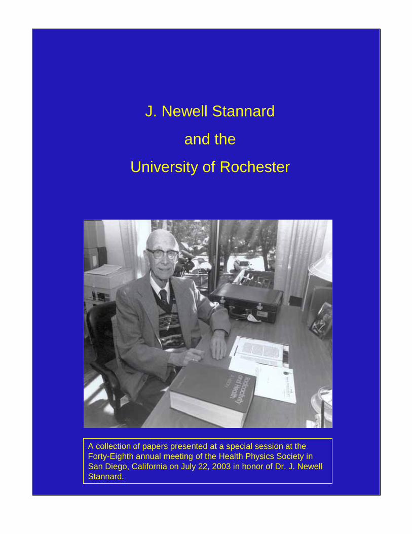
J. Newell Stannard
and the
University of Rochester
A collection of papers presented at a special session at the Forty-Eighth annual meeting of the Health Physics Society in San Diego, California on July 22, 2003 in honor of Dr. J. NewellStannard.
J. Newell Stannard and the University of Rochester
Contents Prologue and Acknowledgment……………………………………………....................1 Photograph of Participants ……………………………………………………………...3 Introduction ……………………………………………………………………………..5 William Bair Sketch of the University of Rochester Atomic Energy Project ………………………...7 J. Newell Stannard University of Rochester and the Health Physics Opportunity …………………………11 Paul Rohwer Firm Foundations for Understanding Radionuclide Dosimetry and Health Effects ……………………………………………………………………..17 Bruce Boecker Advances in Aerosol Science for Radiation Protection ……………………………….27 Otto Raabe Field Studies of Plutonium and Fission Products in Animals …………………………37 Robert Thomas Radon, Smoking, and Lung Cancer: We’ve Come a Long Way Baby! .........................51 Jan Johnson The Chernobyl Accident: Predictions vs. Reality ……………………………………..59 Marvin Goldman Selected Publications of Dr. J. Newell Stannard ………………………………………63
1
Prologue and Acknowledgement My first graduate student, Bill Bair was invited by the chairman of the Health Physics Society’s History Committee, Sydney Porter, to organize a session at the San Diego Meeting of the Health Physics Society, July 2003, to acknowledge and honor me and the University of Rochester for their many contributions to radiation protection through education and research. In consultation with Bob Thomas, Bill selected speakers to represent the contributions of the educational program at the University of Rochester made to the profession of radiation protection through all of its graduates.
The early sections of Bill Bair’s introduction to the session and of my short sketch of the Rochester Project, also describe, respectively, how the special session came about and my delight and gratitude at the honor bestowed on me. However, circumstances beyond our control forced a change in plans for the disposition of the papers that were presented. It was planned to be a series of papers by some of my students presenting aspects of their career work that began during their graduate years. It was planned to be quite informal like an afternoon of old friends reminiscing on old times and old work.
However, the planners knew that the papers would likely contain data that
had not been previously presented in the open literature. They decided to have the session video-taped and published informally. Arrangements were made, or so we thought, to have the session taped professionally. However on the day before the session we found that no professional was available for the recording so we recruited my son-in-law Jack Frazier and daughter Sue Frazier who fell to and did a yeomen job recording the session.
Unfortunately none of us amateurs knew that to use a personal video to
record in a large meeting room, special equipment was needed, as was amply proven when we viewed the tapes. It was a perfectly good picture but the audio portion was so badly distorted that we could not reconstruct a single paper.
Many solutions were contemplated but it took John Taschner and his
stubborn desire to salvage the session that solutions were eventually arrived at. Both John and I had believed that there were ways to clean up a distorted audiotape and John made a business of investigating it. He even took some special classes on the subject. After digitally enhancing the audio, John transcribed each session then persuaded each speaker to produce a manuscript from the transcriptions he had made. Fortunately two speakers had manuscripts we could use which made the task easier. After the individual manuscripts were completed, Dianne Eppler, Newell’s step-daughter edited, assembled and published the final product.
.
2
I cannot begin to express my admiration and gratitude to those who did so much to provide a detailed and first hand view of this fraction of the Rochester project’s work. Lest there be any confusion this session and their papers detail only a small fraction of the University contributions to basic radiobiology. A very large fraction of the Atomic Energy Project’s and Department’s work was in and belongs to basic biomedical science. The work on aerosol science has the broadest application to general science.
At the close of his paper Bruce Boecker addressed me with the remark “You
have made us what we are today, I hope you’re satisfied” I was not quick enough then to give the obvious positive reply “loud and clear” Instead I will do it here with a resounding “Yes, completely”. However, I must say that these students only needed some opportunities which I helped provide for them. They made themselves. J. Newell Stannard
3
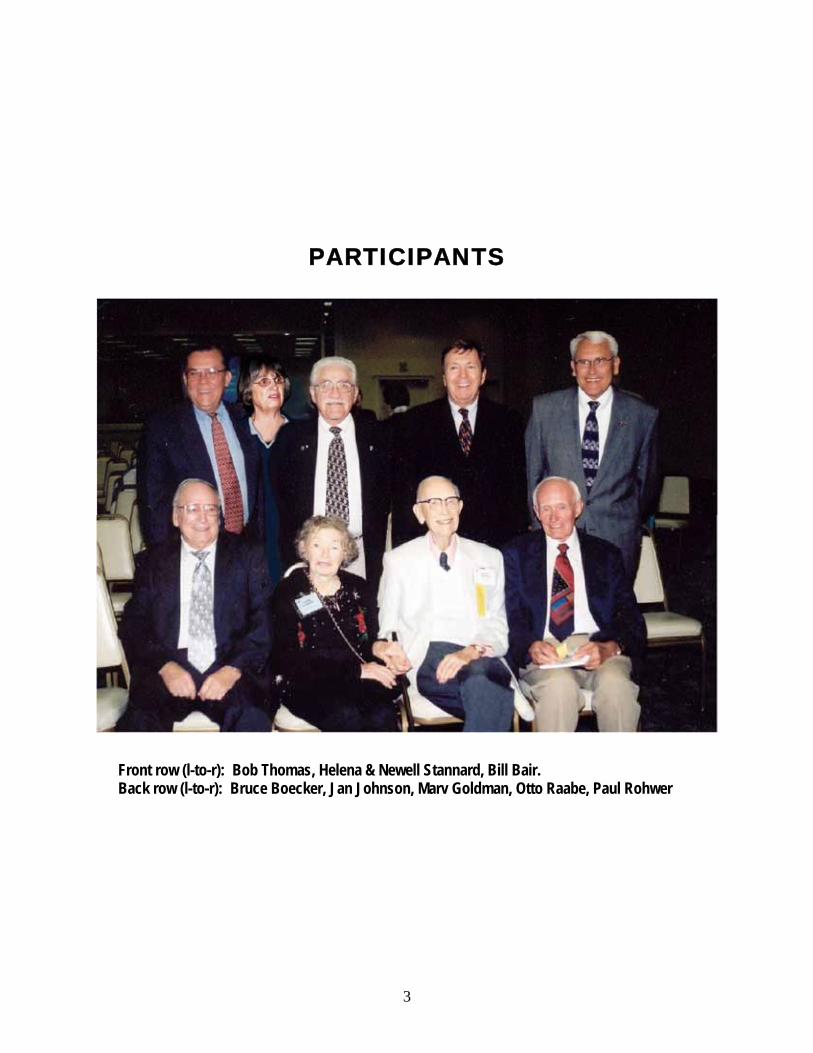
PARTICIPANTS
Front row (l-to-r): Bob Thomas, Helena & Newell Stannard, Bill Bair. Back row (l-to-r): Bruce Boecker, Jan Johnson, Marv Goldman, Otto Raabe, Paul Rohwer

4
5
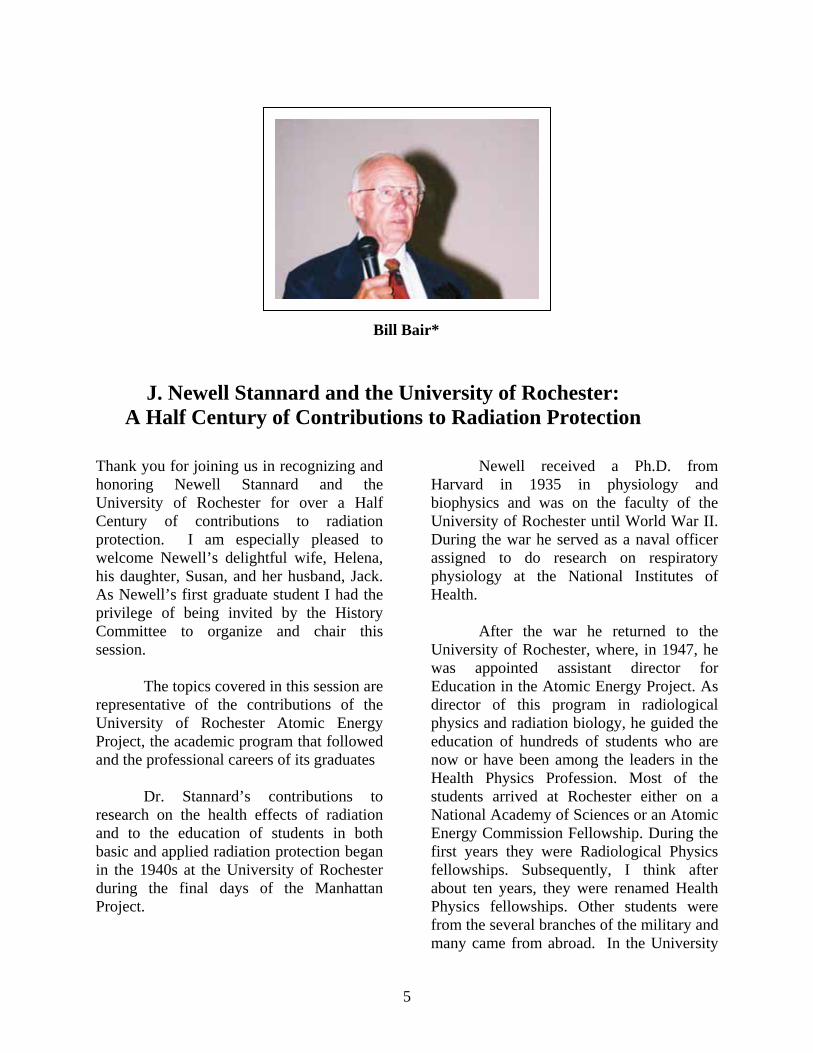
Bill Bair*
J. Newell Stannard and the University of Rochester: A Half Century of Contributions to Radiation Protection
Thank you for joining us in recognizing and honoring Newell Stannard and the University of Rochester for over a Half Century of contributions to radiation protection. I am especially pleased to welcome Newell’s delightful wife, Helena, his daughter, Susan, and her husband, Jack. As Newell’s first graduate student I had the privilege of being invited by the History Committee to organize and chair this session.
The topics covered in this session are representative of the contributions of the University of Rochester Atomic Energy Project, the academic program that followed and the professional careers of its graduates
Dr. Stannard’s contributions to research on the health effects of radiation and to the education of students in both basic and applied radiation protection began in the 1940s at the University of Rochester during the final days of the Manhattan Project.
Newell received a Ph.D. from Harvard in 1935 in physiology and biophysics and was on the faculty of the University of Rochester until World War II. During the war he served as a naval officer assigned to do research on respiratory physiology at the National Institutes of Health.
After the war he returned to the University of Rochester, where, in 1947, he was appointed assistant director for Education in the Atomic Energy Project. As director of this program in radiological physics and radiation biology, he guided the education of hundreds of students who are now or have been among the leaders in the Health Physics Profession. Most of the students arrived at Rochester either on a National Academy of Sciences or an Atomic Energy Commission Fellowship. During the first years they were Radiological Physics fellowships. Subsequently, I think after about ten years, they were renamed Health Physics fellowships. Other students were from the several branches of the military and many came from abroad. In the University
6
Of Rochester’s School of Medicine & Dentistry, Newell developed the world’s first Ph.D. program in Radiation Biology. Many of us are thankful for the opportunities that program provided. The number of Rochester graduates who have made significant contributions to radiation protection and related professions in industry, education, research, medicine, and government is beyond any attempt to measure. The legacy of Newell Stannard and the University of Rochester is huge and still growing. Ten years ago, the Sierra Nevada and Northern California Chapters of the Health Physics Society initiated a lecture series in his honor. The lecture is given each year in April at the combined meeting of the two chapters at Lake Tahoe. The September issue of Health Physics will feature Newell and re-print some of his papers.
We who have had the privilege of being Newell’s student, his colleague, his friend and his confident, are to be envied. It would have been difficult to ignore his high standards of scholarship, integrity, leadership and all the attributes that define an outstanding human being.
On behalf of the Health Physics Society, I want to thank you, Newell, for more than a half century of contributions to Radiation Protection and to those of us in the profession.
(A Standing Ovation for Newell!!)
With the exception of the first speaker, all speakers are graduates of the University of Rochester and Dr. Stannard’s graduate program. Since their professional
accomplishments in radiation protection are indicative of the success of Newell’s Rochester Program, I will say a bit more than usual about them. I will not attempt to describe all of their numerous national and international committees and advisory groups. All are Fellow Class Members of the Society. _____________________________ *Bill Bair was Newell Stannard’s first PhD graduate student. He received the world’s first PhD. in Radiation Biology in 1954 from the University of Rochester School of Medicine & Dentistry. His research was on the effects of radiation on baker’s yeast. His entire professional career was at Hanford in Richland, Washington where he led research on the health effects of inhaling radioactive materials, especially plutonium and other transuranic elements. Recognition of his early work included The Ernest Orlando Lawrence Award in 1970 by the Atomic Energy Commission. He was a long time member of the National Council on Radiation Protection and Measurements and of the International Commission on Radiological Protection, chairing the task group on The Human Respiratory Tract Model for Radiological Protection. He is a past president of the Health Physics Society, recipient of the Society’s Distinguished Scientific Achievement Award in 1991 and a Fellow Class Member. At retirement in 1994 he was Manager of the Life Sciences Center at the Battelle, Pacific Northwest National Laboratory in Richland.
7
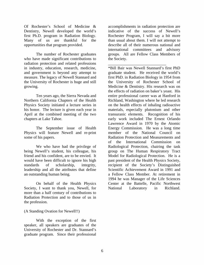
J. Newell Stannard
Sketch of the Rochester Project Before we get into details of the Rochester Project, I want to say how pleased and grateful I am for this special session. When one begins to approach the end of the line, it’s wonderful to have some special events occurring, even a brass band playing. I can hear one today. I cannot find words to tell my colleagues and friends how much this means to me. I am particularly grateful to two of my first Ph.D. students, Bill Bair and Bob Thomas, for both preparing the program and arranging other details, and also to George Anastas, John Taschner, Sydney Porter, Ron Kathren, Martha McDougall, The Board and Program Committee and the History Committee for paving the way and facilitating in many ways.
Also, I want to make it abundantly clear that this Rochester Operation was by no means a one-man show. The personnel were talented & enthusiastic, and all put their shoulders to the wheel, first during the war years, and then during the years of organizing and implementing the graduate-training program. There were no Prima Donnas.
Everyone knew what his job was, and did it well.
The University of Rochester Atomic Energy project was organized in the mid-1940s as a laboratory under the Manhattan Engineer District. It differed from most of the other Manhattan District labs, in that it was entirely biomedical and research-oriented. It really had little to do with the actual development of the atomic bomb.
During the war years the lab focused strongly on the toxicology of uranium in all its forms particularly inhalation toxicology. It was clear to the Manhattan District people that they would be handling large quantities of uranium ore and also uranium in high specific activity. The operation involved large numbers of animals. In fact, the “temporary” building, which was built across the street from the medical school, had as many as 300 people working on three shifts on the programmatic research. When published in the post-war years under the aegis of the National Nuclear Energy Series, the operation was labeled one of the most
8
complete and thorough toxicology studies of any single element. In many respects that characterization still holds.
In addition to the toxicology of uranium, there was work done on polonium, radium and plutonium, on toxicology of fluorine and fluorides, the development of basic instruments, and studies of radon with reference to the exposure of uranium miners. There was a large genetic study in the mouse on the effects of X-rays, which was a template for the large “megamouse” experiment done post-war at the Oak Ridge National Laboratory. There was also considerable basic radiobiological research.
At the end of WWII, with the dissolution of the Manhattan District and the development of the Atomic Energy Commission, the research of the project continued along much the same lines as in the war period with emphasis on inhalation toxicology. However, there was less urgency and it was possible to do several long-term experiments. One of these was a long-term experiment on inhalation of natural uranium in various forms, with a view to the possibility of developing lung cancer. It was surprising how much uranium and how long it took to produce any evidence of lung cancer from exposure to natural uranium. It had to be categorized as a very weak carcinogen.
There was a long-term
experiment on the production of sperm in dogs receiving X irradiation. The work done on polonium during the war continued with life-span studies of both the metabolism and effects, and there was considerable emphasis on understanding the details of radiation biology. The work on radon continued.
In the early post-war years it was Bill Bale, the head of the biophysics division of the project, who pointed out the importance of the daughter products of radon in determining the effects of radon exposure. This was an important finding, which was buried, with undue modesty, in a “memorandum to the files.”
New projects were added. Some of them developed from work that was already underway particularly aerosol science. Many of them, but not all, were requests from the AEC. There was also a large effort on the physiology and pathology of Flash Burns operated largely by the Department of Surgery.
However, the really new development was the addition of a graduate teaching program. The fact that the facility was on campus of a University, and that the faculty was interested seriously in developing a program, which we called the “Applications of Atomic Energy to Life Sciences,” gave the idea impetus and it gained support. The University designated the project as the “Department of Radiation Biology,” it became a full-fledged department of the medical school, and the faculty had the same privileges as in any other department. Degrees were already being offered in Biophysics. To this was added the MS and Ph.D. degree in Radiation Biology and also degrees in Pharmacology and Toxicology. It was our purpose originally not to offer degrees in new subjects, but instead to have them be in one of the classical pre-clinical sciences. However, that turned out to be impractical, because it would take far too long. When the department was authorized to give PhD’s in
9
Radiation Biology, it was the first such degree in the world and Bill Bair was the first recipient of such a degree in the world. The second was Bob Thomas. Student support came from fellowship programs supported by the AEC and later the grant programs of the Public Health Service. Also the Armed Services and foreign governments sent senior officers for degree programs and the department hosted what was called the “Advanced Course in Nuclear Science for Medical Officers.” The Defense Atomic Support Agency supported it for a variety of medical and paramedical officers from all the services and I will say that having the military existence added a certain flavor to the graduate student body that we probably never would have had otherwise. It was an interesting and quite unique group that almost marched to class and startled some of the audience at commencement when some of the officers, in full dress uniform and the obligatory sword at their side, clanked noisily over the wooden steps leading to the platform on the way to obtain their diplomas..
It was a very diversified student group and a delight to work with. These people were anxious to get on with their lives and careers after the interruptions of the war and meant business. The department had a registration of around 70 graduate students at any given time during the peak operational years.
In addition to the graduate degree program, the department (which was later named the Department of Radiation, Biology and Biophysics), along with the Department of Pharmacology and Toxicology, took part in programs related to environmental
problems, both in the field and in the laboratory. You will hear more about these later today.
All told, about 1,000 individuals went through the teaching program at Rochester. By state university standards that’s not a large group but by the standards we worked with, it was large and all consuming. Research was going on nearly everywhere and many students were being bitten by the research bug. It was perhaps this mix of programmatic research and basic science that made the project and the department almost unique.
Tucked away in the snowy fastnesses of Western New York, the Rochester Project had an unusual cohesiveness. People were doing work they wanted to do. Financial support was steady and reliable so long as the requests met reasonable definitions of the overall mission and were reasonable in amount. Administration was benign and respected. Colleagues were talented and congenial and we were all feeling the thrill of teaching a new field and enjoying it. I am thankful that it was my privilege to be a part of it.

10
11
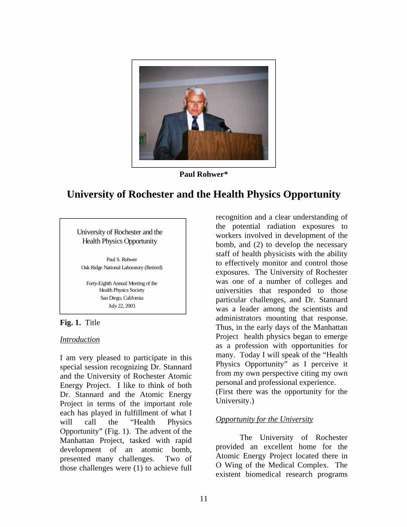
Paul Rohwer*
University of Rochester and the Health Physics Opportunity
Fig. 1. Title Introduction I am very pleased to participate in this special session recognizing Dr. Stannard and the University of Rochester Atomic Energy Project. I like to think of both Dr. Stannard and the Atomic Energy Project in terms of the important role each has played in fulfillment of what I will call the “Health Physics Opportunity” (Fig. 1). The advent of the Manhattan Project, tasked with rapid development of an atomic bomb, presented many challenges. Two of those challenges were (1) to achieve full
recognition and a clear understanding of the potential radiation exposures to workers involved in development of the bomb, and (2) to develop the necessary staff of health physicists with the ability to effectively monitor and control those exposures. The University of Rochester was one of a number of colleges and universities that responded to those particular challenges, and Dr. Stannard was a leader among the scientists and administrators mounting that response. Thus, in the early days of the Manhattan Project health physics began to emerge as a profession with opportunities for many. Today I will speak of the “Health Physics Opportunity” as I perceive it from my own perspective citing my own personal and professional experience. (First there was the opportunity for the University.) Opportunity for the University
The University of Rochester provided an excellent home for the Atomic Energy Project located there in O Wing of the Medical Complex. The existent biomedical research programs
University of Rochester and the Health Physics Opportunity
Paul S. RohwerOak Ridge National Laboratory (Retired)
Forty-Eighth Annual Meeting of the Health Physics SocietySan Diego, California
July 22, 2003
12
and expertise were renowned. The urgency of the war effort provided incentive and resources rarely if ever previously experienced. The Atomic Energy Project assisted the AEC and other federal agencies by focusing academic and research staff and facilities directly on preparation of needed health physicists. Simultaneously the Project’s continued generation of an array of biomedical research results helped facilitate improved understanding of biological effects of ionizing radiation and behavior of radioactive materials in biological systems. The AEC Project was the source of much outstanding fundamental research in radiation biology and biophysics. The project also produced many health physicists who have become prolific contributors to our science and who have achieved positions of prominence in our profession. Witness the other presenters in this session as examples. With these products came increased recognition for the university. (Next came the opportunities for university faculty and staff.) Opportunity for the Faculty and Staff
Dr. Stannard and the University of Rochester Atomic Energy Project were well matched. He was instrumental in development and operation of the Health Physics Program ultimately serving as Associate Dean of Graduate Studies. He like other faculty and staff associated with the project benefited from the additional resources available for education and research due to the focus, intensity, and urgency of the war effort. In addition to being a highly respected research scientist and excellent program administrator, Dr. Stannard was a very sage, caring, and
soft-spoken advisor and mentor who attracted top students to the project. His many graduate students and the outstanding careers they have enjoyed attest to his guidance capabilities. I believe that Dr. Stannard’s own example of professional participation and contribution provides subtle mentoring that goes beyond his students to every health physicist who has ever known him. (Ultimately came the opportunities for students) Opportunity for the Students (Particularly this one)
Now I will go to the opportunity provided by Rochester for people like myself. When I came to Rochester it was a special opportunity for me because when I arrived I met my wife there. Sandy was employed there but I didn’t know her at the time. So not only did I meet my wife there but found that she was a member of Dr. Stannard’s staff. So in addition to achieving a master’s degree, I was finding myself asking Dr. Stannard for her hand as well as her father.
Following my military service I completed my undergraduate degree in physics at Drake University with no particular career goal in mind. During my senior year at Drake I began seeking employment at a number of prospective employers including Brookhaven National Laboratory. In the course of that search my physics professor pointed out to me on the job opportunity bulletin board an advertisement describing AEC Fellowships in health physics. I tore off one of the attached request cards and mailed it in to get further details and an application form. I subsequently applied
13
and was accepted, quite to my surprise. Thanks to the wisdom of the fellowship selection panel at Oak Ridge Institute for Nuclear Studies and my own good fortune I was assigned to the University of Rochester. Thus, following a brief stint as a temporary summer employee at Brookhaven, I arrived in Rochester in the fall 1960 as an incoming AEC Health Physics Fellow having had some prior introduction at Drake and at Brookhaven to radiation and things radioactive. However, it was there at the University of Rochester Atomic Energy Project that the Health Physics Opportunity really materialized in earnest for me.
The program Dr. Stannard guided at Rochester provided a broad spectrum of educational and research opportunities for graduate students. Students had ready access to graduate offerings in various departments of the School of Medicine and Dentistry as well as at the nearby University River Campus. The classroom, library, and laboratory facilities were excellent. Professors and staff daily engaged in research were the principal lecturers. The health physics student body was diverse with a sprinkling of individuals from foreign countries, but largely composed of fellowship students supported by the Atomic Energy Commission, the Defense Atomic Support Agency, and the Public Health Service. In general the academic and research environment of the Atomic Energy Project had a comfortable feel. The teacher-student interactions tended to be relaxed and the overall career guidance superb. Here I come back to Dr. Stannard’s excellence as an advisor and mentor. Although he was not my thesis advisor or a member of my
research committee, I have been a frequent benefactor of his council. Three examples of the cogent advice I received from him continue to be vivid in my memory. The first example is his strong encouragement that I apply for an AEC Advanced Health Physics Fellowship and return to Rochester to seek a Ph.D. The second example is the series of suggestions he made for the composition of my Ph.D. thesis committee with Dr. Thomas R. Noonan as its chair. And the third example is his advice that I visit Oak Ridge National Laboratory while in Oak Ridge interviewing with another potential employer located there. A direct result of the suggested visit to ORNL was a subsequent job offer followed by a 32-year career at the Laboratory.
Now I will provide a brief insight to the career I enjoyed at Oak Ridge National Laboratory. At times my activities were mostly R&D oriented and at other times more applied. In general during the course of my career I experienced decreasing R&D involvement, increasing focus on applied work, and increasing management responsibilities. My R&D experience was primarily in development of methodology for radiation dose estimation, analysis, and interpretation. During my span of involvement the objective of our dosimetry activities expanded from estimation and measurement of radiation-worker exposures to include evaluation of potential exposures of the general population from environmental releases of radioactivity. R&D activities I participated in included the following: (1) development and extension of “reference man” as an update to it’s predecessor “standard man”, (2)
14
computerization of environmental dispersion models and dose models to support proposed peaceful applications of nuclear explosives in the Plowshare Program, (3) development of models and parameters to facilitate age-dependent internal dose calculations, (4) development of models and parameters to simulate radionuclide movement in environmental food-chain pathways leading to man, (5) summation of dose across all radiation sources and all exposure pathways and modes, and (6) conversion of estimates of radiation dose into estimates of risk. The education I received at the University of Rochester was ideal preparation for all of the health physics tasks I was assigned throughout my career. Although I was not involved in biomedical research activities at ORNL, I always found the biomedical background from my Rochester days to be reassuring and a sound basis for my judging the significance of potential radiation exposures associated with the health physics problems I encountered and the related decisions I needed to make at the Laboratory. The Rochester experience also instilled a strong sense of professional pride and participation. (Will the opportunities continue into the future?) Opportunity for the Future
The Health Physics Opportunity still exists even though the profession has matured, resources for education and research have become scarce, and some of the challenges are different. Projections of future professional needs in health physics indicate an emerging critical shortage of appropriately educated and experienced individuals. The Society Scientific and Public Issues Committee, in a position statement
prepared for use in informing congressional offices of this problem, refers to the shortage of health physicists as the “Human Capital Crisis.” To obtain a more current and comprehensive assessment of manpower needs facing the health physics profession Society President John Frazier recently established a Health Physics Manpower Assessment Team chaired by one of the Society’s directors. With the current severely decreased federal support for research and scholarship programs in health physics, the number of health physics students and the number of colleges and universities with health physics programs have declined. The Society is vigorously addressing the need for more educational opportunities in health physics. The subject has been included in information presented at congressional briefings and the Society’s Congressional and Agency Liaison continues to work this issue. One immediate goal of these efforts is to assure that current draft energy bills in congress that include support for education in nuclear sciences also contain language that is specific to health physics. Success in achieving such goals requires that we be a strong resourceful organization. I encourage all health physicists to be active members of the Health Physics Society and help support our profession in meeting the challenges and creating enhanced opportunities. Additional health physicists are needed now and more will be needed in the future. Legacies like those exemplified by the University of Rochester Health Physics Program and Dr. J. Newell Stannard’s lifetime of dedication and contribution to our field must be built upon.
15
In closing I would like to speak for myself and all of the University of Rochester health physics students not privileged to be a speaker in this session today as I say: Dr. Stannard, thank you for pursuing the “Health Physics Opportunity”, and for helping so many of us pursue it also. _________________________________ * Paul Rohwer received his PhD in Radiation Biology from the University of Rochester in 1966. He joined the Oak Ridge National Laboratory in 1966 and remained there until his retirement in 1998 after serving in several health and environmental positions. At the time of his retirement he was the Associate Director of the Life /Sciences Division. Paul is a past president of the Health Physics Society and a certified health physicist.

16
17
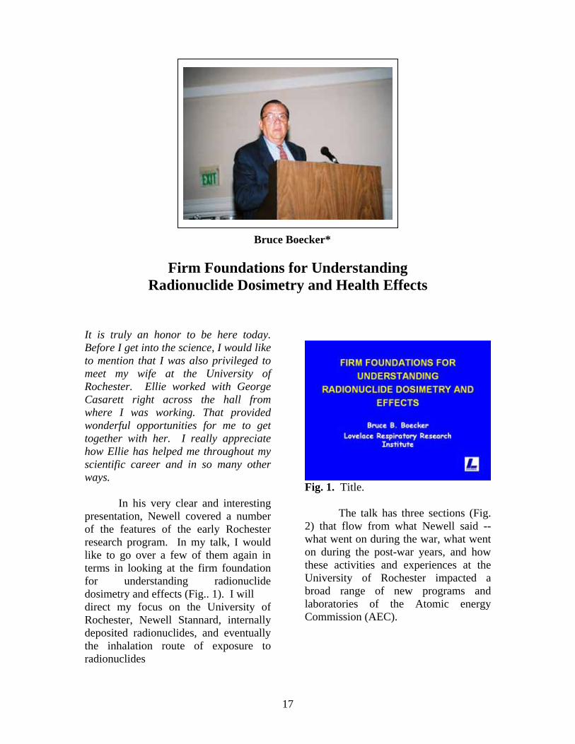
Bruce Boecker*
Firm Foundations for Understanding Radionuclide Dosimetry and Health Effects
It is truly an honor to be here today. Before I get into the science, I would like to mention that I was also privileged to meet my wife at the University of Rochester. Ellie worked with George Casarett right across the hall from where I was working. That provided wonderful opportunities for me to get together with her. I really appreciate how Ellie has helped me throughout my scientific career and in so many other ways.
In his very clear and interesting presentation, Newell covered a number of the features of the early Rochester research program. In my talk, I would like to go over a few of them again in terms in looking at the firm foundation for understanding radionuclide dosimetry and effects (Fig.. 1). I will direct my focus on the University of Rochester, Newell Stannard, internally deposited radionuclides, and eventually the inhalation route of exposure to radionuclides
Fig. 1. Title.
The talk has three sections (Fig. 2) that flow from what Newell said -- what went on during the war, what went on during the post-war years, and how these activities and experiences at the University of Rochester impacted a broad range of new programs and laboratories of the Atomic energy Commission (AEC).
18
Fig. 2. Heritage of Radionuclide Inhalation Studies.
The first part of my talk will address what went on in the Manhattan Engineer District Project during WW II (Fig. 3)
Fig. 3. Manhattan Engineer District (world War II).
I thought this was an interesting comment by General Groves, the Commanding Officer of the Manhattan Engineer District (Fig. 4). He described the work of the Manhattan Project as a “generation of scientific development compressed into three years.” I’ve always thought of that in reference to the development of the atomic bomb and all of the physical things that were required. However, here we’re talking about also compressing radiobiological and toxicological research into that same
very short time frame. That’s also very difficult and challenging when one
Fig. 4. Accomplishments of the Manhattan Project (MED). considers the broad range of one-year studies that were conducted in laboratory animals. Here, it wasn’t possible to compress time very much, but instead, many studies had to be run in parallel. I think that is where much of the effort went.
The work with inhaled radionuclides was spread among elements of the Metallurgical Laboratory, essentially at Berkeley and Chicago, and at the University of Rochester (Fig. 5).
Fig. 5. MED WWII Research on Inhaled Radionuclides.
19
As Newell said, the emphasis at Rochester was on uranium and polonium. The polonium work was directed to its radiotoxicology compared with that for plutonium and radium (Fig. 6). I’ll talk a little bit more about the
Fig. 6. WWII Elemental Emphasis at Rochester. uranium work to which Newell eluded (Fig. 7). It was a very broad and intense program that involved multiple approaches including many routes of administration, a number of species of laboratory animals, and a broad range of physical and chemical forms.
Fig. 7. MED Uranium Toxicology Studies at Rochester (1).
These inhalation studies, which ranged in duration from three days to two years, included many one-year studies. So that’s part of how the
compression we talked about earlier was dealt with. Embedded in all these studies was the important issue of uranium’s chemical toxicity versus its radiological effects.
Collectively, these results (Fig. 8) defined the metabolism and biological effects of many soluble and insoluble forms of uranium. They were published in the National Nuclear Energy Series that Newell mentioned. It’s important to remember that these resources are still available today. They serve as a very important foundation for issues such as the dispersal of depleted uranium in battlefield environments. So here we have results obtained some fifty years ago that are still very important to us today.
Fig. 8. MED Uranium Toxicology Studies at Rochester (2).
In the post-war period, a few people that were involved in the uranium studies continued on with the large five-year study at Rochester (Figs. 9 and 10). It was focused on one agent, insoluble UO2 dust, and three species -- rats, dogs and monkeys. At the end of the five- year exposure period, the accumulation of uranium was primarily in the lung and tracheobronchial lymph nodes as one might expect from the insolubility of the material. There were few obvious
20
biological effects present at the end of five years of inhalation exposure.
Fig. 9. University of Rochester Post-War Studies.
Fig. 10. 5-yr study of inhaled UO2. Because of the insoluble nature the UO2, chemical toxicity of uranium did not come into play. During the six-year follow-up of these animals, fibrosis was found in the lungs and tracheobronchial lymph nodes in both the dogs and monkeys. Fibrosis in the lymph nodes of the monkeys was more pronounced than that in the dogs. Also of interest was the observation of a few pulmonary cancers. As Newell said, “How much work had to go in to produce a very few cancers!” The lasting message that I think this study gave to the field of radiobiology and toxicology is the importance of studying animals over their whole lifetime. A one-year study
tells you something and a five-year study tells you more, but unless you study an animal over its entire lifetime, you may miss some of the late-occurring biological effects. That was one of the important lessons that flowed into the eventual planning and execution of life-span studies at other laboratories.
A Radioactive Inhalation Section was formed at Rochester in the 1950s (Fig. 11). Newell served as its head from 1952 to 1959. That was an important endeavor because it involved
Fig. 11. Radioactive Inhalation Section. the design and use of what was called the Alpha Laboratory. Although this laboratory was built and operated in a university environment, it was designed to safely use relatively high levels of airborne, long-lived, alpha-emitting radionuclides. The laboratory used easily fabricated and replaceable glove boxes; the layout of the rooms and corridors was based on “clean” and “dirty” areas, and the airflow passed from areas of low airborne concentrations to higher concentration areas, as the means of controlling radioactive contamination. The inhalation studies were primarily with 210Po, 238Pu, 239Pu, and 222Rn.
The polonium studies (Fig. 12) comprised an extensive series of studies

21
on the metabolism, dosimetry, and biological effects of internally deposited 210Po. As we consider scientific
Fig. 12. 210Po studies. resources for today’s needs, one of the key features related to this work was the publication of the Polonium Supplement in Radiation Research in 1964. This supplement contains some 26 manuscripts on various aspects of polonium such as those listed in Fig. 12: physicochemical studies, distribution and excretion, etc. One of the unique features of this supplement was that all of these manuscripts were published in the open literature for the first time in this supplement. This meant that some manuscripts sat around for a while, while others were being finished up. In today’s “publish or perish” regime, I don’t think that would work out very well but it was a useful thing to do in this case because this collection of polonium reports in one place became a very useful resource for subsequent investigators.
We’ll now move to the third part of this talk, the influence of the Rochester program on the development of new programs and laboratories by the Atomic Energy Commission (Fig. 13).
Fig. 13. New AEC Programs and Laboratories.
In this brief description of our nuclear environment in the decade between 1950 and 1960 (Fig. 14), one can see that it was substantially different than it is today, fifty years later.
Fig. 14. Our Nuclear Environment, 1950-1960.
At that point in time, the AEC was still projecting a large increase in nuclear power. Fuel reprocessing was a viable option. There was fallout in the environment from atmospheric nuclear weapons testing. There was concern about nuclear war and its associated patterns of nuclear fallout. People were building bomb shelters in their backyards for protection from this fallout. New nuclear technologies were under study such as aircraft nuclear propulsion and space nuclear propulsion.
22
I think Bob Thomas will touch on some studies related to these topics later in the program.
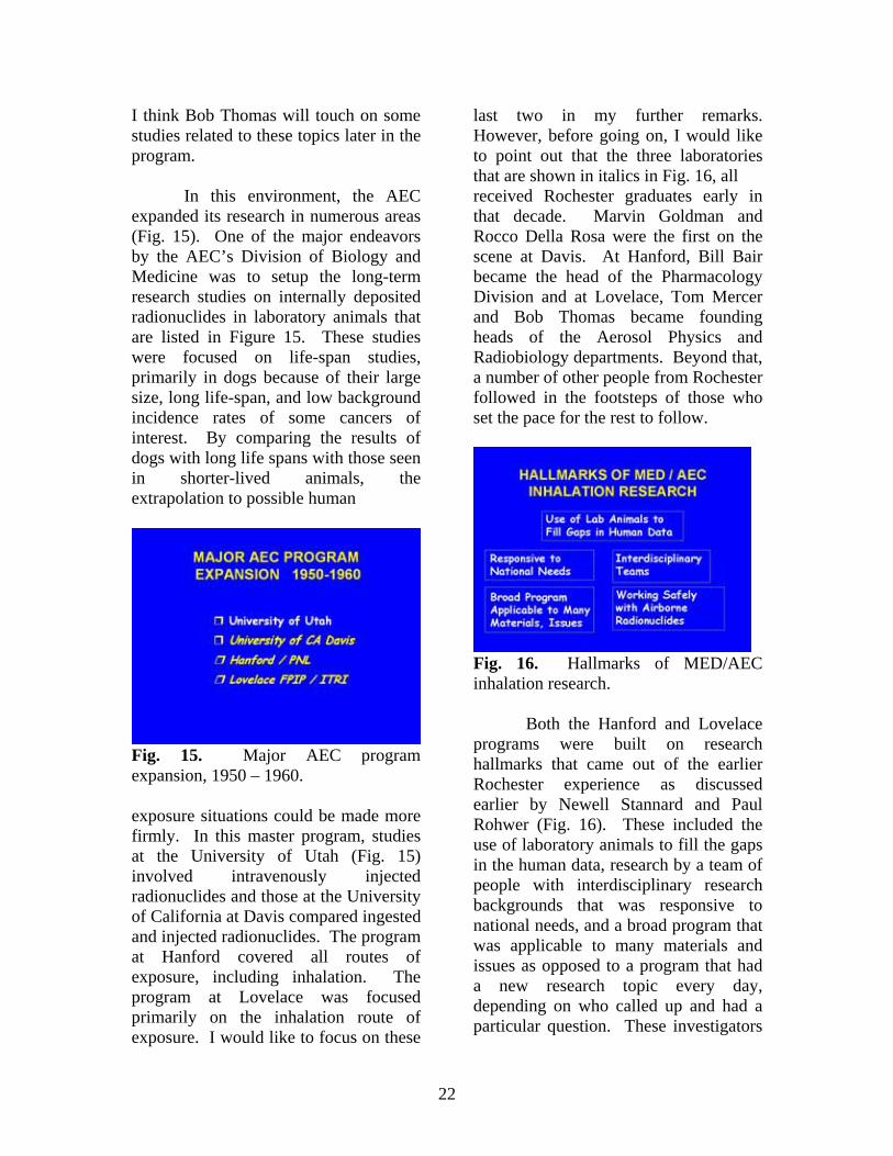
In this environment, the AEC expanded its research in numerous areas (Fig. 15). One of the major endeavors by the AEC’s Division of Biology and Medicine was to setup the long-term research studies on internally deposited radionuclides in laboratory animals that are listed in Figure 15. These studies were focused on life-span studies, primarily in dogs because of their large size, long life-span, and low background incidence rates of some cancers of interest. By comparing the results of dogs with long life spans with those seen in shorter-lived animals, the extrapolation to possible human
Fig. 15. Major AEC program expansion, 1950 – 1960. exposure situations could be made more firmly. In this master program, studies at the University of Utah (Fig. 15) involved intravenously injected radionuclides and those at the University of California at Davis compared ingested and injected radionuclides. The program at Hanford covered all routes of exposure, including inhalation. The program at Lovelace was focused primarily on the inhalation route of exposure. I would like to focus on these
last two in my further remarks. However, before going on, I would like to point out that the three laboratories that are shown in italics in Fig. 16, all received Rochester graduates early in that decade. Marvin Goldman and Rocco Della Rosa were the first on the scene at Davis. At Hanford, Bill Bair became the head of the Pharmacology Division and at Lovelace, Tom Mercer and Bob Thomas became founding heads of the Aerosol Physics and Radiobiology departments. Beyond that, a number of other people from Rochester followed in the footsteps of those who set the pace for the rest to follow.
Fig. 16. Hallmarks of MED/AEC inhalation research.
Both the Hanford and Lovelace programs were built on research hallmarks that came out of the earlier Rochester experience as discussed earlier by Newell Stannard and Paul Rohwer (Fig. 16). These included the use of laboratory animals to fill the gaps in the human data, research by a team of people with interdisciplinary research backgrounds that was responsive to national needs, and a broad program that was applicable to many materials and issues as opposed to a program that had a new research topic every day, depending on who called up and had a particular question. These investigators
23
built broad programs that, in turn, would build a broad knowledge base that could be used in many ways. These concepts follow from some of the things we were talking about a little bit earlier and of course, the concept of working safely with airborne materials.
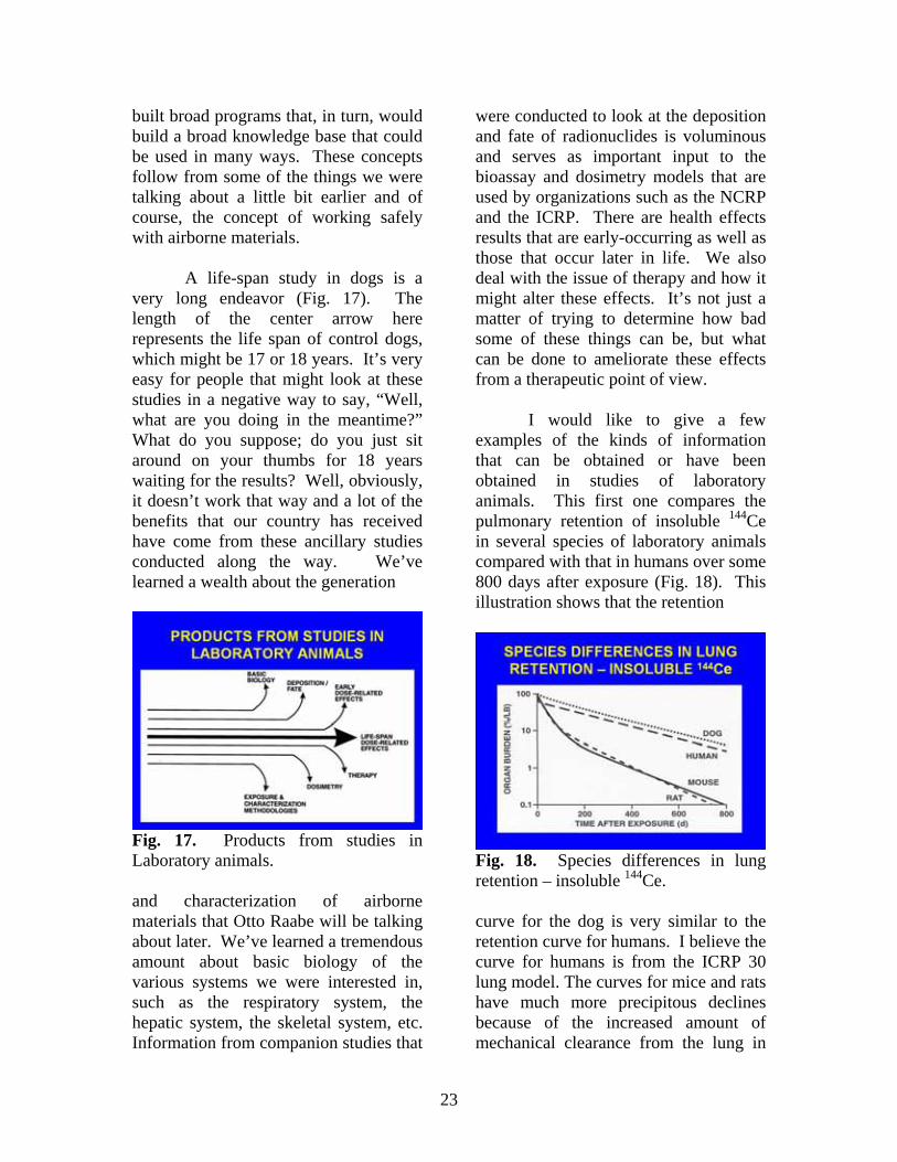
A life-span study in dogs is a very long endeavor (Fig. 17). The length of the center arrow here represents the life span of control dogs, which might be 17 or 18 years. It’s very easy for people that might look at these studies in a negative way to say, “Well, what are you doing in the meantime?” What do you suppose; do you just sit around on your thumbs for 18 years waiting for the results? Well, obviously, it doesn’t work that way and a lot of the benefits that our country has received have come from these ancillary studies conducted along the way. We’ve learned a wealth about the generation
Fig. 17. Products from studies in Laboratory animals. and characterization of airborne materials that Otto Raabe will be talking about later. We’ve learned a tremendous amount about basic biology of the various systems we were interested in, such as the respiratory system, the hepatic system, the skeletal system, etc. Information from companion studies that
were conducted to look at the deposition and fate of radionuclides is voluminous and serves as important input to the bioassay and dosimetry models that are used by organizations such as the NCRP and the ICRP. There are health effects results that are early-occurring as well as those that occur later in life. We also deal with the issue of therapy and how it might alter these effects. It’s not just a matter of trying to determine how bad some of these things can be, but what can be done to ameliorate these effects from a therapeutic point of view.
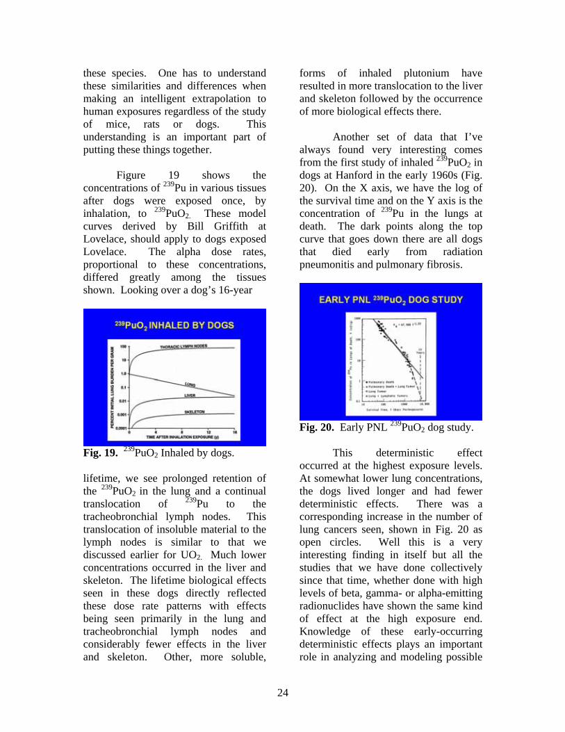
I would like to give a few examples of the kinds of information that can be obtained or have been obtained in studies of laboratory animals. This first one compares the pulmonary retention of insoluble 144Ce in several species of laboratory animals compared with that in humans over some 800 days after exposure (Fig. 18). This illustration shows that the retention
Fig. 18. Species differences in lung retention – insoluble 144Ce. curve for the dog is very similar to the retention curve for humans. I believe the curve for humans is from the ICRP 30 lung model. The curves for mice and rats have much more precipitous declines because of the increased amount of mechanical clearance from the lung in
24
these species. One has to understand these similarities and differences when making an intelligent extrapolation to human exposures regardless of the study of mice, rats or dogs. This understanding is an important part of putting these things together.
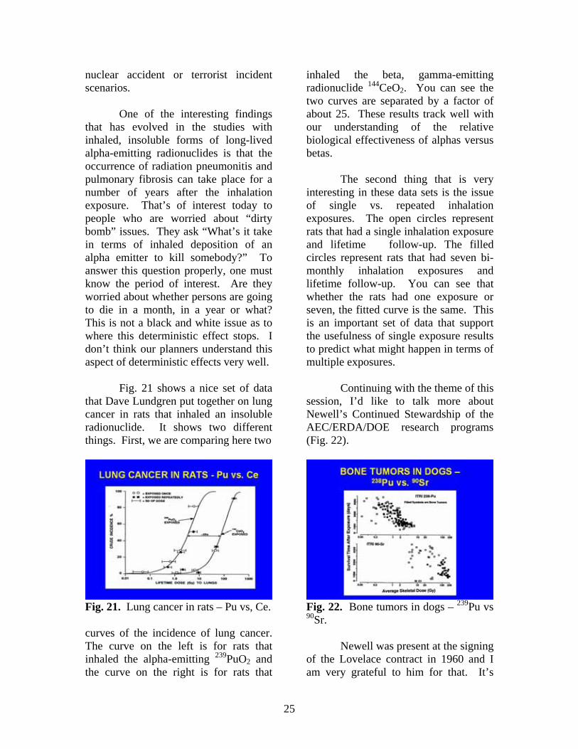
Figure 19 shows the concentrations of 239Pu in various tissues after dogs were exposed once, by inhalation, to 239PuO2. These model curves derived by Bill Griffith at Lovelace, should apply to dogs exposed Lovelace. The alpha dose rates, proportional to these concentrations, differed greatly among the tissues shown. Looking over a dog’s 16-year
Fig. 19. 239PuO2 Inhaled by dogs. lifetime, we see prolonged retention of the 239PuO2 in the lung and a continual translocation of 239Pu to the tracheobronchial lymph nodes. This translocation of insoluble material to the lymph nodes is similar to that we discussed earlier for UO2. Much lower concentrations occurred in the liver and skeleton. The lifetime biological effects seen in these dogs directly reflected these dose rate patterns with effects being seen primarily in the lung and tracheobronchial lymph nodes and considerably fewer effects in the liver and skeleton. Other, more soluble,
forms of inhaled plutonium have resulted in more translocation to the liver and skeleton followed by the occurrence of more biological effects there.
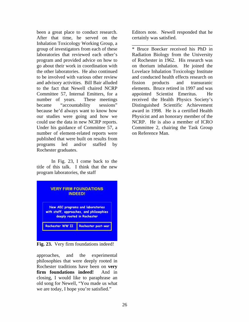
Another set of data that I’ve always found very interesting comes from the first study of inhaled 239PuO2 in dogs at Hanford in the early 1960s (Fig. 20). On the X axis, we have the log of the survival time and on the Y axis is the concentration of 239Pu in the lungs at death. The dark points along the top curve that goes down there are all dogs that died early from radiation pneumonitis and pulmonary fibrosis.
Fig. 20. Early PNL 239PuO2 dog study.
This deterministic effect occurred at the highest exposure levels. At somewhat lower lung concentrations, the dogs lived longer and had fewer deterministic effects. There was a corresponding increase in the number of lung cancers seen, shown in Fig. 20 as open circles. Well this is a very interesting finding in itself but all the studies that we have done collectively since that time, whether done with high levels of beta, gamma- or alpha-emitting radionuclides have shown the same kind of effect at the high exposure end. Knowledge of these early-occurring deterministic effects plays an important role in analyzing and modeling possible
25
nuclear accident or terrorist incident scenarios.
One of the interesting findings that has evolved in the studies with inhaled, insoluble forms of long-lived alpha-emitting radionuclides is that the occurrence of radiation pneumonitis and pulmonary fibrosis can take place for a number of years after the inhalation exposure. That’s of interest today to people who are worried about “dirty bomb” issues. They ask “What’s it take in terms of inhaled deposition of an alpha emitter to kill somebody?” To answer this question properly, one must know the period of interest. Are they worried about whether persons are going to die in a month, in a year or what? This is not a black and white issue as to where this deterministic effect stops. I don’t think our planners understand this aspect of deterministic effects very well.
Fig. 21 shows a nice set of data that Dave Lundgren put together on lung cancer in rats that inhaled an insoluble radionuclide. It shows two different things. First, we are comparing here two
Fig. 21. Lung cancer in rats – Pu vs, Ce. curves of the incidence of lung cancer. The curve on the left is for rats that inhaled the alpha-emitting 239PuO2 and the curve on the right is for rats that
inhaled the beta, gamma-emitting radionuclide 144CeO2. You can see the two curves are separated by a factor of about 25. These results track well with our understanding of the relative biological effectiveness of alphas versus betas.
The second thing that is very interesting in these data sets is the issue of single vs. repeated inhalation exposures. The open circles represent rats that had a single inhalation exposure and lifetime follow-up. The filled circles represent rats that had seven bi-monthly inhalation exposures and lifetime follow-up. You can see that whether the rats had one exposure or seven, the fitted curve is the same. This is an important set of data that support the usefulness of single exposure results to predict what might happen in terms of multiple exposures.
Continuing with the theme of this session, I’d like to talk more about Newell’s Continued Stewardship of the AEC/ERDA/DOE research programs (Fig. 22).
Fig. 22. Bone tumors in dogs – 239Pu vs 90Sr.
Newell was present at the signing of the Lovelace contract in 1960 and I am very grateful to him for that. It’s
26
been a great place to conduct research. After that time, he served on the Inhalation Toxicology Working Group, a group of investigators from each of these laboratories that reviewed each other’s program and provided advice on how to go about their work in coordination with the other laboratories. He also continued to be involved with various other review and advisory activities. Bill Bair alluded to the fact that Newell chaired NCRP Committee 57, Internal Emitters, for a number of years. These meetings became “accountability sessions” because he’d always want to know how our studies were going and how we could use the data in new NCRP reports. Under his guidance of Committee 57, a number of element-related reports were published that were built on results from programs led and/or staffed by Rochester graduates.
In Fig. 23, I come back to the title of this talk. I think that the new program laboratories, the staff
Fig. 23. Very firm foundations indeed! approaches, and the experimental philosophies that were deeply rooted in Rochester traditions have been on very firm foundations indeed! And in closing, I would like to paraphrase an old song for Newell, “You made us what we are today, I hope you’re satisfied.”
Editors note. Newell responded that he certainly was satisfied. _________________________________ * Bruce Boecker received his PhD in Radiation Biology from the University of Rochester in 1962. His research was on thorium inhalation. He joined the Lovelace Inhalation Toxicology Institute and conducted health effects research on fission products and transuranic elements. Bruce retired in 1997 and was appointed Scientist Emeritus. He received the Health Physics Society’s Distinguished Scientific Achievement award in 1998. He is a certified Health Physicist and an honorary member of the NCRP. He is also a member of ICRO Committee 2, chairing the Task Group on Reference Man.
27
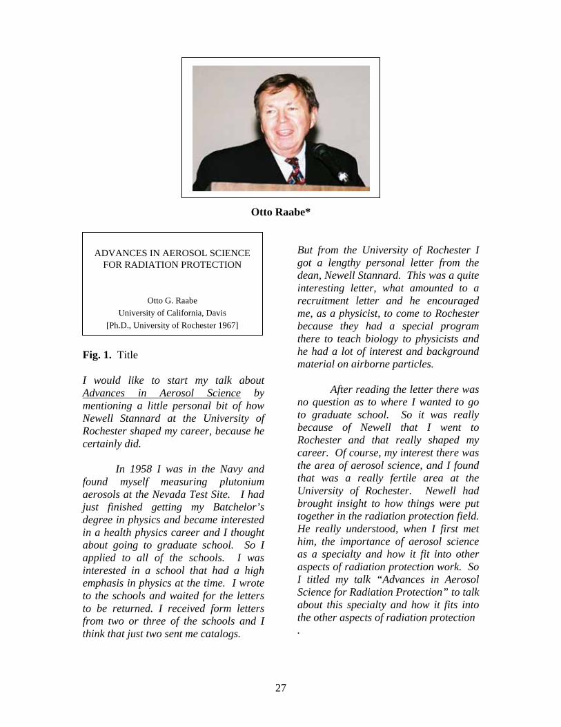
Otto Raabe*
Fig. 1. Title I would like to start my talk about Advances in Aerosol Science by mentioning a little personal bit of how Newell Stannard at the University of Rochester shaped my career, because he certainly did.
In 1958 I was in the Navy and found myself measuring plutonium aerosols at the Nevada Test Site. I had just finished getting my Batchelor’s degree in physics and became interested in a health physics career and I thought about going to graduate school. So I applied to all of the schools. I was interested in a school that had a high emphasis in physics at the time. I wrote to the schools and waited for the letters to be returned. I received form letters from two or three of the schools and I think that just two sent me catalogs.
But from the University of Rochester I got a lengthy personal letter from the dean, Newell Stannard. This was a quite interesting letter, what amounted to a recruitment letter and he encouraged me, as a physicist, to come to Rochester because they had a special program there to teach biology to physicists and he had a lot of interest and background material on airborne particles.
After reading the letter there was no question as to where I wanted to go to graduate school. So it was really because of Newell that I went to Rochester and that really shaped my career. Of course, my interest there was the area of aerosol science, and I found that was a really fertile area at the University of Rochester. Newell had brought insight to how things were put together in the radiation protection field. He really understood, when I first met him, the importance of aerosol science as a specialty and how it fit into other aspects of radiation protection work. So I titled my talk “Advances in Aerosol Science for Radiation Protection” to talk about this specialty and how it fits into the other aspects of radiation protection .
ADVANCES IN AEROSOL SCIENCE FOR RADIATION PROTECTION
Otto G. RaabeUniversity of California, Davis
[Ph.D., University of Rochester 1967]
28
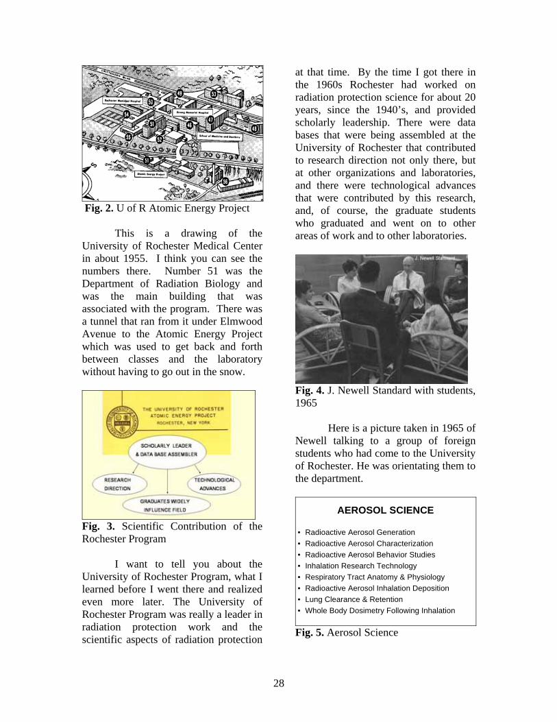
Fig. 2. U of R Atomic Energy Project
This is a drawing of the University of Rochester Medical Center in about 1955. I think you can see the numbers there. Number 51 was the Department of Radiation Biology and was the main building that was associated with the program. There was a tunnel that ran from it under Elmwood Avenue to the Atomic Energy Project which was used to get back and forth between classes and the laboratory without having to go out in the snow.
Fig. 3. Scientific Contribution of the Rochester Program
I want to tell you about the University of Rochester Program, what I learned before I went there and realized even more later. The University of Rochester Program was really a leader in radiation protection work and the scientific aspects of radiation protection
at that time. By the time I got there in the 1960s Rochester had worked on radiation protection science for about 20 years, since the 1940’s, and provided scholarly leadership. There were data bases that were being assembled at the University of Rochester that contributed to research direction not only there, but at other organizations and laboratories, and there were technological advances that were contributed by this research, and, of course, the graduate students who graduated and went on to other areas of work and to other laboratories.
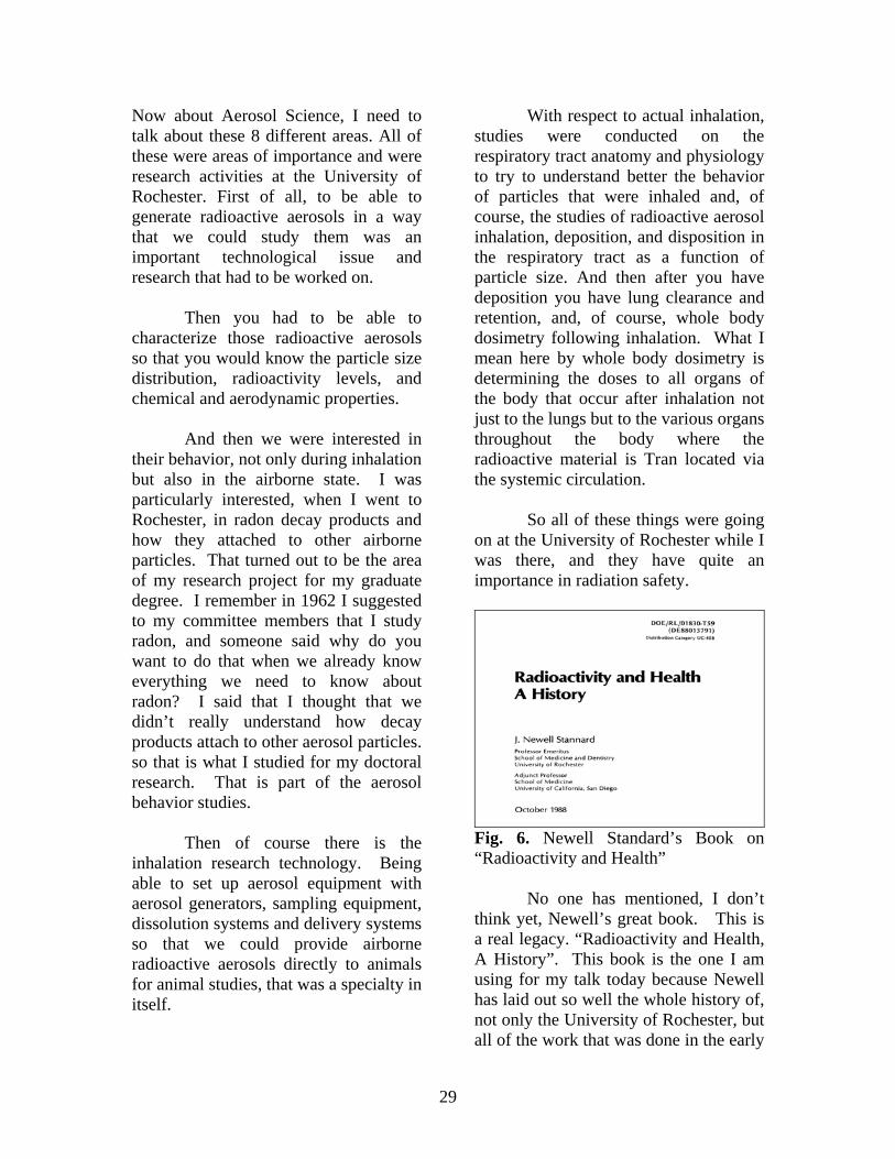
Fig. 4. J. Newell Standard with students, 1965
Here is a picture taken in 1965 of Newell talking to a group of foreign students who had come to the University of Rochester. He was orientating them to the department.
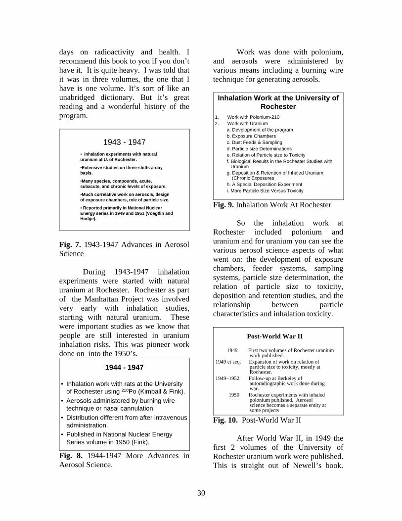
AEROSOL SCIENCE
• Radioactive Aerosol Generation• Radioactive Aerosol Characterization• Radioactive Aerosol Behavior Studies• Inhalation Research Technology• Respiratory Tract Anatomy & Physiology• Radioactive Aerosol Inhalation Deposition• Lung Clearance & Retention• Whole Body Dosimetry Following Inhalation
Fig. 5. Aerosol Science
29
Now about Aerosol Science, I need to talk about these 8 different areas. All of these were areas of importance and were research activities at the University of Rochester. First of all, to be able to generate radioactive aerosols in a way that we could study them was an important technological issue and research that had to be worked on.
Then you had to be able to characterize those radioactive aerosols so that you would know the particle size distribution, radioactivity levels, and chemical and aerodynamic properties.
And then we were interested in their behavior, not only during inhalation but also in the airborne state. I was particularly interested, when I went to Rochester, in radon decay products and how they attached to other airborne particles. That turned out to be the area of my research project for my graduate degree. I remember in 1962 I suggested to my committee members that I study radon, and someone said why do you want to do that when we already know everything we need to know about radon? I said that I thought that we didn’t really understand how decay products attach to other aerosol particles. so that is what I studied for my doctoral research. That is part of the aerosol behavior studies.
Then of course there is the inhalation research technology. Being able to set up aerosol equipment with aerosol generators, sampling equipment, dissolution systems and delivery systems so that we could provide airborne radioactive aerosols directly to animals for animal studies, that was a specialty in itself.
With respect to actual inhalation, studies were conducted on the respiratory tract anatomy and physiology to try to understand better the behavior of particles that were inhaled and, of course, the studies of radioactive aerosol inhalation, deposition, and disposition in the respiratory tract as a function of particle size. And then after you have deposition you have lung clearance and retention, and, of course, whole body dosimetry following inhalation. What I mean here by whole body dosimetry is determining the doses to all organs of the body that occur after inhalation not just to the lungs but to the various organs throughout the body where the radioactive material is Tran located via the systemic circulation.
So all of these things were going on at the University of Rochester while I was there, and they have quite an importance in radiation safety.
Fig. 6. Newell Standard’s Book on “Radioactivity and Health”
No one has mentioned, I don’t think yet, Newell’s great book. This is a real legacy. “Radioactivity and Health, A History”. This book is the one I am using for my talk today because Newell has laid out so well the whole history of, not only the University of Rochester, but all of the work that was done in the early
30
days on radioactivity and health. I recommend this book to you if you don’t have it. It is quite heavy. I was told that it was in three volumes, the one that I have is one volume. It’s sort of like an unabridged dictionary. But it’s great reading and a wonderful history of the program.
1943 - 1947• Inhalation experiments with natural uranium at U. of Rochester.
•Extensive studies on three-shifts-a-day basis.
•Many species, compounds, acute, subacute, and chronic levels of exposure.
•Much correlative work on aerosols, design of exposure chambers, role of particle size.
• Reported primarily in National Nuclear Energy series in 1949 and 1951 (Voegtlin and Hodge).
Fig. 7. 1943-1947 Advances in Aerosol Science
During 1943-1947 inhalation experiments were started with natural uranium at Rochester. Rochester as part of the Manhattan Project was involved very early with inhalation studies, starting with natural uranium. These were important studies as we know that people are still interested in uranium inhalation risks. This was pioneer work done on into the 1950’s.
1944 - 1947
• Inhalation work with rats at the University of Rochester using 210Po (Kimball & Fink).
• Aerosols administered by burning wire technique or nasal cannulation.
• Distribution different from after intravenous administration.
• Published in National Nuclear Energy Series volume in 1950 (Fink).
Fig. 8. 1944-1947 More Advances in Aerosol Science.
Work was done with polonium, and aerosols were administered by various means including a burning wire technique for generating aerosols.
Inhalation Work at the University of Rochester
1. Work with Polonium-2102. Work with Uranium
a. Development of the programb. Exposure Chambersc. Dust Feeds & Samplingd. Particle size Determinationse. Relation of Particle size to Toxicityf. Biological Results in the Rochester Studies with
Uraniumg. Deposition & Retention of Inhaled Uranium
(Chronic Exposuresh. A Special Deposition Experimenti. More Particle Size Versus Toxicity
Fig. 9. Inhalation Work At Rochester
So the inhalation work at Rochester included polonium and uranium and for uranium you can see the various aerosol science aspects of what went on: the development of exposure chambers, feeder systems, sampling systems, particle size determination, the relation of particle size to toxicity, deposition and retention studies, and the relationship between particle characteristics and inhalation toxicity.
Post-World War II
1949 First two volumes of Rochester uranium work published.
1949 et seq. Expansion of work on relation of particle size to toxicity, mostly at Rochester.
1949–1952 Follow-up at Berkeley of autoradiographic work done during war.
1950 Rochester experiments with inhaled polonium published. Aerosol science becomes a separate entity at some projects
Fig. 10. Post-World War II
After World War II, in 1949 the first 2 volumes of the University of Rochester uranium work were published. This is straight out of Newell’s book.
31
“In 1949 we had the expansion of work on the relation between particle size and toxicity.” So back 50 years ago the University of Rochester was working on important issues in aerosol technology and understanding the relationship of particle size and inhalation toxicology. In 1950 Rochester’s experiments on inhaled polonium were published and aerosol science became a separate entity at some projects .
The Postwar Years Begin
A. Continuing Work at Former Manhattan Project Laboratories
B. Aerosol Science1. Development of Liaison2. Generalization of Results in Aerosol Science
a. Role of Particle Size in Depositionb. Practicesc. Role of Physiological Factors in Depositiond. Retention and Clearancee. Mechanisms of Lung Clearancef. Special Considerations for Radioactive Aerosols
Fig. 11. Post-War Years Begin
In the postwar years, we have aerosol science continued as an important separate entity: the role of particle size in deposition, of physiological factors in deposition, retention and clearance studies, mechanisms for lung clearance in relation to particle size and special consideration for radioactive aerosols. Of course, aerosol science was important in industrial hygiene and other work at that time. But, the project put together what we knew about aerosol physics with what we needed to know about radioactive materials in the airborne state.
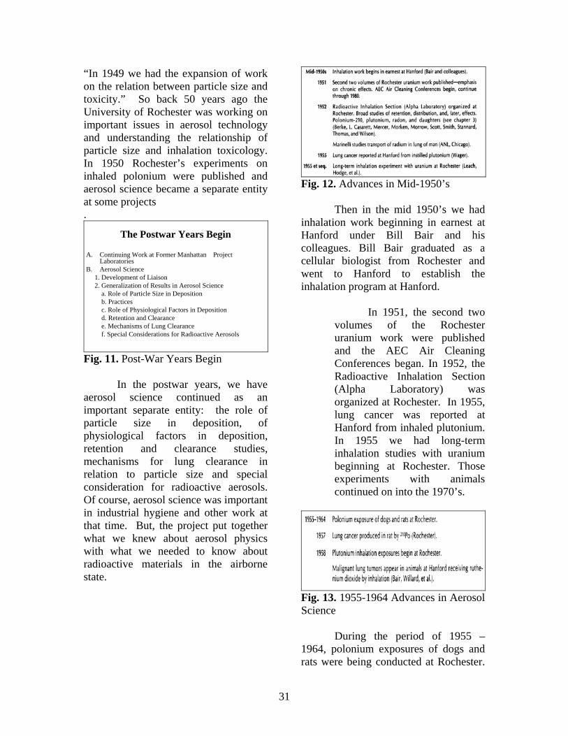
Fig. 12. Advances in Mid-1950’s
Then in the mid 1950’s we had inhalation work beginning in earnest at Hanford under Bill Bair and his colleagues. Bill Bair graduated as a cellular biologist from Rochester and went to Hanford to establish the inhalation program at Hanford.
In 1951, the second two volumes of the Rochester uranium work were published and the AEC Air Cleaning Conferences began. In 1952, the Radioactive Inhalation Section (Alpha Laboratory) was organized at Rochester. In 1955, lung cancer was reported at Hanford from inhaled plutonium. In 1955 we had long-term inhalation studies with uranium beginning at Rochester. Those experiments with animals continued on into the 1970’s.
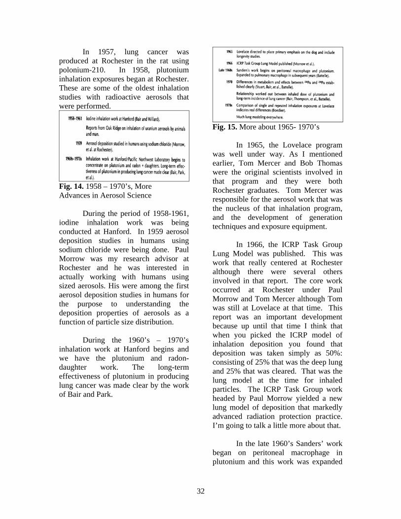
Fig. 13. 1955-1964 Advances in Aerosol Science
During the period of 1955 – 1964, polonium exposures of dogs and rats were being conducted at Rochester.
32
In 1957, lung cancer was produced at Rochester in the rat using polonium-210. In 1958, plutonium inhalation exposures began at Rochester. These are some of the oldest inhalation studies with radioactive aerosols that were performed.
Fig. 14. 1958 – 1970’s, More Advances in Aerosol Science
During the period of 1958-1961, iodine inhalation work was being conducted at Hanford. In 1959 aerosol deposition studies in humans using sodium chloride were being done. Paul Morrow was my research advisor at Rochester and he was interested in actually working with humans using sized aerosols. His were among the first aerosol deposition studies in humans for the purpose to understanding the deposition properties of aerosols as a function of particle size distribution.
During the 1960’s – 1970’s inhalation work at Hanford begins and we have the plutonium and radon-daughter work. The long-term effectiveness of plutonium in producing lung cancer was made clear by the work of Bair and Park.
Fig. 15. More about 1965- 1970’s
In 1965, the Lovelace program was well under way. As I mentioned earlier, Tom Mercer and Bob Thomas were the original scientists involved in that program and they were both Rochester graduates. Tom Mercer was responsible for the aerosol work that was the nucleus of that inhalation program, and the development of generation techniques and exposure equipment.
In 1966, the ICRP Task Group Lung Model was published. This was work that really centered at Rochester although there were several others involved in that report. The core work occurred at Rochester under Paul Morrow and Tom Mercer although Tom was still at Lovelace at that time. This report was an important development because up until that time I think that when you picked the ICRP model of inhalation deposition you found that deposition was taken simply as 50%: consisting of 25% that was the deep lung and 25% that was cleared. That was the lung model at the time for inhaled particles. The ICRP Task Group work headed by Paul Morrow yielded a new lung model of deposition that markedly advanced radiation protection practice. I’m going to talk a little more about that.
In the late 1960’s Sanders’ work began on peritoneal macrophage in plutonium and this work was expanded
33
to pulmonary macrophage in the years that followed. In 1970 the differences in metabolism and effects between plutonium-238 and plutonium-239 had been clearly established by Stuart (a Rochester graduate) and Bair at Battelle. Also in the 1970s were the comparison studies of single and repeated inhalation exposures that were done at Lovelace which Bruce Boecker has just reviewed.
Fig. 16. The 1966 ICRP Task Group Lung Model
Here is the original report that was published in the Health Physics Journal, “Deposition and Retention Models for Internal Dosimetry of the Human Respiratory tract”. This was the Task Group on Lung Dynamics that really revolutionized the way we as health physicists thought about inhalation deposition and clearance.
Fig. 17. Compartmental Clearance Model for Inhaled Particles
Compartmental models such as this that tracked the material that deposited in the various parts of the respiratory tract and various organs of the body were now available for use in internal dosimetry.
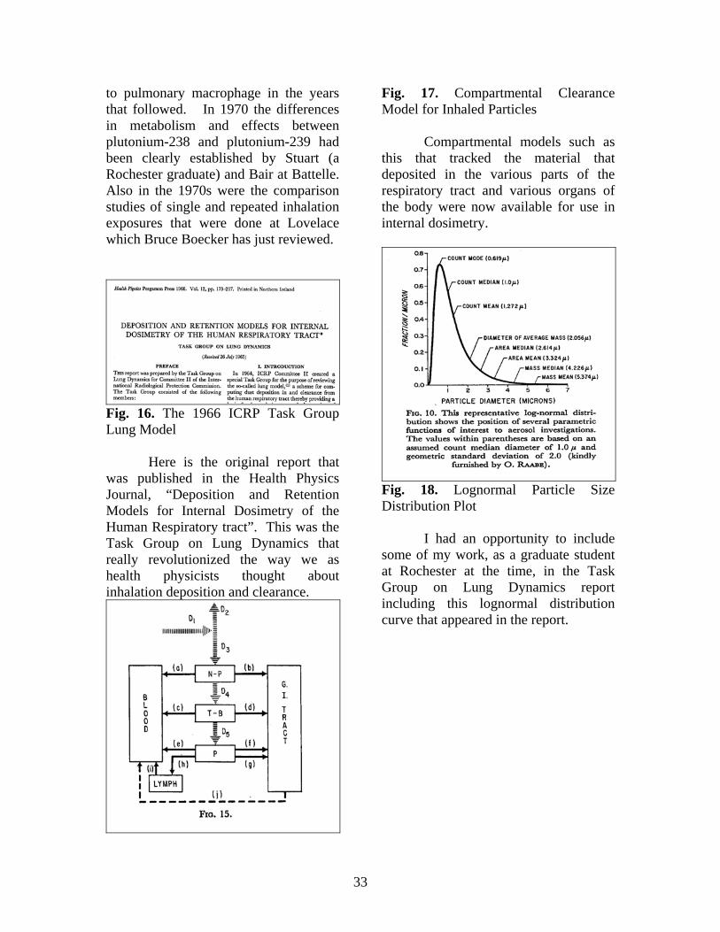
Fig. 18. Lognormal Particle Size Distribution Plot
I had an opportunity to include some of my work, as a graduate student at Rochester at the time, in the Task Group on Lung Dynamics report including this lognormal distribution curve that appeared in the report.
34
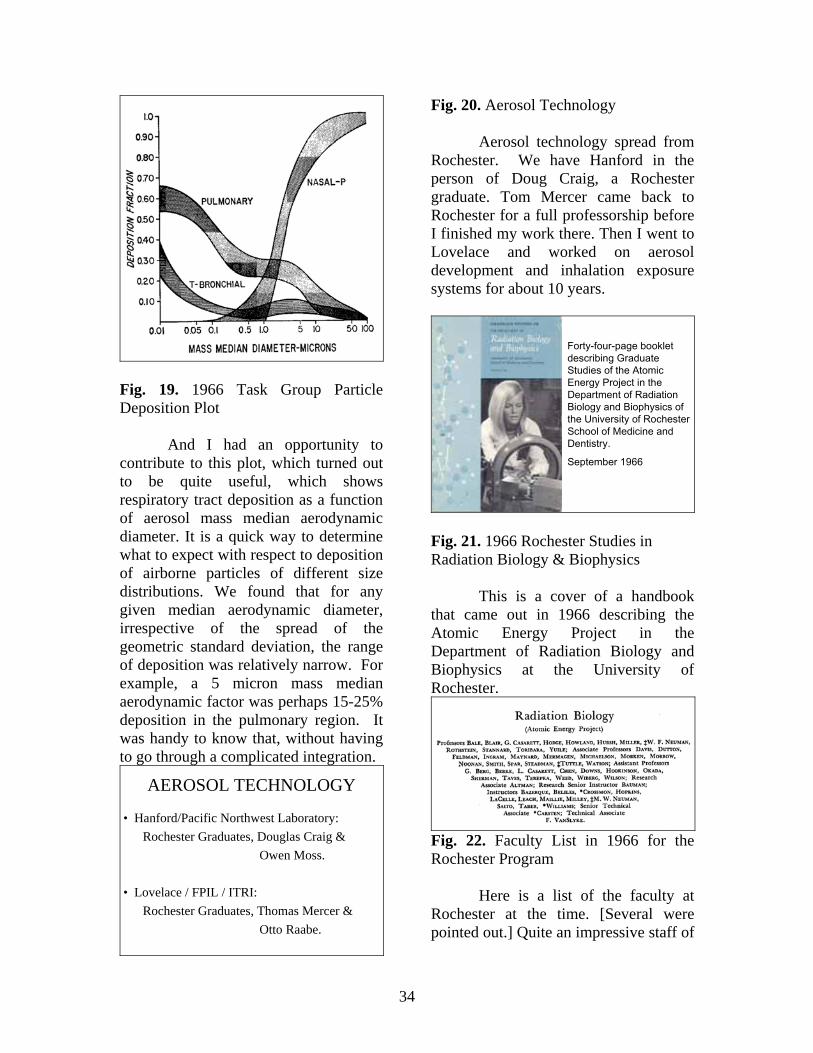
Fig. 19. 1966 Task Group Particle Deposition Plot
And I had an opportunity to contribute to this plot, which turned out to be quite useful, which shows respiratory tract deposition as a function of aerosol mass median aerodynamic diameter. It is a quick way to determine what to expect with respect to deposition of airborne particles of different size distributions. We found that for any given median aerodynamic diameter, irrespective of the spread of the geometric standard deviation, the range of deposition was relatively narrow. For example, a 5 micron mass median aerodynamic factor was perhaps 15-25% deposition in the pulmonary region. It was handy to know that, without having to go through a complicated integration.
AEROSOL TECHNOLOGY
• Hanford/Pacific Northwest Laboratory:Rochester Graduates, Douglas Craig &
Owen Moss.
• Lovelace / FPIL / ITRI:Rochester Graduates, Thomas Mercer &
Otto Raabe.
Fig. 20. Aerosol Technology
Aerosol technology spread from Rochester. We have Hanford in the person of Doug Craig, a Rochester graduate. Tom Mercer came back to Rochester for a full professorship before I finished my work there. Then I went to Lovelace and worked on aerosol development and inhalation exposure systems for about 10 years.
Forty-four-page booklet describing Graduate Studies of the Atomic Energy Project in the Department of Radiation Biology and Biophysics of the University of Rochester School of Medicine and Dentistry.
September 1966
Fig. 21. 1966 Rochester Studies in Radiation Biology & Biophysics
This is a cover of a handbook that came out in 1966 describing the Atomic Energy Project in the Department of Radiation Biology and Biophysics at the University of Rochester.
Fig. 22. Faculty List in 1966 for the Rochester Program
Here is a list of the faculty at Rochester at the time. [Several were pointed out.] Quite an impressive staff of
35
people like Stannard, Hodge and Casarett were in the University of Rochester Department of Radiation Biology and Biophysics in 1966.
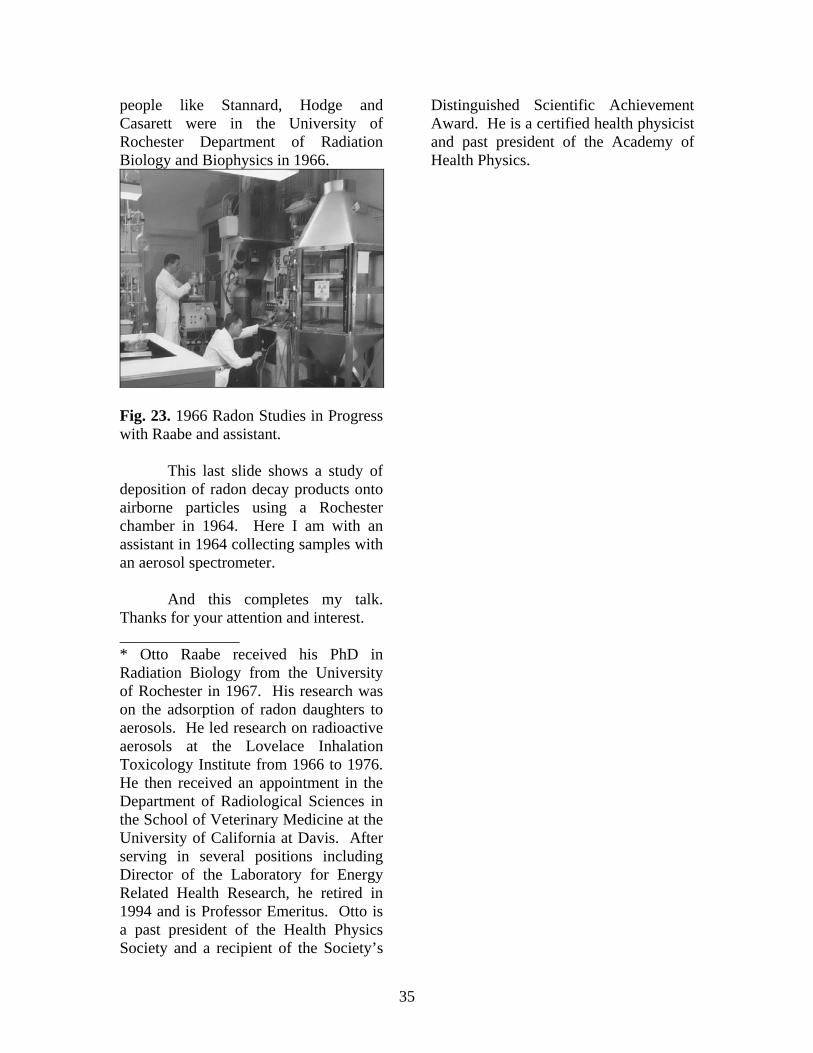
Fig. 23. 1966 Radon Studies in Progress with Raabe and assistant.
This last slide shows a study of deposition of radon decay products onto airborne particles using a Rochester chamber in 1964. Here I am with an assistant in 1964 collecting samples with an aerosol spectrometer.
And this completes my talk. Thanks for your attention and interest. _______________ * Otto Raabe received his PhD in Radiation Biology from the University of Rochester in 1967. His research was on the adsorption of radon daughters to aerosols. He led research on radioactive aerosols at the Lovelace Inhalation Toxicology Institute from 1966 to 1976. He then received an appointment in the Department of Radiological Sciences in the School of Veterinary Medicine at the University of California at Davis. After serving in several positions including Director of the Laboratory for Energy Related Health Research, he retired in 1994 and is Professor Emeritus. Otto is a past president of the Health Physics Society and a recipient of the Society’s
Distinguished Scientific Achievement Award. He is a certified health physicist and past president of the Academy of Health Physics.

36
37
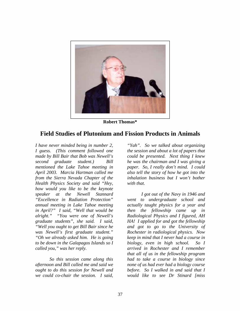
Robert Thomas*
Field Studies of Plutonium and Fission Products in Animals
I have never minded being in number 2, I guess. (This comment followed one made by Bill Bair that Bob was Newell’s second graduate student.) Bill mentioned the Lake Tahoe meeting in April 2003. Marcia Hartman called me from the Sierra Nevada Chapter of the Health Physics Society and said “Hey, how would you like to be the keynote speaker at the Newell Stannard “Excellence in Radiation Protection” annual meeting in Lake Tahoe meeting in April?” I said, “Well that would be alright.” “You were one of Newell’s graduate students”, she said. I said, “Well you ought to get Bill Bair since he was Newell’s first graduate student.” “Oh we already asked him. He is going to be down in the Galapagos Islands so I called you,” was her reply.
So this session came along this afternoon and Bill called me and said we ought to do this session for Newell and we could co-chair the session. I said,
“Yah”. So we talked about organizing the session and about a lot of papers that could be presented. Next thing I knew he was the chairman and I was giving a paper. So, I really don’t mind. I could also tell the story of how he got into the inhalation business but I won’t bother with that.
I got out of the Navy in 1946 and went to undergraduate school and actually taught physics for a year and then the fellowship came up in Radiological Physics and I figured, AH HA! I applied for and got the fellowship and got to go to the University of Rochester in radiological physics. Now keep in mind that I never had a course in biology, even in high school. So I arrived in Rochester and I remember that all of us in the fellowship program had to take a course in biology since none of us had ever had a biology course before. So I walked in and said that I would like to see Dr Stinard [miss
38
pronounced]. They all looked at me and laughed.
Anyway, it was decided that I would go back to Rochester and do graduate work after the summer health physics training at the Brookhaven National Laboratory. Newell said it was foolish to waste my time getting a master’s degree if I was going to get a PhD. I had not thought about that since I assumed everyone was getting a master’s degree after a year and then going to work. He said that it would not benefit me in the long run to get a master’s degree and that I should get a PhD. And I appreciated his advice. It only took me 5 calendar years to do it.
Bill forgot to tell you that I was also the president of the NRS – National Renegade Society. I have been around a number of the national laboratories and picked up the title of being a renegade.
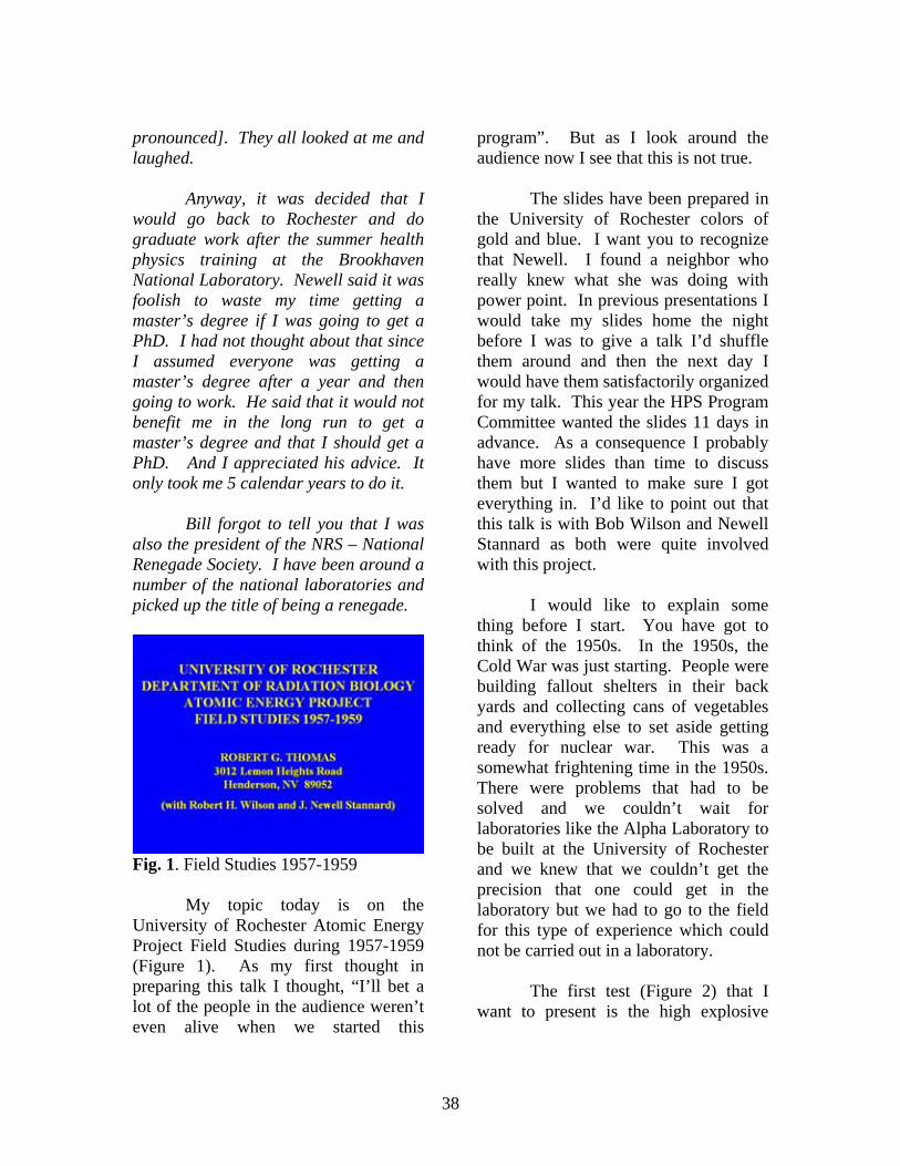
Fig. 1. Field Studies 1957-1959
My topic today is on the University of Rochester Atomic Energy Project Field Studies during 1957-1959 (Figure 1). As my first thought in preparing this talk I thought, “I’ll bet a lot of the people in the audience weren’t even alive when we started this
program”. But as I look around the audience now I see that this is not true.
The slides have been prepared in the University of Rochester colors of gold and blue. I want you to recognize that Newell. I found a neighbor who really knew what she was doing with power point. In previous presentations I would take my slides home the night before I was to give a talk I’d shuffle them around and then the next day I would have them satisfactorily organized for my talk. This year the HPS Program Committee wanted the slides 11 days in advance. As a consequence I probably have more slides than time to discuss them but I wanted to make sure I got everything in. I’d like to point out that this talk is with Bob Wilson and Newell Stannard as both were quite involved with this project.
I would like to explain some thing before I start. You have got to think of the 1950s. In the 1950s, the Cold War was just starting. People were building fallout shelters in their back yards and collecting cans of vegetables and everything else to set aside getting ready for nuclear war. This was a somewhat frightening time in the 1950s. There were problems that had to be solved and we couldn’t wait for laboratories like the Alpha Laboratory to be built at the University of Rochester and we knew that we couldn’t get the precision that one could get in the laboratory but we had to go to the field for this type of experience which could not be carried out in a laboratory.
The first test (Figure 2) that I want to present is the high explosive
39
detonation of a nuclear weapon at the Nevada Test Site in 1957 (Project 57).
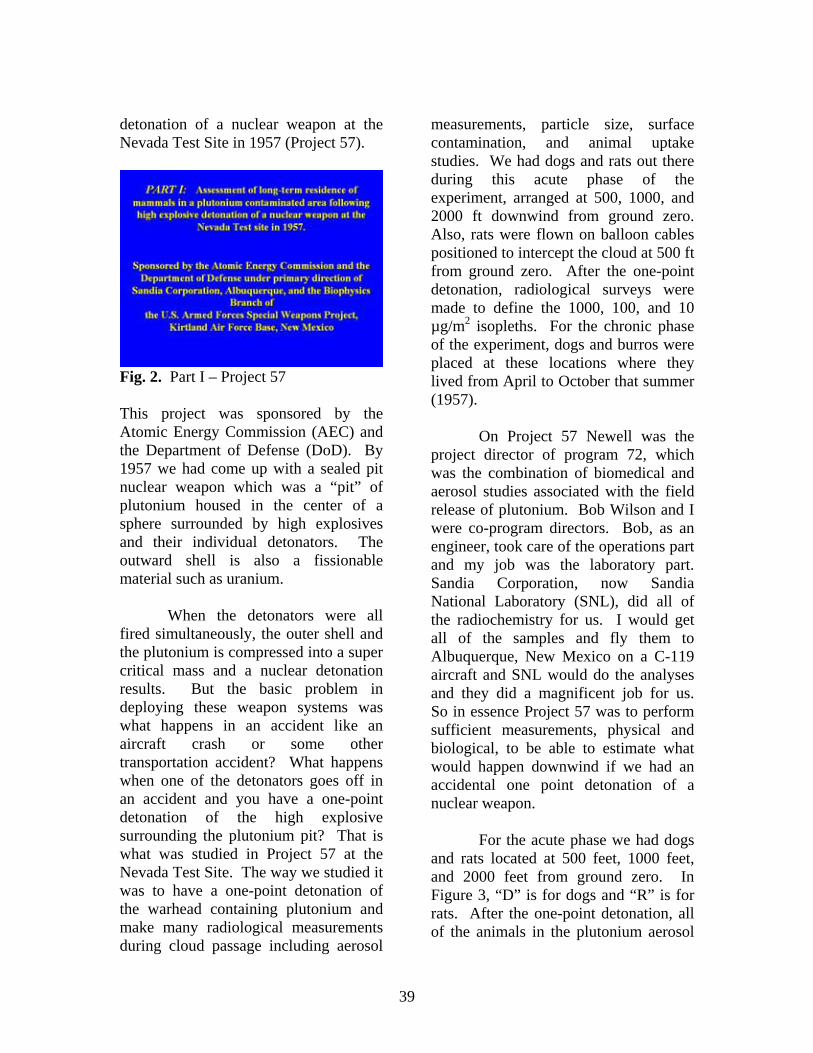
Fig. 2. Part I – Project 57 This project was sponsored by the Atomic Energy Commission (AEC) and the Department of Defense (DoD). By 1957 we had come up with a sealed pit nuclear weapon which was a “pit” of plutonium housed in the center of a sphere surrounded by high explosives and their individual detonators. The outward shell is also a fissionable material such as uranium.
When the detonators were all fired simultaneously, the outer shell and the plutonium is compressed into a super critical mass and a nuclear detonation results. But the basic problem in deploying these weapon systems was what happens in an accident like an aircraft crash or some other transportation accident? What happens when one of the detonators goes off in an accident and you have a one-point detonation of the high explosive surrounding the plutonium pit? That is what was studied in Project 57 at the Nevada Test Site. The way we studied it was to have a one-point detonation of the warhead containing plutonium and make many radiological measurements during cloud passage including aerosol
measurements, particle size, surface contamination, and animal uptake studies. We had dogs and rats out there during this acute phase of the experiment, arranged at 500, 1000, and 2000 ft downwind from ground zero. Also, rats were flown on balloon cables positioned to intercept the cloud at 500 ft from ground zero. After the one-point detonation, radiological surveys were made to define the 1000, 100, and 10 µg/m2 isopleths. For the chronic phase of the experiment, dogs and burros were placed at these locations where they lived from April to October that summer (1957).
On Project 57 Newell was the project director of program 72, which was the combination of biomedical and aerosol studies associated with the field release of plutonium. Bob Wilson and I were co-program directors. Bob, as an engineer, took care of the operations part and my job was the laboratory part. Sandia Corporation, now Sandia National Laboratory (SNL), did all of the radiochemistry for us. I would get all of the samples and fly them to Albuquerque, New Mexico on a C-119 aircraft and SNL would do the analyses and they did a magnificent job for us. So in essence Project 57 was to perform sufficient measurements, physical and biological, to be able to estimate what would happen downwind if we had an accidental one point detonation of a nuclear weapon.
For the acute phase we had dogs and rats located at 500 feet, 1000 feet, and 2000 feet from ground zero. In Figure 3, “D” is for dogs and “R” is for rats. After the one-point detonation, all of the animals in the plutonium aerosol
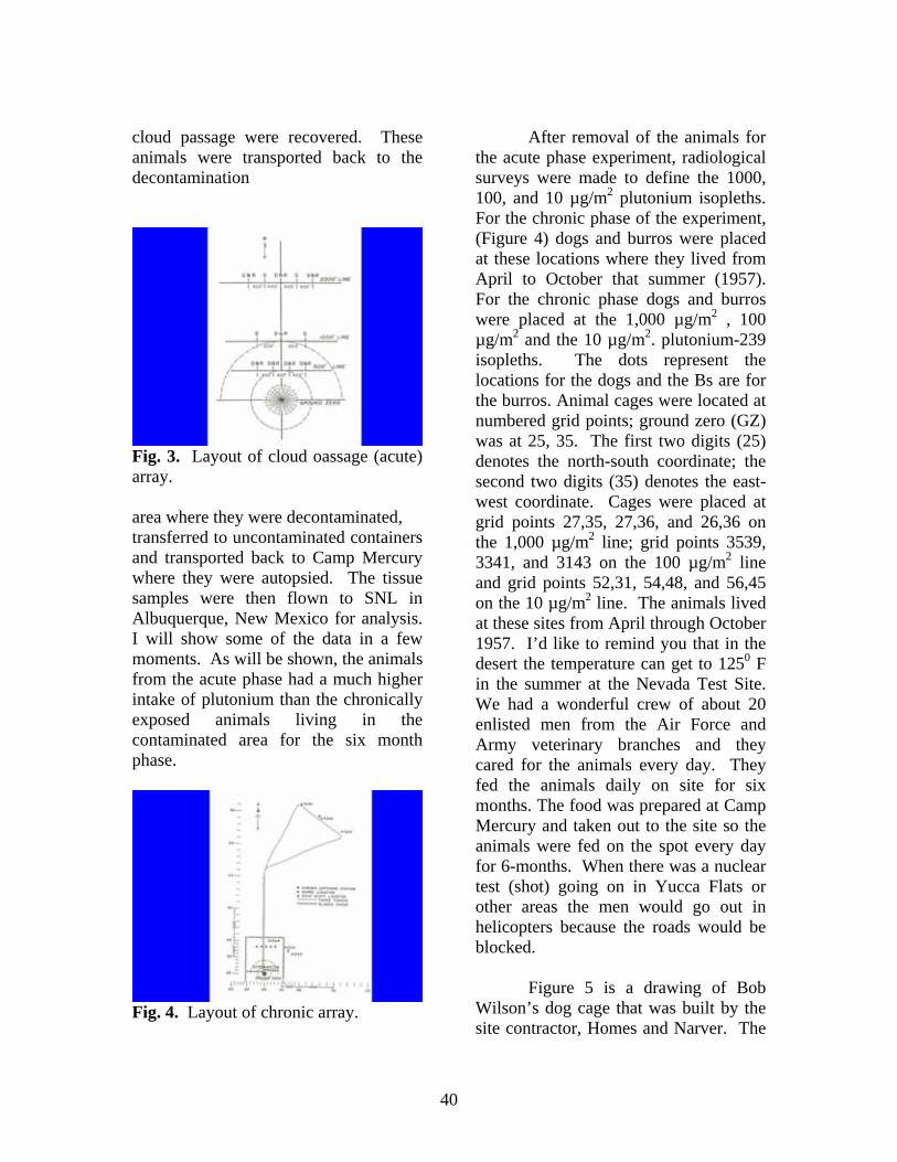
40
cloud passage were recovered. These animals were transported back to the decontamination
Fig. 3. Layout of cloud oassage (acute) array. area where they were decontaminated, transferred to uncontaminated containers and transported back to Camp Mercury where they were autopsied. The tissue samples were then flown to SNL in Albuquerque, New Mexico for analysis. I will show some of the data in a few moments. As will be shown, the animals from the acute phase had a much higher intake of plutonium than the chronically exposed animals living in the contaminated area for the six month phase.
Fig. 4. Layout of chronic array.
After removal of the animals for the acute phase experiment, radiological surveys were made to define the 1000, 100, and 10 µg/m2 plutonium isopleths. For the chronic phase of the experiment, (Figure 4) dogs and burros were placed at these locations where they lived from April to October that summer (1957). For the chronic phase dogs and burros were placed at the 1,000 µg/m2 , 100 µg/m2 and the 10 µg/m2. plutonium-239 isopleths. The dots represent the locations for the dogs and the Bs are for the burros. Animal cages were located at numbered grid points; ground zero (GZ) was at 25, 35. The first two digits (25) denotes the north-south coordinate; the second two digits (35) denotes the east-west coordinate. Cages were placed at grid points 27,35, 27,36, and 26,36 on the 1,000 µg/m2 line; grid points 3539, 3341, and 3143 on the 100 µg/m2 line and grid points 52,31, 54,48, and 56,45 on the 10 µg/m2 line. The animals lived at these sites from April through October 1957. I’d like to remind you that in the desert the temperature can get to 1250 F in the summer at the Nevada Test Site. We had a wonderful crew of about 20 enlisted men from the Air Force and Army veterinary branches and they cared for the animals every day. They fed the animals daily on site for six months. The food was prepared at Camp Mercury and taken out to the site so the animals were fed on the spot every day for 6-months. When there was a nuclear test (shot) going on in Yucca Flats or other areas the men would go out in helicopters because the roads would be blocked.
Figure 5 is a drawing of Bob Wilson’s dog cage that was built by the site contractor, Homes and Narver. The
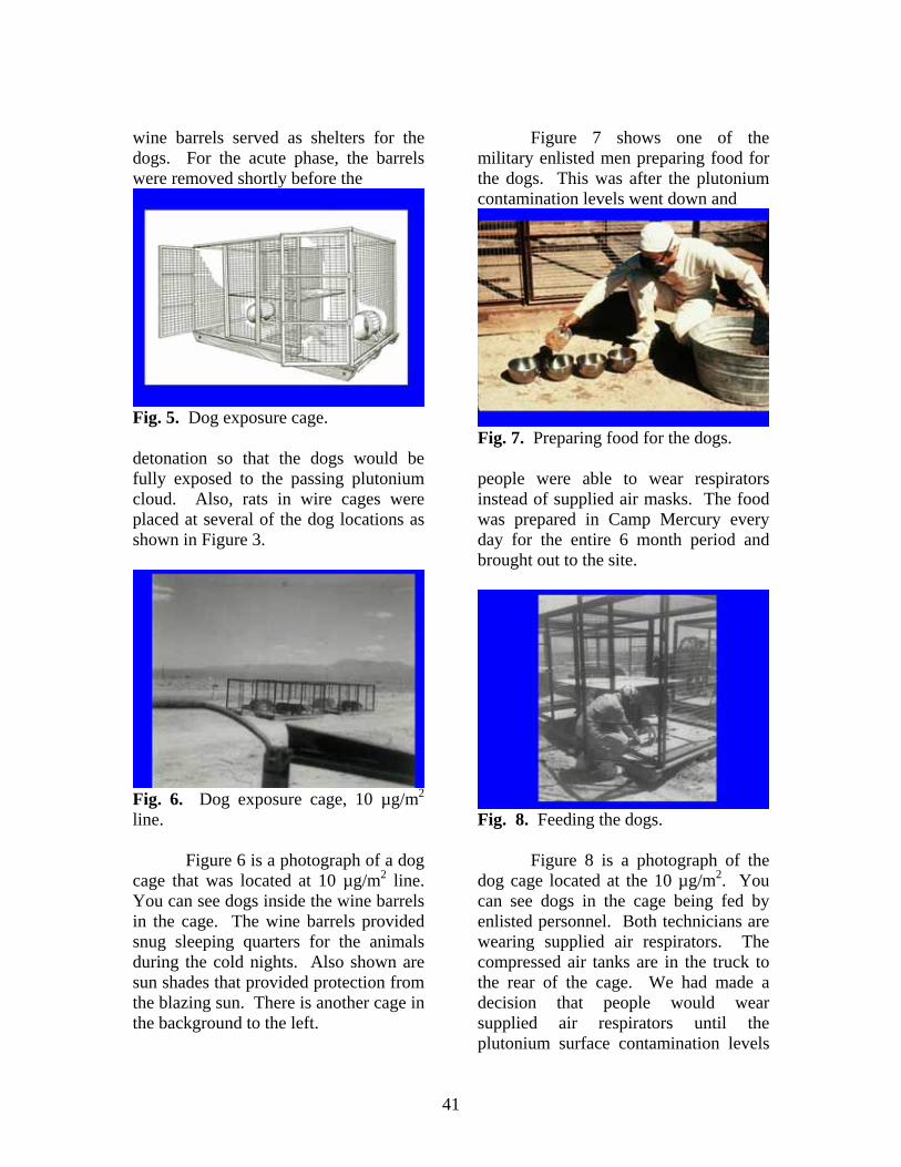
41
wine barrels served as shelters for the dogs. For the acute phase, the barrels were removed shortly before the
Fig. 5. Dog exposure cage. detonation so that the dogs would be fully exposed to the passing plutonium cloud. Also, rats in wire cages were placed at several of the dog locations as shown in Figure 3.
Fig. 6. Dog exposure cage, 10 µg/m2 line.
Figure 6 is a photograph of a dog cage that was located at 10 µg/m2 line. You can see dogs inside the wine barrels in the cage. The wine barrels provided snug sleeping quarters for the animals during the cold nights. Also shown are sun shades that provided protection from the blazing sun. There is another cage in the background to the left.
Figure 7 shows one of the military enlisted men preparing food for the dogs. This was after the plutonium contamination levels went down and
Fig. 7. Preparing food for the dogs. people were able to wear respirators instead of supplied air masks. The food was prepared in Camp Mercury every day for the entire 6 month period and brought out to the site.
Fig. 8. Feeding the dogs.
Figure 8 is a photograph of the dog cage located at the 10 µg/m2. You can see dogs in the cage being fed by enlisted personnel. Both technicians are wearing supplied air respirators. The compressed air tanks are in the truck to the rear of the cage. We had made a decision that people would wear supplied air respirators until the plutonium surface contamination levels
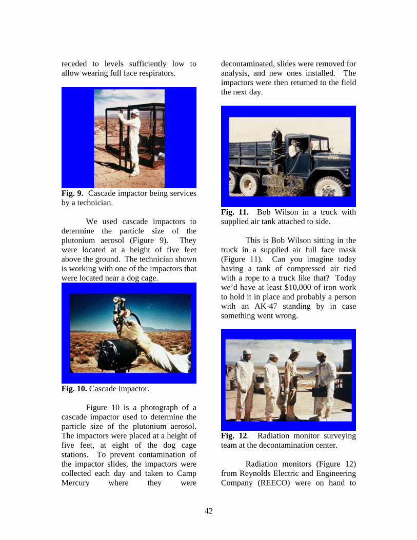
42
receded to levels sufficiently low to allow wearing full face respirators.
Fig. 9. Cascade impactor being services by a technician.
We used cascade impactors to determine the particle size of the plutonium aerosol (Figure 9). They were located at a height of five feet above the ground. The technician shown is working with one of the impactors that were located near a dog cage.
Fig. 10. Cascade impactor.
Figure 10 is a photograph of a cascade impactor used to determine the particle size of the plutonium aerosol. The impactors were placed at a height of five feet, at eight of the dog cage stations. To prevent contamination of the impactor slides, the impactors were collected each day and taken to Camp Mercury where they were
decontaminated, slides were removed for analysis, and new ones installed. The impactors were then returned to the field the next day.
Fig. 11. Bob Wilson in a truck with supplied air tank attached to side.
This is Bob Wilson sitting in the truck in a supplied air full face mask (Figure 11). Can you imagine today having a tank of compressed air tied with a rope to a truck like that? Today we’d have at least $10,000 of iron work to hold it in place and probably a person with an AK-47 standing by in case something went wrong.
Fig. 12. Radiation monitor surveying team at the decontamination center.
Radiation monitors (Figure 12) from Reynolds Electric and Engineering Company (REECO) were on hand to
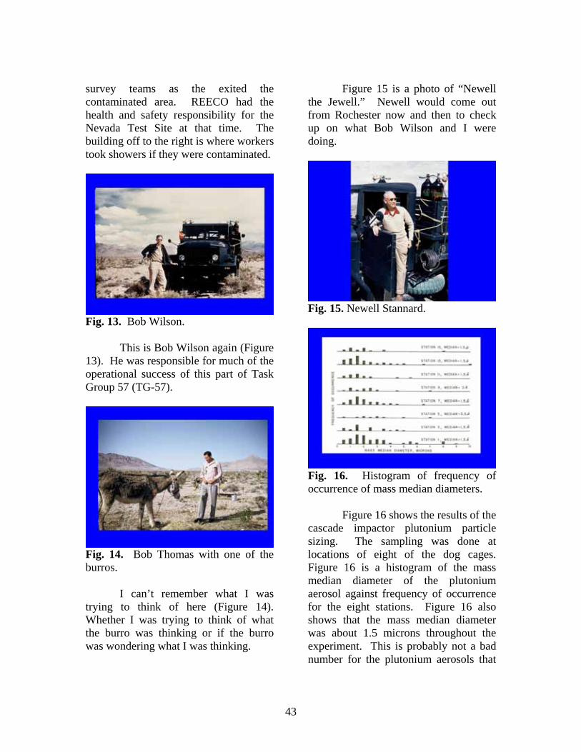
43
survey teams as the exited the contaminated area. REECO had the health and safety responsibility for the Nevada Test Site at that time. The building off to the right is where workers took showers if they were contaminated.
Fig. 13. Bob Wilson.
This is Bob Wilson again (Figure 13). He was responsible for much of the operational success of this part of Task Group 57 (TG-57).
Fig. 14. Bob Thomas with one of the burros.
I can’t remember what I was trying to think of here (Figure 14). Whether I was trying to think of what the burro was thinking or if the burro was wondering what I was thinking.
Figure 15 is a photo of “Newell the Jewell.” Newell would come out from Rochester now and then to check up on what Bob Wilson and I were doing.
Fig. 15. Newell Stannard.
Fig. 16. Histogram of frequency of occurrence of mass median diameters.
Figure 16 shows the results of the cascade impactor plutonium particle sizing. The sampling was done at locations of eight of the dog cages. Figure 16 is a histogram of the mass median diameter of the plutonium aerosol against frequency of occurrence for the eight stations. Figure 16 also shows that the mass median diameter was about 1.5 microns throughout the experiment. This is probably not a bad number for the plutonium aerosols that
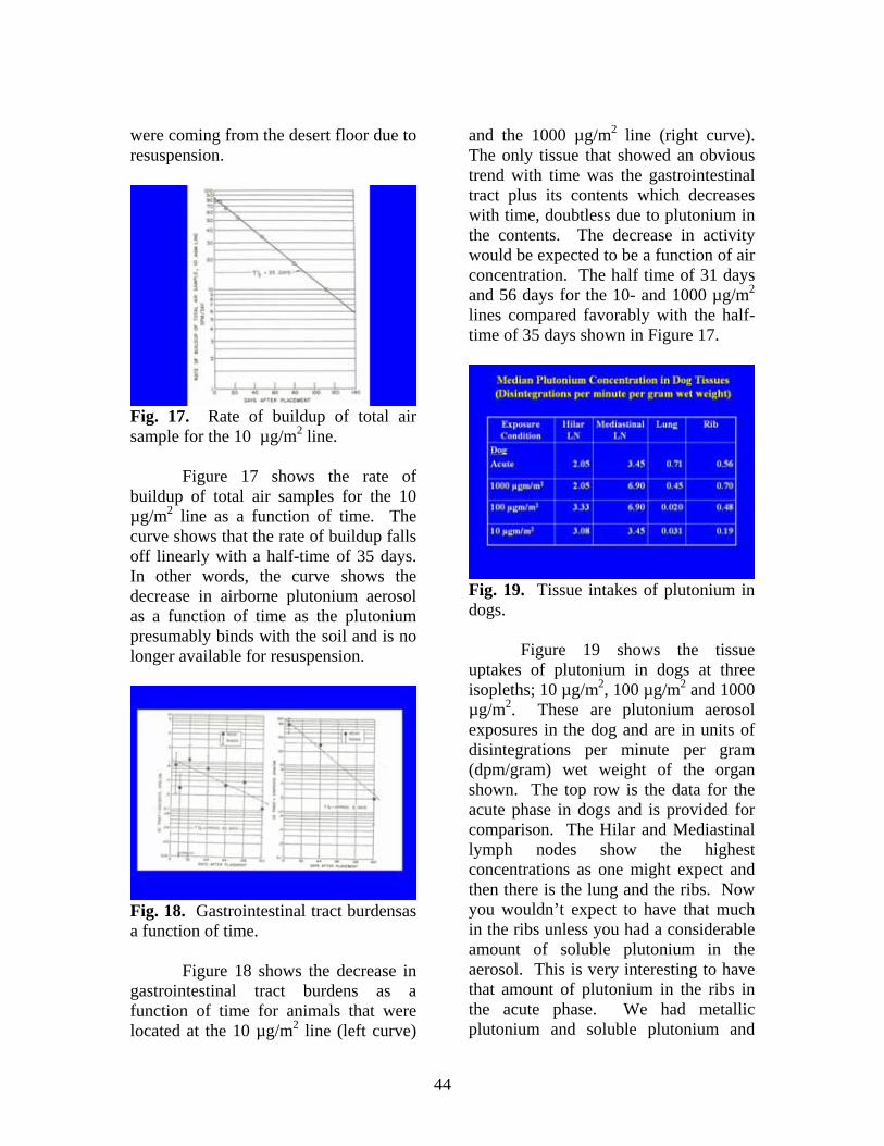
44
were coming from the desert floor due to resuspension.
Fig. 17. Rate of buildup of total air sample for the 10 µg/m2 line.
Figure 17 shows the rate of buildup of total air samples for the 10 µg/m2 line as a function of time. The curve shows that the rate of buildup falls off linearly with a half-time of 35 days. In other words, the curve shows the decrease in airborne plutonium aerosol as a function of time as the plutonium presumably binds with the soil and is no longer available for resuspension.
Fig. 18. Gastrointestinal tract burdensas a function of time.
Figure 18 shows the decrease in gastrointestinal tract burdens as a function of time for animals that were located at the 10 µg/m2 line (left curve)
and the 1000 µg/m2 line (right curve). The only tissue that showed an obvious trend with time was the gastrointestinal tract plus its contents which decreases with time, doubtless due to plutonium in the contents. The decrease in activity would be expected to be a function of air concentration. The half time of 31 days and 56 days for the 10- and 1000 µg/m2 lines compared favorably with the half-time of 35 days shown in Figure 17.
Fig. 19. Tissue intakes of plutonium in dogs.
Figure 19 shows the tissue uptakes of plutonium in dogs at three isopleths; 10 µg/m2, 100 µg/m2 and 1000 µg/m2. These are plutonium aerosol exposures in the dog and are in units of disintegrations per minute per gram (dpm/gram) wet weight of the organ shown. The top row is the data for the acute phase in dogs and is provided for comparison. The Hilar and Mediastinal lymph nodes show the highest concentrations as one might expect and then there is the lung and the ribs. Now you wouldn’t expect to have that much in the ribs unless you had a considerable amount of soluble plutonium in the aerosol. This is very interesting to have that amount of plutonium in the ribs in the acute phase. We had metallic plutonium and soluble plutonium and
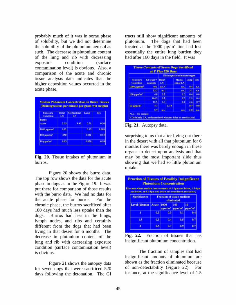
45
probably much of it was in some phase of solubility, but we did not determine the solubility of the plutonium aerosol as such. The decrease in plutonium content of the lung and rib with decreasing exposure condition (surface contamination level) is obvious. Also, a comparison of the acute and chronic tissue analysis data indicates that the higher deposition values occurred in the acute phase.
Fig. 20. Tissue intakes of plutonium in burros.
Figure 20 shows the burro data. The top row shows the data for the acute phase in dogs as in the Figure 19. It was put there for comparison of those results with the burro data. We had no data for the acute phase for burros. For the chronic phase, the burros sacrificed after 180 days had much less uptake than the dogs. Burros had less in the lungs, lymph nodes, and ribs and certainly different from the dogs that had been living in that desert for 6 months. The decrease in plutonium content of the lung and rib with decreasing exposure condition (surface contamination level) is obvious.
Figure 21 shows the autopsy data for seven dogs that were sacrificed 520 days following the detonation. The GI
tracts still show significant amounts of plutonium. The dogs that had been located at the 1000 µg/m2 line had lost essentially the entire lung burden they had after 160 days in the field. It was
Fig. 21. Autopsy data. surprising to us that after living out there in the desert with all that plutonium for 6 months there was barely enough in these organs to detect upon analysis and that may be the most important slide thus showing that we had so little plutonium uptake.
Fig. 22. Fraction of tissues that has insignificant plutonium concentration.
The fraction of samples that had insignificant amounts of plutonium are shown as the fraction eliminated because of non-detectability (Figure 22). For instance, at the significance level of 1.5
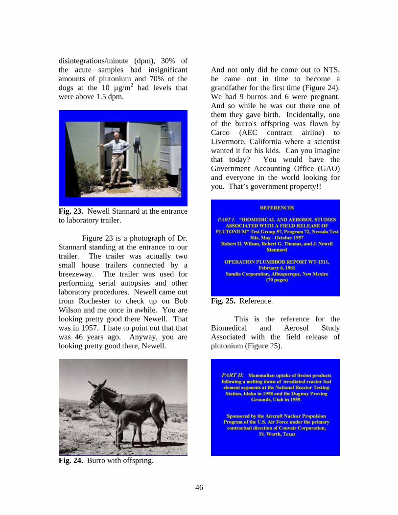
46
disintegrations/minute (dpm), 30% of the acute samples had insignificant amounts of plutonium and 70% of the dogs at the 10 µg/m2 had levels that were above 1.5 dpm.
Fig. 23. Newell Stannard at the entrance to laboratory trailer.
Figure 23 is a photograph of Dr. Stannard standing at the entrance to our trailer. The trailer was actually two small house trailers connected by a breezeway. The trailer was used for performing serial autopsies and other laboratory procedures. Newell came out from Rochester to check up on Bob Wilson and me once in awhile. You are looking pretty good there Newell. That was in 1957. I hate to point out that that was 46 years ago. Anyway, you are looking pretty good there, Newell.
Fig. 24. Burro with offspring.
And not only did he come out to NTS, he came out in time to become a grandfather for the first time (Figure 24). We had 9 burros and 6 were pregnant. And so while he was out there one of them they gave birth. Incidentally, one of the burro's offspring was flown by Carco (AEC contract airline) to Livermore, California where a scientist wanted it for his kids. Can you imagine that today? You would have the Government Accounting Office (GAO) and everyone in the world looking for you. That’s government property!!
Fig. 25. Reference.
This is the reference for the Biomedical and Aerosol Study Associated with the field release of plutonium (Figure 25).
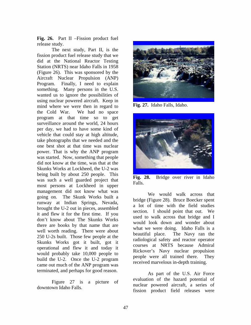
47
Fig. 26. Part II –Fission product fuel release study.
The next study, Part II, is the fission product fuel release study that we did at the National Reactor Testing Station (NRTS) near Idaho Falls in 1958 (Figure 26). This was sponsored by the Aircraft Nuclear Propulsion (ANP) Program. Finally, I need to explain something. Many persons in the U.S. wanted us to ignore the possibilities of using nuclear powered aircraft. Keep in mind where we were then in regard to the Cold War. We had no space program at that time so to get surveillance around the world, 24 hours per day, we had to have some kind of vehicle that could stay at high altitude, take photographs that we needed and the one best shot at that time was nuclear power. That is why the ANP program was started. Now, something that people did not know at the time, was that at the Skunks Works at Lockheed, the U-2 was being built by about 250 people. This was such a well guarded project that most persons at Lockheed in upper management did not know what was going on. The Skunk Works built a runway at Indian Springs, Nevada, brought the U-2 out in pieces, assembled it and flew it for the first time. If you don’t know about The Skunks Works there are books by that name that are well worth reading. There were about 250 U-2s built. Those few people at the Skunks Works got it built, got it operational and flew it and today it would probably take 10,000 people to build the U-2. Once the U-2 program came out much of the ANP program was terminated, and perhaps for good reason.
Figure 27 is a picture of
downtown Idaho Falls.
Fig. 27. Idaho Falls, Idaho.
Fig. 28. Bridge over river in Idaho Falls.
We would walk across that bridge (Figure 28). Bruce Boecker spent a lot of time with the field studies section. I should point that out. We used to walk across that bridge and I would look down and wonder about what we were doing. Idaho Falls is a beautiful place. The Navy ran the radiological safety and reactor operator courses at NRTS because Admiral Rickover’s Navy nuclear propulsion people were all trained there. They received marvelous in-depth training.
As part of the U.S. Air Force evaluation of the hazard potential of nuclear powered aircraft, a series of fission product field releases were
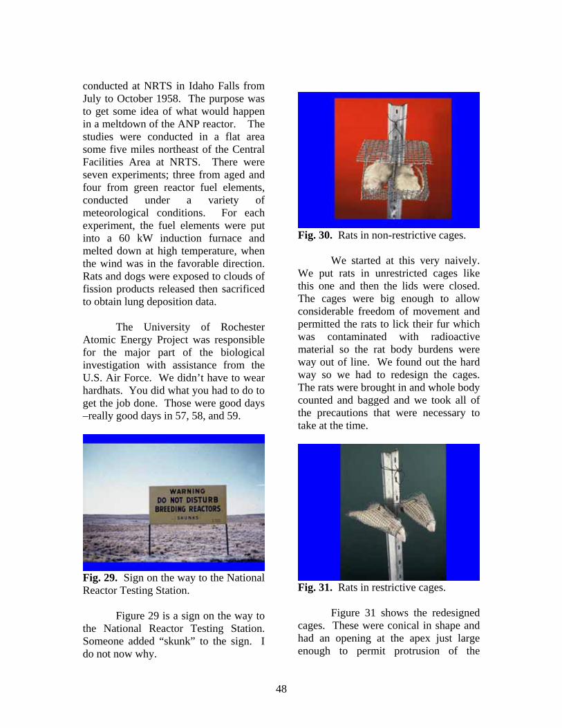
48
conducted at NRTS in Idaho Falls from July to October 1958. The purpose was to get some idea of what would happen in a meltdown of the ANP reactor. The studies were conducted in a flat area some five miles northeast of the Central Facilities Area at NRTS. There were seven experiments; three from aged and four from green reactor fuel elements, conducted under a variety of meteorological conditions. For each experiment, the fuel elements were put into a 60 kW induction furnace and melted down at high temperature, when the wind was in the favorable direction. Rats and dogs were exposed to clouds of fission products released then sacrificed to obtain lung deposition data.
The University of Rochester Atomic Energy Project was responsible for the major part of the biological investigation with assistance from the U.S. Air Force. We didn’t have to wear hardhats. You did what you had to do to get the job done. Those were good days –really good days in 57, 58, and 59.
Fig. 29. Sign on the way to the National Reactor Testing Station.
Figure 29 is a sign on the way to the National Reactor Testing Station. Someone added “skunk” to the sign. I do not now why.
Fig. 30. Rats in non-restrictive cages.
We started at this very naively. We put rats in unrestricted cages like this one and then the lids were closed. The cages were big enough to allow considerable freedom of movement and permitted the rats to lick their fur which was contaminated with radioactive material so the rat body burdens were way out of line. We found out the hard way so we had to redesign the cages. The rats were brought in and whole body counted and bagged and we took all of the precautions that were necessary to take at the time.
Fig. 31. Rats in restrictive cages.
Figure 31 shows the redesigned cages. These were conical in shape and had an opening at the apex just large enough to permit protrusion of the
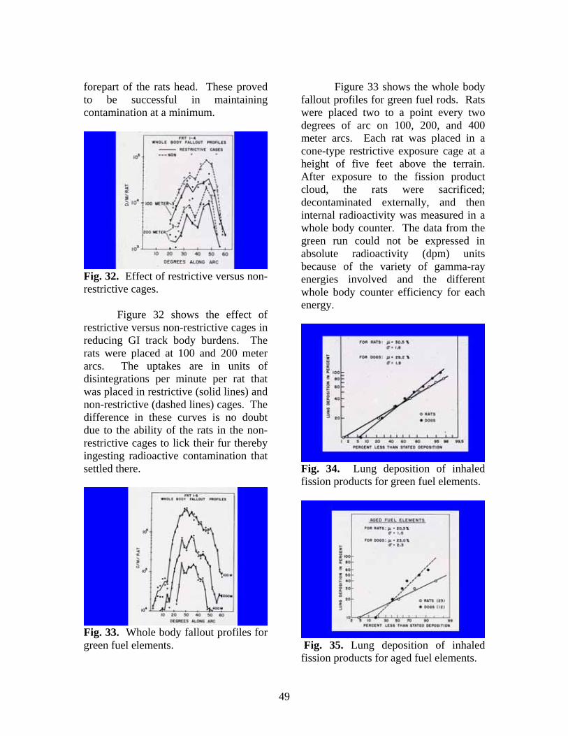
49
forepart of the rats head. These proved to be successful in maintaining contamination at a minimum.
Fig. 32. Effect of restrictive versus non-restrictive cages.
Figure 32 shows the effect of restrictive versus non-restrictive cages in reducing GI track body burdens. The rats were placed at 100 and 200 meter arcs. The uptakes are in units of disintegrations per minute per rat that was placed in restrictive (solid lines) and non-restrictive (dashed lines) cages. The difference in these curves is no doubt due to the ability of the rats in the non-restrictive cages to lick their fur thereby ingesting radioactive contamination that settled there.
Fig. 33. Whole body fallout profiles for green fuel elements.
Figure 33 shows the whole body fallout profiles for green fuel rods. Rats were placed two to a point every two degrees of arc on 100, 200, and 400 meter arcs. Each rat was placed in a cone-type restrictive exposure cage at a height of five feet above the terrain. After exposure to the fission product cloud, the rats were sacrificed; decontaminated externally, and then internal radioactivity was measured in a whole body counter. The data from the green run could not be expressed in absolute radioactivity (dpm) units because of the variety of gamma-ray energies involved and the different whole body counter efficiency for each energy.
Fig. 34. Lung deposition of inhaled fission products for green fuel elements.
Fig. 35. Lung deposition of inhaled fission products for aged fuel elements.
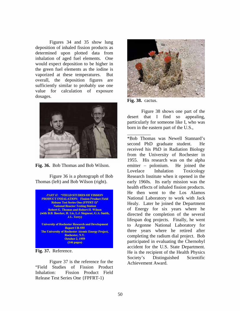
50
Figures 34 and 35 show lung
deposition of inhaled fission products as determined upon plotted data from inhalation of aged fuel elements. One would expect deposition to be higher in the green fuel elements as the iodine is vaporized at these temperatures. But overall, the deposition figures are sufficiently similar to probably use one value for calculation of exposure dosages.
Fig. 36. Bob Thomas and Bob Wilson.
Figure 36 is a photograph of Bob Thomas (left) and Bob Wilson (right).
Fig. 37. Reference.
Figure 37 is the reference for the “Field Studies of Fission Product Inhalation: Fission Product Field Release Test Series One {FPFRT-1)
Fig. 38. cactus.
Figure 38 shows one part of the desert that I find so appealing, particularly for someone like I, who was born in the eastern part of the U.S., __________ *Bob Thomas was Newell Stannard’s second PhD graduate student. He received his PhD in Radiation Biology from the University of Rochester in 1955. His research was on the alpha emitter – polonium. He joined the Lovelace Inhalation Toxicology Research Institute when it opened in the early 1960s. Its early mission was the health effects of inhaled fission products. He then went to the Los Alamos National Laboratory to work with Jack Healy. Later he joined the Department of Energy for six years where he directed the completion of the several lifespan dog projects. Finally, he went to Argonne National Laboratory for three years where he retired after completing the radium dial project. Bob participated in evaluating the Chernobyl accident for the U.S. State Department. He is the recipient of the Health Physics Society’s Distinguished Scientific Achievement Award.
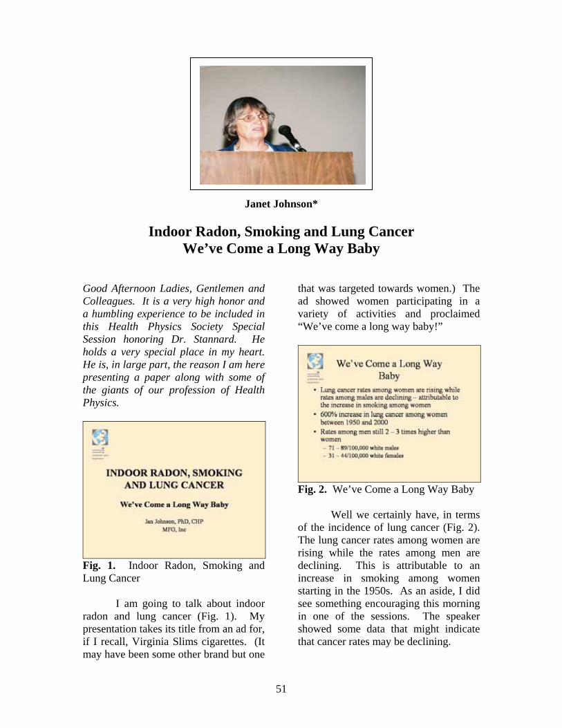
51
Janet Johnson*
Indoor Radon, Smoking and Lung Cancer We’ve Come a Long Way Baby
Good Afternoon Ladies, Gentlemen and Colleagues. It is a very high honor and a humbling experience to be included in this Health Physics Society Special Session honoring Dr. Stannard. He holds a very special place in my heart. He is, in large part, the reason I am here presenting a paper along with some of the giants of our profession of Health Physics.
Fig. 1. Indoor Radon, Smoking and Lung Cancer
I am going to talk about indoor radon and lung cancer (Fig. 1). My presentation takes its title from an ad for, if I recall, Virginia Slims cigarettes. (It may have been some other brand but one
that was targeted towards women.) The ad showed women participating in a variety of activities and proclaimed “We’ve come a long way baby!”
Fig. 2. We’ve Come a Long Way Baby
Well we certainly have, in terms of the incidence of lung cancer (Fig. 2). The lung cancer rates among women are rising while the rates among men are declining. This is attributable to an increase in smoking among women starting in the 1950s. As an aside, I did see something encouraging this morning in one of the sessions. The speaker showed some data that might indicate that cancer rates may be declining.
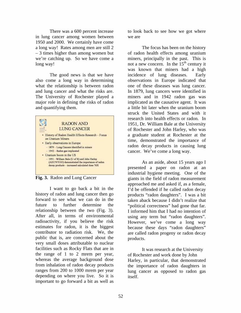
52
There was a 600 percent increase in lung cancer among women between 1950 and 2000. We certainly have come a long way! Rates among men are still 2 – 3 times higher than among women but we’re catching up. So we have come a long way!
The good news is that we have also come a long way in determining what the relationship is between radon and lung cancer and what the risks are. The University of Rochester played a major role in defining the risks of radon and quantifying them.
Fig. 3. Radon and Lung Cancer
I want to go back a bit in the history of radon and lung cancer then go forward to see what we can do in the future to further determine the relationship between the two (Fig. 3). After all, in terms of environmental radioactivity, if you believe the risk estimates for radon, it is the biggest contributor to radiation risk. We, the public that is, are concerned about the very small doses attributable to nuclear facilities such as Rocky Flats that are in the range of 1 to 2 mrem per year, whereas the average background dose from inhalation of radon decay products ranges from 200 to 1000 mrem per year depending on where you live. So it is important to go forward a bit as well as
to look back to see how we got where we are
The focus has been on the history of radon health effects among uranium miners, principally in the past. This is not a new concern. In the 15th century it was known that miners had a high incidence of lung diseases. Early observations in Europe indicated that one of these diseases was lung cancer. In 1879, lung cancers were identified in miners and in 1942 radon gas was implicated as the causative agent. It was a little bit later when the uranium boom struck the United States and with it research into health effects or radon. In 1951, Dr. William Bale at the University of Rochester and John Harley, who was a graduate student at Rochester at the time, demonstrated the importance of radon decay products in causing lung cancer. We’ve come a long way.
As an aside, about 15 years ago I presented a paper on radon at an industrial hygiene meeting. One of the giants in the field of radon measurement approached me and asked if, as a female, I’d be offended if he called radon decay products “radon daughters”. I was a bit taken aback because I didn’t realize that “political correctness” had gone that far. I informed him that I had no intention of using any term but “radon daughters”. However, we’ve come a long way because these days “radon daughters” are called radon progeny or radon decay products.
It was research at the University of Rochester and work done by John Harley, in particular, that demonstrated the importance of radon daughters in lung cancer as opposed to radon gas itself.
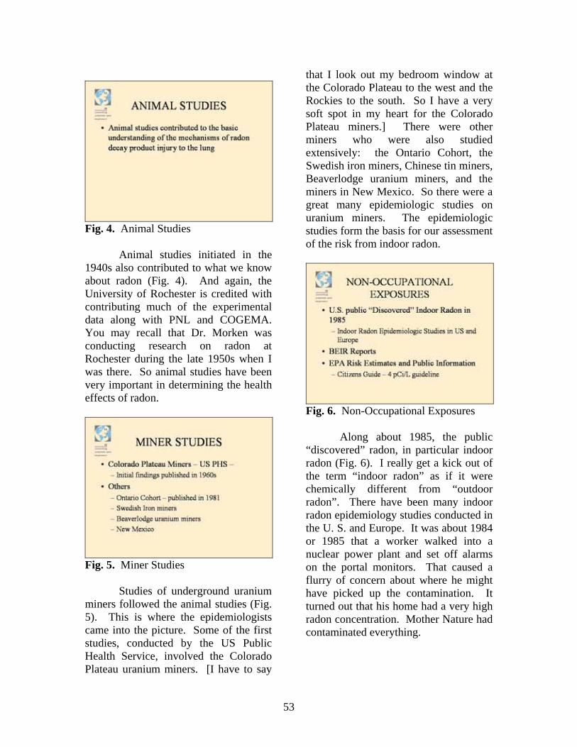
53
Fig. 4. Animal Studies
Animal studies initiated in the 1940s also contributed to what we know about radon (Fig. 4). And again, the University of Rochester is credited with contributing much of the experimental data along with PNL and COGEMA. You may recall that Dr. Morken was conducting research on radon at Rochester during the late 1950s when I was there. So animal studies have been very important in determining the health effects of radon.
Fig. 5. Miner Studies
Studies of underground uranium miners followed the animal studies (Fig. 5). This is where the epidemiologists came into the picture. Some of the first studies, conducted by the US Public Health Service, involved the Colorado Plateau uranium miners. [I have to say
that I look out my bedroom window at the Colorado Plateau to the west and the Rockies to the south. So I have a very soft spot in my heart for the Colorado Plateau miners.] There were other miners who were also studied extensively: the Ontario Cohort, the Swedish iron miners, Chinese tin miners, Beaverlodge uranium miners, and the miners in New Mexico. So there were a great many epidemiologic studies on uranium miners. The epidemiologic studies form the basis for our assessment of the risk from indoor radon.
Fig. 6. Non-Occupational Exposures
Along about 1985, the public “discovered” radon, in particular indoor radon (Fig. 6). I really get a kick out of the term “indoor radon” as if it were chemically different from “outdoor radon”. There have been many indoor radon epidemiology studies conducted in the U. S. and Europe. It was about 1984 or 1985 that a worker walked into a nuclear power plant and set off alarms on the portal monitors. That caused a flurry of concern about where he might have picked up the contamination. It turned out that his home had a very high radon concentration. Mother Nature had contaminated everything.
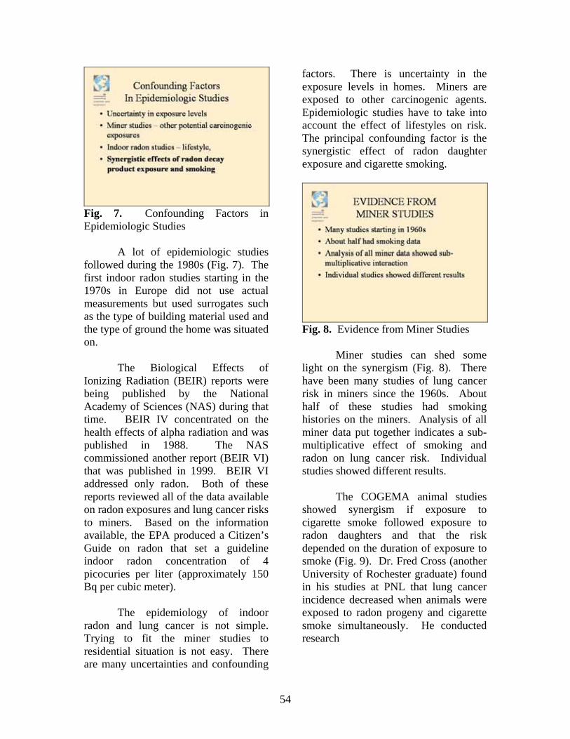
54
Fig. 7. Confounding Factors in Epidemiologic Studies
A lot of epidemiologic studies followed during the 1980s (Fig. 7). The first indoor radon studies starting in the 1970s in Europe did not use actual measurements but used surrogates such as the type of building material used and the type of ground the home was situated on.
The Biological Effects of Ionizing Radiation (BEIR) reports were being published by the National Academy of Sciences (NAS) during that time. BEIR IV concentrated on the health effects of alpha radiation and was published in 1988. The NAS commissioned another report (BEIR VI) that was published in 1999. BEIR VI addressed only radon. Both of these reports reviewed all of the data available on radon exposures and lung cancer risks to miners. Based on the information available, the EPA produced a Citizen’s Guide on radon that set a guideline indoor radon concentration of 4 picocuries per liter (approximately 150 Bq per cubic meter).
The epidemiology of indoor radon and lung cancer is not simple. Trying to fit the miner studies to residential situation is not easy. There are many uncertainties and confounding
factors. There is uncertainty in the exposure levels in homes. Miners are exposed to other carcinogenic agents. Epidemiologic studies have to take into account the effect of lifestyles on risk. The principal confounding factor is the synergistic effect of radon daughter exposure and cigarette smoking.
Fig. 8. Evidence from Miner Studies
Miner studies can shed some light on the synergism (Fig. 8). There have been many studies of lung cancer risk in miners since the 1960s. About half of these studies had smoking histories on the miners. Analysis of all miner data put together indicates a sub-multiplicative effect of smoking and radon on lung cancer risk. Individual studies showed different results.
The COGEMA animal studies showed synergism if exposure to cigarette smoke followed exposure to radon daughters and that the risk depended on the duration of exposure to smoke (Fig. 9). Dr. Fred Cross (another University of Rochester graduate) found in his studies at PNL that lung cancer incidence decreased when animals were exposed to radon progeny and cigarette smoke simultaneously. He conducted research
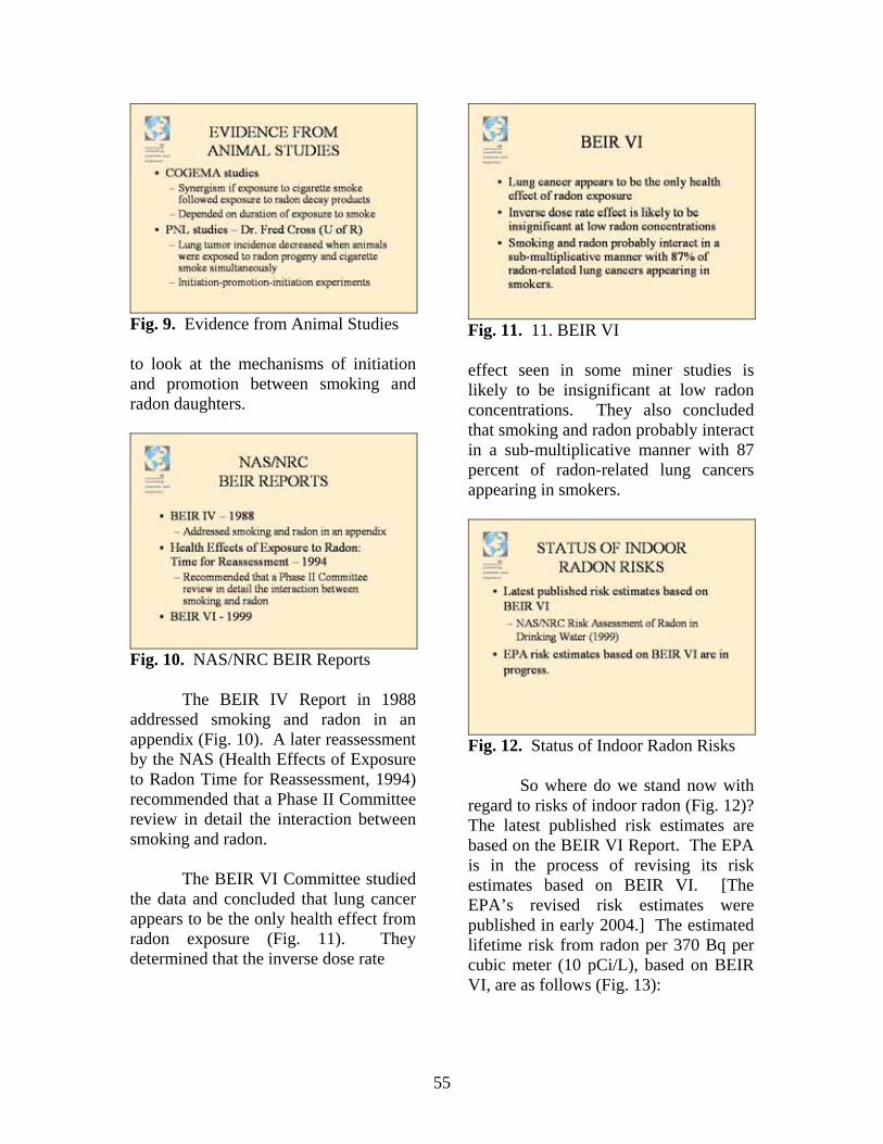
55
Fig. 9. Evidence from Animal Studies
to look at the mechanisms of initiation and promotion between smoking and radon daughters.
Fig. 10. NAS/NRC BEIR Reports
The BEIR IV Report in 1988 addressed smoking and radon in an appendix (Fig. 10). A later reassessment by the NAS (Health Effects of Exposure to Radon Time for Reassessment, 1994) recommended that a Phase II Committee review in detail the interaction between smoking and radon.
The BEIR VI Committee studied the data and concluded that lung cancer appears to be the only health effect from radon exposure (Fig. 11). They determined that the inverse dose rate
Fig. 11. 11. BEIR VI
effect seen in some miner studies is likely to be insignificant at low radon concentrations. They also concluded that smoking and radon probably interact in a sub-multiplicative manner with 87 percent of radon-related lung cancers appearing in smokers.
Fig. 12. Status of Indoor Radon Risks
So where do we stand now with regard to risks of indoor radon (Fig. 12)? The latest published risk estimates are based on the BEIR VI Report. The EPA is in the process of revising its risk estimates based on BEIR VI. [The EPA’s revised risk estimates were published in early 2004.] The estimated lifetime risk from radon per 370 Bq per cubic meter (10 pCi/L), based on BEIR VI, are as follows (Fig. 13):
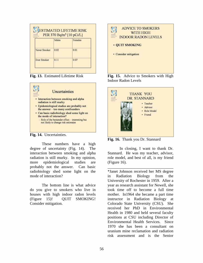
56
Fig. 13. Estimated Lifetime Risk
Fig. 14. Uncertainties.
These numbers have a high degree of uncertainty (Fig. 14). The interaction between smoking and alpha radiation is still murky. In my opinion, more epidemiological studies are probably not the answer. Can basic radiobiology shed some light on the mode of interaction?
The bottom line is what advice do you give to smokers who live in houses with high indoor radon levels (Figure 15)! QUIT SMOKING! Consider mitigation.
Fig. 15. Advice to Smokers with High Indoor Radon Levels
Fig. 16. Thank you Dr. Stannard
In closing, I want to thank Dr. Stannard. He was my teacher, advisor, role model, and best of all, is my friend (Figure 16). _________________________________ *Janet Johnson received her MS degree in Radiation Biology from the University of Rochester in 1959. After a year as research assistant for Newell, she took time off to become a full time mother. In1964 she became a part time instructor in Radiation Biology at Colorado State University (CSU). She received her PhD in Environmental Health in 1980 and held several faculty positions at CSU including Director of Environmental Health Services. Since 1970 she has been a consultant on uranium mine reclamation and radiation risk assessment and is the Senior
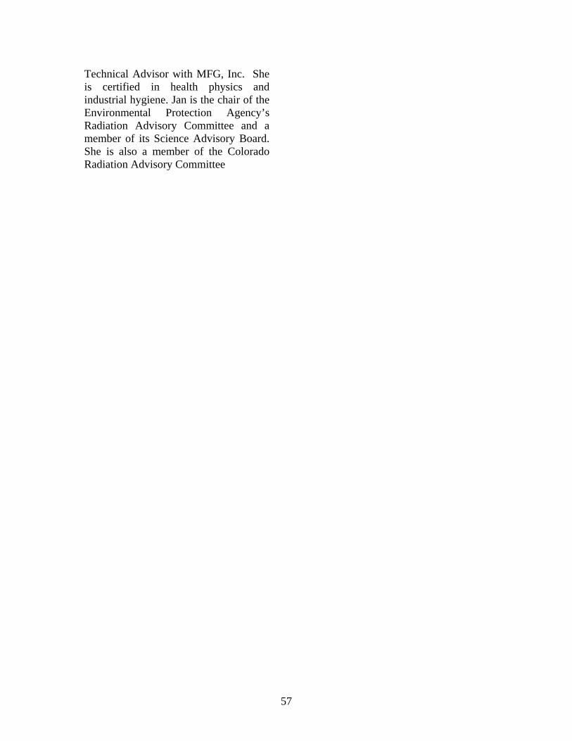
57
Technical Advisor with MFG, Inc. She is certified in health physics and industrial hygiene. Jan is the chair of the Environmental Protection Agency’s Radiation Advisory Committee and a member of its Science Advisory Board. She is also a member of the Colorado Radiation Advisory Committee

58
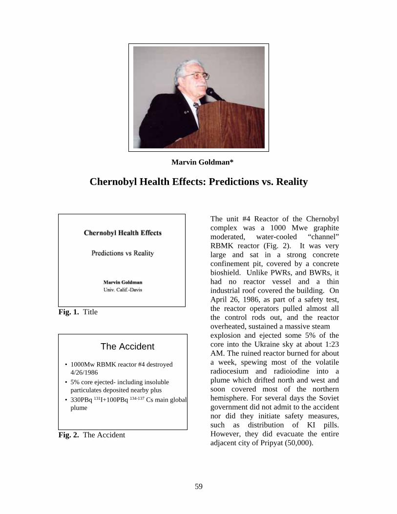
59
Marvin Goldman*
Chernobyl Health Effects: Predictions vs. Reality
Fig. 1. Title
Fig. 2. The Accident
The unit #4 Reactor of the Chernobyl complex was a 1000 Mwe graphite moderated, water-cooled “channel” RBMK reactor (Fig. 2). It was very large and sat in a strong concrete confinement pit, covered by a concrete bioshield. Unlike PWRs, and BWRs, it had no reactor vessel and a thin industrial roof covered the building. On April 26, 1986, as part of a safety test, the reactor operators pulled almost all the control rods out, and the reactor overheated, sustained a massive steam explosion and ejected some 5% of the core into the Ukraine sky at about 1:23 AM. The ruined reactor burned for about a week, spewing most of the volatile radiocesium and radioiodine into a plume which drifted north and west and soon covered most of the northern hemisphere. For several days the Soviet government did not admit to the accident nor did they initiate safety measures, such as distribution of KI pills. However, they did evacuate the entire adjacent city of Pripyat (50,000).
The Accident
• 1000Mw RBMK reactor #4 destroyed 4/26/1986
• 5% core ejected- including insoluble particulates deposited nearby plus
• 330PBq 131I+100PBq 134-137 Cs main global plume
60
Fig. 3. Acute, Non-Stochastic Health Effects.
Almost the entire local fire brigade was killed by radiation and burns; 31 died in the two months after exposure, including two who were killed that night (Fig. 3). About 200 more were reported to have acute radiation syndrome (ARS). The actual data are exactly what one might expect. The number with ARS was 145, including the fatalities. Since then, the survivors have been followed for more than 15 years, and although seven more have died, none of the deaths seem to be due to radiation exposure.
Fig. 4. Predicted Radiation Doses and Impacts.
We performed a global integration of all of the data and we used the collective dose to estimate that there might be 28,000 lethality’s (Fig. 4). We
did attempt to put together the statistics on all of the populations involved. There were 116 thousand USSR evacuees. There are 75 million people who were in the European part of the USSR and 225 million people in the rest of the USSR. The increased risk to all these populations was so small relative to the natural fatal cancer expectation for the populations shown in the slide, that there is no epidemiological test to ever indicate that the accident increased cancer cases.
Fig. 5. Actual Health Effects
However, the actual results that are seen today in the Ukraine claims that anywhere from 4000 to 7000 liquidator have died; almost all are most likely not caused by their radiation exposures (Fig. 5).
Radioiodine has induced thyroid
cancers in about 3000 children. The highest risk is in the youngest children that were exposed through their mother’s milk. The number of fatalities was about three. I expect the number to ultimately reach about 5,000 cases. The data already show that the younger the child, the greater the sensitivity and the shorter the latency; in some cases as little as three years after exposure.
Predicted Population Health Impacts
28~600,0001,2003,500No. Hemisphere
0.0241,0001.1226USA
0.6450,000272,600Asia (other)
1372,000580400Europe (other)
2.528,000110225Asian USSR
119,40047075European USSR
0.4117160.116USSR evacuees
Fatal CancerRad. (10+3)
Fatal CancerNatural(10+3)
Coll.Dose(10+3 pGy)
Population(10+6)
28~600,0001,2003,500No. Hemisphere
0.0241,0001.1226USA
0.6450,000272,600Asia (other)
1372,000580400Europe (other)
2.528,000110225Asian USSR
119,40047075European USSR
0.4117160.116USSR evacuees
Fatal CancerRad. (10+3)
Fatal CancerNatural(10+3)
Coll.Dose(10+3 pGy)
Population(10+6)
Acute, Non Stochastic Health Effects
-Predicted• 31 dead, incl. 2
immediately• ~200 radiation
syndrome
– Actual• 31 dead, incl. 2
immediately• 145 radiation
syndrome• 7 more died; none
seem to be due to radiation
Actual Health Effects• Ukraine claims 4000+ liquidator deaths; almost all are most likely
not radiation caused.• Radiocesium was too dilute to induce cancers• Radioiodine has induced childhood thyroid cancers in about 3000
5 yr minimum latency; highest risk in youngest;
probably another 1000 to come; 3 fatalities
• Widespread “radiophobia”-stress, fear and disbelief in populations independent of dose.
61
There was widespread “radiophobia”. There was a tremendous psychological effect, especially with the background of disbelief, disinformation, rumor and distrust of authorities. To this day people are still ending children away to summer camps to “help clean them out”.
The environmental impact was
confined to the immediate exclusion zone, although some countries took extreme measures such as killing reindeer herds when the radiocesium in the meat was thought to be too high. I did not think this was justified. The conifer forest downwind from the plant was killed by radioactive particulate exposure. We noticed this “red forest effect” within several weeks using Landsat-5 infrared Satellite imagery and used the date to get preliminary estimated of local doses.
Thus in my estimation, the Chernobyl accident, while a true tragedy, really did not cause the massive death and destruction initially predicted. It was a true wakeup call for the nuclear industry, for all emergency responders and a confirmation of just how small our planet really is.
Finally, I want to thank Newell Stannard for getting me started on this odyssey way back in 1952. Thanks, Newell. _________________________________ * Marv Goldman received his PhD in Radiation Biology from the University of Rochester in 1956. His research was on strontium-90 health effects in monkeys. In the late 1960’s he joined Leo Bustad’s strontium and radium lifespan dog study in the Department of Radiological Sciences in the School of
Veterinary Medicine at the University of California Davis. In the mid 1970’s he became the director of the Department of Radiological Sciences. He retired in 1990 as Director of the Laboratory for Energy Related Health research. Marv is Professor emeritus of Radiological Sciences. He is a past president of the Health Physics Society and the recipient of the Society’s Distinguish Scientific Achievement award. He received the E.O. Lawrence Award in recognition of his work on the health risks of internally deposited radioactive strontium. In 1986 he chaired a DOE committee that prepared an initial estimate of the world wide health consequences of the Chernobyl accident. Since then he has been a member of a DOE Scientific Review Group on international health programs in Russia.

62
63
Selected Publications of J. N. Stannard
Books and Major Works 1. J. N. Stannard: Radioactivity and Health: A History, Ray W. Baalman, Editor. National Technical **
Information Service Number DE 880137911, DOE/RL/01830-T59, Library of Congress Number 88-600-371; ISBN 0-87079-590-2. 2010 pages, October 1988. Reprinted in three volumes by Battelle Press, Columbus, Ohio. Volume 1: Laboratory Research; Volume 2: Environmental Aspects; Volume 3: Applied Aspects, Instrumentation, Nuclear Medicine and Conclusions. Instrumentation Chapters by H.L. Andrews and R.L. Kathren.
2. Monograph “Metabolism and Biological Effects of an Alpha Particle Emitter, Polonium-210.”
Supplement 5 to Radiation Research, 1964. Editor, with George W. Casarett and author or co-author of eleven papers therein. (See Part B).
3. Co-editor and Author: Handbook of Experimental Pharmacology, Volume 36, entitled “Uranium, *
Plutonium, and the Transplutonic Elements,” Springer-Verlag, New York and Berlin. (Other Editors: H.C. Hodge and J.B. Hursh), 1973. Author of two chapters on Plutonium. See Publications Part B. 4. Co-editor and organizer with M. Miller of Scientific program and book for 1975 Rochester
International Toxicology Conference on “Radioisotopes in the Aquatic Environment,” Am Arbor Science Publishers. Inc, 1976
Publications 1. J.N. Stannard and E.M. Russ, “Estimation of Critical Dead Space in Respiratory Protective Devices,” PRESENTED to Symposium on Military Physiology, Am. Physiol. Soc., Washington, D.C., December 1947. In: J. Applied Physiol., 1: 326-332 (1948). 2. T.J.B. Stier and J.N. Stannard, “A Kinetic Analysis of the Endogenous Respiration of Bakers’ Yeast,” J. Gen. Physiol., 19: 461-477 (1936). * 3. T.J.B. Stier and J.N. Stannard, “The Metabolic Systems Involved in Dissimilation of Carbohydrate Reserves in Bakers’ Yeast.” J. Gen. Physiol., 19: 479-494 (1936). 4. J.N. Stannard, O.R. McCoy and W.B. Latchford, “Studies on the Metabolism of Trichinella Spiralis Larvae,” Amer. J. Hyg., 27: 666-682 (1938). 5. J.N. Stannard, “Separation of the Resting and Activity Oxygen Consumption’s of Frog Muscle by Means of Sodium Azide,” Paper in Amer. J. Physiol., 126: 196-213 (1939).
6. R.T. Clark, Jr., J.N. Stannard and W.O. Fenn, “ The Burning of CO to CO-2 by Isolated Tissues as shown by the Use of Radioactive Carbon.” Amer. J. Physiol., 161: 40-46 (1950).
64
7. R.G. Thomas, K.I. Altman, J.N. Stannard and K. Saloman, “Polonium-induced Anemia in Rats Studied with Alpha C14-Glycine,” Rad. Res., 1: 180-188 (1954)
8. J.C. Gallimore, G.A. Boyd and J.N. Stannard, “An Autoradiographic Study of the Distribution of Polonium in the Rat during 24 Hours after Intravenous Injection, Anat. Rec., 118: 253-274 (1954). 9. W.J. Bair and J.N. Stannard, “Role of Electrolytes and Starvation in Altering Apparent Radiosensitivity of Bakers’ Yeast,” PRESENTED to Am. Physiol. Soc., Atlantic City, N.J., April 1954. In: J. Gen. Physiol., 38: 493-504 (1955). 10. J.N. Stannard, “Is the Concept of ‘Critical Organ’ Valid in Determining the Maximum Permissible Level for Exposure to Radioactive Materials,” Intl. Conf. on the Peaceful Uses of Atomic Energy, Geneva, August, 1955. In: Proceedings, UN-250, Vol. 13, Legal Administrative, Health and Safety Aspects of Large-Scale Use of Nuclear Energy, 265-209, New York, 1956. 11. J.N. Stannard and P.E. Morrow, 1956: “Retention of Sub-Micron Aerosols in the Human Respiratory Tract.” In: The Shorter Term Biological Hazards of a Fallout Field. Symposium, December 12-14, 1956. (Published in 1958) U.S.A. Atomic Energy Commission and Department of Defense. Eds. G.M. Dunning and J.A. Hilcken, pp 189-195.
12. A.K. Bruce and J.N. Stannard, “The Effect of X-Irradiation upon Potassium Retentivity of Yeast,” J. Cell. Comp Physiol. 51: 325-340 (1958).
13. J.N. Stannard, “Training Programs and Challenges in Nuclear Energy and the Life Sciences,” Second Intl. Conf. on the Peaceful Uses of Atomic Energy, Geneva, Sept. 1958. In: Proceedings, UN-1542, Vol. 1, Progress in Atomic Energy, 318-320 (1958).
14. J.N. Stannard, “An Evaluation of Inhalation Hazards in the Nuclear Energy Industry,” PRESENTED to Second Intl. Conference on Peaceful Uses of Atomic Energy, Geneva, 1958. In: Proceedings, UN-738, Vol. 23, Experience in Radiological Protection, 306-312, New York (1958). Also in: Progress in Nuclear Energy Series V11, Vol. 2: Medical Sciences, 70-82 and Series X11, Vol. 1: Health Physics, 40-51, Pergamon Press, New York (1959).
15. F.A. Smith, P.E. Morrow, F.R. Gibb, R.J. Della Rosa, L.J. Casarett, D.A. Morken and J.N. Stannard, “Distribution and Excretion Studies in Dogs Exposed to an Aerosol Containing Polonium-210,” PRESENTED to Am. Ind. Hyg. Assoc., Atlantic City, N.J. April 1958. In: A.I.H.A. Journal, 22: 201-208 (1961). 16. R.J. Della Rosa, F.A. Smith and J.N. Stannard, “The Renal Excretion of Strontium and Calcium in Dogs,” PRESENTED to Am. Soc. Pharm. Exptl. Therap., Atlantic City, N.J., April 1959. In: Intl. J. Rad. Biol., 3: 557-578 (1961).
17. R.H. Wilson, R.G. Thomas and J.N. Stannard, “Biomedical and Aerosol Studies Associated with a Field Release of Plutonium,” TG-1511, Operation Plumbbob, Sandia Corp. (1961); I acted as director for Program 72, Test Group 57, and this is the final report of this program.
65
18. J.N. Stannard, “Gentlemen, Scholars and Scientists,” Banquet address Symposium on Inhaled Radioactive Particles and Gases, Hanford Laboratories. Richland Wash, May 1964. In: Health Phys., 10: 867-871 (1964). 19. J.N. Stannard, “Distribution and Excretion of Polonium-210. I. Comparison of Oral and Intravenous Routes in the Rat,” Rad. Res., Supplement 5: 49-59 (1964). 20. J.N. Stannard, “Distribution and Excretion of Polonium-210. 111. Long-Term Retention and Distribution in the Rat,” Rad. Res., Supplement 5: 67-79 (1964). 21. J.N. Stannard and R.C. Baxter, “Distribution and Excretion of Polonium-210. 1V. On a Multiple Dose Regimen,” Rad. Res., Supplement 5: 80-92 (1964). 22. R.G. Thomas and J.N. Stannard, “Distribution and Excretion of Polonium-210. V1. After Intratracheal Administration in the Rat,” Rad. Res., Supplement 5: 106-123 (1964). 23. J.N. Stannard, H.A. Blair and R.C. Baxter, “Mortality, Life Span and Growth of Rats with a Maintained Body Burden of Polonium,” Rad. Res., Supplement 5: 228-245 (1964). 24. J.N. Stannard and G.W. Casarett, “Concluding Comments on Biological Effects of Alpha Emitters in Soft Tissue as Exemplified by Experiments with Polonium - 210,” Rad. Res., Supplement 5: 398-434 (1964). 25. B.O. Stuart and J.N. Stannard, “The Radioprotection of Saccharomyces Cerevisiae by Cystamine,” Rad. Res., 28: No. 3, 609-623 (1966). 26. J.N. Stannard (co-author with members of Committee M-4) National Council on Radiation Protection, “Dose Effect Modifying Factors in Radiation Protection,” Brookhaven National Laboratory Report 50073 (T-471), August 1967 27. J.N. Stannard, “Evaluation of Health Hazards to the Public Associated with Nuclear Power Plant Operations,” Proceedings of Conference on Nuclear Power and the Public, University of Minnesota Press, 90-106 (1970). 28. V.P. Bond, R.H. Chamberlain, H. Curtis, H.I. Friedell, E.F. Oakberg, H.H. Rossi and J.N. Stannard, “Basic Radiation Protection Criteria.” NCRP Report No. 39, National Council on Radiation Protection and Measurements, Washington, D.C., (1971), (H.M. Parker, Comm, Chairman, L.S. Taylor, Co-Chairman).
29. Handbook of Experimental Pharmacology, Vol 36, “Uranium, Plutonium and Transplutonic Elements,” Ed. by H.C. Hodge, J.N. Stannard and J.B. Hursh. Springer-Verlag, New York, Heidelberg and Berlin, 1973, 21 chapters, 993 pages, 243 figures.
• J.N. Stannard, “Biomedical Aspects of Plutonium, Discovery, Development, Projections,” Ibid. Chap. 8, pp. 309-322.
• J.N. Stannard, “Plutonium in the Environment,” Ibid. Chap. 15, pp. 669-688.
66
30. J.N. Stannard, “Toxicology of Radionuclides,” Ann. Rev. of Pharmacol., Vol. 13 325-357, Annual Reviews, Inc., Palo Alto, 1973.
31. J.N. Stannard, “The Concepts of Critical Organ and Radiation Dose as Applied to Plutonium.” Health Physics 29: 539-550 (1975).
32. National Council on Radiation Protection and Measurements. Report No. 46, “Alpha-Emitting Particles in Lungs.” Prepared by W.J. Bair, A. Kellerer, J.N. Stannard and R.C. Thompson for the Council. NCRP, Washington (1975).
33. J.N. Stannard, “Zealous Companions in Research” (The Graduate Studies Program). Chapter 7 in To Each His Furthest Star, 50th Anniversary Volume of University of Rochester Medical Center. Published by W.F. Humphrey Press for University of Rochester, 1975, pp. 120-144, 1975.
34. J.N. Stannard, “Internal Emitter Research and Standard Setting,” 25th Anniversary + Symposium on Radiation Protection Standards. Health Physics Society, July 22, 1980. Seattle, Washington. Published. Health Physics. Volume 41, 703-708, Number 5, 1981.
35. J.N. Stannard, “Breathing is an Old Habit,” Proceedings of The Hanford Biology + Symposium on Pulmonary Toxicology of Respirable Particles, October 1979. DOE Symposium Series 53, 1979.
36. J.N. Stannard, “What was going on in the Internal Emitters Program.” Paper in Volume “Health Physics, A backward Glance,” Edited by Ronald Kathren and Paul Ziemer, Pergamon Press, New York - Oxford, 1980, pp. 21-35.
37. J.N. Stannard, “Fundamentals of Radiation Toxicology,” Chapter in CRC Handbook of Radiation Measurement and Protection, Allen Brodsky, Ed., Section A., Vol 11, Biological and Mathematical Information pp. 73-99. CRC Press, Boca Raton, Florida (1982).
38. J.N. Stannard, “Uranium and Man.” Keynote address at Conference on Uranium + sponsored by the University of Washington and the U.S. Uranium Registry of the Hanford Environmental Health Foundation. August, 1984. Report USUR-05 HEHF-47, 1986, pp. D-1 through D-26.
39. J.N. Stannard, “Some Health Aspects of Nuclear Energy: Who Minded the Store and + What Did They Do?” Parker Lecture, the 27th Hanford Biology Symposium, Richland, WA, October 1988. Published in Multilevel Health Effects Research: From Molecules to Man, Ed: J. Park and R. Pelroy, Battelle Press, Columbus, OH pp. 1-29, 1989.
40. J.N. Stannard, “NCRP Report No. 84” In DOE Bioassay and Internal Dose Workshop. + Albuqueque, N.M., Jan 20, 1986. Published as PNL-SA-14043/Conf-860146, pp. 41-51.
41. J.N. Stannard, “Radiation Protection and the Internal Emitter Saga.” The Lauritson + S. Taylor Lecture No. 14. The National Council on Radiation Protection and Measurements, Bethesda, MD. April 4, 1990. Published Nov. 15, 1990.
67
42. J.N. Stannard, “History of Radiobiological Research at the National Laboratories” + Radiation Protection Dosimetry, 60, 363-368, 1995.
43. J.N. Stannard and Ronald L. Kathren, “Radiation Protection and Medical Practices with Special Reference to Health Physicists and the Health Physics Society”, Health Physics 69, 837-844, 1995 (Roentgen Centennial Issue).
44. Prepared a document entitled “Our Heritage - What, Where, and Who” that summarized the location and purpose of the archival and artifactual collections in our field. Copies are available from the HPS Secretariat and it is on the Health Physics
Web page http://www2.hps.org/hps/heritage htm.
_____________________________ * Work at Harvard University ** Work at National Institute of Health + Work at University of California, San Diego