iv. genesis of the eeg
TRANSCRIPT

IV. Genesis of the EEG

Readings:
Elul, R. The Genesis of the EEG. International Review of Neurobiology, 15:227-272 (1972).
Speckmann, E-J. & Elger, C.E. Neurophysiological Basis of the EEG and of DC Potentials. In: E. Niedermeyer & F. Lopes da Silva, eds. Electroencephalography: Basic Principles, Clinical Applications and Related Fields. Urban & Schwarzenberg, Baltimore-Munich, pp. 1-13 (1982). Lopes da Silva, F. & Van Rotterdam, A. Biophysical Aspects of EEG and MEG Generation. In: E. Niedermeyer & F. Lopes da Silva, eds. Electroencephalography: Basic Principles, Clinical Applications and Related Fields. Urban & Schwarzenberg, Baltimore-Munich, pp. 15-26 (1982). Gloor, P. Neuronal Generators and the Problem of Localization in Electroencephalography: Application of Volume Conductor Theory to Electroencephalography. Journal of Clinical Neurophysiology, 2(4):327-354 (1985).
Plonsey, R. Volume-conductor Fields. Chapter 5 in Bioelectric Phenomena. McGraw Hill, New York, pp. 202-275 (1969).

Unitary Generators
The EEG (electroencephalogram) refers to recording of the spontaneous electrical activity of the brain. It is usually recorded with electrodes on the scalp, although it may be recorded from electrodes placed directly on or in the brain itself. The EEG is considered to be a macroscopic phenomenon, i.e. it results from activity of large populations of neurons.
What is the relation between the neuronal generators and the macroscopic EEG?
We want to know how potentials generated by single neurons in the brain relate to the macroscopic level of the EEG.

We first look at the cellular basis of the EEG.
There are no visible pulses (spikes) in the EEG. In early days of EEG recording, prior to the advent of microelectrode recording, dendritic potentials were not yet appreciated. It was postulated that the gross EEG represented an envelope of spike activity in the underlying tissue (Adrian & Matthews 1934).

An essential consequence of the spike-envelope model would be that large EEG waves would signify intense pulse activity, and lulls in the gross activity should correspond to a decrease in firing by individual cells or at least a decrease in synchrony. This hypothesis was tested when microelectrode recording became available. By recording differentially between extracellular microelectrodes, with high common-mode rejection, it is possible to isolate the activity coming from the small volume of brain tissue between the two electrode tips.

This hypothesis would predict that, as the tips of the recording electrodes are moved closer together, the recorded differential activity should become less wave-like and increasingly spike-like. When recordings of this type are made, it is found that they are no more spike-like at 30 um separation than at several cm separation. This contradicts the spike-envelope hypothesis. Whatever the generator of wave activity, it must be smaller than the 30 um separation, i.e. it must be of cellular dimensions. Therefore, wavelike activity is observed all the way down to the cellular level.

Certainly, spikes are observed in extracellular microelectrode recordings, but they do not replace wave activity. Rather they are seen in addition to it. Wave activity has become synonymous with dendritic activity. Furthermore, intracellular recording shows that neurons have background activity of up to 20 mV. Since the largest extracellular waves are no more than 2 mV, neurons themselves can generate EEG waves (i.e. intracellular recording is not passively reflecting extracellular wave activity).

Current Flow During Synaptic Activation (Dipole Model) •Intracellular recording in cerebral cortex shows variations in potential in different regions of the same cell during spontaneous EEG activity. •This indicates that there are multiple sites of origin of wave activity on the surface of each neuron, namely the synapses. •During the EPSP, there is an inward flow of positive charge at the synapse, and outward flow across surrounding membrane.

•During the IPSP, there is outward flow at the synapse and inward flow across surrounding membrane. •In cortical tissue, synapses at the distal end of the apical dendrite of elongated cells such as pyramidal cells have a different effect than at the base near the soma. The effect also depends on the sign of the synapse.
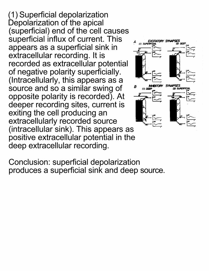
(1) Superficial depolarization Depolarization of the apical (superficial) end of the cell causes superficial influx of current. This appears as a superficial sink in extracellular recording. It is recorded as extracellular potential of negative polarity superficially. (Intracellularly, this appears as a source and so a similar swing of opposite polarity is recorded). At deeper recording sites, current is exiting the cell producing an extracellularly recorded source (intracellular sink). This appears as positive extracellular potential in the deep extracellular recording. Conclusion: superficial depolarization produces a superficial sink and deep source.

(2) Deep depolarization Depolarization of the deep end of the cell (e.g. soma) causes influx of current there. This creates a deep sink that is recorded as negative extracellular polarity. Superficially, the efflux of current creates a source that appears as positive polarity extracellularly. (3) Superficial hyperpolarization Hyperpolarization of the superficial end produces a source which gives rise to a positive deflection. The entering deep current produces a sink, thus giving rise to a negative deflection. the source-sink pair is the same as for deep depolarization.

(4) Deep hyperpolarization Likewise, deep hyperpolarization produces a source-sink pair as in superficial depolarization.

Current Source Density
Viewed from the extracellular space, the recorded field potential results from the flow of current in closed loops from sources to sinks. The same relation that was used to deduce current flow in the axon can be used for the dendrites. The extracellular potential can be recorded in a line along the axis of the pyramidal cells (the z direction).

The first derivative of potential with depth (i.e. the potential gradient in the z direction) is proportional to longitudinal current in that direction. (Current flows when potential differences exist). The first derivative of longitudinal current (second derivative of potential) is proportional to current crossing the membrane. (Differences in longitudinal current must be balanced by transmembrane current).

Thus, the second spatial derivative of potential In the z direction is proportional to the current density (Current Source Density): jm =
1rd 2v z( )dv 2

Populations of Unitary Neuronal Generators
How do the extracellular potentials of neurons behave in a population of generators?
We have seen that cortical neurons produce fluctuating current flow across the cell membrane. The currents generated by individual neurons may combine or cancel in the extracellular medium surrounding active neurons.

A closed field of potential results when the current flow is confined within the population. It may occur: (a) if the neurons are stellate in morphology with dendrites extending radially outward (A in figure). (b) if the neurons have single dendrites, but the dendrites are randomly oriented, or are oriented radially inward (B in figure). Outside of the population, the sources and sinks cancel and zero potential is recorded.

An open field of potential results when the sources and sinks combine, permitting the spread of current in the volume of the brain. This may occur if the neurons all have similar orientation and morphological polarization (e.g. cortical pyramidal cells) (C in figure).
Commonly, there may be a mixed population, resulting in a hybrid open-closed field (D in figure).

Cellular Generation of the EEG
What is the principal neuronal generator source of the scalp-recorded EEG?
EEG generation requires that the cells generate an open field of sufficient strength to be observed at the scalp. The generating population of cells must meet certain criteria:
1. The individual cellular generators in the population must have a polarized geometry.
2. The individual generators must be aligned in parallel.
3. The population must be relatively near the scalp. 4. The population must be capable of synchronized
activity.

The population of cortical pyramidal neurons generally meets these criteria:
1. Pyramidal cells are elongated.
2. They are arranged in parallel with their apical dendrites oriented perpendicular to the cortical surface.
3. The cortex is the structure closest to the surface of the head.
4. Pyramidal cells receive synchronous input on fiber paths from subcortical and cortical sources.

Even a single action potential from a single thalamic input neuron would synchronously activate a fairly large number (potentially thousands) of pyramidal cells. They would be depolarized at roughly the same location on their dendritic shaft. Thus, all the neurons in this population would generate a dipole field of the same orientation at the same time.

Apical depolarization of pyramidal cells produces a superficial sink and a deep source.
Current flows in closed loops along the interior of the dendritic trunk and with paths throughout the volume conductor.
The density of current is greatest intracellularly. The extracellular current density drops off with distance from the neuron.

The Neuronal Dipole Generator Making the assumption that the cerebral tissue is homogeneous, isopotential lines are drawn
perpendicular to lines of current.
The zero isopotential line is halfway between the center of mass of all sources and that of all sinks.
Potential changes most rapidly between the two poles of the "dipole".

In the figure, points A and B are on lines that differ by 500 uV, whereas C and D (while closer to the neuron) are on the same isopotential line.

In the cortex, we assume that: 1) Large numbers of pyramidal cell generators are
aligned in parallel dipole sheets. 2) The cell generators in the sheet are synchronously
active.
The solid angle principle is an approximation that allows us to estimate the potential
measured in a homogeneous medium at a
distance from an active dipole sheet: P =
±e4π
Ω

The potential measured with
respect to a neutral reference is
proportional to the potential
across the dipole layer, and the
solid angle subtended by the dipole layer at point P.
On the positive side of the dipole, the recorded potential is positive.
On the negative side, the recorded potential is negative. Halfway between the poles, on the zero isopotential plane, the recorded potential is zero.

The ratio of the observed potential to the dipole strength is proportional to the solid angle. P
±e=Ω4π
The solid angle is proportional to the active area. Therefore, the greater the active area, the larger is the observed potential.

(1) For a flat tangential dipole sheet, the shape of the potential field is elongated in the tangential direction. This situation exists on the crown of cortical gyri. Potential recorded on a distant surface (e.g. scalp) shows a bell-shaped curve: the potential is maximal over the active region and falls off with distance.

(2) The cerebral cortex is a highly convoluted structure with gyri and sulci. For a dipole surface on one wall of a sulcus, the solid angle at a point directly over the sulcus on the scalp is zero. Moving this point in 1 direction yields a negative potential, and in the other direction a positive potential.

(3) If a generator symmetrically
occupied both walls of a sulcus, no scalp signal would be recorded because the positive and negative sides of the dipole would cancel.
(4) For an active surface on the crown of a gyrus that also extends into the sulci on both sides, the effect of the tangential surface is dominant, i.e. the potential at the surface shows a bell-shaped curve.
However the curve falls off faster than with a flat sheet because of partial cancellation due to the distant sulcal wall.