iv fluid for nursing staff
TRANSCRIPT

INTRAVENOUS FLUIDS & ELECTROLYTES
Dr Somendra Shukla


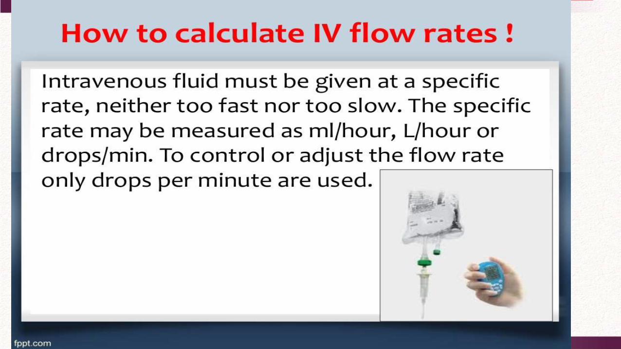

How to calculate IV flow rates !
What is a drop factor?
Drop factor is the number of drops in one milliliter used in IV fluid administration (also called drip factor). A number of different drop factors are available but the Commonest are:
1- 10 drops/ml (blood set)
2- 15 drops / ml (regular set)
3- 60 drops / ml (microdrop, burette)

How to calculate IV flow rates ?
The formula for working out flow rates is:
Example:
1500 ml IV Saline is ordered over 12 hours. Using a drop factor of 15 drops / ml, how many drops per minute need to be delivered?
volume (ml) X drop factor (gtts / ml)
---------------------------------------------
time (min)
= gtts / min
(flow rate)
1500 (ml) X 15 (drop / ml)
---------------------------------------------------
12 x 60 (gives us total minutes)
= 31 drop/ minute
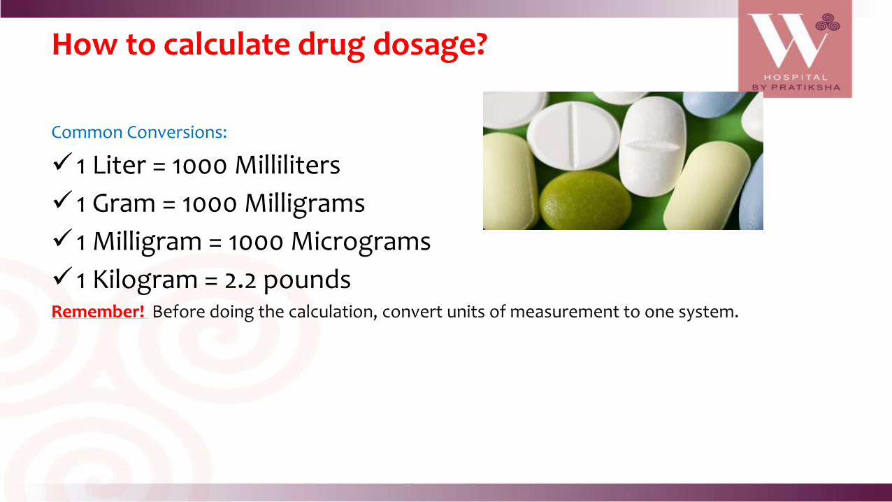
How to calculate drug dosage?
Common Conversions:
1 Liter = 1000 Milliliters
1 Gram = 1000 Milligrams
1 Milligram = 1000 Micrograms
1 Kilogram = 2.2 poundsRemember! Before doing the calculation, convert units of measurement to one system.

How to calculate drug dosage?
Example:
The ordered dose is Ceftriaxone 750 mg IV. the container contain 1g in a 10 ml vial.
How to calculate?
You should convert first g to mg , then :
(D) 750 mg X (V) 10 ml = 7.5 ml
(H) 1000 mg

How to calculate drug dosage?
D
Hx V = Amount to Give
D = dose ordered or desired doseH = dose on container label or dose on handV = form and amount in which drug comes (tablet, capsule, liquid)


Aims1. To gain peripheral venous access in
order to:• administer blood products, medicationsnutritional components
2. To minimize the risk of complications when initiating IV therapy through:• careful choice of IV site• good insertion technique• aseptic preparation of infusions
Key points
1. Only nurses who have been certified as
competent in the insertion of IV cannula will
perform this procedure.
2. Where the patient is less than 5 years of
the IV cannula will be inserted by a medical
practitioner. The exception will be in the case
neonates where neonatal trained nurses
may insert an IV cannula if directed by a
medical officer.

3. In the case of two unsuccessful attempts at insertion, the operator will seek the assistance of another experienced nurse for one additional attempt. After a total three unsuccessful attempts the assistance of a medical practitioner will be sought.Selection of Catheter Site Choose a suitable vein. In adults, use long straight veins in an upper extremity away from the joints for catheter insertion - in preference to sites on the lower extremities. If possible avoid veins in the dominant hand and use distal veins first.


Veins of the Hand1. Digital Dorsal veins2. Dorsal Metacarpal veins3. Dorsal venous network4. Cephalic vein5. Basilic vein
1. Digital Dorsal veins
2. Dorsal Metacarpal veins
3. Dorsal venous network
4. Cephalic vein
5. Basilic vein
Veins of the Forearm1. Cephalic vein2. Median Cubital vein3. Accessory Cephalic Vein4. Basilic vein5. Cephalic vein6. Median antebrachial vein

1. Check for the doctors order.
2. Identify the pt.
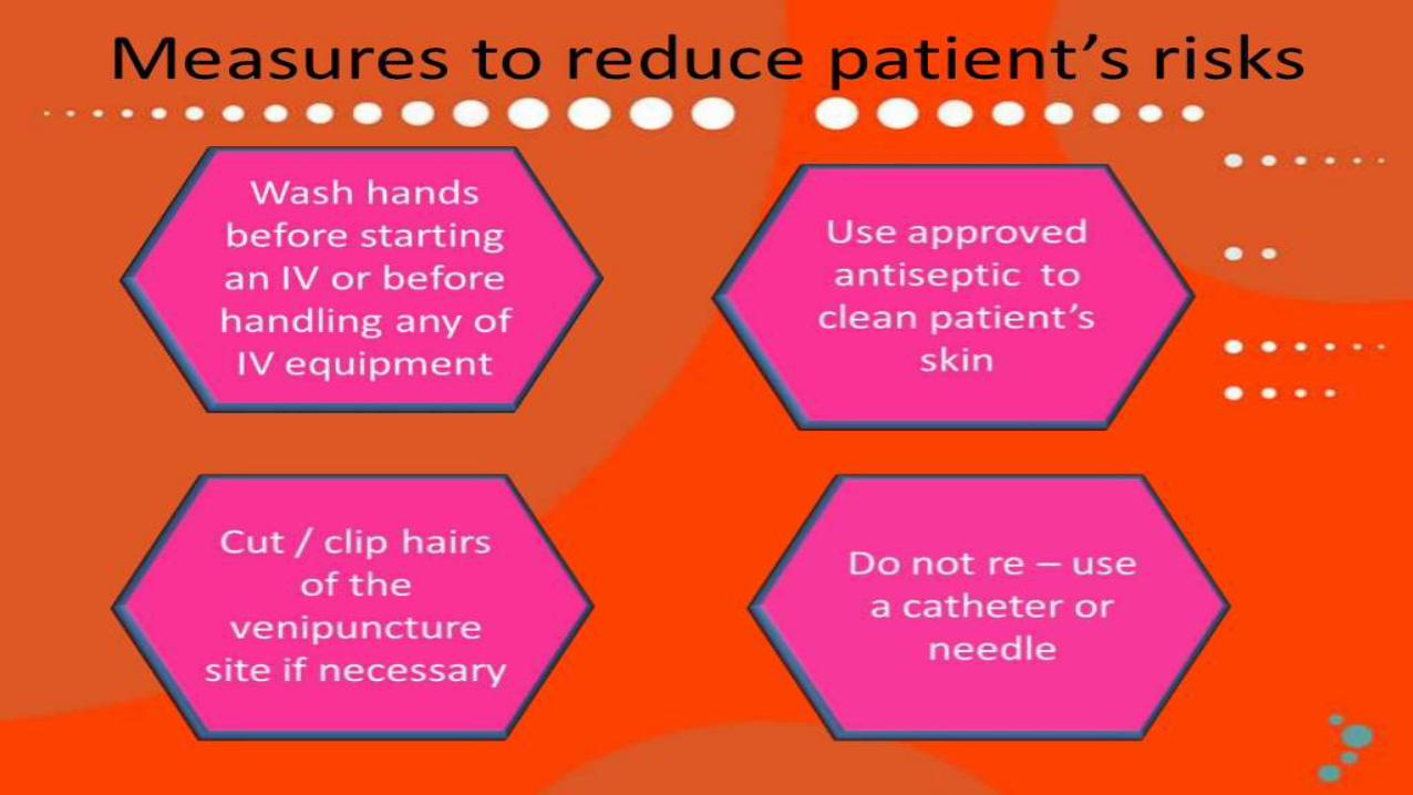
3. Wash hands prior to insertion.
6. Bring all the necessary materials @ pt. bed sides.
7. Explain procedure to the patient.
8. Prepare IV infusion , open the seal of the IV bottles and close the IV clamp. Spike the infusate aseptically and fill the drip chamber to at least half & prime the tubing prior to insertion. Remove air bubbles if any & cover the distal end of tubing.

9. Don gloves and select appropriate venipuncture site.
10. Apply torniquet 2-6 inches above proposed insertion site.
11. Disinfect the selected site with skin cotton balls with alcohol and allow to dry. Do not touch the skin with the fingers after preparation solution has been applied and maintain aseptic technique while doing the procedure remains the cornerstone of prevention of cannula related infections.
12. Inspect the cannula before insertion to ensure that
the needle is fully inserted into the plastic cannula
and that the cannula tip is not damaged.
13. Ensure the bevel of the cannula is facing upwards
to facilitate piercing of the skin by the bevel.
14. Using the appropriate cannula, pierce the skin w/ needle positioned on a 15-30 degree angle. Insert the needle and the cannula into the vein & apply gentle traction on skin may stabilize the vein under the skin.
15. Partially withdraw the needle and advance the
cannula.
16. Release the torniquet. Apply gentle pressure over the vein just proximal to the entry site to prevent blood flow. Remove the needle from within the plastic catheter.
17. Quickly connect end of the infusing tubing to the end cannula, secure connection and regulate the IV fluids
18. Cover the intravenous and surrounding area with a transparent dressing ensure that insertion site and the area proximal to the site are visible for inspection purposes.
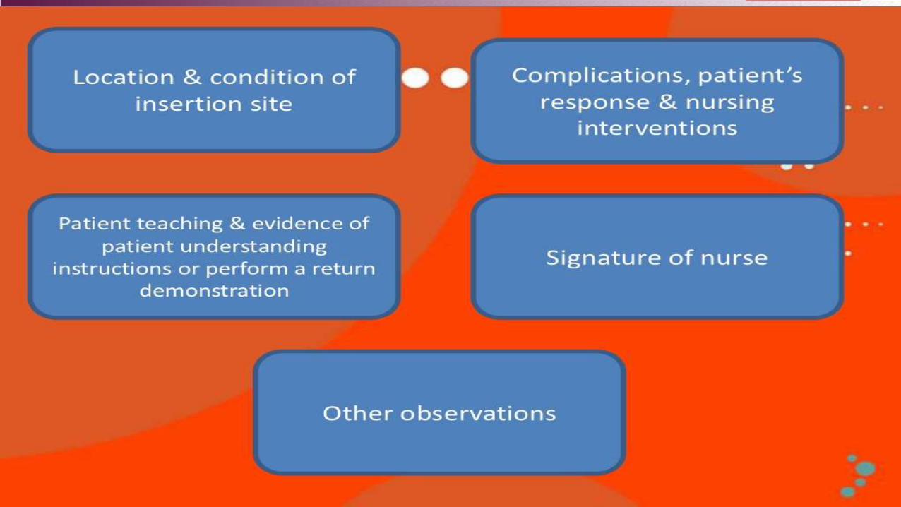
19. Make an IV tag and write the name of the pt, type of IV fluids, incorporation if any, date and
time IV fluid was started.
20. Dispose of all sharps in the appropriate sharps container, remove gloves and wash hands

Local Complication:
1. Phlebitis is irritation of a vein that is not caused by infection, but from the mere presence of a foreign body (the IV catheter) or the fluids or medication being given. Symptoms are warmth, swelling, pain, and redness around the vein. The IV device must be removed and if necessary re-inserted into another extremity.
To ease your patient's discomfort, apply warm packs.
Document your patient's condition and interventions.
If indicated, insert a new catheter at a different
site, preferably on the opposite arm, using a
vein or a smaller device and restart
the infusion.
2. Thrombophlebitis is similar to phlebitis but a
thrombus (or clot) is in addition involved. As the
IV cannula stays inside your body, it may irritate
the vein leading the body to trigger its clotting
mechanisms. This occurs when the catheter
unintentionally enters the tissue surrounding the
blood vessel. In this case the IV fluid and
associated medications will go into the tissues and
there will be a lump as an IV has been inserted

3. Infiltration - occurs when I.V. fluid leaks into surrounding tissue. It's commonly caused by improper placement or dislodgment of the catheter. Patient movement may cause the catheter to slip out or through the lumen of the vessel. t is characterized by coolness and pallor to the skin as well as local edema It is usually not painful. It is treated by removing the intravenous access device and elevating the affected limb so that the collected fluids can drain away. Infiltration is one of the most common adverse effects of IV therapy and is usually not serious ..

4. Hematoma is a collection of blood caused by
internal bleeding. This happens when the catheter punctures through the vein and causes a hematoma
5. Extravazation the leaking of vesicant drugs (such as antineoplastics) into surrounding tissue, can cause severe local tissue damage, resulting in delayed healing, infection, tissue necrosis, disfigurement, loss of function,
6. Infection -Any break in the skin carries a risk of infection. Although IV insertion is a sterile procedure, skin-dwelling organisms such as candidaalbicans may enter through the insertion site around the catheter, or bacteria may be accidentally introduced inside the catheter from contaminated equipment.

7. Venous Spasm - A sudden involuntary contraction of a vein or an artery resulting in temporary cessation of blood flow through a vessel.
Systemic Complication:1. Septicemia: a febrile disease process that results from the presence of microorganisms or
their toxic products in the circulatory system.2. Fluid overload & Pulmonary edema - caused by infusing excessive amounts of isotonic or
hypertonic crystalloid solutions to rapidly, failure to monitor the IV infusion or too-rapid infusion of any fluid in a patient compromised by cardiopulmonary or renal disease

3. Air embolism -Air entering the central vein, which is quickly trapped in the blood as it flows forward. Prevention is the key.
4. Shock - occurs when a foreign substance usually a medication is rapidly introduced into the circulation
5. Catheter embolism - a piece of the catheter
off and travels through the vascular system.
Treatment : Discontinue IV, apply a tourniquet
above the site, take appropriate emergency
measures, inspect catheter for rough edges that
might indicate loss of fragments. Obtain order for x-ray to determine if fragments are present.