isuog basic training · determining fetal lie ant post maternal r maternal l. basic training key...
TRANSCRIPT
Basic Training
ISUOG Basic Training Distinguishing Between Normal and Abnormal
Appearances of the Fetal Anatomy
Basic Training Basic training
Learning Objective At the end of the lecture you will be able to:
• Compare the differences between the ultrasound appearances of
normal fetal anatomy and of the more common structural fetal
abnormalities.
Basic Training
Key Questions
• Which abnormalities can be excluded by obtaining normal HC and AC
sections in the 2nd or 3rd trimester fetus?
• What are principal differences in ultrasound appearances between a
structurally normal fetus and a fetus with open spina bifida?
• How can the AC section be used to exclude the most common abdominal
wall and gastrointestinal defects?
• What are the typical ultrasound features of lower urinary tract obstruction?
Basic Training
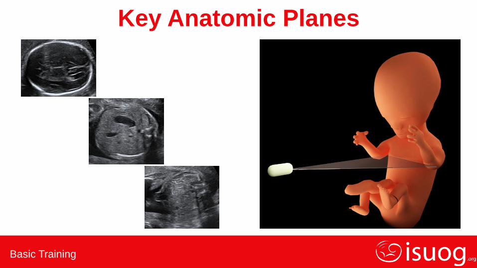
Key Anatomic Planes
Basic Training
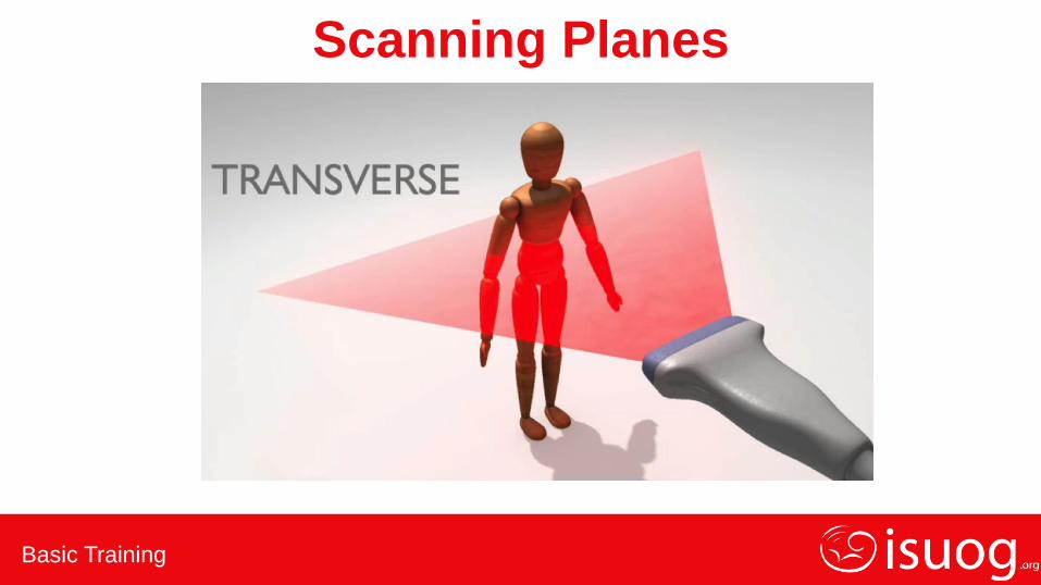
Scanning Planes

Basic Training
Scanning Planes
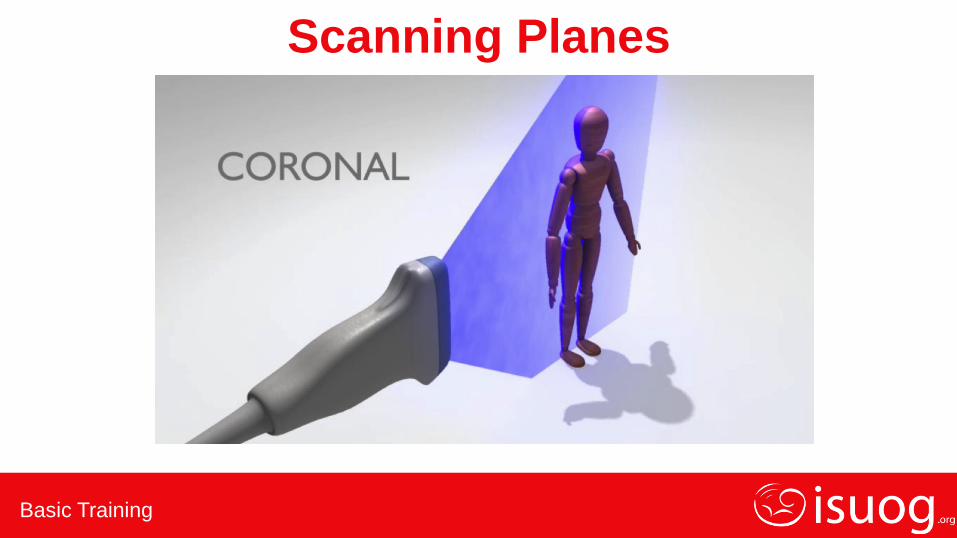
Basic Training
Scanning Planes

Basic Training
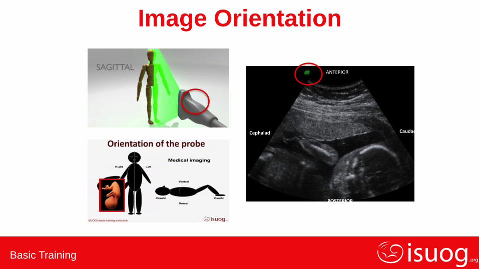
Image Orientation
ANTERIOR
POSTERIOR
RIGHT LEFT
Basic Training
Image Orientation
ANTERIOR
POSTERIOR
Cephalad Caudad
Basic Training
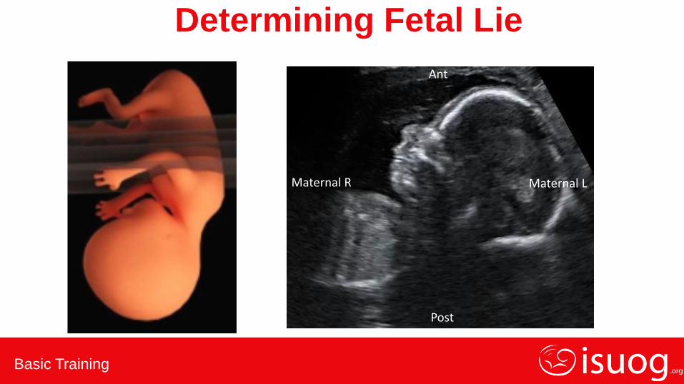
Determining Fetal Lie
Ant
Post
Maternal R Maternal L
Basic Training
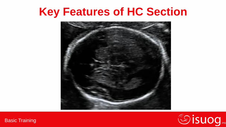
Key Features of HC Section
Basic Training
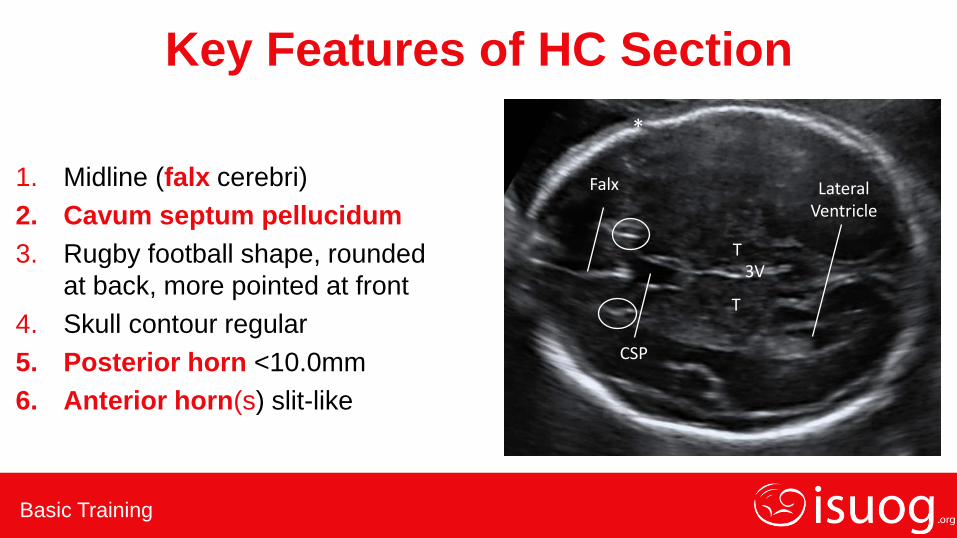
Key Features of HC Section
1. Midline (falx cerebri)
2. Cavum septum pellucidum
3. Rugby football shape, rounded
at back, more pointed at front
4. Skull contour regular
5. Posterior horn <10.0mm
6. Anterior horn(s) slit-like
CSP
T
T
Falx Lateral Ventricle
3V
*
Basic Training
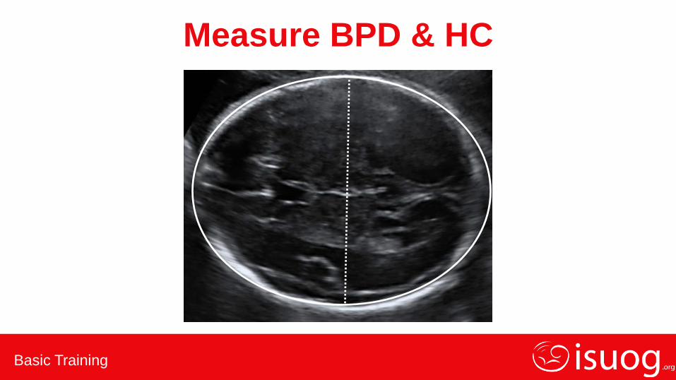
Measure BPD & HC
Basic Training
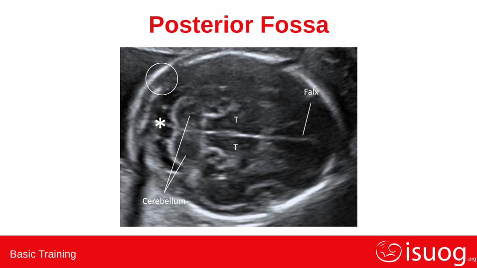
Posterior Fossa
*
Cerebellum
T
T
Falx

Basic Training
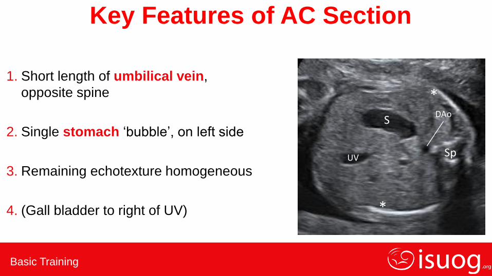
Key Features of AC Section
Basic Training
Key Features of AC Section
1. Short length of umbilical vein,
opposite spine
2. Single stomach ‘bubble’, on left side
3. Remaining echotexture homogeneous
4. (Gall bladder to right of UV)
DAo S
Sp UV
*
*
Basic Training
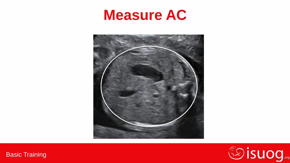
Measure AC
Basic Training
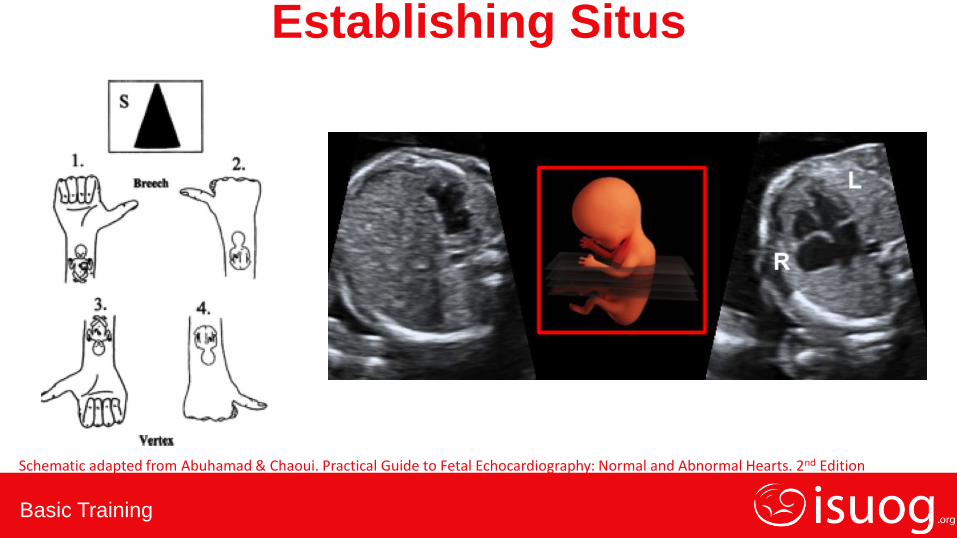
Schematic adapted from Abuhamad & Chaoui. Practical Guide to Fetal Echocardiography: Normal and Abnormal Hearts. 2nd Edition
L
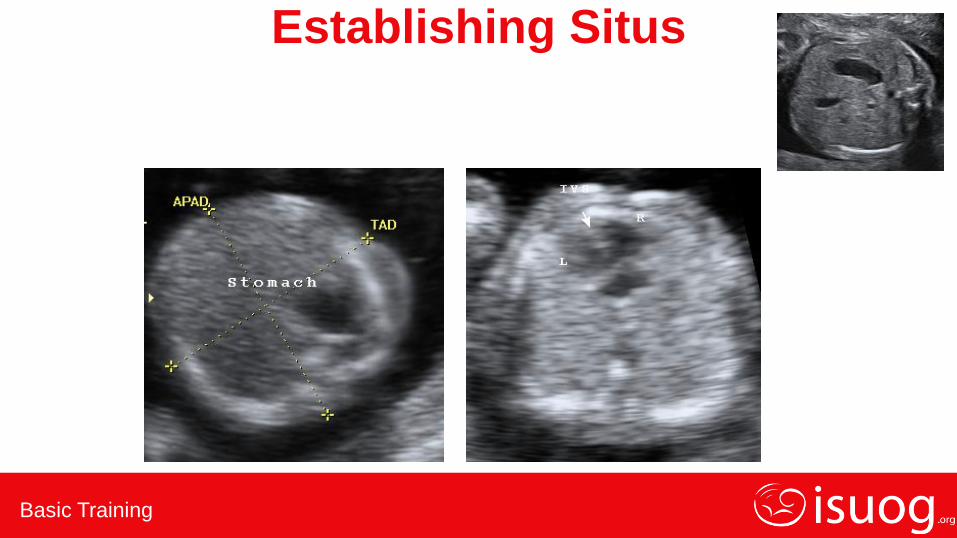
Establishing Situs
Basic Training
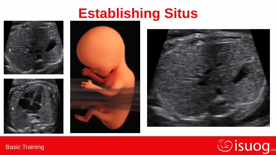
Establishing Situs

Basic Training
First Establish Fetal Position

Basic Training
4 Chamber View
• Easy view to obtain
• No specialized skill needed
• Obtainable in all fetal positions
• Rules out 60% CHD
• Easy slide up from AC with full rib
• Starting point for the sweep

Basic Training
4 Chamber View – Normal Appearance • Right ventricle is the most anterior, below
the sternum
• Left atrium is closest to the spine and the
most central structure in the chest
• Tricuspid valve is more apical than the mitral
valve
• Flap of the foramen ovale is in the left atrium
• Moderator band is in the right ventricle
• Crux seen
R L
Sp
DAo

Basic Training
Kidneys – Normal Appearance
Basic Training
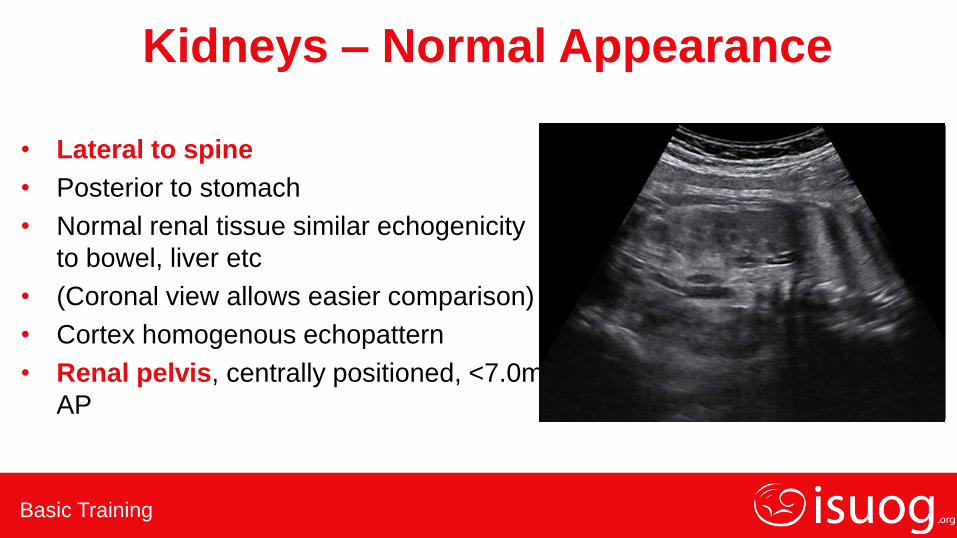
Kidneys – Normal Appearance
• Lateral to spine
• Posterior to stomach
• Normal renal tissue similar echogenicity
to bowel, liver etc
• (Coronal view allows easier comparison)
• Cortex homogenous echopattern
• Renal pelvis, centrally positioned, <7.0mm
AP
Sp
* *
Basic Training
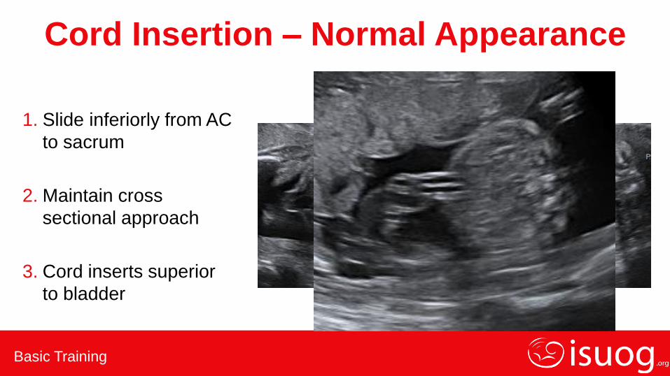
Cord Insertion – Normal Appearance
1. Slide inferiorly from AC
to sacrum
2. Maintain cross
sectional approach
3. Cord inserts superior
to bladder
Basic Training
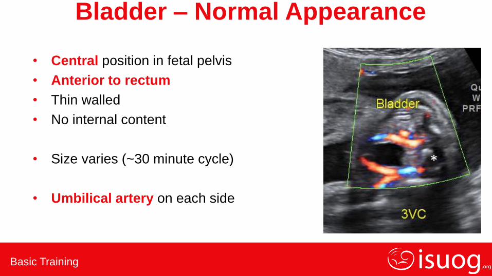
Bladder – Normal Appearance
• Central position in fetal pelvis
• Anterior to rectum
• Thin walled
• No internal content
• Size varies (~30 minute cycle)
• Umbilical artery on each side
*
Basic Training
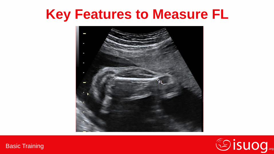
Key Features to Measure FL
Basic Training
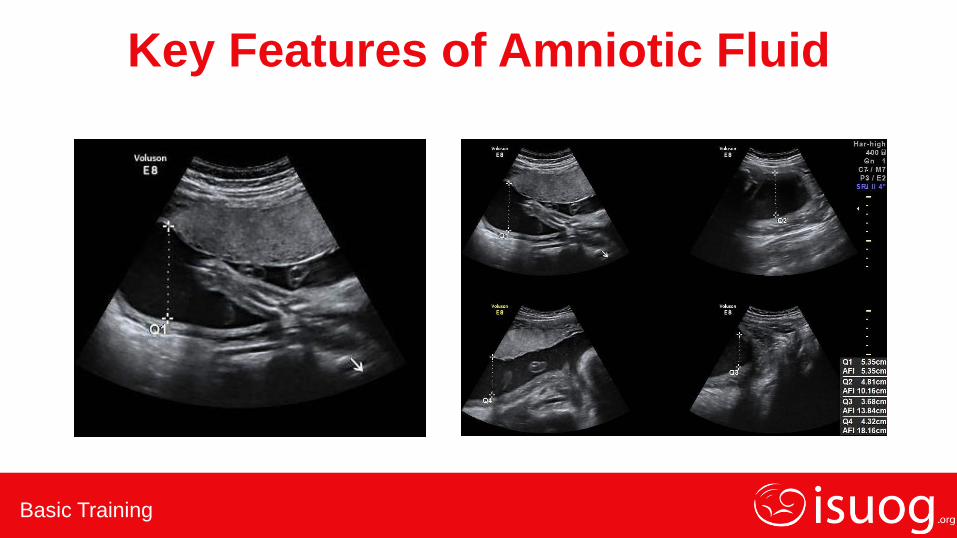
Key Features of Amniotic Fluid
Basic Training
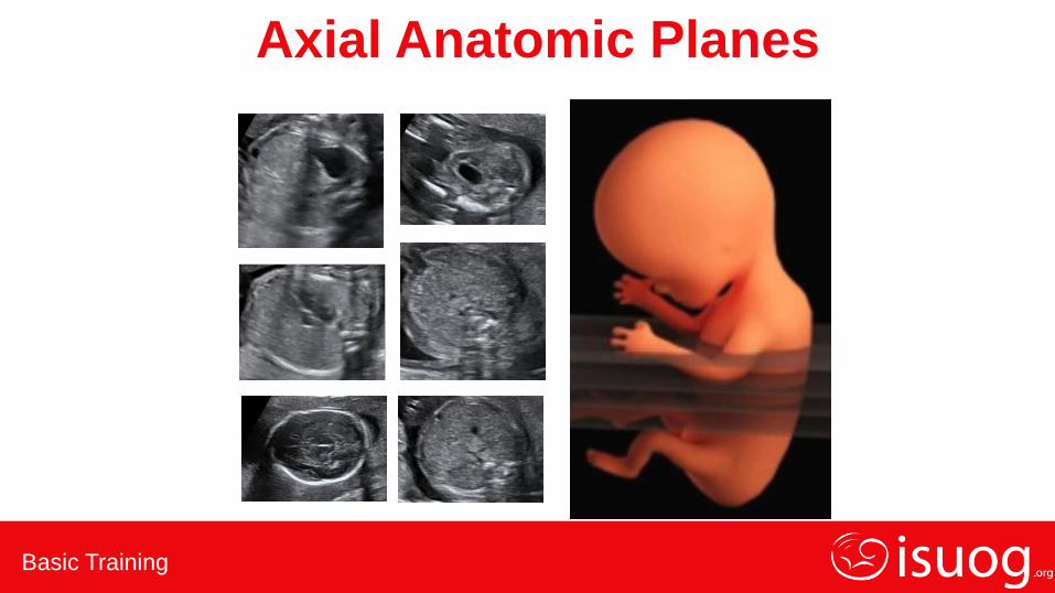
Axial Anatomic Planes
Basic Training
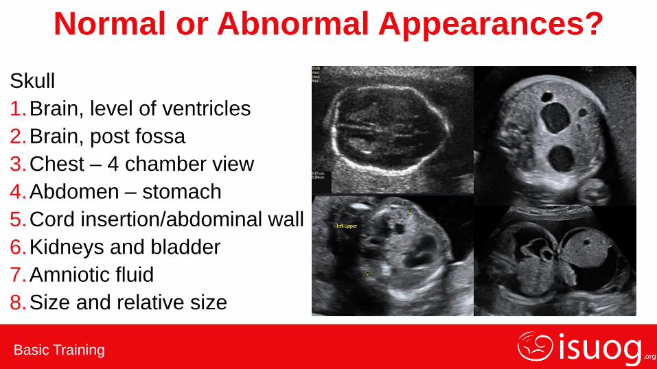
Normal or Abnormal Appearances?
Skull
1.Brain, level of ventricles
2.Brain, post fossa
3.Chest – 4 chamber view
4.Abdomen – stomach
5.Cord insertion/abdominal wall
6.Kidneys and bladder
7.Amniotic fluid
8.Size and relative size
Basic Training
Normal or Abnormal Appearances?
1. Skull
2. Brain, level of ventricles
3. Brain, post fossa
4. Chest – 4 chamber view
5. Abdomen – stomach
6. Cord insertion/abdominal wall
7. Kidneys and bladder
8. Amniotic fluid
9. Size and relative size
Basic Training
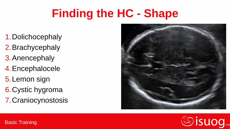
Finding the HC - Shape
1.Dolichocephaly
2.Brachycephaly
3.Anencephaly
4.Encephalocele
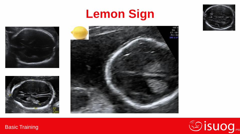
5.Lemon sign
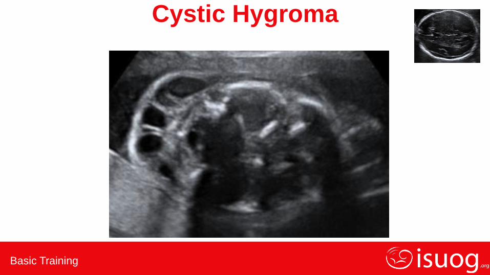
6.Cystic hygroma
7.Craniocynostosis
Basic Training
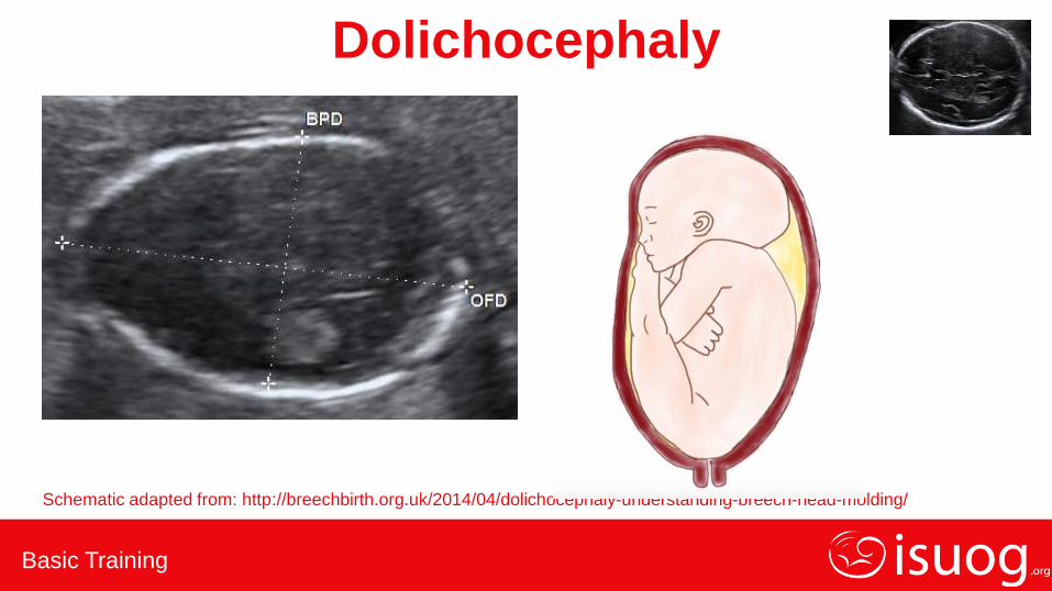
Dolichocephaly
Schematic adapted from: http://breechbirth.org.uk/2014/04/dolichocephaly-understanding-breech-head-molding/
Basic Training
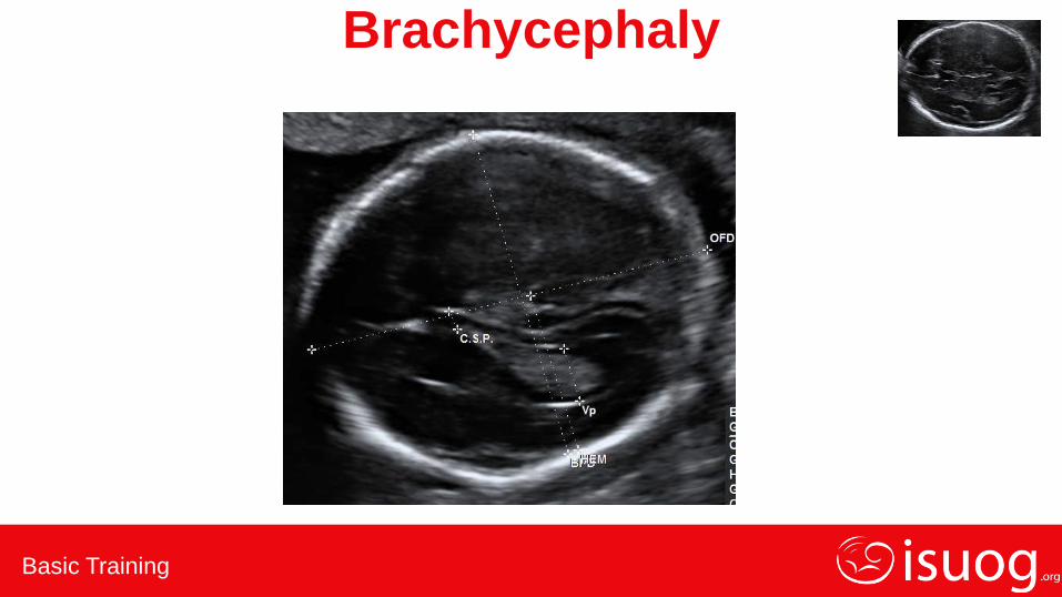
Brachycephaly

Basic Training
Anencephaly
Basic Training
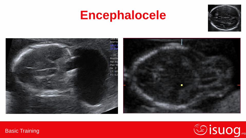
Encephalocele
Basic Training
Lemon Sign
Basic Training
Cystic Hygroma

Basic Training
Craniocynostosis
Basic Training
Normal or Abnormal Appearances?
1.Skull
2.Brain, level of ventricles
3.Brain, post fossa
4.Chest – 4 chamber view
5.Abdomen – stomach
6.Cord insertion/abdominal wall
7.Kidneys and bladder
8.Amniotic fluid
9.Size and relative size

Basic Training
Finding the HC – Intracranial Structures
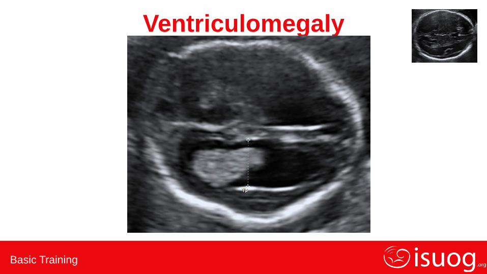
1.Ventriculomegaly
2.Holoprosencephaly
Basic Training
Ventriculomegaly

Basic Training
Holoprosencephaly
Basic Training
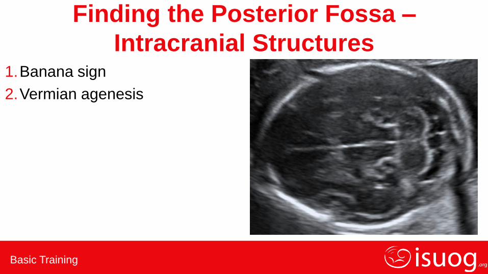
Finding the Posterior Fossa –
Intracranial Structures 1.Banana sign
2.Vermian agenesis

Basic Training
Banana Sign
Basic Training
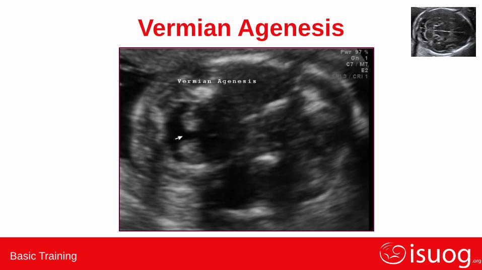
Vermian Agenesis
Basic Training
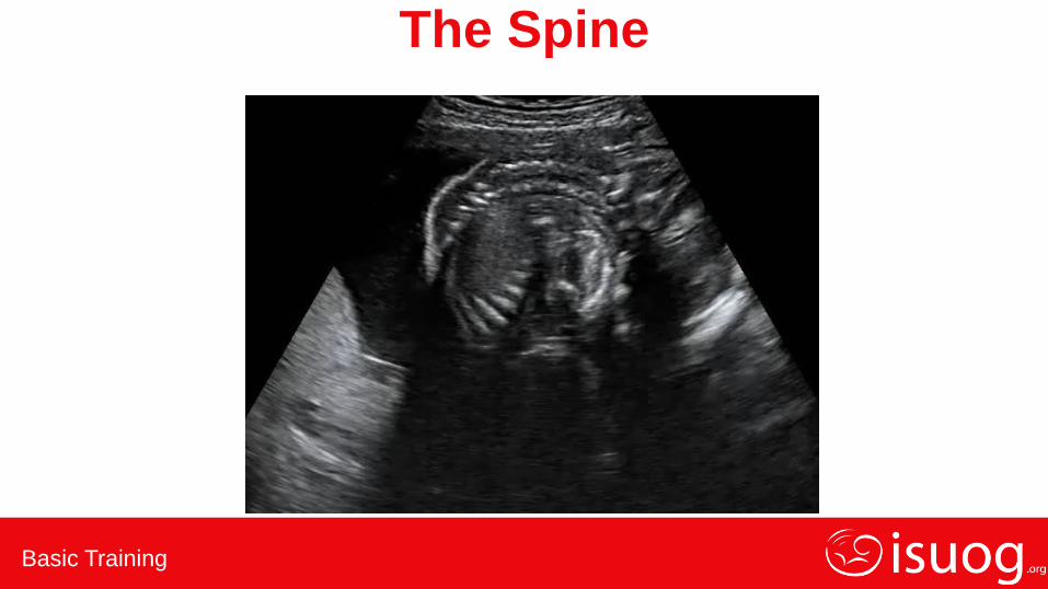
The Spine
Basic Training
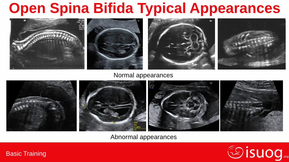
Open Spina Bifida Typical Appearances
Normal appearances
Abnormal appearances

Basic Training
Normal or Abnormal Appearances?
1.Skull
2.Brain, level of ventricles
3.Brain, post fossa
4.Chest – 4 chamber view
5.Abdomen – stomach
6.Cord insertion/abdominal wall
7.Kidneys and bladder
8.Amniotic fluid
9.Size and relative size
Basic Training
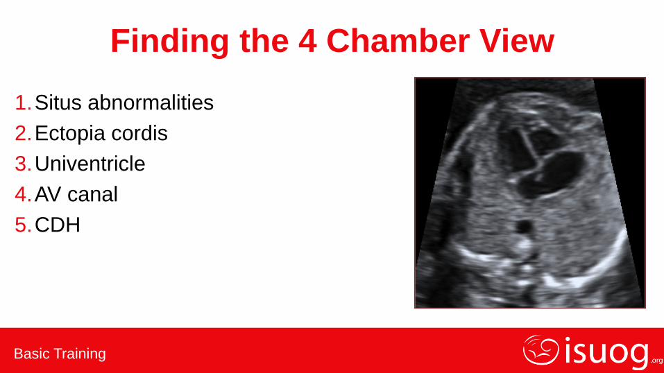
Finding the 4 Chamber View
1.Situs abnormalities
2.Ectopia cordis
3.Univentricle
4.AV canal
5.CDH

Basic Training
Abnormal Situs
Basic Training
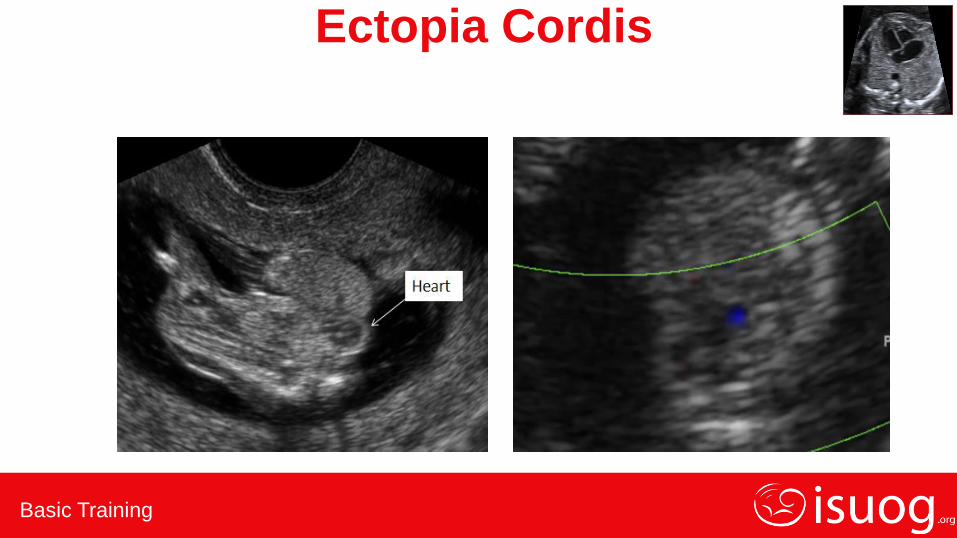
Ectopia Cordis
Basic Training
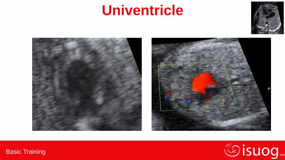
Univentricle
Basic Training
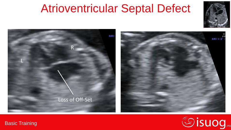
R
L
Loss of Off-Set
Atrioventricular Septal Defect
Basic Training
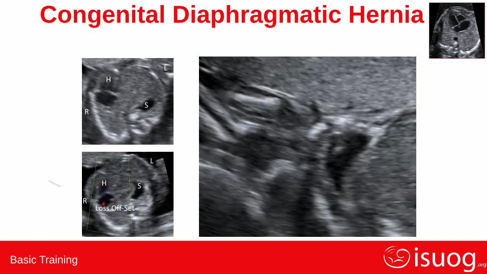
R
L
H
S
Loss Off-Set R
L
H S
Congenital Diaphragmatic Hernia
Basic Training
Normal or Abnormal Appearances?
1.Skull
2.Brain, level of ventricles
3.Brain, post fossa
4.Chest – 4 chamber view
5.Abdomen – stomach
6.Cord insertion/abdominal wall
7.Kidneys and bladder
8.Amniotic fluid
9.Size and relative size
Basic Training
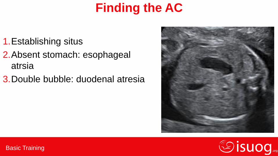
Finding the AC
1.Establishing situs
2.Absent stomach: esophageal
atrsia
3.Double bubble: duodenal atresia
Basic Training
Establishing Situs
Basic Training
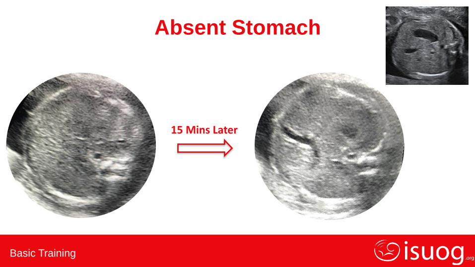
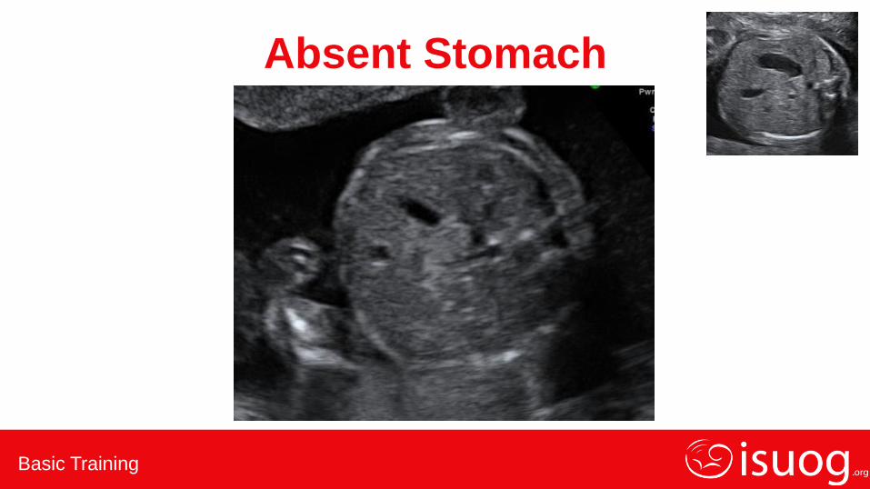
Absent Stomach
15 Mins Later
Basic Training
Absent Stomach

Basic Training
Double Bubble Sign
Basic Training
Normal or Abnormal Appearances?
1.Skull
2.Brain, level of ventricles
3.Brain, post fossa
4.Chest – 4 chamber view
5.Abdomen – stomach
6.Cord insertion/abdominal wall
7.Kidneys and bladder
8.Amniotic fluid
9.Size and relative size

Basic Training
Cord Insertion/Abdominal Wall
1.Normal gut herniation
2.Omphalocele
3.Gastroschisis

Basic Training
Normal Gut Herniation
Fetuses have exompholos at 9-10 weeks that resolves by 12 weeks

Basic Training
Omphalocele
Abnormal cord insertion
• Cord inserts into apex of defect
• Contains liver +/- bowel etc
• Membrane covered
Basic Training
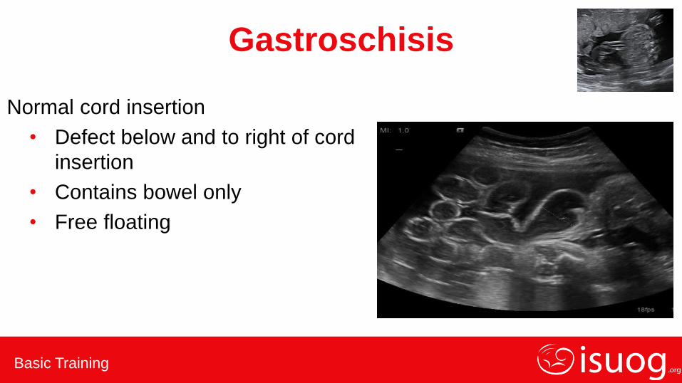
Gastroschisis
Normal cord insertion
• Defect below and to right of cord
insertion
• Contains bowel only
• Free floating
Basic Training
Normal or Abnormal Appearances?
1.Skull
2.Brain, level of ventricles
3.Brain, post fossa
4.Chest – 4 chamber view
5.Abdomen – stomach
6.Cord insertion/abdominal wall
7.Kidneys and bladder
8.Amniotic fluid
9.Size and relative size

Basic Training
Kidneys and Bladder

Basic Training
Kidneys and Bladder
1.Renal agenesis
2.Hydronephrosis
3.Bladder outlet obstruction
Basic Training
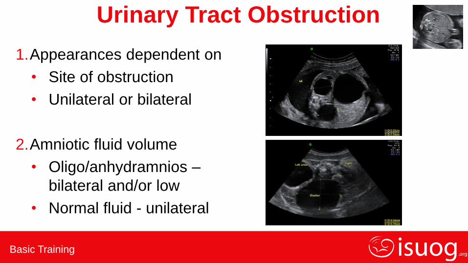
Urinary Tract Obstruction
1.Appearances dependent on
• Site of obstruction
• Unilateral or bilateral
2.Amniotic fluid volume
• Oligo/anhydramnios –
bilateral and/or low
• Normal fluid - unilateral
Basic Training
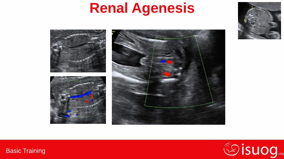
Renal Agenesis
Basic Training
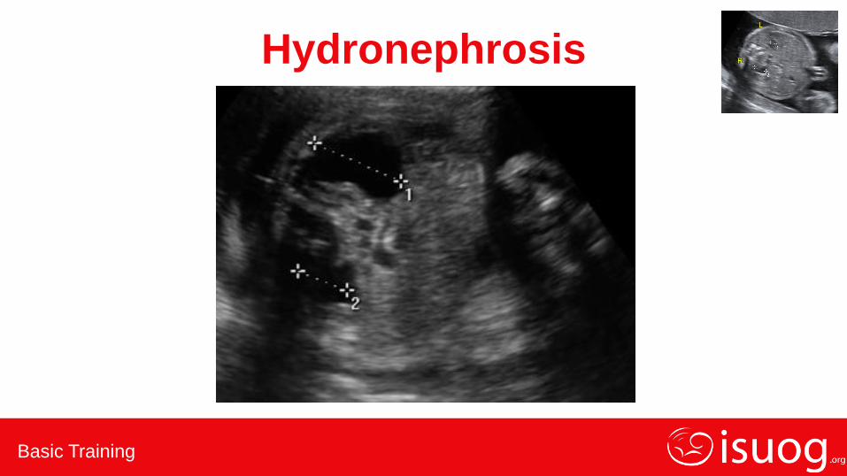
Hydronephrosis

Basic Training
Bladder Outlet Obstruction

Basic Training
Bladder Outlet Obstruction

Basic Training
Normal or Abnormal Appearances?
1.Skull
2.Brain, level of ventricles
3.Brain, post fossa
4.Chest – 4 chamber view
5.Abdomen – stomach
6.Cord insertion/abdominal wall
7.Kidneys and bladder
8.Amniotic fluid
9.Size and relative size

Basic Training
Oligohydramnios: Causes
Ultrasound in Obstetrics & Gynecology: A Practical Approach. Abuhamad et al 2014

Basic Training
Polyhydramnios: Causes
Ultrasound in Obstetrics & Gynecology: A Practical Approach. Abuhamad et al 2014
Basic Training
Normal or abnormal appearances?
1.Skull
2.Brain, level of ventricles
3.Brain, post fossa
4.Chest – 4 chamber view
5.Abdomen – stomach
6.Cord insertion/abdominal wall
7.Kidneys and bladder
8.Amniotic fluid
9.Size and relative size

Basic Training
Size and Relative Size

Basic Training
Key points
1. The key to identifying abnormalities is understanding the
range of normal appearances at differing gestations
2. It is important to develop a consistent approach to each
scan, rather than scanning randomly
3. Find the long axis of the fetus first and assess the
appearances
Basic Training
Key points 4. Then assess the fetal anatomy in cross section starting
with the head, assess skull and intracranial anatomy,
measure the HC
5. Slide through the chest to the abdomen, assess situs,
chest contents and upper abdomen, measure AC
6. Find FL by continuing to slide through lower abdomen
and pelvis, assess abdominal wall, cord insertion,
kidneys, bladder, spine and skin covering

Basic Training
Conclusions Distinguishing between normal and abnormal ultrasound
appearances requires:
• The development of a consistent scanning technique
• Paying rigorous attention to the quality of sections obtained
• Understanding how to manipulate the probe to improve poor
sections
• Appreciating how the range of normal appearances, and
therefore potentially abnormal appearances, changes with
gestation