issue 4 serious allergic conditions of the ocular surface · jodi i. luchs, md patients with atopic...
TRANSCRIPT
to obtain cme credit for this activity, go to http://cme.ufl .edu/toai Topics in Ocular antiinflammatOries 1Supported by an unrestricted educational grant from Bausch + Lomb, Inc.
Serious Allergic Conditions of the Ocular Surface
Within ophthalmology, the past decade has been marked by an increasing awareness of the importance of the ocular surface and the potentially disastrous consequences of chronic, severe infl ammation. Of the allergy-related infl ammatory states that aff ect the eye, vernal keratoconjunctivitis (VKC) and atopic keratoconjunctivitis (AKC) are the most serious and pose the greatest overall threat to corneal health and vision.
In hot, dry climates—including parts of the Mediterranean region, Africa, and the Middle East—these aggressive, chronic ocular surface infl ammatory diseases are relatively common and contribute signifi cantly to ocular morbidity and corneal blindness. Fortunately, both conditions are comparatively rare in the US, although their apparent prevalence may be increasing due to better access to healthcare and the migration of susceptible populations to the US.1,2
Treatment of VKC and AKC patients requires a high level of vigilance for several reasons: 1) compared to milder ocular allergic conditions, such as seasonal or perennial allergic con-junctivitis (SAC and PAC), in VKC and AKC the underlying immune defect is more severe and the clinical manifestation more aggressive; 2) while there is some variability in disease severity over time (due to seasonal exposures and other fac-
tors), infl ammation in VKC and AKC tends to be unrelenting and chronic; and 3) the risk of disease-related and treatment-related complications is far greater in VKC and AKC.
associationsAs with many allergic conditions, patients with seri-
ous ocular allergic conditions commonly have overlapping immune-related disorders. Patients with VKC and AKC are
See INSIDE for:Issues in Long-term Antiinfl ammatory Therapy by Michael B. Raizman, MD
ISSUE 4
JoDi i. lucHs, MD Patients with atopic or vernal keratoconjunctivitis are at risk for vision loss from the eff ects of continuous ocular surface infl ammation. Prompt diagnosis, adequate antiinfl ammatory treatment, including the use of cell-active agents, and careful monitoring are mainstays of eff ective management.
A CONTINUINGMEDICAL EDUCATION
PUBLICATIONCME
CONTINUING MEDICAL EDUCATION
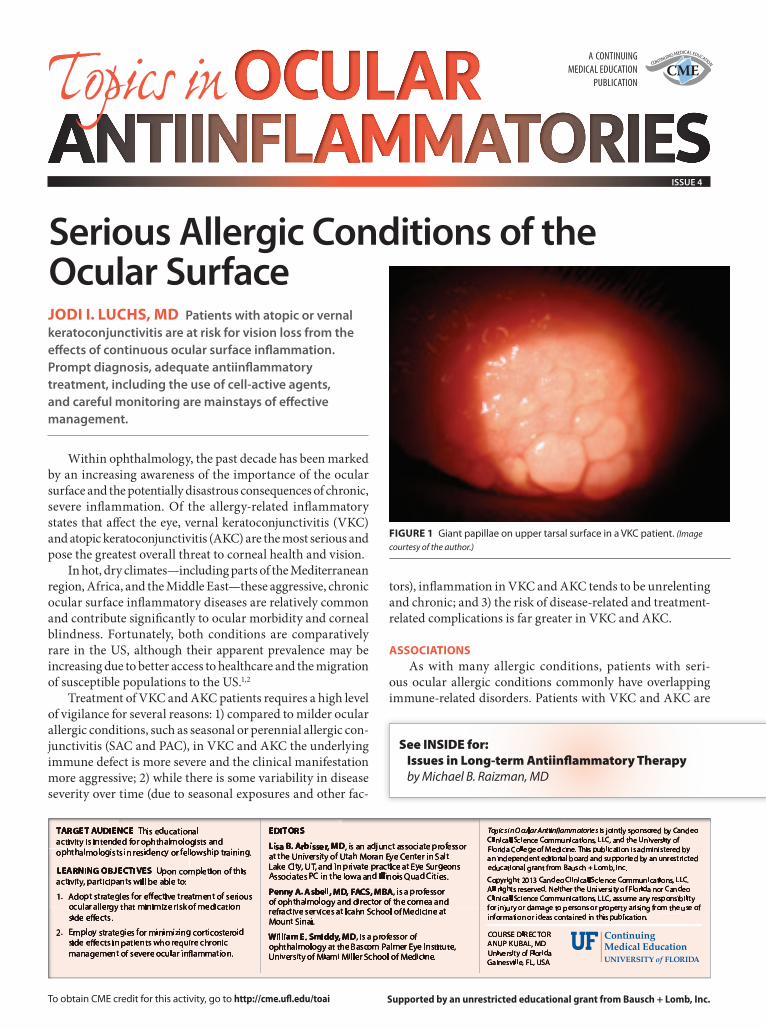
FiGure 1 Giant papillae on upper tarsal surface in a VKc patient. (Image
courtesy of the author.)

2 Topics in Ocular antiinflammatOries to obtain cme credit for this activity, go to http://cme.ufl .edu/toai
stateMent oF neeDthe indications for topical ophthalmic antiinfl ammatory drugs (both steroidal and nonsteroidal) are evolving rapidly, as new agents and new applications emerge. many of these are novel—eg, the perioperative use of nonsteroidal antiinfl ammatory drugs (nsaids) to prevent cystoid macular edema—and/or fl y in the face of older thinking—eg, the use of steroids to calm infl ammation and reduce the risk of melting or scarring from infection. neither of these important applications is on-label.
in addition, new steroidal and nonsteroidal agents continue to come to market, expanding the utility of both classes. antiinfl ammatory drugs are now used for: the treatment of ocular surface disease and allergic conjunctivitis; prevention of perioperative pain and infl ammation in ocular surgery; infection management; cystoid macular edema prophylaxis following cataract surgery; haze prevention in PrK; and much more.
What has regrettably not followed this expansion of indications, formulations, and new molecular entities are protocols for drug selection and use.1 these are vital because signifi cant diff erences in safety, tolerability, and effi cacy exist between and within both antiinfl ammatory drug classes. c-20 ester steroids, for example, have a demonstrated lower risk of intraocular pressure (iOP) elevation than ketone steroids.2,3 since a range of steroid formulations and concentrations is available, clinicians need up-to-date information about the indications and optimum uses for each.3
although topical nsaid formulations have been associated with signifi cant adverse events (keratopathy ranging from superfi cial punctate keratitis to corneal melt), recent work shows these to be uncommon and less severe with newer formulations.4 indeed, novel nsaids make use of lower concentrations and less frequent dosing, potentially impacting safety profi les and reducing risk from long-term use.5
although both are “antiinfl ammatory,” steroids and nsaids act at diff erent points in the infl ammatory cascade, and thus off er opportunities for physicians to fi ne-tune their drug selection. and although they are frequently used together, whether or not the two drug classes can act synergistically is controversial. in the context of recent clinical data, a clear mechanistic understanding of each drug class generally—and of newer formulations specifi cally—will equip clinicians to make eff ective, evidence-based prescribing decisions across the many situations that call for ocular infl ammation control.
reFerences1. dua Hs, attre r. treatment of post-operative
infl ammation following cataract surgery —a review. European Ophthalmic Review. 2012;6(2):98-103.
2. comstock tl, decory H. advances in corticosteroid therapy for ocular infl ammation: loteprednol etabonate. International Journal of Infl ammation. 2012; doi:10.1155/2012/789623.
3. fong r, leitritz m, siou-mermet r, erb t. loteprednol etabonate gel 0.5% for postoperative pain and infl ammation after cataract surgery: results of a multicenter trial. Clin Ophthalmol. 2012;6:1113-24.
4. singer m, cid md, luth J, et al. incidence of corneal melt in clinical practice: our experience vs a meta-analysis of the literature. Clin Exp Ophthalmol. 2012;s1:003.
5. cable m. comparison of bromfenac 0.09% Qd to nepafenac 0.1% tid after cataract surgery: pilot evaluation of visual acuity, macular volume, and retinal thickness at a single site. clin Ophthalmol. 2012;6:997-1004.
oFF-laBel use stateMent this work discusses off -label uses of antiinfl ammatory medications.
General inForMation this cme activity is sponsored by the university of florida college of medicine and is supported by an unrestricted educational grant from Bausch + lomb, inc.
Directions: select one answer to each question in the exam (questions 1–10) and in the evaluation (questions 11–16). the university of florida college of medicine designates this activity for a maximum of 1.0 AMA PRA Category 1 Credit™. there is no fee to participate in this activity. in order to receive cme credit, participants should read the report, and then take the posttest. a score of 80% is required to qualify for cme credit. estimated time
to complete the activity is 60 minutes. On completion, tear out or photocopy the answer sheet and send it to:
university of florida cme Offi cePO Box 100233, Gainesville, fl 32610-0233PHONE: 352-733-0064 FAX: 352-733-0007
Or you can take the test online at http://cme.ufl .edu/toaisystem requirements for this activity are: For PC users: Windows® 2000, xP, 2003 server, or Vista; internet explorer® 6.0 or newer, or mozilla® firefox® 2.0 or newer (Javascript™ and Java™ enabled). For Mac® users: mac Os® x 10.4 (tiger®) or newer; safari™ 3.0 or newer, mozilla® firefox® 2.0 or newer; (Javascript™ and Java™ enabled).
internet connection required: cable modem, dsl, or better.
Date oF oriGinal release november 2013. approved for a period of 12 months.
accreDitation stateMent this activity has been planned and implemented in accordance with the essential areas and Policies of the accreditation council for continuing medical education (accme) through the joint sponsorship of the university of florida college of medicine and candeo clinical/science communications, llc. the university of florida college of medicine is accredited by the accme to provide continuing medical education for physicians.
creDit DesiGnation stateMent the university of florida college of medicine designates this enduring material for a maximum of 1.0 AMA PRA Category 1 Credit™. Physicians should only claim the credit commensurate with the extent of their participation in the activity.
Faculty anD Disclosure stateMents lisa B. arbisser, MD (faculty advisor), is an adjunct associate professor at the university of utah moran eye center in salt lake city, ut, and an ophthalmologist at eye surgeons associates Pc in Bettendorf, ia. she states that in the past 12 months, she has participated in a stand-alone Bausch + lomb advisory board meeting.
Penny a. asbell, MD, Facs, MBa (faculty advisor), is a professor of ophthalmology and director of the cornea and refractive services at icahn school of medicine at mount sinai. she states that in the past 12 months, she has been a consultant for r-tech, senju, and Bausch + lomb, has given cme lectures for merck, and has received a research grant from alcon.
William e. smiddy, MD (faculty advisor), is a professor of ophthalmology at the Bascom Palmer eye institute, university of miami miller school of medicine. He states that in the past 12 months, he has attended a steering committee meeting of alimera scientifi c.
Jodi i. luchs, MD, is an assistant professor of ophthalmology at the Hofstra university school of medicine, and co-director of the department of refractive surgery at north shore/long island Jewish Health system, Great neck, nY. He practices at south shore eye care, llP, Wantagh, nY. He states that in the past 12 months, he has received grant/research support from allergan, alcon, Bausch + lomb, and shire; that he has been a consultant for allergan, Bausch + lomb, nicox, santen, and eyegate; and that he has been on the speakers bureau for allergan and Bausch + lomb.
Michael B. raizman, MD, is a cornea specialist and refractive surgeon at Ophthalmic consultants of Boston in Boston, ma. He is the director of the cornea and cataract service at new england eye center, tufts university school of medicine. He states that in the past 12 months, he has been a consultant to alcon, allergan, Bausch + lomb, and eyeGate.
DisclaiMer Participants have an implied responsibility to use the newly acquired information to enhance patient outcomes and professional development. the information presented in this activity is not meant to serve as a guideline for patient care. Procedures, medications, and other courses of diagnosis and treatment discussed or suggested in this activity should not be used by clinicians without evaluation of their patients’ conditions and possible contraindications or dangers in use, applicable manufacturer’s product information, and comparison with recommendations of other authorities.
coMMercial suPPorters this activity is supported by an unrestricted educational grant from Bausch + lomb, inc.
TOPICS IN OCULAR ANTIINFLAMMATORIES, ISSUE 4more likely than unaffected peers to have comorbid atopic conditions, in-cluding asthma, eczema, and rhinitis. Approximately 55% to 60% of VKC and AKC patients are skin test-positive for allergens, indicating the presence of circulating allergen-specifi c IgE.
A family history of atopy is also common in VKC and AKC patients.1,3 Studies in Europe have found increased rates of autoimmune conditions, such as psoriasis and thyroiditis, in family members of VKC patients.4,5
PatHoPHysioloGyVKC and AKC are managed dif-
ferently from milder forms of ocular allergy. Th e main objective in treating SAC and PAC is to alleviate patients’ ocular itching, typically by prescrib-ing a mast cell stabilizer/antihistamine combination agent. If the itch is severe or there are additional signs or symptoms, a pulse of low-dose corticosteroid (eg, loteprednol etabonate 0.2%) can be help-ful. Identifying and avoiding allergen triggers can also be a viable strategy in SAC and PAC.
By contrast, while treating symp-toms and avoiding triggers are im-portant, the goal of treatment in VKC and AKC is to suppress or rebalance overactive immune mechanisms on a long-term basis while monitoring closely for complications.
Th ese vital clinical distinctions stem from key diff erences in pathophysiol-ogy.1 SAC and PAC-related symptoms are thought to result mainly from IgE-related mast cell degranulation. While genetic-based immune dysregulation may be present in individuals with SAC, SAC is primarily an allergen-driven condition. In contrast, VKC and AKC are complex, immune-driven diseases.
Research points to the importance of T helper (Th ) lymphocytes and eosino-phils (as well as other cell types includ-ing mast cells) in the pathophysiology of VKC and AKC.6 Exposure to outside antigens may exacerbate the process; however, it is an underlying defect (or defects) in regulation of the infl amma-tory cascade that drives the pathology in VKC and AKC.
Th e immunopathology of VKC and

to obtain cme credit for this activity, go to http://cme.ufl .edu/toai Topics in Ocular antiinflammatOries 3
conjunctivitis. AKC patients also complain of burning, tear-ing, foreign body sensation, and photophobia. Th e eyes may appear infl amed, red, and show a mucous discharge. Eczema on the eyelids or elsewhere is helpful in making the diagnosis but is not always present.
Long-term infl ammation can aff ect the lids, conjunctiva, and cornea, leading to corneal thinning and scarring, symblepha-ron, severe keratoconus, and potentially to corneal blindness.10,11
AKC involves degranulation of conjunctival mast cells and eosinophils on a regular basis; this results in an ongoing release of large quantities of chemokines, cytokines, lytic proteins, and growth factors that cause ongoing infl ammation, cellular damage and tissue remodeling on conjunctival and corneal surfaces.6-8
Although commonly discussed side by side, VKC and AKC are distinct diseases with diff erent types of underlying immune dysfunction.7 VKC has been associated with a pau-city of Th 1 cells, increased numbers of Th 2 cells, and excess eosinophilic production of IL-3, IL-5, and IL-6.1,6,7 AKC is associated with overstimulation of Th 1 cells and excess eo-sinophilic production of IL-4 and IL-8.6-8
clinical cHaracteristics oF vKcConsistent with their immunohistopathology, VKC and
AKC share some clinical manifestations. VKC, named for its springtime (“vernal”) onset in the fi rst few years of the disease, commonly aff ects children in the fi rst two decades of life, with boys more oft en aff ected than girls. Children tend to “outgrow” the disease by the end of puberty, but remain at risk for the development of AKC later in life.1
VKC can be quite dramatic in its presentation, which may be characterized by severe bilateral ocular itch, red conjunc-tivae, swelling, tearing, photophobia, foreign body sensation, and the presence of a thick ropey discharge. Lids are typically spared.1 Patients will oft en be referred from a pediatrician aft er failing to improve following one or more rounds of topical antibiotic for suspected bacterial conjunctivitis.
VKC may be predominantly tarsal, limbal, or mixed in presentation. Th e hallmark characteristic of the tarsal type is giant cobblestone papillae on the upper tarsal conjunctiva (Figure 1). In the limbal type, small white dots (Trantas dots) on the limbus represent collections of eosinophils.2 Corneal involvement, more common when there is tarsal involvement, may include superfi cial punctate keratitis, small epithelial defects, or larger oval-shaped defects (shield ulcers) (Figure 2). Centrally located shield ulcers can scar and cause vision loss; they are typically sterile but can become infected.9 Corneal involvement can also lead to keratoconus and keratoglobus; permanent visual acuity reduction occurs in about 6% of VKC patients.1,2,10
Presentation: aKcAKC is the ocular manifestation of a system-wide immune
dysregulation that also aff ects the skin (atopic dermatitis or eczema), nose (allergic rhinitis), and bronchioles (asthma). It typically presents in patients in their 20s through 40s and who have a history of atopic disease, and it may occur among patients formerly aff ected by VKC.
Compared with VKC, the presentation in AKC is subtle, most notably including bilateral itch (generally worse than the itch associated with SAC or PAC) and diff use fi ne papillae on the upper and lower tarsal conjunctivae. Th is fi ne papil-lary reaction remains present even when symptoms are well controlled, a fi nding that distinguishes AKC from allergic
core concePts ✦ aKc and VKc are uncommon but serious infl ammatory
disorders of the ocular surface with potentially sight-threatening consequences.
✦ VKc is characterized by a severe presentation, typically in the fi rst or second decade of life: giant papillae on the upper tarsal plate, thick ropey discharge, and trantas dots. corneal shield ulcers can compromise vision.
✦ a majority of patients with aKc have comorbid non-ocular manifestations of atopy.
✦ aKc can lead to debilitating conjunctival and corneal cicatrization and symblepharon development; careful monitoring of the ocular surface is required.
✦ Patients with aKc or VKc may have allergen-specifi c environmental triggers; however an underlying immune system abnormality, likely t-cell-related, is the basis of the disease.
✦ Baseline management consists of topical antihistamine/mast cell stabilizers and systemic antihistamine; some patients require chronic or intermittent topical corticosteroids.
✦ calcineurin inhibitors may be used for long-term maintenance of immune control on the ocular surface.
✦ strategic use of corticosteroids to manage infl ammation can help prevent bad outcomes.
FiGure 2 shield ulcer associated with VKc. (Image courtesy of the author.)
4 Topics in Ocular antiinflammatOries to obtain cme credit for this activity, go to http://cme.ufl .edu/toai
ManaGeMent: vKcAntiinfl ammatory treatment helps prevent the develop-
ment of shield ulcers in patients with VKC. Topical dual-acting antihistamine/mast cell stabilizing agents and oral antihista-mines form the foundation of treatment. Immune modulators that target T cells, such as cyclosporine or tacrolimus, may be used for long term adjunctive management.7,12
Short courses of topical ocular corticosteroids are intermit-tently necessary for fl ares of infl ammation associated with the presence of very large papillae, which signal an increased risk for shield ulcers. Corticosteroids are also needed in the treat-ment of shield ulcers, which are usually sterile. (An antibiotic may be used adjunctively to cover for infection.) Th e challenge for clinicians is to achieve adequate infl ammatory control with the least cumulative corticosteroid exposure.
To reduce corticosteroid exposure, it may be useful to gain the upper hand quickly with high potency corticosteroids and minimize risk for rebound infl ammation via a carefully moni-tored taper. For the treatment of giant papillae, my practice is to start with an agent such as topical ocular prednisolone acetate or difl uprednate, tapering the dose once the papillae respond. I may introduce an adjunctive agent such as cyclosporine dur-ing the corticosteroid taper. Careful patient monitoring helps prevent rebound infl ammation.
Some patients may require continual low-dose corticoste-roids, for example, a drop every other day to maintain control of infl ammation. One must rely on clinical judgment and make every eff ort to use agents with the best safety profi les at the lowest eff ective doses.
ManaGeMent: aKcA similar management strategy applies to the treatment
of AKC. Patients are generally maintained on topical anti-histamine/mast cell stabilizing agents, systemic antihista-mines, topical cyclosporine, with corticosteroids added for fl are-ups. Some patients require chronic corticosteroids. Th e main management diff erence between AKC and VKC is the need for more frequent monitoring of the ocular surface in AKC. Patients with signs of disease progression—including shortening of the fornix, progressive conjunctival scarring, corneal keratinization or neovascularization, or limbal stem cell defi ciency—require more aggressive management in the form of systemic immunosuppression.
Used adjunctively, immunotherapy may be benefi cial to the subset of patients who are skin-test-positive for specifi c allergens.
tHeraPeutic aDvancesTh e development of topical antihistamine/mast cell stabi-
lizing agents has enabled signifi cant improvement in the ability to control infl ammation associated with AKC and VKC. Th e calcineurin inhibiting agent cyclosporine is available in topical ophthalmic formulation and is commonly used off -label for the treatment of serious ocular allergic conditions; higher con-centrations of cyclosporine can be pharmacy-compounded. Tacrolimus is another calcineurin inhibiting immunosup-
pressant under investigation for topical ophthalmic use and is showing promise.7,12
Th e introduction of topical ophthalmic loteprednol has broadened topical corticosteroid options. Its ester-based chemical structure allows for rapid deactivation on the ocu-lar surface which reduces overall patient exposure and risk for side eff ects.13 Its high therapeutic index makes it a strong choice for patients with repeated needs for corticosteroids over the long term.
In the pipeline is a new class of agents known as selective glucocorticoid receptor agonists (SEGRAs), designed to be a safer version of corticosteroids. SEGRAs are structurally similar to steroids and bind the same glucocorticoid recep-tor ubiquitous in cells; however, SEGRAs elicit only some of corticosteroids’ downstream actions. SEGRAs maintain the transrepressive actions of steroids, which underlie their anti-infl ammatory effi cacy, but are “dissociated” from transactiva-tion, which is thought to underlie side eff ects such as increased intraocular pressure and cataract formation.14 Should they arrive on the market, SEGRAs could have a substantial impact on the treatment of chronic ocular infl ammation.
Jodi I. Luchs, MD, is an assistant professor of ophthalmology at the Hofstra University School of Medicine, and co-director of the department of refractive surgery at North Shore/Long Island Jewish Health System, Great Neck, NY. He practices at South Shore Eye Care, LLP, Wantagh, NY. He states that in the past 12 months, he has received grant/research support from Allergan, Alcon, Bausch + Lomb, and Shire; that he has been a consultant for Allergan, Bausch + Lomb, NiCox, Santen, and Eyegate; and that he has been on the speakers bureau for Allergan and Bausch + Lomb. Medical writer Noelle Lake, MD, assisted in the preparation of this article.
reFerences 1. Kumar S. Vernal keratoconjunctivitis: a major review. Acta Ophthalmol.
2009;87:133-47. 2. Leonardi A, Bonini S. Is visual function aff ected in severe ocular allergies?
Curr Opin Allergy Clin Immunol. 2013;13:558-62. 3. Bielory L, Goodman PE, Fisher EM. Allergic ocular disease. Clin Rev Allergy
Immunol. 2001;20:183-200. 4. Tesse R, Spadavecchia L, Fanelli P, et al. New insights into childhood Vernal
keratoconjunctivitis-associated factors. Pediatr Allergy Immunol. 201;23:682-5. 5. Zicari AM, Nebbioso M, Lollobrigida V, et al. Vernal keratoconjunctivitis
atopy and autoimmunity. Eur Rev Med Pharmacol Sci. 2013;17:1419-23. 6. Sy H, Bielory L. Atopic keratoconjunctivitis. Allergy Asthma Proc. 2013;34:33-41 7. Mantelli F, Calder VL, Bonini S. � e Anti-Infl ammatory Eff ects of � erapies
for Ocular Allergy. J Ocul Pharmacol � er. 2013 Sep 17. 8. Rachdan D, Anijeet DR, Shah S. Atopic keratoconjunctivitis: present day
diagnosis. Br J Ophthalmol. 2012;96:1361-2. 9. Gedik S, Akova YA, Gür S. Secondary bacterial keratitis associated with
shield ulcer caused by vernal conjunctivitis. Cornea. 2006;25:974-6. 10. Hall A, Leonardi A. Mechanisms of corneal allergic reaction: new options
for treatment. Exp Rev Ophthalmol. 2010;5:545-56. 11. Cingu AK, Cinar Y, Turkcu FM, et al. Eff ects of vernal and allergic conjunc-
tivitis on severity of keratoconus. Int J Ophthalmol. 2013;6:370-4. 12. Vichyanond P, Kosrirukvongs P. Use of cyclosporine A and tacrolimus in
treatment of vernal keratoconjunctivitis. Curr Allergy Asthma Rep. 2013 Jun;13:308-14.
13. Novak GD, Howes J, Crockett S, et al. Change in intraocular pressure during long-term use of loteprednol etabonate. J Glaucoma. 1998;7:266-9.
14. Schäcke H, Berger M, Rehwinkel H, Asadullah K. Selective glucocorticoid receptor agonists (SEGRAs): novel ligands with an improved therapeutic index. Mol Cell Endocrinol. 2007;275:109-17.

to obtain cme credit for this activity, go to http://cme.ufl .edu/toai Topics in Ocular antiinflammatOries 5
Issues in Long-term Antiinfl ammatory TherapyMicHael B. raiZMan, MD Corticosteroids are highly eff ective antiinfl ammatory agents that enable clinicians to treat ocular infl ammation aggressively. However, the potency of corticosteroid agents must be balanced against their potential to cause vision-threatening side eff ects, especially in prolonged antiinfl ammatory treatment. Appropriate corticosteroid use involves more than just minimizing side eff ects; rather corticosteroid treatment should aim to preserve vision by controlling both infl ammation and complications.
Corticosteroids were introduced to ophthalmology more than 50 years ago, and because they are our most potent an-tiinfl ammatory agents they have been a mainstay in the treat-ment of infl ammatory eye disease ever since.1 Corticosteroid effi cacy, however, comes at a price: their broad spectrum of action can give rise to serious adverse side eff ects, including glaucoma and posterior subcapsular cataract. Th e dangers increase with prolonged use.
Despite these risks, long-term corticosteroid use (ie, use for more than 3 months) continues to be an essential aspect of managing a number of serious ocular conditions, including post-graft corneas to prevent rejection, noninfectious uveitis, postoperative infl ammation, and retinal conditions that re-quire chronic infl ammatory control. To eff ectively preserve vision and reduce ocular morbidity in these situations, the clinician must have strategies for minimizing risk and be prepared to deal with complications, should they occur.
aDverse eFFects
Chronic corticosteroid therapy, local or systemic, can generate a variety of ocular side eff ects. Among these, elevated intraocular pressure (IOP) and cataract formation are most common. Other ocular side eff ects of corticosteroids include delayed epithelial and stromal healing, exacerbation of mi-crobial infection, and potentiation of herpes simplex viral infections.2
Corticosteroids diff er in their propensity to induce com-plications, and the risk of adverse eff ects typically parallels the agent’s antiinfl ammatory potency.2 Stronger corticosteroids, such as prednisolone, dexamethasone, and difl uprednate, are more likely to cause serious side eff ects and are less safe for chronic antiinfl ammatory therapy. Th ey are usually indicated for acute and severe infl ammation. Other corticosteroids, such as loteprednol etabonate and fl uorometholone, are less potent but more suitable for maintenance treatment because of
their lower propensity to cause elevated pressure and cataract formation.3-5 In addition to the corticosteroid’s strength, the risk of side eff ects is also related to the dosage, duration of treatment, and in the case of topical treatment, ocular penetra-tion.2,6 Loteprednol is particularly safe, as it is converted to an inactive metabolite in the anterior chamber.
route oF aDMinistrationOphthalmic corticosteroids can be given topically, in-
travitreally, by subconjunctival or periocular injection, or systemically. Topical drops are the most commonly used corticosteroid therapy, but drug administered this way does not penetrate in adequate concentration beyond the crystalline lens. Treatment of profound infl ammation in the posterior segment typically requires use of intraocular, periocular, or oral corticosteroids.6
Locally administered corticosteroids are generally not associated with systemic side eff ects. Th ere is, however, a po-tential risk for systemic absorption and adrenal suppression.2 No matter the route of administration, including systemic, all corticosteroid therapies cause the same adverse eff ects in the eye, primarily IOP elevation and cataract formation.
intraocular HyPertensionCorticosteroids aff ect the trabecular meshwork and elevate
the IOP by increasing aqueous humor outfl ow resistance.7,8 While potent corticosteroids may elevate IOP within a few
core concePts ✦ despite the potential for serious ocular and systemic side
eff ects, corticosteroids remain the fi rst line therapy in patients with severe chronic ocular infl ammatory disease.
✦ elevated iOP and cataract formation are two major ocular side eff ects of long-term corticosteroid use. they can usually be managed by standard means.
✦ corticosteroids induce the same ocular side eff ects, irrespective of the route of administration.
✦ the risk of iOP elevation and cataract is roughly proportional to an agent’s antiinfl ammatory potency, the dosage, duration of treatment, and for topical agents, ocular penetration.
✦ Predisposing risk factors for corticosteroid-induced hypertension and glaucoma include POaG, family history of POaG, diabetes, high myopia, connective tissue disease, and young age.
✦ immunosuppressive therapy, including use of novel biologic agents, off ers an alternative treatment option for patients with severe chronic intraocular infl ammation or patients resistant to or intolerant of corticosteroids.
6 Topics in Ocular antiinflammatOries to obtain cme credit for this activity, go to http://cme.ufl .edu/toai
on the lowest possible dose that controls the infl ammation can minimize adverse eff ects. Patients on long-term corticosteroid therapy, particularly elderly patients, should receive calcium and vitamin D to prevent osteoporosis.
intravitreal treatMent
Intravitreal corticosteroids are highly eff ective and now of-ten used in the treatment of chronic non-infectious uveitis and macular edema due to retinal vein occlusion and aft er cataract surgery. Th ey can be delivered by injection (eg, triamcinolone) or implantation of a sustained-release device (fl uocinolone or dexamethasone) into the vitreous.
Use of intravitreal corticosteroids, however, is associated with signifi cantly higher risk of IOP elevation and cataract formation. Cataract formation is common with single injec-tions of triamcinolone, and the risk increases with multiple injections. Almost all eyes with the long-acting fl uocinolone acetonide implant develop marked cataract within 3 years, and up to 40% may require trabeculectomy.22
Adverse eff ects of intravitreal therapy can also arise from the injection or implantation procedure. Th ese side eff ects, including endophthalmitis, bulbar perforation, choroidal injection, intravitreal hemorrhage, and retinal detachment, can occur following any intraocular injection but fortunately are very rare.6
Periocular injection of corticosteroids also carries inherent risks such as ptosis, bulbar perforation, choroidal injection, and he morrhage. Th ough less potent than intravitreal injec-tions, the risk of cataract and elevated intraocular pressure is much lower, making periocular injections useful alternatives in many situations.
FactorinG in tHe risKCorticosteroid-induced glaucoma and cataract are sight-
threatening complications, but so is severe ocular infl amma-tion. In many cases, the severity of the ocular infl ammation leaves the clinician with little or no alternative to corticosteroid use. Th e clinician must evaluate each patient individually and weigh the clinical benefi ts of the antiinfl ammatory therapy against the known risks.
If controlling a patient’s serious chronic infl ammation requires aggressive corticosteroid treatment, clinicians should initiate treatment and manage the side eff ects, should they oc-cur, as needed with standard means. Of course, there are some situations in which the risk of glaucoma or cataract makes sustained corticosteroid therapy unacceptable. In children, for example, it is best to avoid cataract or glaucoma surgery. If ocular infl ammation is threatening the young patient’s vision, and the risk of elevated IOP or cataract is high, it may be the wiser course to consider an alternative, non-corticosteroid treatment such as systemic immunosuppressive therapy.
iMMunosuPPressive tHeraPyImmunosuppressive therapy can benefi t patients by bet-
ter controlling ocular infl ammation and/or reducing the risk
weeks, weaker steroids typically require a longer treatment period to do so.9
Corticosteroid responsiveness varies widely among indi-viduals in the normal population. When treated with topical corticosteroids for 4 to 6 weeks, about 4 to 6% of the population will develop a signifi cant IOP rise (greater than 15 mmHg) and are considered “high responders.”10,11 One-third of the population will have a moderate pressure elevation (6 to 15 mmHg). Th e rest of the population are “nonreponders,” with an IOP rise less than 6 mmHg.
Certain risk factors predispose a patient to corticosteroid-induced hypertension. Th ese include primary open angle glaucoma (POAG) or a family history of POAG, diabetes, high myopia, and connective tissue disease.12-17 Young children are more likely to be responders than adults.7 Corticosteroid should be used with caution (if at all) in at-risk patients, and careful monitoring of IOP is necessary.
Corticosteroid-induced IOP elevations generally return to baseline upon withdrawal of the treatment, but in some cases the ocular hypertension may persist and result in glaucoma-tous optic nerve damage and visual fi eld loss. Th e reversibility of corticosteroid-induced hypertension may be associated with duration of the corticosteroid treatment.18 Similar to POAG, corticosteroid-induced glaucoma is managed with glaucoma medications and laser or surgical intervention.
cataract ForMation Th e association between corticosteroid therapy and poste-
rior subcapsular cataract is well established.19 Corticosteroids are thought to cause cataract by binding to lens proteins with subsequent oxidation.2 Th e relative risk of corticosteroid agents is not fully understood but is thought to depend on length of administration and the agent’s antiinfl ammatory potency.2 But even weaker corticosteroids such as fl uorometholone have been reported to cause cataract aft er 4 months of topical treatment.20
oral corticosteroiDs Oral corticosteroids can raise the IOP and cause cataract;
and they also have many systemic side eff ects: gastric ulcers, weight gain, psychological disturbances, osteoporosis, diabe-tes, hypertension, and growth suppression in children, among others.21
Because oral therapy is associated with signifi cant systemic side eff ects, clinicians have become increasingly interested in the local delivery of corticosteroids to the eye. But oral corti-costeroids continue to play an important role in the manage-ment of severe chronic infl ammation, particularly when the patient has an underlying systemic disease or when the ocular disease is bilateral. One advantage of oral therapy is that it can be easily discontinued, whereas intraocular or periocular corticosteroids can be diffi cult to remove if problems arise.
Long-term use of moderate- and high-dose systemic cor-ticosteroids has become much less common due to the risk of side eff ects and the availability of steroid sparing alternatives. When using systemic corticosteroid, maintaining the patient
to obtain cme credit for this activity, go to http://cme.ufl .edu/toai Topics in Ocular antiinflammatOries 7
of corticosteroid side eff ects. Immunosuppressives play an important role in the treatment of severe chronic intraocular inflammatory disease, particularly in patients with poor response to corticosteroid treatment or at risk of serious cor-ticosteroid side eff ects.23 Certain conditions such as Behçet’s disease require early use of immunosuppressive medications. Immunosuppressive agents are recommended for patients who require chronic oral corticosteroid therapy (more than 3 months) at doses greater than 5 to 10 mg per day.23
More clinicians are now becoming familiar with systemic immunosuppressive medications and there is a trend to use these agents more frequently, both as primary therapy and as secondary corticosteroid-sparing agents. Conventional im-munosuppressive agents, including antimetabolites, alkylating agents, and T-cell inhibitors, can have serious side eff ects and the patients should be closely monitored in follow-up care.
Newer biologic agents such as tumor necrosis factor (TNF)-α inhibitors further expand the treatment options for sight-threatening ocular infl ammation. Biologic therapy off ers more specifi c suppression of damaging immune responses by targeting particular cytokines, chemokines, and cellular receptors and may be associated with fewer side eff ects.
Michael B. Raizman, MD, is a cornea specialist and refractive surgeon at Ophthalmic Consultants of Boston in Boston, MA. He is the director of the cornea and cataract service at New England Eye Center, Tufts University School of Medicine. He states that in the past 12 months, he has been a consultant to Alcon, Allergan, Bausch + Lomb, and EyeGate. Medical writer Ying Guo, MBBS, PhD, assisted in the preparation of this article.
reFerences 1. Raizman M. Corticosteroid therapy of eye disease. Fifty years later. Arch
Ophthalmol. 1996;114(8):1000-1. 2. McGhee CN, Dean S, Danesh-Meyer H. Locally administered ocular cor-
ticosteroids: benefi ts and risks. Drug Saf. 2002;25(1):33-55. 3. McGhee CN. Pharmacokinetics of ophthalmic corticosteroids. Br J Ophthal-
mol. 1992;76(11):681-4.
4. Pavesio CE, Decory HH. Treatment of ocular infl ammatory conditions with loteprednol etabonate. Br J Ophthalmol. 2008;92(4):455-9.
5. Pfl ugfelder SC, Maskin SL, Anderson B, et al. A randomized, double-masked, placebo-controlled, multicenter comparison of loteprednol etabonate ophthalmic suspension, 0.5%, and placebo for treatment of keratocon-junctivitis sicca in patients with delayed tear clearance. Am J Ophthalmol. 2004;138(3):444-57.
6. Tempest-Roe S, Joshi L, Dick AD, et al. Local therapies for infl amma-tory eye disease in translation: past, present and future. BMC Ophthalmol. 2013;13(1):39.
7. Jones R 3rd, Rhee DJ. Corticosteroid-induced ocular hypertension and glaucoma: a brief review and update of the literature. Curr Opin Ophthalmol.2006;17(2):163-7.
8. Clark AF, Wordinger RJ. � e role of steroids in outfl ow resistance. Exp Eye Res. 2009;88(4):752-9.
9. François J. Corticosteroid glaucoma. Ann Ophthalmol. 1977; 9(9): 1075-80. 10. Armaly MF. Statistical attributes of the steroid hypertensive response in the
clinically normal eye: the demonstration of three levels of response. Invest Ophthalmol. 1965;4:187-97.
11. Becker B. Intraocular pressure response to topical corticosteroids. Invest Ophthalmol. 1965;4:198-205
12. Armaly MF. Eff ect of corticosteroids on intraocular pressure and fl uid dynam-ics: 2. � e eff ect of dexamethasone in the glaucomatous eye. Arch Ophthalmol. 1963;70:492-9
13. Becker B, Hahn KA. Topical corticosteroids and hereditary in glaucoma. Am J Ophthalmol. 1964;57:543-51.
14. Becker B. Diabetes mellitus and primary open-angle glaucoma. � e XXVII Edward Jackson Memorial Lecture. Am J Ophthalmol. 1971;1(1 Part 1):1-16.
15. Podos SM, Becker B, Morton WR. High myopia and primary open-angle glaucoma. Am J Ophthalmol. 1966;62(6):1038-43.
16. Gaston H, Absolon MJ, � urtle OA, et al. Steroid responsiveness in connec-tive tissue diseases. Br J Ophthalmol. 1983;67(7):487-90.
17. Chang DF, Tan JJ, Tripodis Y. Risk factors for steroid response among cataract patients. J Cataract Refract Surg. 2011;37(4):675-81.
18. Clark AF. Basic sciences in clinical glaucoma: steroids, ocular hypertension, and glaucoma. J Glaucoma. 1995;4(5):354-69.
19. Urban Jr RC, Cotlier E. Corticosteroid-induced cataracts. Surv Ophthalmol. 1986;31(2):102-10.
20. BilgihanK, GurelikG, Akata F, et al. Fluorometholone-induced cataract after photorefractive keratectomy. Ophthalmologica.1997;211(6):394-6
21. Lightman S, Kok H: Developments in the treatment of uveitis. Expert Opin Investig Drugs. 2002;11(1):59-67.
22. Jaff e GJ, Martin D, Callanan D, et al. Fluocinolone acetonide implant (Retisert) for noninfectious posterior uveitis: thirty-four-week results of a multicenter randomized clinical study. Ophthalmology. 2006;113(6):1020-7.
23. Jabs DA, Rosenbaum JT, Foster CS, et al. Guidelines for the use of immu-nosuppressive drugs in patients with ocular infl ammatory disorders: recom-mendations of an expert panel. Am J Ophthalmol. 2000;130(4):492-513.
8 Topics in Ocular antiinflammatOries to obtain cme credit for this activity, go to http://cme.ufl.edu/toai
1. Which of the following characteristics differentiate(s) VKC and AKC from allergic conjunctivitis? A. Mast cell involvement B. Severity of symptoms C. Danger of treatment-related
complications D. B and C
2. Giant papillae on the upper tarsal conjunctiva is a distinguishing feature of: A. Seasonal allergic conjunctivitis B. VKC C. Symblepharon D. All of the above
3. What percentage of the population will have significant IOP increases after a month on steroid drops? A. Less than 1% B. 4% to 6% C. 15% to 30% D. Over 30%
4. A corticosteroid’s ability to cause serious side effects is related to its: A. Antiinflammatory potency B. Dosage C. Length of administration D. All of the above
examination answer sheet Topics in ocular anTiinflammaTories | issue 4
evaluation:1=Poor 2=fair 3=satisfactory 4=Good 5=Outstanding
11. extent to which the activity met the identified Objective 1: 1 2 3 4 5 Objective 2: 1 2 3 4 5
12. rate the overall effectiveness of how the activity: related to my practice: 1 2 3 4 5 Will influence how i practice: 1 2 3 4 5 Will help me improve patient care: 1 2 3 4 5 stimulated my intellectual curiosity: 1 2 3 4 5 Overall quality of material: 1 2 3 4 5 Overall met my expectations: 1 2 3 4 5 avoided commercial bias/influence: 1 2 3 4 5
13. Will the information presented cause you to make any changes in your practice? Yes no
14. if yes, please describe: __________________________
________________________________________________
15. How committed are you to making these changes? 1 2 3 4 5
16. are future activities on this topic important to you? Yes no
if you wish to receive credit for this activity, please fill in the following information. retain a copy for your records. Please Print clearly
________________________________________________________________first name last name deGree
________________________________________________________________OrGanizatiOn/institute
________________________________________________________________address line 1
________________________________________________________________address line 2
________________________________________________________________citY state ziP
________________________________________________________________PHOne fax
________________________________________________________________e-mail address
this cme activity is jointly sponsored by the university of florida and candeo clinical/science communications, llc, and supported by an unrestricted educational grant from Bausch + lomb, inc. mail to: university of florida cme Office, PO Box 100233, Gainesville, fl 32610-0233. directiOns: select the one best answer for each ques-tion in the exam above (Questions 1–10). Participants must score at least 80% on the questions and complete the entire evaluation (Questions 11–16) to receive cme credit. cme exam expires October 31, 2014.
1. A B C D
2. A B C D
3. A B C D
4. A B C D
5. A B C D
6. A B C D
7. A B C D
8. A B C D
9. A B C D
10. A B C D
ansWers:
5. Which dietary supplements are recommended for patients on long-term systemic corticosteroid therapy? A. Calcium and vitamin D B. Vitamins A and E C. Omega-3 fatty acids D. None of the above is correct
6. Which of the following is NOT true of shield ulcers? A. Etiology is always infectious B. More common in tarsal VKC than
limbal VKC C. Can lead to permanent visual loss D. Respond to treatment with topical
corticosteroids
7. Which of the following best describes the pathophysiologic basis for VKC and AKC? A. Allergen-driven seasonal event B. Immune-driven; exacerbations
may be allergen-related C. Medicamentosa: allergic reaction
to topical medication D. None of the above
8. Which of the following conditions is NOT known to predispose a patient to corticosteroid-induced ocular hypertension? A. POAG B. High blood pressure C. High myopia D. Diabetes
9. Which of the following statements is NOT correct? A. Systemic corticosteroids can
elevate IOP and cause cataract B. Stronger corticosteroids are
generally more likely to cause side effects
C. There is zero risk of systemic side effects from topical corticosteroids
D. Intravitreal therapy has significantly higher rates of ocular hypertension and cataract
10. Which of the following is NOT a component of AKC management at present? A. Systemic antihistamines B. Selective glucocorticoid receptor
agonists (SEGRAs) C. Topical dual acting antihistamine/
mast cell stabilizers D. Calcineurin inhibitors
this cme program is sponsored by the university of florida college of medicine and supported by an unrestricted educational grant from Bausch + lomb, inc. Directions: select the one best answer to each question in the exam (Questions 1–10) and in the evaluation (Questions 11–16) below by circling one letter for each answer. Participants must score at least 80% on the questions and complete the entire evaluation section on the form below. the university of florida college of medicine designates this enduring material for a maximum of 1.0 AMA PRA Category 1 Credit™. there is no fee to participate in this activity. You can take the test online at http://cme.ufl.edu/toai.
examination Questions Topics in ocular anTiinflammaTories | issue 4