issn 1948-0210 (online) world journal of stem cells
TRANSCRIPT
World Journal ofStem Cells
ISSN 1948-0210 (online)
World J Stem Cells 2020 December 26; 12(12): 1439-1690
Published by Baishideng Publishing Group Inc

WJSC https://www.wjgnet.com I December 26, 2020 Volume 12 Issue 12
World Journal of
Stem CellsW J S CContents Monthly Volume 12 Number 12 December 26, 2020
REVIEW
Brain tumors: Cancer stem-like cells interact with tumor microenvironment1439
Liu HL, Wang YN, Feng SY
Effect of metformin on stem cells: Molecular mechanism and clinical prospect1455
Jiang LL, Liu L
Novel insights for improving the therapeutic safety and efficiency of mesenchymal stromal cells1474
Najar M, Martel-Pelletier J, Pelletier JP, Fahmi H
Noninvasive in vivo cell tracking using molecular imaging: A useful tool for developing mesenchymal stem cell-based cancer treatment
1492
Rajendran RL, Jogalekar MP, Gangadaran P, Ahn BC
Prospects for the therapeutic development of umbilical cord blood-derived mesenchymal stem cells1511
Um S, Ha J, Choi SJ, Oh W, Jin HJ
Mesenchymal stem cells secretome: The cornerstone of cell-free regenerative medicine1529
González-González A, García-Sánchez D, Dotta M, Rodríguez-Rey JC, Pérez-Campo FM
Minibrain-related kinase/dual-specificity tyrosine-regulated kinase 1B implication in stem/cancer stem cells biology
1553
Kokkorakis N, Gaitanou M
ORIGINAL ARTICLE
Basic Study
Acupuncture accelerates neural regeneration and synaptophysin production after neural stem cells transplantation in mice
1576
Zhao L, Liu JW, Kan BH, Shi HY, Yang LP, Liu XY
Spinal cord injury regeneration using autologous bone marrow-derived neurocytes and rat embryonic stem cells: A comparative study in rats
1591
Sadat-Ali M, Al-Dakheel DA, Ahmed A, Al-Turki HA, Al-Omran AS, Acharya S, Al-Bayat MI
6-gingerol protects nucleus pulposus-derived mesenchymal stem cells from oxidative injury by activating autophagy
1603
Nan LP, Wang F, Liu Y, Wu Z, Feng XM, Liu JJ, Zhang L
Stem cells from human exfoliated deciduous teeth ameliorate concanavalin A-induced autoimmune hepatitis by protecting hepatocytes from apoptosis
1623
Zhou YK, Zhu LS, Huang HM, Cui SJ, Zhang T, Zhou YH, Yang RL

WJSC https://www.wjgnet.com II December 26, 2020 Volume 12 Issue 12
World Journal of Stem CellsContents
Monthly Volume 12 Number 12 December 26, 2020
Influence of donor age on the differentiation and division capacity of human adipose-derived stem cells1640
Horinouchi CD, Barisón MJ, Robert AW, Kuligovski C, Aguiar AM, Dallagiovanna B
Umbilical cord-derived mesenchymal stem cells preconditioned with isorhamnetin: potential therapy for burn wounds
1652
Aslam S, Khan I, Jameel F, Zaidi MB, Salim A
Effects of normobaric cyclic hypoxia exposure on mesenchymal stem-cell differentiation–pilot study on bone parameters in elderly
1667
Camacho-Cardenosa M, Quesada-Gómez JM, Camacho-Cardenosa A, Leal A, Dorado G, Torrecillas-Baena B, Casado-Díaz A

WJSC https://www.wjgnet.com III December 26, 2020 Volume 12 Issue 12
World Journal of Stem CellsContents
Monthly Volume 12 Number 12 December 26, 2020
ABOUT COVER
Editorial Board Member of World Journal of Stem Cells, Dr. Mohammed Grawish is a Distinguished Professor at Mansoura University and Vice-Dean for Community Services and Environmental Affairs at Delta University for Science and Technology (Egypt). Dr. Grawish received his Bachelor’s degree (1990), Master’s degree in Oral Biology (1998), and his PhD (2003) from the Faculty of Dentistry, Mansoura University. After, he worked as Lecturer in the Al-Gabl Al-Garby University (2005-2008; Gehrian, Libya) and as Associate Professor in the King Saud University (2011-2013; Riyadh, Saudi Arabia). His ongoing research interests focus mainly on the appropriate therapeutic use of stem cells in dentistry, the design and characterization of biomaterials as scaffold materials for loading stem cells, and the application of complementary and alternative medicine as an adjunctive treatment to traditional medicine for oral diseases. (L-Editor: Filipodia)
AIMS AND SCOPE
The primary aim of World Journal of Stem Cells (WJSC, World J Stem Cells) is to provide scholars and readers from various fields of stem cells with a platform to publish high-quality basic and clinical research articles and communicate their research findings online. WJSC publishes articles reporting research results obtained in the field of stem cell biology and regenerative medicine, related to the wide range of stem cells including embryonic stem cells, germline stem cells, tissue-specific stem cells, adult stem cells, mesenchymal stromal cells, induced pluripotent stem cells, embryonal carcinoma stem cells, hemangioblasts, lymphoid progenitor cells, etc.
INDEXING/ABSTRACTING
The WJSC is now indexed in Science Citation Index Expanded (also known as SciSearch®), Journal Citation Reports/Science Edition, Biological Abstracts, BIOSIS Previews, PubMed, and PubMed Central. The 2020 Edition of Journal Citation Reports® cites the 2019 impact factor (IF) for WJSC as 3.231; IF without journal self cites: 3.128; Ranking: 18 among 29 journals in cell and tissue engineering; Quartile category: Q3; Ranking: 113 among 195 journals in cell biology; and Quartile category: Q3.
RESPONSIBLE EDITORS FOR THIS ISSUE
Production Editor: Yan-Xia Xing; Production Department Director: Yun-Xiaojian Wu; Editorial Office Director: Ze-Mao Gong.
NAME OF JOURNAL INSTRUCTIONS TO AUTHORS
World Journal of Stem Cells https://www.wjgnet.com/bpg/gerinfo/204
ISSN GUIDELINES FOR ETHICS DOCUMENTS
ISSN 1948-0210 (online) https://www.wjgnet.com/bpg/GerInfo/287
LAUNCH DATE GUIDELINES FOR NON-NATIVE SPEAKERS OF ENGLISH
December 31, 2009 https://www.wjgnet.com/bpg/gerinfo/240
FREQUENCY PUBLICATION ETHICS
Monthly https://www.wjgnet.com/bpg/GerInfo/288
EDITORS-IN-CHIEF PUBLICATION MISCONDUCT
Shengwen Calvin Li, PhD, MPhil., FRSM, Tong Cao, Carlo Ventura https://www.wjgnet.com/bpg/gerinfo/208
EDITORIAL BOARD MEMBERS ARTICLE PROCESSING CHARGE
https://www.wjgnet.com/1948-0210/editorialboard.htm https://www.wjgnet.com/bpg/gerinfo/242
PUBLICATION DATE STEPS FOR SUBMITTING MANUSCRIPTS
December 26, 2020 https://www.wjgnet.com/bpg/GerInfo/239
COPYRIGHT ONLINE SUBMISSION
© 2020 Baishideng Publishing Group Inc https://www.f6publishing.com
© 2020 Baishideng Publishing Group Inc. All rights reserved. 7041 Koll Center Parkway, Suite 160, Pleasanton, CA 94566, USA
E-mail: [email protected] https://www.wjgnet.com

WJSC https://www.wjgnet.com 1511 December 26, 2020 Volume 12 Issue 12
World Journal of
Stem CellsW J S CSubmit a Manuscript: https://www.f6publishing.com World J Stem Cells 2020 December 26; 12(12): 1511-1528
DOI: 10.4252/wjsc.v12.i12.1511 ISSN 1948-0210 (online)
REVIEW
Prospects for the therapeutic development of umbilical cord blood-derived mesenchymal stem cells
Soyoun Um, Jueun Ha, Soo Jin Choi, Wonil Oh, Hye Jin Jin
ORCID number: Soyoun Um 0000-0003-0034-114X; Jueun Ha 0000-0003-2451-4885; Soo Jin Choi 0000-0002-2351-9935; Wonil Oh 0000-0002-6349-738X; Hye Jin Jin 0000-0002-1209-2083.
Author contributions: Um S and Jin HJ wrote the manuscript; Ha J and Choi SJ and Oh W collected the data and provided the resources. All authors have read and approve the final manuscript.
Conflict-of-interest statement: The authors declare no conflict of interest.
Open-Access: This article is an open-access article that was selected by an in-house editor and fully peer-reviewed by external reviewers. It is distributed in accordance with the Creative Commons Attribution NonCommercial (CC BY-NC 4.0) license, which permits others to distribute, remix, adapt, build upon this work non-commercially, and license their derivative works on different terms, provided the original work is properly cited and the use is non-commercial. See: http://creativecommons.org/Licenses/by-nc/4.0/
Manuscript source: Invited manuscript
Specialty type: Cell biology
Soyoun Um, Research Team for Immune Cell Therapy, Biomedical Research Institute, MEDIPOST Co., Ltd., Seongnam 13494, South Korea
Jueun Ha, Research Team for Osteoarthritis, Biomedical Research Institute, MEDIPOST Co., Ltd., Seongnam 13494, South Korea
Soo Jin Choi, Wonil Oh, Hye Jin Jin, Biomedical Research Institute, MEDIPOST Co., Ltd., Seongnam 13494, South Korea
Corresponding author: Hye Jin Jin, PhD, Director, Senior Researcher, Biomedical Research Institute, MEDIPOST Co., Ltd., 21, Daewangpangyo-ro 644-gil, Bundang-gu, Seongnam 13494, South Korea. [email protected]
AbstractUmbilical cord blood (UCB) is a primitive and abundant source of mesenchymal stem cells (MSCs). UCB-derived MSCs have a broad and efficient therapeutic capacity to treat various diseases and disorders. Despite the high latent self-renewal and differentiation capacity of these cells, the safety, efficacy, and yield of MSCs expanded for ex vivo clinical applications remains a concern. However, immunomodulatory effects have emerged in various disease models, exhibiting specific mechanisms of action, such as cell migration and homing, angiogenesis, anti-apoptosis, proliferation, anti-cancer, anti-fibrosis, anti-inflammation and tissue regeneration. Herein, we review the current literature pertaining to the UCB-derived MSC application as potential treatment strategies, and discuss the concerns regarding the safety and mass production issues in future applications.
Key Words: Umbilical cord blood; Mesenchymal stem cell; Stem cell therapy; Immunomodulation; Regenerative medicine; Therapeutic cell manufacturing processing
©The Author(s) 2020. Published by Baishideng Publishing Group Inc. All rights reserved.
Core Tip: Umbilical cord blood (UCB) is a primitive and rich source of mesenchymal stem cells (MSCs). UCB-derived MSCs have the potential of exerting profound immunomodulatory effects with the secretion of factors and cytokines. However, the safety and yield of UCB-derived MSCs are still a concern. Next-generation stem cell

Um S et al. Therapeutic development of UCB-MSCs
WJSC https://www.wjgnet.com 1512 December 26, 2020 Volume 12 Issue 12
Country/Territory of origin: South Korea
Peer-review report’s scientific quality classificationGrade A (Excellent): A Grade B (Very good): 0 Grade C (Good): 0 Grade D (Fair): 0 Grade E (Poor): 0
Received: June 30, 2020 Peer-review started: June 30, 2020 First decision: October 21, 2020 Revised: October 23, 2020 Accepted: November 11, 2020 Article in press: November 11, 2020 Published online: December 26, 2020
P-Reviewer: Long X S-Editor: Liu M L-Editor: A P-Editor: Xing YX
therapy is necessary, referring to the mass production of efficient stem cells based on the fundamental technology, to improve whole cell processing.
Citation: Um S, Ha J, Choi SJ, Oh W, Jin HJ. Prospects for the therapeutic development of umbilical cord blood-derived mesenchymal stem cells. World J Stem Cells 2020; 12(12): 1511-1528URL: https://www.wjgnet.com/1948-0210/full/v12/i12/1511.htmDOI: https://dx.doi.org/10.4252/wjsc.v12.i12.1511
INTRODUCTIONRegenerative medicine is a medical technology specialty whereby tissues and organs, irreparably damaged by injury or disease, are restored through reconstruction or replacement in order to reestablish normal function. A current approach within the field of regenerative medicine is the development of stem cell research. Over the past 50 years, stem cell biology has advanced and focused on finding new sources of stem cells. Understanding the characteristics and the therapeutic potential of stem cells forms the basis for future prospective research in evaluating this field of regenerative medicine for clinical benefit. Mesenchymal stem cells (MSCs), known as mesenchymal stromal cells or medical signaling cells, are multipotent stromal cells that have the potential to differentiate into various cell types[1]; thus are attractive candidates for regenerative medicine. Recently, adipose tissue (AT), bone marrow (BM), dental pulp, peripheral blood, menstrual blood, fallopian tube, cord blood, liver, and lung MSCs have gained much attention due to the high proliferation and differentiation capacity of the cells obtained from these sources[2-4].
Human umbilical cord blood (UCB) contain the youngest and most primitive MSCs, and a rich source is obtained at birth[5]. The collection of UCB is relatively easy, with no risk to the mother or baby as it does not require invasive procedures for procurement, and is ethically non-controversial. UCB collected after birth can be frozen and banked for future clinical use, without losing viability nor function[6]. Moreover, UCB has a low risk of transmitting viral infections and somatic mutations after clinical transplantation[7]. Both public and private cord blood banks have been developed to store umbilical cord blood for future use. Currently there are around twenty public cord blood banks worldwide[8-11]. The main advantages of UCB-MSC result from their properties in self-renewal, multipotency, hypo-immunogenicity, non-tumorigenicity, and immunomodulation; therefore have a broad therapeutic potential[12-16]. Despite the similar spindle-shaped morphology, UCB-derived MSCs have unique and significant advantages over adult source-derived MSCs[14,17-19]. UCB-derived MSCs are easier to obtain than BM stem cells, which are the most widely studied and harvested. The MSC proliferation rates and yield per unit volume in UCB is greater than that in BM. Further, transplantation of MSCs derived from UCB results in fewer immune system incompatibilities, such as graft-versus-host disease (GvHD). UCB-derived cells exhibit class I human leukocyte antigen (HLA), showing inherent immunoprivileged properties[6,20,21]. A typical UCB unit of approximately 100 mL includes approximately 1000 to 5000 MSCs[22]. UCB-derived MSCs are more tolerant of HLA mismatches than those derived from BM[23,24]. Additionally, the capability of storing MSCs in a bank allows UCB-derived MSCs to be used “off-the-shelf” for the treatment of various diseases. More than 5 million cord blood samples are stored in private cord blood banks for the treatment of blood and immune system disorders[11]. To date, UCB-derived MSCs have been used in around 133 clinical trials (ClinicalTrials.gov). Regardless of the success achieved in MSC clinical trials, manufacturing a therapeutic cell-based product is and will remain a challenge. Herein, we discuss the many concerns surrounding potential and current clinical applications of UCB-derived MSCs. Nonetheless, we foresee that the use of UCB-derived MSCs will continue to increase and diversify within the field of regenerative medicine.

Um S et al. Therapeutic development of UCB-MSCs
WJSC https://www.wjgnet.com 1513 December 26, 2020 Volume 12 Issue 12
CHARACTERISTICS OF UCB-MSCSSurface markers and self-renewalAccording to the International Society for Cellular Therapy guidelines, multipotent human MSCs show fibroblastic morphology with adherent properties during culture conditions. In addition, they express positive (≥ 95%) immunophenotypic markers CD105, CD73, and CD90, as well as negative expression (≤ 2%) of CD11b, CD14, CD19, CD34, CD45, CD79, and HLA-DR[25]. Several studies support the finding that MSCs derived from both UCB and BM express the same surface markers and differentiation capacity. However, MSCs derived from UCB have a faster population doubling time and higher fibroblast colony-forming units frequency (CFUF) in comparison to MSCs derived from BM[14,18,26]. Despite the similar cell surface immunophenotypes, the higher proliferation rate increases the potential therapeutic value. MSCs are heterogeneous populations of cells, and the diversity of the existing tissue sources adds to its complexity. The origin of the tissue can affect the secretion of MSC factors. Donor age is a critical factor affecting MSC efficacy. MSCs obtained from neonatal tissues show a longer lifespan than those obtained from adult tissues, such as adipose tissue and BM. Interestingly, UCB-derived MSCs are reported to have the lowest CFUF frequency but can be cultured for the longest period and show the highest proliferation capacity[14,18,27].
Senescence and apoptosisThe optimal MSC characteristics for clinical selection and application are slow senescence and low apoptosis rates. The function of MSCs appear to decrease with age; therefore, understanding the MSC aging process is critical for the development of therapeutic interventions to enhance the repair processes. Earlier passages of cultured MSCs are reported to have better colony efficiency[28,29]. Comparative analysis showed significantly higher CD146 expression in UCB-derived MSCs, compared to BM- and umbilical cord (UC)-MSCs[30]. Suppression of CD146 accelerates cellular senescence in MSCs, correlating with studies that showed high levels of CD146 expression delayed the cellular senescence of UCB-derived MSCs compared to other source-derived MSCs[31]. CD106 expression was weakly positive in UCB-derived MSCs, whereas umbilical cord vein - and umbilical cord Wharton’s jelly - MSC lacked cell surface CD106 expression[18,30,32]. Furthermore, a significantly higher expression of HLA-ABC on the cell surface of UCB-derived MSCs was shown, compared to umbilical cord tissue-derived MSCs[33]. Comparative studies on the cellular senescence of BM-, AT, and UCB-derived MSCs demonstrated that MSCs derived from UCB had significantly lower expressions of senescence markers p53, p21, and p16. Dramatically increased senescence-associated β-galactosidase expression in BM- and AT-derived MSCs was observed in UCB-derived MSCs at the same passage[14]. Telomere lengths shorten after each division cycle, undergoing cellular senescence[34-37]. UCB-derived MSCs have demonstrated greater telomerase activity and longer telomere length, associated with shorter doubling time, than adult tissue-derived MSCs[38]. A higher proportion of UCB-derived MSCs in the quiescent state (G0/G1) was observed, compared to BM-derived MSCs, which possess shorter cell cycles. Taken together, the longer telomere activities and higher expression of senescence-related genes in UCB-derived MSCs results in a higher proliferative potential, maintaining the self-renewal abilities of stem cells compared to other source-derived stem cells[39,40].
MECHANISMS OF ACTIONHoming and migrationIn cell-based therapies, homing and migration of MSCs to sites of injury and tumors is a critical mechanism for delivering trophic signals[41,42]. Chemoattraction to inflammation sites facilitates trafficking of MSCs, adhesion, and infiltration to injured site. Accumulated chemokines and cytokines, platelet-derived growth factor, vascular endothelial growth factor (VEGF), stromal cell-derived factor (SDF)-1, and inflammatory cytokines, stimulate the mobilization of MSCs[41,43,44]. Chemokine receptors, C-X-C chemokine receptor type 1 (CXCR1) and CXCR4, expressed on UCB-derived MSCs are attracted to the accumulation of chemokines and cytokines at target sites[45-47]. Previous independent reports demonstrated that secretion of SDF-1 by UCB-derived MSCs plays a pivotal role in mobilization and homing via protein kinase B (PKB/Akt), extracellular signal-regulated kinase (ERK), and p38 signaling

Um S et al. Therapeutic development of UCB-MSCs
WJSC https://www.wjgnet.com 1514 December 26, 2020 Volume 12 Issue 12
pathways[48]. Subsequent adhesion of UCB-derived MSCs is brought about by adhesion molecules, vascular cell adhesion molecule, CD62, and integrins[18,44,49]. MSCs finally infiltrate into the site aided by enzymatic proteins, matrix metalloproteinase (MMP)-2 and MT1-MMP[43].
Tissue regeneration Although MSCs appear to have similar potential for differentiation, significant differences have also been observed. Many studies have provided insights into the distinct differentiation capacities of UCB-derived MSCs, compared to BM- or adipo-derived MSCs[14,32]. Induction of osteogenesis in UCB-derived MSCs demonstrated higher increases in Alizarin Red S and developed alkaline phosphatase activity than other adult tissue-derived MSCs, respectively[17]. Gene expression profiles showed that UCB-derived MSCs had osteogenic key transcription factors, Runx2 and Osterix, similar to BM-derived MSCs[50]. Runx2 expression peaked at 3-7 d in induced BM-derived MSCs, far ahead of UCB-derived MSCs[51]. Significant increase in Runx2 gene expression in UCB-derived MSCs has been reported in polyglcolic acid scaffolds[52]. Similarly, the arginine-glycine-aspartic acid on 3-dimensional polyurethane scaffolds and GHK peptides (Gly-His-Lys) on oxidized alginate hydrogel scaffolds have bolstered attempts to harness the osteogenic differentiation potential of UCB-derived MSCs, expressing enhanced alkaline phosphatase activity and osteogenic gene markers[53,54]. In addition, osteo-induction efficiency of UCB-derived MSCs was analyzed and assessed by metabolomics analysis of osteogenic differentiation. This revealed that UCB-derived MSCs showed sensitivity to osteogenic agents[55]. UCB-derived MSCs from bone defects promoted new bone formation in osteoporotic models, similar to non-osteoporotic bone regeneration[56,57].
While comparing the therapeutic potentials of other adult tissue- and UCB-derived MSCs, a similar pattern in the extent and level of chondrogenic differentiation capacity was demonstrated[58,59]. Moreover, similar increases in proteoglycans were detected by safranin O staining[14]. Chondrogenic differentiation of UCB-derived MSCs has been shown by collagen type 2a1 (COL2a1) antibody staining[60]. Except for cartilage oligomeric matrix protein, chondrogenesis-related gene markers, SOX9, Runx2, AGC1, and COL10a1 were not significantly different between BM- and UCB-derived MSCs, as shown by microarray analysis[15]. Using UCB-derived MSCs 3D culture systems, increased levels of mature chondrocyte-specific markers, COL2a1, COL2b, and ACAN were detected[61,62]. In rabbit and rat models, cartilage repair was observed after transplantation of UCB-derived MSCs with hyaluronic acid hydrogel composites[63-65]. Additionally, hypoxia triggered the chondrogenesis of UCB-derived MSCs in the presence of bone morphogenetic protein (BMP)-2 and transforming growth factor (TGF)-β1[66].
Interestingly, several studies demonstrated that UCB-derived MSCs showed low levels of adipogenic differentiation capacity, in contrast to BM- and AT-derived MSCs[14,17,18,67]. It is difficult to induce adipogenic differentiation in UCB-derived MSCs, to reveal the production of fat droplets, identified by Oil red O staining[68,69]. However, microarray results revealed the up-regulation of adipogenesis-related genes, such as LPL and PPARγ, in UCB-MSC, which was corroborated by quantitative RT-PCR analysis[70]. Additionally, calcium induction increased the adipogenic differentiation capacity of UCB-derived MSCs via Wnt5a/β-catenin signaling pathways[71,72].
UCB-derived MSCs can also differentiate into neural-like cells. Similar developmental and functional characteristics to neurons were observed in neuronal differentiated UCB-derived MSCs, which expressed neuronal transcription factors mammalian achaete scute homolog-1, distal-less homeobox 1 (DLX1), and DLX2, and is reported to develop into human cortical GABAergic neurons[73]. Neuronal differentiation of UCB-derived MSCs, showing glial fibrillary acidic protein (GFAP) and nestin gene expression, was demonstrated together with a combination of chemical and growth factors during neuronal induction[74]. Disialoganglioside 2 proteins regulate neuronal differentiation of UCB-derived MSCs[75]. After transplanting UCB-derived MSCs intravenously into the animal brain area, only a small portion of MSCs remained, and expressed the neuronal markers neuron-specific nuclear protein, microtubule-associated protein-2, GFAP and class III beta-tubulin[76,77]. Brain-derived neurotrophic factor (BDNF) mediates and activates the mitogen-activated protein kinases/ERK and PI3K/Akt-dependent signaling pathways to stimulate the neural differentiation of UCB-derived MSCs[78]. Inducing the differentiation of UCB-derived MSCs with antioxidants, tropical factors, and stimulated microgravity microenvironments, resulted in the differentiation of neuronal-like cells, such as oligodendrocytes, neurons and astrocytes[79-81].
UCB-derived MSCs can also differentiate into cells of the cardiomyocyte lineage.

Um S et al. Therapeutic development of UCB-MSCs
WJSC https://www.wjgnet.com 1515 December 26, 2020 Volume 12 Issue 12
Myocardial proteins with or without application of oscillating pressure augmented cardiac-specific genes, α-MHC, Cx43, cTNT, and ANP. Consistent with in vitro data, the transplantation of UCB-derived MSCs into acute myocardial infarction models, lead to improved cardiac function and expression of cardiomyocyte-specific markers after 8 wk[82]. VEGF, which induces angiogenesis, was also engineered into UCB-derived MSCs to control the VEGF level and applied to a rat myocardial infarction (MI) model. The VEGF-inducible MSCs showed significant improvement in left ventricle ejection fraction. Fractional shortening with decreased MI size, fibrosis, and increased muscle thickness protected the cardiomyocytes from MI damage[83]. TGF-β1, a key anti-fibrosis factor can be secreted by UCB-derived MSCs and acts upon the tissue by improving both muscle and skin regeneration after cleft repair[84]. VEGF, IL-10, and tumor necrosis factor-stimulated gene (TSG)-6 secreted by UCB-MSCs also affects wound healing in a severe burn model in rats[85].
ImmunomodulationThe differentiation capacity of UCB-derived MSCs suggests that umbilical cord blood is a highly appropriate cell source for regenerative purposes. Additionally, the main advantage of UCB-derived MSCs in injured tissue regeneration is immunomodulation (Figure 1). It is well known that MSCs release growth factors and cytokines along with extracellular vesicles to modulate immune responses. Among different origin-derived MSCs, UCB-derived MSCs showed higher immune modulatory effects by both direct immune cell contact and secretion factors[86]. Secreted factors from UCB-derived MSCs affect angiogenic properties in animal models. UCB-derived MSCs possess unique paracrine properties that can affect angiogenesis, as confirmed by the formation of vascular tubular structures[87]. Thrompospondin-2 secreted by UCB-derived MSCs also induced chondrogenic differentiation[88,89]. Growth/differentiation factor -15 secretion from UCB-derived MSCs is reported to induce hippocampal neurogenesis and synaptic activity in an Alzheimer’s disease mouse model[90]. Secreted proteins, decorin and progranulin, from UCB-derived MSCs induced anti-apoptotic and anti-neurotoxic activity of amyloid-β42, which is involved in the pathogenesis of Alzheimer’s disease[91]. Intercellular adhesion molecule (ICAM)-1 secreted by UCB-derived MSCs also reduced amyloid β plaques in Alzheimer’s disease mouse model[92]. Another group also demonstrated that cortical neurogenesis was enhanced by sequential induction of UCB-derived MSCs[93]. GDNF, BDNF, and VEGF secreted by UCB-derived MSCs significantly increased the neurogenic and neurorescue effects in an ischemic stroke rat model[94]. In monocrotaline-induced pulmonary artery hypertension, the secretory factors, ICAM-1 and MMP-2, from UCB-derived MSCs inhibited immune reactions[95]. Monocyte chemoattractant protein (MCP)-1 secreted by UCB-derived MSCs downregulated BMI-1 proteins to control senescence[96]. Notwithstanding the immunosuppressive effect of MSCs, it was recently shown that UCB-derived MSCs can be used in cancer therapy. Tumor necrosis factor-related apoptosis-inducing ligand-secreting UCB-derived MSCs delivered the gene to treat intracranial glioma[97]. Similar to BM-derived MSCs, TSG-6 secreted by UCB-MSCs controlled the anti-inflammatory reaction by inhibiting the activation of P38 and JNK signaling[98,99].
Another aspect of immunomodulation is triggered by direct contact of immune cells with UCB-derived MSCs. Macrophage polarization-mediated paracrine factors from UCB-derived MSCs were determined using bronchopulmonary dysplasia (BPD) model. Decorin secreted by MSCs attenuated the anti-inflammatory reaction of macrophages, polarized toward an anti-inflammatory phenotype via CD44. Knockdown of decorin on UCB-derived MSCs showed less recovery of lung alveolarization in BPD model[100]. Similarly, UCB-derived MSCs also released pentraxin-related protein (PTX3), while interacting with macrophages in hyperoxic lung injury rat model. PTX3 secretion induced increased cell survival levels, lung alveolarization, and Dectin-1 Levels along with anti-inflammatory cytokine release in macrophages of the BPD model[101]. Decorin-overexpressing UCB-derived MSCs revealed decreased levels of inflammatory cytokines, MCP-1, MCP-3, MIP-2, and eotaxin by targeting pro-fibrotic factors and T-regulatory cells[102]. UCB-derived MSCs pretreated with IFN-γ suppressed the functional activity of mature dendritic cells, stimulating T-lymphocyte proliferation after direct contact[103].
CURRENT APPROACH FOR THERAPEUTIC EFFECTSNew culture conditions of UCB-MSCs for cellular therapy benefits Recent applications of stem cells have been presented as potential therapeutic

Um S et al. Therapeutic development of UCB-MSCs
WJSC https://www.wjgnet.com 1516 December 26, 2020 Volume 12 Issue 12
Figure 1 Mechanisms of action mediated by umbilical cord blood-derived mesenchymal stem cells. SDF: Stromal cell-derived factor; VCAM: Vascular cell adhesion molecule; MMP: Matrix metalloproteinase; HGF: Hepatocyte growth factor; PDGF: Platelet-derived growth factor; MCP: Monocyte chemoattractant protein; FGF: Fibroblast growth factor; PTX: Pentraxin-related protein; TGF: Transforming growth factor; TRAIL: Tumor necrosis factor-related apoptosis-inducing ligand; GDF: Growth/differentiation factor; ICAM: Intercellular adhesion molecule; BDNF: Brain-derived neurotrophic factor; VEGF: Vascular endothelial growth factor; FGF: Fibroblast growth factor; UCB: Umbilical cord blood; MSC: Mesenchymal stem cell.
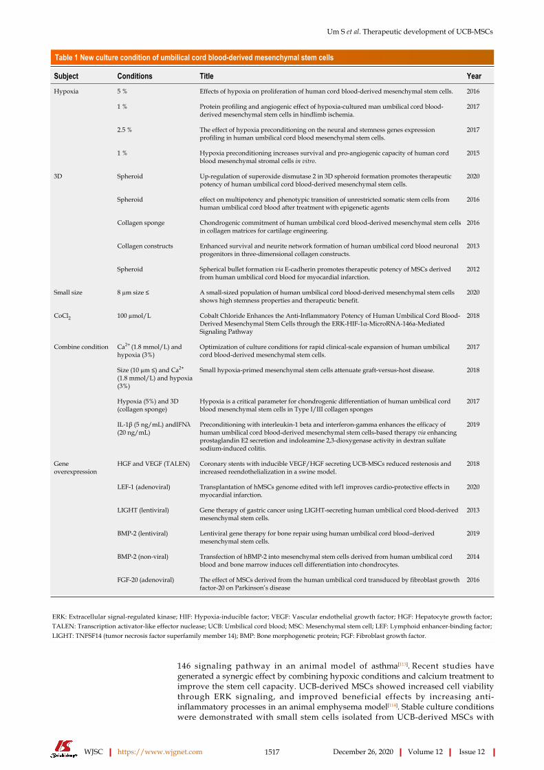
strategies for incurable diseases. However, unsolved issues still remain regarding early senescence during cell culture and low treatment efficacy after transplantation. To apply UCB-derived MSCs to clinical settings, several conditions need to be verified. Firstly, acquiring a large number of MSCs, is related to improved proliferation and delayed senescence. Secondly, a highly efficient cell culture condition is needed to enhance the therapeutic efficacy of MSCs. The current verified and developed culture conditions are described in Table 1.
The preexisting cell culture is mostly based on normoxia conditions (20%). However, recent culture conditions have changed to favor a hypoxic state with 1 to 5% oxygen, similar to oxygen deficiency of the body in a biotic environment. Hypoxia improved cell proliferation, neurogenic gene expression, and stem cell capacity of UCB-derived MSCs. In particular, apoptosis and enhanced angiogenesis of MSCs promote therapeutic efficacy in a mouse hindlimb ischemia model[77,104-106]. The classic characteristics of MSCs are adherence to cell monolayers in two-dimensional cell cultures. Recent reports have shown that three-dimensional cell culture techniques, including aggregation, microcarrier formation, spheroid formation, and sponge form, increase cell viability, stem cell potential, and differentiation capacity on osteogenesis, adipogenesis, chondrogenesis, and neurogenesis of UCB-derived MSCs. Moreover, the therapeutic efficacy of UCB-derived MSCs is improved in several disease-related animal models[61,65,107,108]. In an MI animal model, the therapeutic benefits of MSCs formed by spherical bullets were affected by the increased secretion of various paracrine factors[109]. With spherical bullets or aggregation formation of UCB-derived MSCs, high levels of paracrine factors are stimulated by increased protein interactions between SOD2 or E-cadherin[61,109].
Another aspect to consider regarding cell collection, is the size of MSCs. Cell size may compromise the therapeutic efficacy of UCB-derived MSCs. A small size ranging between 7 to 10 μm showed a high MSCs proliferation rate, referred to as recycling stem cells or rapid stem cells[110,111]. UCB-derived MSCs from neonatal tissue are small in size compared to other adult tissues, such as BM and adipose tissue[112]. The efficacy of small stem cells isolated from UCB-derived MSCs to have high proliferative rates, enhanced stem cell capacity, and delayed senescence has been confirmed. In an animal model of emphysema, the therapeutic efficacy of small cells on UCB-derived MSCs has been proven[112].
Additional reports on the improvement of the stem cell capacity have been confirmed after pretreatment with cobalt chloride (CoCl2). The anti-inflammatory function of UCB-derived MSCs increased with CoCl2 via the ERK-HIF-1α-MicroRNA-
Um S et al. Therapeutic development of UCB-MSCs
WJSC https://www.wjgnet.com 1517 December 26, 2020 Volume 12 Issue 12
Table 1 New culture condition of umbilical cord blood-derived mesenchymal stem cells
Subject Conditions Title Year
5 % Effects of hypoxia on proliferation of human cord blood-derived mesenchymal stem cells. 2016
1 % Protein profiling and angiogenic effect of hypoxia-cultured man umbilical cord blood-derived mesenchymal stem cells in hindlimb ischemia.
2017
2.5 % The effect of hypoxia preconditioning on the neural and stemness genes expression profiling in human umbilical cord blood mesenchymal stem cells.
2017
Hypoxia
1 % Hypoxia preconditioning increases survival and pro-angiogenic capacity of human cord blood mesenchymal stromal cells in vitro.
2015
Spheroid Up-regulation of superoxide dismutase 2 in 3D spheroid formation promotes therapeutic potency of human umbilical cord blood-derived mesenchymal stem cells.
2020
Spheroid effect on multipotency and phenotypic transition of unrestricted somatic stem cells from human umbilical cord blood after treatment with epigenetic agents
2016
Collagen sponge Chondrogenic commitment of human umbilical cord blood-derived mesenchymal stem cells in collagen matrices for cartilage engineering.
2016
Collagen constructs Enhanced survival and neurite network formation of human umbilical cord blood neuronal progenitors in three-dimensional collagen constructs.
2013
3D
Spheroid Spherical bullet formation via E-cadherin promotes therapeutic potency of MSCs derived from human umbilical cord blood for myocardial infarction.
2012
Small size 8 µm size ≤ A small-sized population of human umbilical cord blood-derived mesenchymal stem cells shows high stemness properties and therapeutic benefit.
2020
CoCl2 100 µmol/L Cobalt Chloride Enhances the Anti-Inflammatory Potency of Human Umbilical Cord Blood-Derived Mesenchymal Stem Cells through the ERK-HIF-1α-MicroRNA-146a-Mediated Signaling Pathway
2018
Ca2+ (1.8 mmol/L) and hypoxia (3%)
Optimization of culture conditions for rapid clinical-scale expansion of human umbilical cord blood-derived mesenchymal stem cells.
2017
Size (10 µm ≤) and Ca2+
(1.8 mmol/L) and hypoxia (3%)
Small hypoxia-primed mesenchymal stem cells attenuate graft-versus-host disease. 2018
Hypoxia (5%) and 3D (collagen sponge)
Hypoxia is a critical parameter for chondrogenic differentiation of human umbilical cord blood mesenchymal stem cells in Type I/III collagen sponges
2017
Combine condition
IL-1β (5 ng/mL) andIFNλ (20 ng/mL)
Preconditioning with interleukin-1 beta and interferon-gamma enhances the efficacy of human umbilical cord blood-derived mesenchymal stem cells-based therapy via enhancing prostaglandin E2 secretion and indoleamine 2,3-dioxygenase activity in dextran sulfate sodium-induced colitis.
2019
HGF and VEGF (TALEN) Coronary stents with inducible VEGF/HGF secreting UCB-MSCs reduced restenosis and increased reendothelialization in a swine model.
2018
LEF-1 (adenoviral) Transplantation of hMSCs genome edited with lef1 improves cardio-protective effects in myocardial infarction.
2020
LIGHT (lentiviral) Gene therapy of gastric cancer using LIGHT-secreting human umbilical cord blood-derived mesenchymal stem cells.
2013
BMP-2 (lentiviral) Lentiviral gene therapy for bone repair using human umbilical cord blood–derived mesenchymal stem cells.
2019
BMP-2 (non-viral) Transfection of hBMP-2 into mesenchymal stem cells derived from human umbilical cord blood and bone marrow induces cell differentiation into chondrocytes.
2014
Gene overexpression
FGF-20 (adenoviral) The effect of MSCs derived from the human umbilical cord transduced by fibroblast growth factor-20 on Parkinson’s disease
2016
ERK: Extracellular signal-regulated kinase; HIF: Hypoxia-inducible factor; VEGF: Vascular endothelial growth factor; HGF: Hepatocyte growth factor; TALEN: Transcription activator-like effector nuclease; UCB: Umbilical cord blood; MSC: Mesenchymal stem cell; LEF: Lymphoid enhancer-binding factor; LIGHT: TNFSF14 (tumor necrosis factor superfamily member 14); BMP: Bone morphogenetic protein; FGF: Fibroblast growth factor.
146 signaling pathway in an animal model of asthma[113]. Recent studies have generated a synergic effect by combining hypoxic conditions and calcium treatment to improve the stem cell capacity. UCB-derived MSCs showed increased cell viability through ERK signaling, and improved beneficial effects by increasing anti-inflammatory processes in an animal emphysema model[114]. Stable culture conditions were demonstrated with small stem cells isolated from UCB-derived MSCs with

Um S et al. Therapeutic development of UCB-MSCs
WJSC https://www.wjgnet.com 1518 December 26, 2020 Volume 12 Issue 12
calcium treatment and hypoxia. Small MSCs primed with hypoxia and calcium improved stem cell capacity and immunomodulatory function in vitro, as well as the therapeutic effectiveness against organ failure in a GvHD animal model using key regulator, polo-like kinase 1[115]. In addition, UCB-derived MSC culture with collagen sponge under hypoxic conditions enhanced chondrogenic differentiation capacity[66]. In vitro pre-conditioning of UCB-derived MSCs with inflammatory cytokines, IL-1β and IFN-γ, suppressed inflammation, and increased the gene expression of PGE2; while the therapeutic effect of MSCs had increased in colitis and cerebral ischemia models[116,117].
Various culture conditions have been verified by past research and technical approaches to obtain low-cost cell therapy products. Emerging studies evaluating new culture conditions need to be expanded consistently to develop successful stem cell therapies for intractable diseases.
Gene editing techniques have been applied to stem cell therapy to improve stem cell efficacy. In particular, most studies on UCB-derived MSCs have used gene overexpression to achieve the desired therapeutic effects (Table 1). UCB-derived MSCs overexpressed with VEGF/hepatocyte growth factor (HGF) using the transcription activator-like effector nuclease (TALEN) system, showed high proliferative rates, cell viability, angiogenesis, and progress in coronary restenosis in a swine model with stent material[118]. Additionally, TALEN-mediated HGF editing in UCB-derived MSCs promoted angiogenesis to improve the tube-formation ability and anti-apoptotic responses to oxidative stress[119]. Overexpression of lymphoid enhancer-binding factor 1 in UCB-derived MSCs using an adenoviral vector increased proliferation and anti-apoptotic effects by improving the cardioprotective effect in an animal model of MI[120]. In addition, TNFSF14 (LIGHT, tumor necrosis factor superfamily member 14) -overexpressed UCB-derived MSCs using a lentiviral system demonstrated suppressed growth and augmented apoptosis of tumors in a gastric cancer model[121]. Similarly, BMP-2 overexpression in UCB-derived MSCs using a lentiviral system, showed high osteogenic differentiation, which was confirmed in an animal model with bone repair[122]. Additionally, non-viral BMP-2 overexpressed in UCB-derived MSCs demonstrated increased chondrogenic marker, ColII, and induced chondrocyte differentiation in a disease model[123]. Overexpression of SRY-related high-mobility group box 9 (SOX9), a cartilage-specific transcription factor, enhanced the chondrogenic differentiation of UCB-derived MSCs[124]. Adenoviral transduction of FGF-20 in Parkinson’s disease (PD) promotes the degradation of the proinflammatory cytokine NF-kB, expressed in nigrostriatal dopaminergic regions in PD patients[125]. In future stem cell approaches, modified therapy must focus on fundamental treatment of the disease; therefore specific target-based modifications will be needed. However, the development of desired gene-edited stem cells will increase the price and safety considerations in manufacturing and quality control processes. The concerns regarding stem cell gene editing warrants further assessments for obtaining viable solutions.
CONCERNSSafety of UCB-MSC treatmentsStem cell therapy is based on adequate availability due to the innate biological characteristics of stem cells, such as self-renewal, differentiation, and motility potential. However, these biological characteristics of stem cells can affect safety issues. The most representative problem is the possibility of inducing tumorigenicity, brought on by chromosomal abnormalities. MSCs, mostly used in stem cell therapy, have a relatively low risk of potential tumorigenicity compared to multipotent stem cells. Emerging studies have demonstrated that tumor formation cannot be avoided due to stem cell characteristics and external conditions[126]. Analytical techniques for testing tumorigenicity are based on in vivo experiments. Additional in vitro tests with karyotyping and molecular and cellular genetic analysis (fluorescence in situ hybridization, chromosomal comparative genomic hybridization, single nucleotide polymorphisim et al) need to be used for genetic stability analysis. Karyotyping analysis demonstrated that UCB-derived MSCs did not have any abnormalities on chromosome until passage 15[112]. In addition, the carcinogenicity evaluation of UCB-derived MSCs confirmed that tumors were not induced in vitro and that tumor formation in vivo was not observed at 13 wk after a single injection of UCB-derived MSCs administered subcutaneously in the internal organs of BALB/c-nude mice[127].
As per the expectation and demand for stem cell therapy in regenerative medicine, the application of various administration routes, such as spinal cord, subcutaneous,

Um S et al. Therapeutic development of UCB-MSCs
WJSC https://www.wjgnet.com 1519 December 26, 2020 Volume 12 Issue 12
intramuscular, and intravenous injection, had increased, followed by confirmation of biological distribution. The Food and Drug Administration recommended that data for biological distribution, mobility, and residual period were needed and retained on the aspect of safety probability. In particular, the confirmation of cell fate after injection is important in order to analyze the mode of action of cell therapy and to decide whether the activation of cell engraftment is necessary and critical. Direct single injection of cells into the topical site of the disease, by intraparenchymal, intratracheal, intramyocardial, and intra-articular routes, demonstrated the residue of cells from 3 to 10 wk[65,92,109,128]. Therapeutic efficacy was observed before the verification of cell distribution, confirming anti-apoptosis, anti-fibrosis, anti-inflammation, and tissue regeneration. The cells injected intravenously in lung disease models, emphysema, and asthma have remained for 7 d[96,112]. The residual cells in the bladder were observed until day 7 after injection for the treatment of cystitis[129]. From the above studies, intravenous administration showed rapid extinction of cells compared to direct injection at the disease site. Intravenous injection also revealed therapeutic efficacy with anti-inflammation, anti-apoptosis, anti-fibrosis, and angiogenesis. Collectively, the results indicate the stability of injected UCB-derived MSCs in various diseases.
Heterogeneity Heterogeneity remains a critical problem, not only for gaining a general understanding of the mechanism by which MSCs maintain their growth rate and undergo differentiation toward specific lineage potentials, but also with respect to achieving better outcomes in therapeutic applications[130]. It is mainly affected by growth media, two-dimensional adherence to plastic culture dishes, and sub-culturing methods; therefore, these processes were repeated until an adequate number of MSCs were obtained for large-scale expansion in vitro[131]. In this context, researchers have tried to establish a standard set of criteria for attaining more homogenous populations of MSCs. Firstly, for clearer cell origins, studies have attempted to clone UCB-MSCs derived from single cells by limiting dilution assays. Single cell-derived clones were identified by evaluating MSC features including growth, surface marker, stemness, and multi-lineage potential. As a result, one clone showed a faster growth rate and higher differentiation potential than the original populations. However, other clone cells showed weak growth ability and differentiation potential compared to the original cells, except for one clone that had superior data[132]. Further, this processing draws attention to the selection criteria as a possible marker related to the excellent MSC clones. Secondly, several protocols have been developed to isolate more homogeneous cells using several specific antigens such as CD143, CD146, and CD271[16,133,134]; however, none of these processing methods have gained wide acceptance, and a unique single marker has not been identified to date. Moreover, to obtain primitive homogeneous, multi or pluripotent stem cells have been introduced with different names in adult hematopoietic tissues, for example, unrestricted somatic stem cells[135], depending on the isolation strategy, ex vivo expansion protocol, and markers employed for their identification; however, these factors remain unclear and are a major obstacle for heterogeneity. Despite such an attempt, there is still no defined culture protocol to overcome MSC heterogeneity.
Low yieldDespite the many advantages of UCB-MSCs, their utility remains controversial due to their low isolation efficiency. Many groups have reported that UCB has a 65%-90% maximum isolation efficiency in various culture protocols, including the depletion of lymphocytes and monocytes from mononuclear cells before cell seeding, delivery time, volume, addition of cytokines supplements or platelet lysate to the medium, density gradient purification, or cultivation of cells under hypoxia[12,18,136-139]. This could also help improve the utility of UCB-MSCs as a therapeutic resource. Further studies should be performed to validate these methods for clinical use.
Current good manufacturing practices for developmentFor advanced therapy development using UCB-derived MSCs, standard operation procedures are necessary, as well as the reliable application into Good Manufacturing Practice procedures. The current established and developed MSC therapy has limitations in commercialization and market expansion because of the high cost caused by the manufacturing process and quality control with conventional static monolayer culture. Therefore, cost reduction from improving the efficacy of cells is essential to develop the next-generation stem cell therapies, based on fundamental technologies over conventional culture. Consequently, evaluation of optimized and

Um S et al. Therapeutic development of UCB-MSCs
WJSC https://www.wjgnet.com 1520 December 26, 2020 Volume 12 Issue 12
innovative manufacturing processes is needed (Figure 2).The study of therapeutic manufacturing focuses on the workflow process for
selecting the outstanding upstream cells. However, various commercialization products have been introduced to academic researchers as well as to industrial companies, in accordance with the higher interest in downstream areas to develop the product. Stem cell therapy is affected not only by the skill of the workforce but also by the massification and automation of equipment to guarantee consistent products. The adhesive characteristics of MSCs make it difficult to expand cells for mass production, making it difficult to develop a bioreactor. However, many companies have developed related and combined bioreactors for the extensive production of stem cells[129,140,141]. This system demonstrated that the scale-up of a stem cell batch in a single progression reduced the cost for production, followed by an increase in the level of quality control with regards to the development of automated systems in manufacturing cell cultures[142]. A bioreactor is defined as a culture system where the organism is controlled and regulated to produce the specific material or cells, by removing the unnecessary metabolic products while culturing the cells and continuously maintaining the proper levels of nutrients and growth factors. For this reason, the selection of the bioreactor, which is appropriate for specific cells, is important in the final manufacturing process to complete the efficient scale-up applied while maintaining the characteristics of cells based on the technical equipment.
Recently developed manufacturing bioreactors, which are fully closed, controllable, and have scalable culture systems, have been used to monitor and control the metabolic state in real time including, dissolved oxygen, glucose, ammonia, pH, and lactate[143-145]. As the collection of large cell quantities takes a long time using manual methods, a reduction in collection times using automated equipment, increases the efficacy and consistency of the quality control. Current commercialized stem cell therapy-based products are temperature sensitive, resulting in a short expiration period. The development of frozen preservation techniques and the appropriate storage of these cell products are important for their viable export overseas. The preservative solution in basic culture media or saline solution, is not appropriate for maintaining long-term cell viability. Several frozen preservative solutions have recently been developed, such as serum-free, Xeno-free media and DMSO. However, these result in a low stability of cells with a low cell recovery rate. Therefore, the development of an efficient preservation system needs to be complemented[146-149]. Ideally, a division system has been developed for the automated manufacturing of frozen storage of bulk cultured cells, allowing improvements in the accuracy, repetition, time consumption, and number of workers needed compared to the current manual workspace.
CONCLUSIONStem cell therapy is an outstanding method for regenerative medicine. With significant advantages, such as self-renewal, differentiation capacity, and immunomodulation, the use of stem cells is appropriate for the treatment of several disorders and diseases. UCB is a primitive and rich source of MSCs. UCB-derived MSCs have the potential of exerting profound immunomodulatory effects with the secretion of factors and cytokines. However, the safety and yield of UCB-derived MSCs are still a concern. Next-generation stem cell therapy is necessary, referring to the mass production of efficient stem cells based on the fundamental technology, to improve whole cell processing. This will solve problems of limited product expansions caused by short expired periods and high production costs. In accordance with the advanced process, a manufacturing system is needed to produce quantity in order to reduce production costs, as well as enhancing yield output, and delivering consistent quality by automated production processes. Overall, advanced manufacturing systems will improve and trigger the commercialization and globalization of stem cell therapy.

Um S et al. Therapeutic development of UCB-MSCs
WJSC https://www.wjgnet.com 1521 December 26, 2020 Volume 12 Issue 12
Figure 2 Workflow for therapeutic cell manufacturing processing.
REFERENCESFriedenstein AJ. Precursor cells of mechanocytes. Int Rev Cytol 1976; 47: 327-359 [PMID: 11195 DOI: 10.1016/s0074-7696(08)60092-3]
1
Perdikogianni C, Dimitriou H, Stiakaki E, Martimianaki G, Kalmanti M. Could cord blood be a source of mesenchymal stromal cells for clinical use? Cytotherapy 2008; 10: 452-459 [PMID: 18821358 DOI: 10.1080/14653240701883079]
2
Baer PC, Koch B, Hickmann E, Schubert R, Cinatl J Jr, Hauser IA, Geiger H. Isolation, Characterization, Differentiation and Immunomodulatory Capacity of Mesenchymal Stromal/Stem Cells from Human Perirenal Adipose Tissue. Cells 2019; 8: 1346 [PMID: 31671899 DOI: 10.3390/cells8111346]
3
Jiang Y, Jahagirdar BN, Reinhardt RL, Schwartz RE, Keene CD, Ortiz-Gonzalez XR, Reyes M, Lenvik T, Lund T, Blackstad M, Du J, Aldrich S, Lisberg A, Low WC, Largaespada DA, Verfaillie CM. Pluripotency of mesenchymal stem cells derived from adult marrow. Nature 2002; 418: 41-49 [PMID: 12077603 DOI: 10.1038/nature00870]
4
Gluckman E, Broxmeyer HA, Auerbach AD, Friedman HS, Douglas GW, Devergie A, Esperou H, Thierry D, Socie G, Lehn P. Hematopoietic reconstitution in a patient with Fanconi's anemia by means of umbilical-cord blood from an HLA-identical sibling. N Engl J Med 1989; 321: 1174-1178 [PMID: 2571931 DOI: 10.1056/NEJM198910263211707]
5
Roura S, Pujal JM, Gálvez-Montón C, Bayes-Genis A. The role and potential of umbilical cord blood in an era of new therapies: a review. Stem Cell Res Ther 2015; 6: 123 [PMID: 26133757 DOI: 10.1186/s13287-015-0113-2]
6
Liao Y, Geyer MB, Yang AJ, Cairo MS. Cord blood transplantation and stem cell regenerative potential. Exp Hematol 2011; 39: 393-412 [PMID: 21238533 DOI: 10.1016/j.exphem.2011.01.002]
7
Badowski MS, Harris DT. Collection, processing, and banking of umbilical cord blood stem cells for transplantation and regenerative medicine. Methods Mol Biol 2012; 879: 279-290 [PMID: 22610565 DOI: 10.1007/978-1-61779-815-3_16]
8
Spartano S, Bianchi M, Murgi E, Giannadrea S, Landini A, Barbagallo O, Screnci M, Girelli G, Zini G, Teofili L. Medicine use in pregnancy and public cord blood bank databases. Pharmacoepidemiol Drug Saf 2014; 23: 1107-1109 [PMID: 25316303 DOI: 10.1002/pds.3693]
9
Haw J, Polzer J, Devine DV. Contextual factors influencing donor recruitment and cord blood collection: perspectives of frontline staff of the Canadian Blood Services' Cord Blood Bank. Transfusion 2019; 59: 1742-1748 [PMID: 30741433 DOI: 10.1111/trf.15185]
10
Dessels C, Alessandrini M, Pepper MS. Factors Influencing the Umbilical Cord Blood Stem Cell Industry: An Evolving Treatment Landscape. Stem Cells Transl Med 2018; 7: 643-650 [PMID: 29777574 DOI: 10.1002/sctm.17-0244]
11
Lee OK, Kuo TK, Chen WM, Lee KD, Hsieh SL, Chen TH. Isolation of multipotent mesenchymal stem cells from umbilical cord blood. Blood 2004; 103: 1669-1675 [PMID: 14576065 DOI: 10.1182/blood-2003-05-1670]
12
Rashnonejad A, Ercan G, Gunduz C, Akdemir A, Tiftikcioglu YO. Comparative analysis of human UCB and adipose tissue derived mesenchymal stem cells for their differentiation potential into brown and white adipocytes. Mol Biol Rep 2018; 45: 233-244 [PMID: 29453764 DOI: 10.1007/s11033-018-4156-1]
13
Jin HJ, Bae YK, Kim M, Kwon SJ, Jeon HB, Choi SJ, Kim SW, Yang YS, Oh W, Chang JW. Comparative analysis of human mesenchymal stem cells from bone marrow, adipose tissue, and umbilical cord blood as sources of cell therapy. Int J Mol Sci 2013; 14: 17986-18001 [PMID: 24005862 DOI: 10.3390/ijms140917986]
14
Markov V, Kusumi K, Tadesse MG, William DA, Hall DM, Lounev V, Carlton A, Leonard J, Cohen RI, Rappaport EF, Saitta B. Identification of cord blood-derived mesenchymal stem/stromal cell populations with distinct growth kinetics, differentiation potentials, and gene expression profiles. Stem Cells Dev 2007; 16: 53-73 [PMID: 17348805 DOI: 10.1089/scd.2006.0660]
15
Amati E, Perbellini O, Rotta G, Bernardi M, Chieregato K, Sella S, Rodeghiero F, Ruggeri M, Astori G. High-throughput immunophenotypic characterization of bone marrow- and cord blood-derived mesenchymal stromal cells reveals common and differentially expressed markers:
16

Um S et al. Therapeutic development of UCB-MSCs
WJSC https://www.wjgnet.com 1522 December 26, 2020 Volume 12 Issue 12
identification of angiotensin-converting enzyme (CD143) as a marker differentially expressed between adult and perinatal tissue sources. Stem Cell Res Ther 2018; 9: 10 [PMID: 29338788 DOI: 10.1186/s13287-017-0755-3]Chang YJ, Shih DT, Tseng CP, Hsieh TB, Lee DC, Hwang SM. Disparate mesenchyme-lineage tendencies in mesenchymal stem cells from human bone marrow and umbilical cord blood. Stem Cells 2006; 24: 679-685 [PMID: 16179428 DOI: 10.1634/stemcells.2004-0308]
17
Kern S, Eichler H, Stoeve J, Klüter H, Bieback K. Comparative analysis of mesenchymal stem cells from bone marrow, umbilical cord blood, or adipose tissue. Stem Cells 2006; 24: 1294-1301 [PMID: 16410387 DOI: 10.1634/stemcells.2005-0342]
18
Malgieri A, Kantzari E, Patrizi MP, Gambardella S. Bone marrow and umbilical cord blood human mesenchymal stem cells: state of the art. Int J Clin Exp Med 2010; 3: 248-269 [PMID: 21072260]
19
Götherström C, Ringdén O, Tammik C, Zetterberg E, Westgren M, Le Blanc K. Immunologic properties of human fetal mesenchymal stem cells. Am J Obstet Gynecol 2004; 190: 239-245 [PMID: 14749666 DOI: 10.1016/j.ajog.2003.07.022]
20
Aggarwal S, Pittenger MF. Human mesenchymal stem cells modulate allogeneic immune cell responses. Blood 2005; 105: 1815-1822 [PMID: 15494428 DOI: 10.1182/blood-2004-04-1559]
21
Rogers I, Casper RF. Umbilical cord blood stem cells. Best Pract Res Clin Obstet Gynaecol 2004; 18: 893-908 [PMID: 15582545 DOI: 10.1016/j.bpobgyn.2004.06.004]
22
Patel DM, Shah J, Srivastava AS. Therapeutic potential of mesenchymal stem cells in regenerative medicine. Stem Cells Int 2013; 2013: 496218 [PMID: 23577036 DOI: 10.1155/2013/496218]
23
Park KS, Lee YS, Kang KS. In vitro neuronal and osteogenic differentiation of mesenchymal stem cells from human umbilical cord blood. J Vet Sci 2006; 7: 343-348 [PMID: 17106225 DOI: 10.4142/jvs.2006.7.4.343]
24
Dominici M, Le Blanc K, Mueller I, Slaper-Cortenbach I, Marini F, Krause D, Deans R, Keating A, Prockop Dj, Horwitz E. Minimal criteria for defining multipotent mesenchymal stromal cells. The International Society for Cellular Therapy position statement. Cytotherapy 2006; 8: 315-317 [PMID: 16923606 DOI: 10.1080/14653240600855905]
25
Carrade DD, Lame MW, Kent MS, Clark KC, Walker NJ, Borjesson DL. Comparative Analysis of the Immunomodulatory Properties of Equine Adult-Derived Mesenchymal Stem Cells(). Cell Med 2012; 4: 1-11 [PMID: 23152950 DOI: 10.3727/215517912X647217]
26
Hollweck T, Marschmann M, Hartmann I, Akra B, Meiser B, Reichart B, Eblenkamp M, Wintermantel E, Eissner G. Comparative analysis of adherence, viability, proliferation and morphology of umbilical cord tissue-derived mesenchymal stem cells seeded on different titanium-coated expanded polytetrafluoroethylene scaffolds. Biomed Mater 2010; 5: 065004 [PMID: 20924136 DOI: 10.1088/1748-6041/5/6/065004]
27
Li Y, Wu Q, Wang Y, Li L, Bu H, Bao J. Senescence of mesenchymal stem cells (Review). Int J Mol Med 2017; 39: 775-782 [PMID: 28290609 DOI: 10.3892/ijmm.2017.2912]
28
Hallows SE, Regnault TR, Betts DH. The long and short of it: the role of telomeres in fetal origins of adult disease. J Pregnancy 2012; 2012: 638476 [PMID: 23094159 DOI: 10.1155/2012/638476]
29
Martin-Rendon E, Sweeney D, Lu F, Girdlestone J, Navarrete C, Watt SM. 5-Azacytidine-treated human mesenchymal stem/progenitor cells derived from umbilical cord, cord blood and bone marrow do not generate cardiomyocytes in vitro at high frequencies. Vox Sang 2008; 95: 137-148 [PMID: 18557828 DOI: 10.1111/j.1423-0410.2008.01076.x]
30
Jin HJ, Kwon JH, Kim M, Bae YK, Choi SJ, Oh W, Yang YS, Jeon HB. Downregulation of Melanoma Cell Adhesion Molecule (MCAM/CD146) Accelerates Cellular Senescence in Human Umbilical Cord Blood-Derived Mesenchymal Stem Cells. Stem Cells Transl Med 2016; 5: 427-439 [PMID: 26941359 DOI: 10.5966/sctm.2015-0109]
31
Lv F, Lu M, Cheung KM, Leung VY, Zhou G. Intrinsic properties of mesemchymal stem cells from human bone marrow, umbilical cord and umbilical cord blood comparing the different sources of MSC. Curr Stem Cell Res Ther 2012; 7: 389-399 [PMID: 22934544 DOI: 10.2174/157488812804484611]
32
Wagner W, Wein F, Seckinger A, Frankhauser M, Wirkner U, Krause U, Blake J, Schwager C, Eckstein V, Ansorge W, Ho AD. Comparative characteristics of mesenchymal stem cells from human bone marrow, adipose tissue, and umbilical cord blood. Exp Hematol 2005; 33: 1402-1416 [PMID: 16263424 DOI: 10.1016/j.exphem.2005.07.003]
33
Harley CB, Futcher AB, Greider CW. Telomeres shorten during ageing of human fibroblasts. Nature 1990; 345: 458-460 [PMID: 2342578 DOI: 10.1038/345458a0]
34
Terai M, Uyama T, Sugiki T, Li XK, Umezawa A, Kiyono T. Immortalization of human fetal cells: the life span of umbilical cord blood-derived cells can be prolonged without manipulating p16INK4a/RB braking pathway. Mol Biol Cell 2005; 16: 1491-1499 [PMID: 15647378 DOI: 10.1091/mbc.e04-07-0652]
35
Whiteman VE, Goswami A, Salihu HM. Telomere length and fetal programming: A review of recent scientific advances. Am J Reprod Immunol 2017; 77 [PMID: 28500672 DOI: 10.1111/aji.12661]
36
Ahmed S, Passos JF, Birket MJ, Beckmann T, Brings S, Peters H, Birch-Machin MA, von Zglinicki T, Saretzki G. Telomerase does not counteract telomere shortening but protects mitochondrial function under oxidative stress. J Cell Sci 2008; 121: 1046-1053 [PMID: 18334557 DOI: 10.1242/jcs.019372]
37
Izadpanah R, Trygg C, Patel B, Kriedt C, Dufour J, Gimble JM, Bunnell BA. Biologic properties of 38

Um S et al. Therapeutic development of UCB-MSCs
WJSC https://www.wjgnet.com 1523 December 26, 2020 Volume 12 Issue 12
mesenchymal stem cells derived from bone marrow and adipose tissue. J Cell Biochem 2006; 99: 1285-1297 [PMID: 16795045 DOI: 10.1002/jcb.20904]Gammaitoni L, Weisel KC, Gunetti M, Wu KD, Bruno S, Pinelli S, Bonati A, Aglietta M, Moore MA, Piacibello W. Elevated telomerase activity and minimal telomere loss in cord blood long-term cultures with extensive stem cell replication. Blood 2004; 103: 4440-4448 [PMID: 14726371 DOI: 10.1182/blood-2003-09-3079]
39
Zimmermann S, Voss M, Kaiser S, Kapp U, Waller CF, Martens UM. Lack of telomerase activity in human mesenchymal stem cells. Leukemia 2003; 17: 1146-1149 [PMID: 12764382 DOI: 10.1038/sj.leu.2402962]
40
De Becker A, Riet IV. Homing and migration of mesenchymal stromal cells: How to improve the efficacy of cell therapy? World J Stem Cells 2016; 8: 73-87 [PMID: 27022438 DOI: 10.4252/wjsc.v8.i3.73]
41
Butcher EC, Picker LJ. Lymphocyte homing and homeostasis. Science 1996; 272: 60-66 [PMID: 8600538 DOI: 10.1126/science.272.5258.60]
42
Majumdar MK, Keane-Moore M, Buyaner D, Hardy WB, Moorman MA, McIntosh KR, Mosca JD. Characterization and functionality of cell surface molecules on human mesenchymal stem cells. J Biomed Sci 2003; 10: 228-241 [PMID: 12595759 DOI: 10.1007/BF02256058]
43
Reinisch A, Etchart N, Thomas D, Hofmann NA, Fruehwirth M, Sinha S, Chan CK, Senarath-Yapa K, Seo EY, Wearda T, Hartwig UF, Beham-Schmid C, Trajanoski S, Lin Q, Wagner W, Dullin C, Alves F, Andreeff M, Weissman IL, Longaker MT, Schallmoser K, Majeti R, Strunk D. Epigenetic and in vivo comparison of diverse MSC sources reveals an endochondral signature for human hematopoietic niche formation. Blood 2015; 125: 249-260 [PMID: 25406351 DOI: 10.1182/blood-2014-04-572255]
44
Qiu Y, Marquez-Curtis LA, Janowska-Wieczorek A. Mesenchymal stromal cells derived from umbilical cord blood migrate in response to complement C1q. Cytotherapy 2012; 14: 285-295 [PMID: 22264191 DOI: 10.3109/14653249.2011.651532]
45
Kim SM, Kim DS, Jeong CH, Kim DH, Kim JH, Jeon HB, Kwon SJ, Jeun SS, Yang YS, Oh W, Chang JW. CXC chemokine receptor 1 enhances the ability of human umbilical cord blood-derived mesenchymal stem cells to migrate toward gliomas. Biochem Biophys Res Commun 2011; 407: 741-746 [PMID: 21439934 DOI: 10.1016/j.bbrc.2011.03.093]
46
Tondreau T, Meuleman N, Stamatopoulos B, De Bruyn C, Delforge A, Dejeneffe M, Martiat P, Bron D, Lagneaux L. In vitro study of matrix metalloproteinase/tissue inhibitor of metalloproteinase production by mesenchymal stromal cells in response to inflammatory cytokines: the role of their migration in injured tissues. Cytotherapy 2009; 11: 559-569 [PMID: 19551542 DOI: 10.1080/14653240903051541]
47
Ryu CH, Park SA, Kim SM, Lim JY, Jeong CH, Jun JA, Oh JH, Park SH, Oh WI, Jeun SS. Migration of human umbilical cord blood mesenchymal stem cells mediated by stromal cell-derived factor-1/CXCR4 axis via Akt, ERK, and p38 signal transduction pathways. Biochem Biophys Res Commun 2010; 398: 105-110 [PMID: 20558135 DOI: 10.1016/j.bbrc.2010.06.043]
48
Oh W, Kim DS, Yang YS, Lee JK. Immunological properties of umbilical cord blood-derived mesenchymal stromal cells. Cell Immunol 2008; 251: 116-123 [PMID: 18495100 DOI: 10.1016/j.cellimm.2008.04.003]
49
Ardeshirylajimi A, Mossahebi-Mohammadi M, Vakilian S, Langroudi L, Seyedjafari E, Atashi A, Soleimani M. Comparison of osteogenic differentiation potential of human adult stem cells loaded on bioceramic-coated electrospun poly (L-lactide) nanofibres. Cell Prolif 2015; 48: 47-58 [PMID: 25495212 DOI: 10.1111/cpr.12156]
50
Peng L, Jia Z, Yin X, Zhang X, Liu Y, Chen P, Ma K, Zhou C. Comparative analysis of mesenchymal stem cells from bone marrow, cartilage, and adipose tissue. Stem Cells Dev 2008; 17: 761-773 [PMID: 18393634 DOI: 10.1089/scd.2007.0217]
51
Wang L, Dormer NH, Bonewald LF, Detamore MS. Osteogenic differentiation of human umbilical cord mesenchymal stromal cells in polyglycolic acid scaffolds. Tissue Eng Part A 2010; 16: 1937-1948 [PMID: 20070186 DOI: 10.1089/ten.TEA.2009.0706]
52
Tahlawi A, Klontzas ME, Allenby MC, Morais JCF, Panoskaltsis N, Mantalaris A. RGD-functionalized polyurethane scaffolds promote umbilical cord blood mesenchymal stem cell expansion and osteogenic differentiation. J Tissue Eng Regen Med 2019; 13: 232-243 [PMID: 30537385 DOI: 10.1002/term.2784]
53
Klontzas ME, Reakasame S, Silva R, Morais JCF, Vernardis S, MacFarlane RJ, Heliotis M, Tsiridis E, Panoskaltsis N, Boccaccini AR, Mantalaris A. Oxidized alginate hydrogels with the GHK peptide enhance cord blood mesenchymal stem cell osteogenesis: A paradigm for metabolomics-based evaluation of biomaterial design. Acta Biomater 2019; 88: 224-240 [PMID: 30772514 DOI: 10.1016/j.actbio.2019.02.017]
54
Klontzas ME, Vernardis SI, Heliotis M, Tsiridis E, Mantalaris A. Metabolomics Analysis of the Osteogenic Differentiation of Umbilical Cord Blood Mesenchymal Stem Cells Reveals Differential Sensitivity to Osteogenic Agents. Stem Cells Dev 2017; 26: 723-733 [PMID: 28418785 DOI: 10.1089/scd.2016.0315]
55
Hong B, Lee S, Shin N, Ko Y, Kim D, Lee J, Lee W. Bone regeneration with umbilical cord blood mesenchymal stem cells in femoral defects of ovariectomized rats. Osteoporos Sarcopenia 2018; 4: 95-101 [PMID: 30775550 DOI: 10.1016/j.afos.2018.08.003]
56
An JH, Park H, Song JA, Ki KH, Yang JY, Choi HJ, Cho SW, Kim SW, Kim SY, Yoo JJ, Baek 57

Um S et al. Therapeutic development of UCB-MSCs
WJSC https://www.wjgnet.com 1524 December 26, 2020 Volume 12 Issue 12
WY, Kim JE, Choi SJ, Oh W, Shin CS. Transplantation of human umbilical cord blood-derived mesenchymal stem cells or their conditioned medium prevents bone loss in ovariectomized nude mice. Tissue Eng Part A 2013; 19: 685-696 [PMID: 23215868 DOI: 10.1089/ten.TEA.2012.0047]Ibrahim AM, Elgharabawi NM, Makhlouf MM, Ibrahim OY. Chondrogenic differentiation of human umbilical cord blood-derived mesenchymal stem cells in vitro. Microsc Res Tech 2015; 78: 667-675 [PMID: 26096638 DOI: 10.1002/jemt.22520]
58
Rakic R, Bourdon B, Demoor M, Maddens S, Saulnier N, Galéra P. Differences in the intrinsic chondrogenic potential of equine umbilical cord matrix and cord blood mesenchymal stromal/stem cells for cartilage regeneration. Sci Rep 2018; 8: 13799 [PMID: 30217993 DOI: 10.1038/s41598-018-28164-9]
59
Contentin R, Demoor M, Concari M, Desancé M, Audigié F, Branly T, Galéra P. Comparison of the Chondrogenic Potential of Mesenchymal Stem Cells Derived from Bone Marrow and Umbilical Cord Blood Intended for Cartilage Tissue Engineering. Stem Cell Rev Rep 2020; 16: 126-143 [PMID: 31745710 DOI: 10.1007/s12015-019-09914-2]
60
Gómez-Leduc T, Hervieu M, Legendre F, Bouyoucef M, Gruchy N, Poulain L, de Vienne C, Herlicoviez M, Demoor M, Galéra P. Chondrogenic commitment of human umbilical cord blood-derived mesenchymal stem cells in collagen matrices for cartilage engineering. Sci Rep 2016; 6: 32786 [PMID: 27604951 DOI: 10.1038/srep32786]
61
Pievani A, Scagliotti V, Russo FM, Azario I, Rambaldi B, Sacchetti B, Marzorati S, Erba E, Giudici G, Riminucci M, Biondi A, Vergani P, Serafini M. Comparative analysis of multilineage properties of mesenchymal stromal cells derived from fetal sources shows an advantage of mesenchymal stromal cells isolated from cord blood in chondrogenic differentiation potential. Cytotherapy 2014; 16: 893-905 [PMID: 24794181 DOI: 10.1016/j.jcyt.2014.02.008]
62
Park YB, Ha CW, Kim JA, Han WJ, Rhim JH, Lee HJ, Kim KJ, Park YG, Chung JY. Single-stage cell-based cartilage repair in a rabbit model: cell tracking and in vivo chondrogenesis of human umbilical cord blood-derived mesenchymal stem cells and hyaluronic acid hydrogel composite. Osteoarthritis Cartilage 2017; 25: 570-580 [PMID: 27789339 DOI: 10.1016/j.joca.2016.10.012]
63
Chung JY, Song M, Ha CW, Kim JA, Lee CH, Park YB. Comparison of articular cartilage repair with different hydrogel-human umbilical cord blood-derived mesenchymal stem cell composites in a rat model. Stem Cell Res Ther 2014; 5: 39 [PMID: 24646697 DOI: 10.1186/scrt427]
64
Lee M, Song BR, Kim DH, Ha J, Lee M, Choi SJ, Oh W, Um S, Jin HJ. Up-Regulation of Superoxide Dismutase 2 in 3D Spheroid Formation Promotes Therapeutic Potency of Human Umbilical Cord Blood-Derived Mesenchymal Stem Cells. Antioxidants (Basel) 2020; 9: 66 [PMID: 31940867 DOI: 10.3390/antiox9010066]
65
Gómez-Leduc T, Desancé M, Hervieu M, Legendre F, Ollitrault D, de Vienne C, Herlicoviez M, Galéra P, Demoor M. Hypoxia Is a Critical Parameter for Chondrogenic Differentiation of Human Umbilical Cord Blood Mesenchymal Stem Cells in Type I/III Collagen Sponges. Int J Mol Sci 2017; 18: 1933 [PMID: 28885597 DOI: 10.3390/ijms18091933]
66
Bieback K, Kern S, Klüter H, Eichler H. Critical parameters for the isolation of mesenchymal stem cells from umbilical cord blood. Stem Cells 2004; 22: 625-634 [PMID: 15277708 DOI: 10.1634/stemcells.22-4-625]
67
Koch TG, Heerkens T, Thomsen PD, Betts DH. Isolation of mesenchymal stem cells from equine umbilical cord blood. BMC Biotechnol 2007; 7: 26 [PMID: 17537254 DOI: 10.1186/1472-6750-7-26]
68
Kögler G, Sensken S, Wernet P. Comparative generation and characterization of pluripotent unrestricted somatic stem cells with mesenchymal stem cells from human cord blood. Exp Hematol 2006; 34: 1589-1595 [PMID: 17046580 DOI: 10.1016/j.exphem.2006.07.011]
69
Sibov TT, Severino P, Marti LC, Pavon LF, Oliveira DM, Tobo PR, Campos AH, Paes AT, Amaro E Jr, F Gamarra L, Moreira-Filho CA. Mesenchymal stem cells from umbilical cord blood: parameters for isolation, characterization and adipogenic differentiation. Cytotechnology 2012; 64: 511-521 [PMID: 22328147 DOI: 10.1007/s10616-012-9428-3]
70
Bae YK, Kwon JH, Kim M, Kim GH, Choi SJ, Oh W, Yang YS, Jin HJ, Jeon HB. Intracellular Calcium Determines the Adipogenic Differentiation Potential of Human Umbilical Cord Blood-Derived Mesenchymal Stem Cells via the Wnt5a/β-Catenin Signaling Pathway. Stem Cells Int 2018; 2018: 6545071 [PMID: 30123291 DOI: 10.1155/2018/6545071]
71
Bae JE, Kang GM, Min SH, Jo DS, Jung YK, Kim K, Kim MS, Cho DH. Primary cilia mediate mitochondrial stress responses to promote dopamine neuron survival in a Parkinson's disease model. Cell Death Dis 2019; 10: 952 [PMID: 31844040 DOI: 10.1038/s41419-019-2184-y]
72
Ali H, Bayatti N, Lindsay S, Dashti AA, Al-Mulla F. Directed differentiation of umbilical cord blood stem cells into cortical GABAergic neurons. Acta Neurobiol Exp (Wars) 2013; 73: 250-259 [PMID: 23823986]
73
Rafieemehr H, Kheirandish M, Soleimani M. Improving the neuronal differentiation efficiency of umbilical cord blood-derived mesenchymal stem cells cultivated under appropriate conditions. Iran J Basic Med Sci 2015; 18: 1100-1106 [PMID: 26949497]
74
Jin HJ, Nam HY, Bae YK, Kim SY, Im IR, Oh W, Yang YS, Choi SJ, Kim SW. GD2 expression is closely associated with neuronal differentiation of human umbilical cord blood-derived mesenchymal stem cells. Cell Mol Life Sci 2010; 67: 1845-1858 [PMID: 20165901 DOI: 10.1007/s00018-010-0292-z]
75
Chen J, Sanberg PR, Li Y, Wang L, Lu M, Willing AE, Sanchez-Ramos J, Chopp M. Intravenous 76

Um S et al. Therapeutic development of UCB-MSCs
WJSC https://www.wjgnet.com 1525 December 26, 2020 Volume 12 Issue 12
administration of human umbilical cord blood reduces behavioral deficits after stroke in rats. Stroke 2001; 32: 2682-2688 [PMID: 11692034 DOI: 10.1161/hs1101.098367]Kheirandish M, Gavgani SP, Samiee S. The effect of hypoxia preconditioning on the neural and stemness genes expression profiling in human umbilical cord blood mesenchymal stem cells. Transfus Apher Sci 2017; 56: 392-399 [PMID: 28428031 DOI: 10.1016/j.transci.2017.03.015]
77
Lim JY, Park SI, Oh JH, Kim SM, Jeong CH, Jun JA, Lee KS, Oh W, Lee JK, Jeun SS. Brain-derived neurotrophic factor stimulates the neural differentiation of human umbilical cord blood-derived mesenchymal stem cells and survival of differentiated cells through MAPK/ERK and PI3K/Akt-dependent signaling pathways. J Neurosci Res 2008; 86: 2168-2178 [PMID: 18438930 DOI: 10.1002/jnr.21669]
78
Wang L, Lu M. Regulation and direction of umbilical cord blood mesenchymal stem cells to adopt neuronal fate. Int J Neurosci 2014; 124: 149-159 [PMID: 23879374 DOI: 10.3109/00207454.2013.828055]
79
Bonaventura G, Chamayou S, Liprino A, Guglielmino A, Fichera M, Caruso M, Barcellona ML. Different Tissue-Derived Stem Cells: A Comparison of Neural Differentiation Capability. PLoS One 2015; 10: e0140790 [PMID: 26517263 DOI: 10.1371/journal.pone.0140790]
80
George S, Hamblin MR, Abrahamse H. Differentiation of Mesenchymal Stem Cells to Neuroglia: in the Context of Cell Signalling. Stem Cell Rev Rep 2019; 15: 814-826 [PMID: 31515658 DOI: 10.1007/s12015-019-09917-z]
81
Chang SA, Lee EJ, Kang HJ, Zhang SY, Kim JH, Li L, Youn SW, Lee CS, Kim KH, Won JY, Sohn JW, Park KW, Cho HJ, Yang SE, Oh WI, Yang YS, Ho WK, Park YB, Kim HS. Impact of myocardial infarct proteins and oscillating pressure on the differentiation of mesenchymal stem cells: effect of acute myocardial infarction on stem cell differentiation. Stem Cells 2008; 26: 1901-1912 [PMID: 18403756 DOI: 10.1634/stemcells.2007-0708]
82
Cho HM, Kim PH, Chang HK, Shen YM, Bonsra K, Kang BJ, Yum SY, Kim JH, Lee SY, Choi MC, Kim HH, Jang G, Cho JY. Targeted Genome Engineering to Control VEGF Expression in Human Umbilical Cord Blood-Derived Mesenchymal Stem Cells: Potential Implications for the Treatment of Myocardial Infarction. Stem Cells Transl Med 2017; 6: 1040-1051 [PMID: 28186692 DOI: 10.1002/sctm.16-0114]
83
Schreurs M, Suttorp CM, Mutsaers HAM, Kuijpers-Jagtman AM, Von den Hoff JW, Ongkosuwito EM, Carvajal Monroy PL, Wagener FADTG. Tissue engineering strategies combining molecular targets against inflammation and fibrosis, and umbilical cord blood stem cells to improve hampered muscle and skin regeneration following cleft repair. Med Res Rev 2020; 40: 9-26 [PMID: 31104334 DOI: 10.1002/med.21594]
84
Liu L, Yu Y, Hou Y, Chai J, Duan H, Chu W, Zhang H, Hu Q, Du J. Human umbilical cord mesenchymal stem cells transplantation promotes cutaneous wound healing of severe burned rats. PLoS One 2014; 9: e88348 [PMID: 24586314 DOI: 10.1371/journal.pone.0088348]
85
Gomez-Salazar M, Gonzalez-Galofre ZN, Casamitjana J, Crisan M, James AW, Péault B. Five Decades Later, Are Mesenchymal Stem Cells Still Relevant? Front Bioeng Biotechnol 2020; 8: 148 [PMID: 32185170 DOI: 10.3389/fbioe.2020.00148]
86
Montemurro T, Viganò M, Ragni E, Barilani M, Parazzi V, Boldrin V, Lavazza C, Montelatici E, Banfi F, Lauri E, Giovanelli S, Baccarin M, Guerneri S, Giordano R, Lazzari L. Angiogenic and anti-inflammatory properties of mesenchymal stem cells from cord blood: soluble factors and extracellular vesicles for cell regeneration. Eur J Cell Biol 2016; 95: 228-238 [PMID: 27139721 DOI: 10.1016/j.ejcb.2016.04.003]
87
Jeong SY, Ha J, Lee M, Jin HJ, Kim DH, Choi SJ, Oh W, Yang YS, Kim JS, Kim BG, Chang JH, Cho DH, Jeon HB. Autocrine Action of Thrombospondin-2 Determines the Chondrogenic Differentiation Potential and Suppresses Hypertrophic Maturation of Human Umbilical Cord Blood-Derived Mesenchymal Stem Cells. Stem Cells 2015; 33: 3291-3303 [PMID: 26235673 DOI: 10.1002/stem.2120]
88
Jeong SY, Kim DH, Ha J, Jin HJ, Kwon SJ, Chang JW, Choi SJ, Oh W, Yang YS, Kim G, Kim JS, Yoon JR, Cho DH, Jeon HB. Thrombospondin-2 secreted by human umbilical cord blood-derived mesenchymal stem cells promotes chondrogenic differentiation. Stem Cells 2013; 31: 2136-2148 [PMID: 23843355 DOI: 10.1002/stem.1471]
89
Kim DH, Lee D, Chang EH, Kim JH, Hwang JW, Kim JY, Kyung JW, Kim SH, Oh JS, Shim SM, Na DL, Oh W, Chang JW. GDF-15 secreted from human umbilical cord blood mesenchymal stem cells delivered through the cerebrospinal fluid promotes hippocampal neurogenesis and synaptic activity in an Alzheimer's disease model. Stem Cells Dev 2015; 24: 2378-2390 [PMID: 26154268 DOI: 10.1089/scd.2014.0487]
90
Kim JY, Kim DH, Kim JH, Yang YS, Oh W, Lee EH, Chang JW. Umbilical cord blood mesenchymal stem cells protect amyloid-β42 neurotoxicity via paracrine. World J Stem Cells 2012; 4: 110-116 [PMID: 23293711 DOI: 10.4252/wjsc.v4.i11.110]
91
Kim JY, Kim DH, Kim JH, Lee D, Jeon HB, Kwon SJ, Kim SM, Yoo YJ, Lee EH, Choi SJ, Seo SW, Lee JI, Na DL, Yang YS, Oh W, Chang JW. Soluble intracellular adhesion molecule-1 secreted by human umbilical cord blood-derived mesenchymal stem cell reduces amyloid-β plaques. Cell Death Differ 2012; 19: 680-691 [PMID: 22015609 DOI: 10.1038/cdd.2011.140]
92
Ali H, Forraz N, McGuckin CP, Jurga M, Lindsay S, Ip BK, Trevelyan A, Basford C, Habibollah S, Ahmad S, Clowry GJ, Bayatti N. In vitro modelling of cortical neurogenesis by sequential induction of human umbilical cord blood stem cells. Stem Cell Rev Rep 2012; 8: 210-223 [PMID: 21678036
93

Um S et al. Therapeutic development of UCB-MSCs
WJSC https://www.wjgnet.com 1526 December 26, 2020 Volume 12 Issue 12
DOI: 10.1007/s12015-011-9287-x]Koh SH, Kim KS, Choi MR, Jung KH, Park KS, Chai YG, Roh W, Hwang SJ, Ko HJ, Huh YM, Kim HT, Kim SH. Implantation of human umbilical cord-derived mesenchymal stem cells as a neuroprotective therapy for ischemic stroke in rats. Brain Res 2008; 1229: 233-248 [PMID: 18634757 DOI: 10.1016/j.brainres.2008.06.087]
94
Lee H, Lee JC, Kwon JH, Kim KC, Cho MS, Yang YS, Oh W, Choi SJ, Seo ES, Lee SJ, Wang TJ, Hong YM. The Effect of Umbilical Cord Blood Derived Mesenchymal Stem Cells in Monocrotaline-induced Pulmonary Artery Hypertension Rats. J Korean Med Sci 2015; 30: 576-585 [PMID: 25931788 DOI: 10.3346/jkms.2015.30.5.576]
95
Jin HJ, Lee HJ, Heo J, Lim J, Kim M, Kim MK, Nam HY, Hong GH, Cho YS, Choi SJ, Kim IG, Shin DM, Kim SW. Senescence-Associated MCP-1 Secretion Is Dependent on a Decline in BMI1 in Human Mesenchymal Stromal Cells. Antioxid Redox Signal 2016; 24: 471-485 [PMID: 26573462 DOI: 10.1089/ars.2015.6359]
96
Kim SM, Lim JY, Park SI, Jeong CH, Oh JH, Jeong M, Oh W, Park SH, Sung YC, Jeun SS. Gene therapy using TRAIL-secreting human umbilical cord blood-derived mesenchymal stem cells against intracranial glioma. Cancer Res 2008; 68: 9614-9623 [PMID: 19047138 DOI: 10.1158/0008-5472.CAN-08-0451]
97
Liu L, Song H, Duan H, Chai J, Yang J, Li X, Yu Y, Zhang X, Hu X, Xiao M, Feng R, Yin H, Hu Q, Yang L, Du J, Li T. TSG-6 secreted by human umbilical cord-MSCs attenuates severe burn-induced excessive inflammation via inhibiting activations of P38 and JNK signaling. Sci Rep 2016; 6: 30121 [PMID: 27444207 DOI: 10.1038/srep30121]
98
Um S, Kim HY, Lee JH, Song IS, Seo BM. TSG-6 secreted by mesenchymal stem cells suppresses immune reactions influenced by BMP-2 through p38 and MEK mitogen-activated protein kinase pathway. Cell Tissue Res 2017; 368: 551-561 [PMID: 28247086 DOI: 10.1007/s00441-017-2581-4]
99
Kwon JH, Kim M, Bae YK, Kim GH, Choi SJ, Oh W, Um S, Jin HJ. Decorin Secreted by Human Umbilical Cord Blood-Derived Mesenchymal Stem Cells Induces Macrophage Polarization via CD44 to Repair Hyperoxic Lung Injury. Int J Mol Sci 2019; 20: 4815 [PMID: 31569732 DOI: 10.3390/ijms20194815]
100
Kim M, Kwon JH, Bae YK, Kim GH, Um S, Ha J, Choi SJ, Oh W, Jin HJ. Soluble PTX3 of Human Umbilical Cord Blood-Derived Mesenchymal Stem Cells Attenuates Hyperoxic Lung Injury by Activating Macrophage Polarization in Neonatal Rat Model. Stem Cells Int 2020; 2020: 1802976 [PMID: 32399038 DOI: 10.1155/2020/1802976]
101
Liu D, Kong F, Yuan Y, Seth P, Xu W, Wang H, Xiao F, Wang L, Zhang Q, Yang Y, Wang H. Decorin-Modified Umbilical Cord Mesenchymal Stem Cells (MSCs) Attenuate Radiation-Induced Lung Injuries via Regulating Inflammation, Fibrotic Factors, and Immune Responses. Int J Radiat Oncol Biol Phys 2018; 101: 945-956 [PMID: 29976507 DOI: 10.1016/j.ijrobp.2018.04.007]
102
Wang M, Yang Y, Yang D, Luo F, Liang W, Guo S, Xu J. The immunomodulatory activity of human umbilical cord blood-derived mesenchymal stem cells in vitro. Immunology 2009; 126: 220-232 [PMID: 18624725 DOI: 10.1111/j.1365-2567.2008.02891.x]
103
Peng L, Shu X, Lang C, Yu X. Effects of hypoxia on proliferation of human cord blood-derived mesenchymal stem cells. Cytotechnology 2016; 68: 1615-1622 [PMID: 25742732 DOI: 10.1007/s10616-014-9818-9]
104
Han KH, Kim AK, Kim MH, Kim DH, Go HN, Kang D, Chang JW, Choi SW, Kang KS, Kim DI. Protein profiling and angiogenic effect of hypoxia-cultured human umbilical cord blood-derived mesenchymal stem cells in hindlimb ischemia. Tissue Cell 2017; 49: 680-690 [PMID: 28958480 DOI: 10.1016/j.tice.2017.09.006]
105
Eleftheriadis T, Pissas G, Sounidaki M, Antoniadis N, Antoniadi G, Liakopoulos V, Stefanidis I. Preconditioning of primary human renal proximal tubular epithelial cells without tryptophan increases survival under hypoxia by inducing autophagy. Int Urol Nephrol 2017; 49: 1297-1307 [PMID: 28417340 DOI: 10.1007/s11255-017-1596-9]
106
Ghanjati F, Santourlidis S. Effect on Multipotency and Phenotypic Transition of Unrestricted Somatic Stem Cells from Human Umbilical Cord Blood after Treatment with Epigenetic Agents. Stem Cells Int 2016; 2016: 7643218 [PMID: 26788071 DOI: 10.1155/2016/7643218]
107
Bercu MM, Arien-Zakay H, Stoler D, Lecht S, Lelkes PI, Samuel S, Or R, Nagler A, Lazarovici P, Elchalal U. Enhanced survival and neurite network formation of human umbilical cord blood neuronal progenitors in three-dimensional collagen constructs. J Mol Neurosci 2013; 51: 249-261 [PMID: 23233347 DOI: 10.1007/s12031-012-9933-z]
108
Lee EJ, Park SJ, Kang SK, Kim GH, Kang HJ, Lee SW, Jeon HB, Kim HS. Spherical bullet formation via E-cadherin promotes therapeutic potency of mesenchymal stem cells derived from human umbilical cord blood for myocardial infarction. Mol Ther 2012; 20: 1424-1433 [PMID: 22453767 DOI: 10.1038/mt.2012.58]
109
Colter DC, Class R, DiGirolamo CM, Prockop DJ. Rapid expansion of recycling stem cells in cultures of plastic-adherent cells from human bone marrow. Proc Natl Acad Sci U S A 2000; 97: 3213-3218 [PMID: 10725391 DOI: 10.1073/pnas.070034097]
110
Colter DC, Sekiya I, Prockop DJ. Identification of a subpopulation of rapidly self-renewing and multipotential adult stem cells in colonies of human marrow stromal cells. Proc Natl Acad Sci U S A 2001; 98: 7841-7845 [PMID: 11427725 DOI: 10.1073/pnas.141221698]
111
Kim M, Bae YK, Um S, Kwon JH, Kim GH, Choi SJ, Oh W, Jin HJ. A Small-Sized Population of Human Umbilical Cord Blood-Derived Mesenchymal Stem Cells Shows High Stemness Properties
112

Um S et al. Therapeutic development of UCB-MSCs
WJSC https://www.wjgnet.com 1527 December 26, 2020 Volume 12 Issue 12
and Therapeutic Benefit. Stem Cells Int 2020; 2020: 5924983 [PMID: 32399043 DOI: 10.1155/2020/5924983]Kwak J, Choi SJ, Oh W, Yang YS, Jeon HB, Jeon ES. Cobalt Chloride Enhances the Anti-Inflammatory Potency of Human Umbilical Cord Blood-Derived Mesenchymal Stem Cells through the ERK-HIF-1α-MicroRNA-146a-Mediated Signaling Pathway. Stem Cells Int 2018; 2018: 4978763 [PMID: 30254683 DOI: 10.1155/2018/4978763]
113
Choi W, Kwon SJ, Jin HJ, Jeong SY, Choi SJ, Oh W, Yang YS, Jeon HB, Jeon ES. Optimization of culture conditions for rapid clinical-scale expansion of human umbilical cord blood-derived mesenchymal stem cells. Clin Transl Med 2017; 6: 38 [PMID: 29019171 DOI: 10.1186/s40169-017-0168-z]
114
Kim Y, Jin HJ, Heo J, Ju H, Lee HY, Kim S, Lee S, Lim J, Jeong SY, Kwon J, Kim M, Choi SJ, Oh W, Yang YS, Hwang HH, Yu HY, Ryu CM, Jeon HB, Shin DM. Small hypoxia-primed mesenchymal stem cells attenuate graft-versus-host disease. Leukemia 2018; 32: 2672-2684 [PMID: 29789652 DOI: 10.1038/s41375-018-0151-8]
115
Yu Y, Yoo SM, Park HH, Baek SY, Kim YJ, Lee S, Kim YL, Seo KW, Kang KS. Preconditioning with interleukin-1 beta and interferon-gamma enhances the efficacy of human umbilical cord blood-derived mesenchymal stem cells-based therapy via enhancing prostaglandin E2 secretion and indoleamine 2,3-dioxygenase activity in dextran sulfate sodium-induced colitis. J Tissue Eng Regen Med 2019; 13: 1792-1804 [PMID: 31293088 DOI: 10.1002/term.2930]
116
Zhu Y, Guan YM, Huang HL, Wang QS. Human umbilical cord blood mesenchymal stem cell transplantation suppresses inflammatory responses and neuronal apoptosis during early stage of focal cerebral ischemia in rabbits. Acta Pharmacol Sin 2014; 35: 585-591 [PMID: 24727940 DOI: 10.1038/aps.2014.9]
117
Chang HK, Kim PH, Kim DW, Cho HM, Jeong MJ, Kim DH, Joung YK, Lim KS, Kim HB, Lim HC, Han DK, Hong YJ, Cho JY. Coronary stents with inducible VEGF/HGF-secreting UCB-MSCs reduced restenosis and increased re-endothelialization in a swine model. Exp Mol Med 2018; 50: 114 [PMID: 30174328 DOI: 10.1038/s12276-018-0143-9]
118
Chang HK, Kim PH, Cho HM, Yum SY, Choi YJ, Son Y, Lee D, Kang I, Kang KS, Jang G, Cho JY. Inducible HGF-secreting Human Umbilical Cord Blood-derived MSCs Produced via TALEN-mediated Genome Editing Promoted Angiogenesis. Mol Ther 2016; 24: 1644-1654 [PMID: 27434585 DOI: 10.1038/mt.2016.120]
119
Cho HM, Lee KH, Shen YM, Shin TJ, Ryu PD, Choi MC, Kang KS, Cho JY. Transplantation of hMSCs Genome Edited with LEF1 Improves Cardio-Protective Effects in Myocardial Infarction. Mol Ther Nucleic Acids 2020; 19: 1186-1197 [PMID: 32069701 DOI: 10.1016/j.omtn.2020.01.007]
120
Zhu X, Su D, Xuan S, Ma G, Dai Z, Liu T, Tang D, Mao W, Dong C. Gene therapy of gastric cancer using LIGHT-secreting human umbilical cord blood-derived mesenchymal stem cells. Gastric Cancer 2013; 16: 155-166 [PMID: 22850801 DOI: 10.1007/s10120-012-0166-1]
121
Bougioukli S, Saitta B, Sugiyama O, Tang AH, Elphingstone J, Evseenko D, Lieberman JR. Lentiviral Gene Therapy for Bone Repair Using Human Umbilical Cord Blood-Derived Mesenchymal Stem Cells. Hum Gene Ther 2019; 30: 906-917 [PMID: 30773946 DOI: 10.1089/hum.2018.054]
122
Wang J, Li J, Deng N, Zhao X, Liu Y, Wang X, Zhang H. Transfection of hBMP-2 into mesenchymal stem cells derived from human umbilical cord blood and bone marrow induces cell differentiation into chondrocytes. Minerva Med 2014; 105: 283-288 [PMID: 25078484]
123
Wang ZH, Li XL, He XJ, Wu BJ, Xu M, Chang HM, Zhang XH, Xing Z, Jing XH, Kong DM, Kou XH, Yang YY. Delivery of the Sox9 gene promotes chondrogenic differentiation of human umbilical cord blood-derived mesenchymal stem cells in an in vitro model. Braz J Med Biol Res 2014; 47: 279-286 [PMID: 24652327 DOI: 10.1590/1414-431X20133539]
124
Jinfeng L, Yunliang W, Xinshan L, Shanshan W, Chunyang X, Peng X, Xiaopeng Y, Zhixiu X, Honglei Y, Xia C, Haifeng D, Bingzhen C. The Effect of MSCs Derived from the Human Umbilical Cord Transduced by Fibroblast Growth Factor-20 on Parkinson's Disease. Stem Cells Int 2016; 2016: 5016768 [PMID: 27274736 DOI: 10.1155/2016/5016768]
125
Barkholt L, Flory E, Jekerle V, Lucas-Samuel S, Ahnert P, Bisset L, Büscher D, Fibbe W, Foussat A, Kwa M, Lantz O, Mačiulaitis R, Palomäki T, Schneider CK, Sensebé L, Tachdjian G, Tarte K, Tosca L, Salmikangas P. Risk of tumorigenicity in mesenchymal stromal cell-based therapies--bridging scientific observations and regulatory viewpoints. Cytotherapy 2013; 15: 753-759 [PMID: 23602595 DOI: 10.1016/j.jcyt.2013.03.005]
126
Park SJ, Kim HJ, Kim W, Kim OS, Lee S, Han SY, Jeong EJ, Park HS, Kim HW, Moon KS. Tumorigenicity Evaluation of Umbilical Cord Blood-derived Mesenchymal Stem Cells. Toxicol Res 2016; 32: 251-258 [PMID: 27437093 DOI: 10.5487/TR.2016.32.3.251]
127
Ahn SY, Chang YS, Kim SY, Sung DK, Kim ES, Rime SY, Yu WJ, Choi SJ, Oh WI, Park WS. Long-term (postnatal day 70) outcome and safety of intratracheal transplantation of human umbilical cord blood-derived mesenchymal stem cells in neonatal hyperoxic lung injury. Yonsei Med J 2013; 54: 416-424 [PMID: 23364976 DOI: 10.3349/ymj.2013.54.2.416]
128
Kim A, Yu HY, Heo J, Song M, Shin JH, Lim J, Yoon SJ, Kim Y, Lee S, Kim SW, Oh W, Choi SJ, Shin DM, Choo MS. Mesenchymal stem cells protect against the tissue fibrosis of ketamine-induced cystitis in rat bladder. Sci Rep 2016; 6: 30881 [PMID: 27481042 DOI: 10.1038/srep30881]
129
Rennerfeldt DA, Van Vliet KJ. Concise Review: When Colonies Are Not Clones: Evidence and Implications of Intracolony Heterogeneity in Mesenchymal Stem Cells. Stem Cells 2016; 34: 1135-
130

Um S et al. Therapeutic development of UCB-MSCs
WJSC https://www.wjgnet.com 1528 December 26, 2020 Volume 12 Issue 12
1141 [PMID: 26840390 DOI: 10.1002/stem.2296]Lee MW, Ryu S, Kim DS, Sung KW, Koo HH, Yoo KH. Strategies to improve the immunosuppressive properties of human mesenchymal stem cells. Stem Cell Res Ther 2015; 6: 179 [PMID: 26445096 DOI: 10.1186/s13287-015-0178-y]
131
Demerdash Z, El Baz H, Ali N, Mahmoud F, Mohamed S, Khalifa R, Hassan M, Shawky S. Cloning of human cord blood-mesenchymal stem cells for isolation of enriched cell population of higher proliferation and differentiation potential. Mol Biol Rep 2020; 47: 3963-3972 [PMID: 32394306 DOI: 10.1007/s11033-020-05489-1]
132
Sacchetti B, Funari A, Remoli C, Giannicola G, Kogler G, Liedtke S, Cossu G, Serafini M, Sampaolesi M, Tagliafico E, Tenedini E, Saggio I, Robey PG, Riminucci M, Bianco P. No Identical "Mesenchymal Stem Cells" at Different Times and Sites: Human Committed Progenitors of Distinct Origin and Differentiation Potential Are Incorporated as Adventitial Cells in Microvessels. Stem Cell Reports 2016; 6: 897-913 [PMID: 27304917 DOI: 10.1016/j.stemcr.2016.05.011]
133
Lv FJ, Tuan RS, Cheung KM, Leung VY. Concise review: the surface markers and identity of human mesenchymal stem cells. Stem Cells 2014; 32: 1408-1419 [PMID: 24578244 DOI: 10.1002/stem.1681]
134
Schira J, Falkenberg H, Hendricks M, Waldera-Lupa DM, Kögler G, Meyer HE, Müller HW, Stühler K. Characterization of Regenerative Phenotype of Unrestricted Somatic Stem Cells (USSC) from Human Umbilical Cord Blood (hUCB) by Functional Secretome Analysis. Mol Cell Proteomics 2015; 14: 2630-2643 [PMID: 26183719 DOI: 10.1074/mcp.M115.049312]
135
Fan X, Liu T, Liu Y, Ma X, Cui Z. Optimization of primary culture condition for mesenchymal stem cells derived from umbilical cord blood with factorial design. Biotechnol Prog 2009; 25: 499-507 [PMID: 19319963 DOI: 10.1002/btpr.68]
136
Laitinen A, Nystedt J, Laitinen S. The isolation and culture of human cord blood-derived mesenchymal stem cells under low oxygen conditions. Methods Mol Biol 2011; 698: 63-73 [PMID: 21431511 DOI: 10.1007/978-1-60761-999-4_6]
137
Dos Santos F, Andrade PZ, Boura JS, Abecasis MM, da Silva CL, Cabral JM. Ex vivo expansion of human mesenchymal stem cells: a more effective cell proliferation kinetics and metabolism under hypoxia. J Cell Physiol 2010; 223: 27-35 [PMID: 20020504 DOI: 10.1002/jcp.21987]
138
Amati E, Sella S, Perbellini O, Alghisi A, Bernardi M, Chieregato K, Lievore C, Peserico D, Rigno M, Zilio A, Ruggeri M, Rodeghiero F, Astori G. Generation of mesenchymal stromal cells from cord blood: evaluation of in vitro quality parameters prior to clinical use. Stem Cell Res Ther 2017; 8: 14 [PMID: 28115021 DOI: 10.1186/s13287-016-0465-2]
139
Jossen V, van den Bos C, Eibl R, Eibl D. Manufacturing human mesenchymal stem cells at clinical scale: process and regulatory challenges. Appl Microbiol Biotechnol 2018; 102: 3981-3994 [PMID: 29564526 DOI: 10.1007/s00253-018-8912-x]
140
Mennan C, Garcia J, Roberts S, Hulme C, Wright K. A comprehensive characterisation of large-scale expanded human bone marrow and umbilical cord mesenchymal stem cells. Stem Cell Res Ther 2019; 10: 99 [PMID: 30885254 DOI: 10.1186/s13287-019-1202-4]
141
Russell AL, Lefavor RC, Zubair AC. Characterization and cost-benefit analysis of automated bioreactor-expanded mesenchymal stem cells for clinical applications. Transfusion 2018; 58: 2374-2382 [PMID: 30203447 DOI: 10.1111/trf.14805]
142
Salehi-Nik N, Amoabediny G, Pouran B, Tabesh H, Shokrgozar MA, Haghighipour N, Khatibi N, Anisi F, Mottaghy K, Zandieh-Doulabi B. Engineering parameters in bioreactor's design: a critical aspect in tissue engineering. Biomed Res Int 2013; 2013: 762132 [PMID: 24000327 DOI: 10.1155/2013/762132]
143
Robb KP, Fitzgerald JC, Barry F, Viswanathan S. Mesenchymal stromal cell therapy: progress in manufacturing and assessments of potency. Cytotherapy 2019; 21: 289-306 [PMID: 30528726 DOI: 10.1016/j.jcyt.2018.10.014]
144
Mesquita TJB, Sargo CR, Fuzer JR Neto, Paredes SAH, Giordano RC, Horta ACL, Zangirolami TC. Metabolic fluxes-oriented control of bioreactors: a novel approach to tune micro-aeration and substrate feeding in fermentations. Microb Cell Fact 2019; 18: 150 [PMID: 31484570 DOI: 10.1186/s12934-019-1198-6]
145
Lallinger G, Zweygarth E, Bell-Sakyi L, Passos LM. Cold storage and cryopreservation of tick cell lines. Parasit Vectors 2010; 3: 37 [PMID: 20388200 DOI: 10.1186/1756-3305-3-37]
146
Jang TH, Park SC, Yang JH, Kim JY, Seok JH, Park US, Choi CW, Lee SR, Han J. Cryopreservation and its clinical applications. Integr Med Res 2017; 6: 12-18 [PMID: 28462139 DOI: 10.1016/j.imr.2016.12.001]
147
Miki T, Wong W, Zhou E, Gonzalez A, Garcia I, Grubbs BH. Biological impact of xeno-free chemically defined cryopreservation medium on amniotic epithelial cells. Stem Cell Res Ther 2016; 7: 8 [PMID: 26758986 DOI: 10.1186/s13287-015-0258-z]
148
Fernandes-Platzgummer A, Carmelo JG, da Silva CL, Cabral JM. Clinical-Grade Manufacturing of Therapeutic Human Mesenchymal Stem/Stromal Cells in Microcarrier-Based Culture Systems. Methods Mol Biol 2016; 1416: 375-388 [PMID: 27236684 DOI: 10.1007/978-1-4939-3584-0_22]
149

Published by Baishideng Publishing Group Inc
7041 Koll Center Parkway, Suite 160, Pleasanton, CA 94566, USA
Telephone: +1-925-3991568
E-mail: [email protected]
Help Desk: https://www.f6publishing.com/helpdesk
https://www.wjgnet.com
© 2020 Baishideng Publishing Group Inc. All rights reserved.