isothiocyanate-enriched moringa seed ... - a healthy leaf
TRANSCRIPT
RESEARCH ARTICLE
Isothiocyanate-enriched moringa seed extract
alleviates ulcerative colitis symptoms in mice
Youjin Kim1,2*, Alex G. Wu3, Asha Jaja-Chimedza2, Brittany L. Graf2, Carrie Waterman4,
Michael P. Verzi3, Ilya Raskin2
1 Nutrasorb, LLC., Freehold, New Jersey, United States of America, 2 Department of Plant Biology, Rutgers,
The State University of New Jersey, New Brunswick, New Jersey, United States of America, 3 Department of
Genetics and the Human Genetics Institute of New Jersey, Rutgers, The State University of New Jersey,
Piscataway Township, New Jersey, United States of America, 4 Department of Nutrition, University of
California-Davis, Davis, California, United States of America
Abstract
Moringa (Moringa oleifera Lam.) seed extract (MSE) has anti-inflammatory and antioxidant
activities. We investigated the effects of MSE enriched in moringa isothiocyanate-1 (MIC-1),
its putative bioactive, on ulcerative colitis (UC) and its anti-inflammatory/antioxidant mecha-
nism likely mediated through Nrf2-signaling pathway. Dextran sulfate sodium (DSS)-induced
acute (n = 8/group; 3% DSS for 5 d) and chronic (n = 6/group; cyclic rotations of 2.5% DSS/
water for 30 d) UC was induced in mice that were assigned to 4 experimental groups: healthy
control (water/vehicle), disease control (DSS/vehicle), MSE treatment (DSS/MSE), or 5-ami-
nosalicyic acid (5-ASA) treatment (positive control; DSS/5-ASA). Following UC induction,
water (vehicle), 150 mg/kg MSE, or 50 mg/kg 5-ASA were orally administered for 1 or 2 wks.
Disease activity index (DAI), spleen/colon sizes, and colonic histopathology were measured.
From colon and/or fecal samples, pro-inflammatory biomarkers, tight-junction proteins, and
Nrf2-mediated enzymes were analyzed at protein and/or gene expression levels. Compared
to disease control, MSE decreased DAI scores, and showed an increase in colon lengths
and decrease in colon weight/length ratios in both UC models. MSE also reduced colonic
inflammation/damage and histopathological scores (modestly) in acute UC. MSE decreased
colonic secretions of pro-inflammatory keratinocyte-derived cytokine (KC), tumor necrosis
factor (TNF)-α, nitric oxide (NO), and myeloperoxidase (MPO) in acute and chronic UC;
reduced fecal lipocalin-2 in acute UC; downregulated gene expression of pro-inflammatory
interleukin (IL)-1, IL-6, TNF-α, and inducible nitric oxide synthase (iNOS) in acute UC; upre-
gulated expression of claudin-1 and ZO-1 in acute and chronic UC; and upregulated GSTP1,
an Nrf2-mediated phase II detoxifying enzyme, in chronic UC. MSE was effective in mitigat-
ing UC symptoms and reducing UC-induced colonic pathologies, likely by suppressing pro-
inflammatory biomarkers and increasing tight-junction proteins. This effect is consistent with
Nrf2-mediated anti-inflammatory/antioxidant signaling pathway documented for other isothio-
cyanates similar to MIC-1. Therefore, MSE, enriched with MIC-1, may be useful in prevention
and treatment of UC.
PLOS ONE | https://doi.org/10.1371/journal.pone.0184709 September 18, 2017 1 / 20
a1111111111
a1111111111
a1111111111
a1111111111
a1111111111
OPENACCESS
Citation: Kim Y, Wu AG, Jaja-Chimedza A, Graf BL,
Waterman C, Verzi MP, et al. (2017)
Isothiocyanate-enriched moringa seed extract
alleviates ulcerative colitis symptoms in mice.
PLoS ONE 12(9): e0184709. https://doi.org/
10.1371/journal.pone.0184709
Editor: Yi-Hsien Hsieh, Institute of Biochemistry
and Biotechnology, TAIWAN
Received: March 22, 2017
Accepted: August 8, 2017
Published: September 18, 2017
Copyright: © 2017 Kim et al. This is an open
access article distributed under the terms of the
Creative Commons Attribution License, which
permits unrestricted use, distribution, and
reproduction in any medium, provided the original
author and source are credited.
Data Availability Statement: All relevant data are
within the paper.
Funding: YK salary was paid by a SBIR grant #
5R43AT008628-02 from the National Institute of
Health National Center for Complementary and
Integrative Health (NIH-NCCIH), funding Nutrasorb,
LLC. IR receives consulting income from
Nutrasorb, LLC. IR was partially funded by a grant
# 2-P50 AT002776-06 from the NIH-NCCIH and
NIH Office of Dietary Supplements (NIH-ODS),
funding the Botanical Dietary Supplements

Introduction
Ulcerative colitis (UC), a type of inflammatory bowel disease (IBD), is a chronic intestinal dis-
order characterized by recurrent inflammation of the gastrointestinal tract. It affects over 1.6
million people in the United States [1–3]. The etiopathogenesis of IBD involves a complex
interplay of genetic, environmental, microbial, and immune factors, which result in altered
barrier properties of the mucous and epithelial layers. These alterations can allow luminal tox-
ins, pathogens, and antigens to penetrate the intestinal mucosa and elicit an overproduction of
pro-inflammatory cytokines and trafficking of effector leukocytes into the intestinal mucosa.
This can ultimately lead to uncontrolled and exaggerated intestinal inflammation [4].
Pharmacological intervention is one of the key approaches for UC treatment. Anti-inflam-
matory drugs such as corticosteroids, 5-aminosalicyic acid (5-ASA) derivatives, and immune-
suppressants can reduce mucosal inflammation and UC-related symptoms [5]. While these
therapies are often effective, their long-term use is limited by significant side effects including
immune suppression [6]. Hence, there is a need to discover new therapeutic options for UC
that are safe and capable of sustaining clinical remission, while improving gut mucosal healing.
In this respect, dietary and medicinal plants containing bioactive phytochemicals are emerging
as promising alternatives to drug therapies for prevention and treatment of UC [7].
Moringa (Moringa oleifera Lam.) is a fast growing, drought-tolerant tree that thrives in tropi-
cal and subtropical regions. It belongs to the Moringaceae family within the order Brassicales
which includes broccoli and other cruciferous vegetables of the Brassicaceae family. Traditionally,
moringa is renowned for its nutritional value and medicinal benefits. All parts of the plant (i.e.,
leaves, seeds, roots, bark, flowers) are edible and used for treatment of a number of acute and
chronic ailments such as inflammation, infection, hypertension, and diabetes [8]. The known
toxicity of moringa is mostly limited to a moringa root, which contains alkaloid spirochin, a
potential neuro-paralytic toxin that can cause paralysis and death [8]. Recently, medicinal bene-
fits of moringa have been confirmed in multiple in vitro and in vivo studies [9–11]. Observed
pharmacological effects of moringa, linked to its anti-inflammatory, anti-bacterial, and antioxi-
dant properties, are attributed to the presence of phytoactive compounds such as flavonoids, phe-
nolic acids, and, most notably, its unique glucosinolates [12]. Moringa glucosinolates can be
converted, via myrosinase, to highly bioactive and stable moringa isothiocyanates (MICs) [9, 12],
of which MIC-1 (4-[(α-L-rhamnosyloxy)benzyl] isothiocyanate) is the most abundant MIC in
moringa seeds. Isothiocyanates from Brassicaceae plants, taxonomically related to moringa, are
well-known anti-inflammatory compounds that likely act via activation of nuclear factor ery-
throid 2-related factor 2 (Nrf2), a key transcription factor involved in anti-inflammatory and
antioxidant responses [13, 14]. A growing body of evidence has supported the anti-inflammatory
effect of MIC-1 by suggesting that MIC-1 inhibited nuclear factor-kappa B (NF-κB) expression
in nude mice [15], and attenuated gene expression of inducible nitric oxide synthase (iNOS) and
interleukin-1β (IL-1β), as well as production of nitric oxide (NO) and tumor necrosis factor-α/β(TNF-α/β) in RAW macrophages [12, 16]. An MIC-1-enriched moringa leaf extract also reduced
IL-1β and TNF-α levels in plasma and various tissues, and ameliorated weight gain, insulin resis-
tance, and hepatic gluconeogenesis in obese mice [17]. Furthermore, MIC-1 is known to activate
NAD(P)H quinone oxidoreductase 1 (NQO1) [18], one of the phase II detoxifying enzymes that
are directly regulated by Nrf2 [13]. Nrf2-deficient mice are susceptible to UC and colorectal can-
cer, which was correlated with downregulation of antioxidant/phase II detoxifying enzymes and
upregulation of pro-inflammatory cytokines and related biomarkers [19]. Therefore, Nrf2 signal-
ing has been suggested to play a role in mediating the inflammatory response involved in UC.
To examine the in vivo efficacy of potential therapeutic agents, various colitogenic sub-
stances, such as dextran sulfate sodium (DSS), di-/tri-nitrobenzene sulfonic acid, oxazolone,
Moringa seed extract alleviates ulcerative colitis
PLOS ONE | https://doi.org/10.1371/journal.pone.0184709 September 18, 2017 2 / 20
Research Center (BDSRC) of Pennington
Biomedical Research Center, and AJ by an NIH
training grant (# T32: 5T32AT004094). The funders
did not have any additional role in the study design,
data collection and analysis, decision to publish, or
preparation of the manuscript. The specific roles of
these authors are articulated in the ‘author
contributions’ section.
Competing interests: YK is employed by
Nutrasorb, LLC, which has licensed the patent
application from its owner - Rutgers University.
The funder provided supports in the form of
salaries for YK, but did not have any additional role
in the study design, data collection and analysis,
decision to publish, or preparation of the
manuscript. The specific roles of these authors are
articulated in the ‘author contributions’ section. IR
receives consulting income from Nutrasorb, LLC.
IR and CW are inventors on a pending US patent
application No. 14683730 titled: Extracts from
Plants of the Moringaceae Family and Methods of
Making. This does not alter our adherence to PLOS
ONE policies on sharing data and materials.

or acetic acid, have been used to establish chemically-induced UC models [20]. Of the experi-
mental UC models, DSS-induced UC in mice is most commonly used due to its simplicity,
affordability, high degree of uniformity, and reproducibility [21]. The DSS model generally
mimics the common clinical features of inflammation and histopathology observed in human
UC [22]. In this experimental model, changes in concentrations and durations of DSS, deliv-
ered in drinking water, can mimic acute or chronic UC [23].
Earlier, our laboratory developed a moringa leaf extract enriched in several MICs, averaging
3–5% (w/w), by extracting fresh moringa leaves in water and utilizing endogenous myrosinase
to convert the precursor glucosinolate to MICs, and reported biological activities of this extract
and stability of MICs contained [12, 17–18]. Subsequently, we developed a method for produc-
ing 40–50% (w/w) MIC-1- enriched moringa seed extract (MSE) and biochemically character-
ized this extract [24]. We also completed a 14-day repeated-dose oral toxicological evaluation
of MSE in male/female rats, in which the no observation of adverse effect level (NOAEL) of
MSE was estimated to equal 100 mg/kg body weight (bw)/d MIC-1. This companion paper
provides evidence that MIC-1 is the main anti-inflammatory bioactive in MSE responsible for
reductions of carrageenan-induced edema in vivo and inflammatory responses in lipopolysac-
charide (LPS)-stimulated RAW macrophages in vitro [24]. Here we investigate the potential of
MSE to relieve UC symptoms in DSS-induced acute and chronic UC models in mice.
Materials and methods
Chemicals and reagents
DSS (molecular weight: 36–50 kDa) was purchased from MP Biomedicals, Inc. (Solon, OH),
and 5-ASA (known as mesalamine) was from TCA America (Portland, OR). Roswell Park
Memorial Institute (RPMI) 1640 medium, heat-inactivated fetal bovine serum (HI-FBS), and
antibiotics (100 U/ml penicillin + 100 μg/ml streptomycin) were purchased from Thermo
Fisher Scientific, Inc. (Waltham, MA). All other chemicals were purchased from Sigma-
Aldrich Co. (St. Louis, MO) unless otherwise noted.
Formulation of MSE
MSE was prepared following the protocol developed in our laboratory [24]. Briefly, moringa
seeds (obtained from Jamaica Moringa Farmers’ Association, Kingston, Jamaica) were ground
to a fine powder using a blender (VitaMix 5200; Cleveland, OH) and mixed with ambient tem-
perature Millipore water in a ratio of seeds (1 g) to water (3 ml). The mixture was then placed
on a shaker and incubated for 2 h at 37˚C. After incubation, ethanol was added to the mixture
in a ratio of seeds (1 g) to ethanol (12 ml), stirred, and filtered. Subsequently, solvents were
removed from the extract by rotary evaporation and lyophilization before being stored at
-20˚C. MSE powder used in this study contained 47% (w/w) of MIC-1. Additional constituents
of MSE consisted of moisture (13.92%), protein (crude; 18.27%), fat (crude; 1.76%), fiber
(crude; 0.60%), ash (4.01%) and carbohydrate (62.4%, including 45.4% total sugar) (NJFL, Inc;
Trenton, NJ).
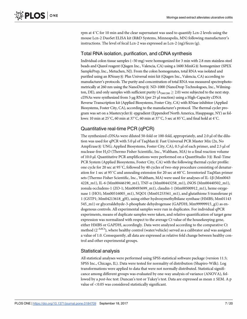
Animals and experimental protocols
All animal protocols used in this study were approved by the Institutional Animal Care and
Use Committee at Rutgers, The State University of New Jersey (No. 14–014), and followed fed-
eral and state laws. Male C57BL/6J mice at 7 wks of age (average weight: 20–22 g) were pur-
chased from Jackson Laboratories (Bar Harbor, ME) and acclimatized to the animal facility for
1 wk with ad libitum access to drinking water and a rodent chow diet (Purina 5001; LabDiet,
Moringa seed extract alleviates ulcerative colitis
PLOS ONE | https://doi.org/10.1371/journal.pone.0184709 September 18, 2017 3 / 20
St. Louis, MO) prior to experiments. Mice were initially housed 4 animals per cage, and main-
tained under standardized conditions (22 ± 2˚C, 12-h light/dark cycle, and 50–60% humidity).
Experiments began with mice at 8 wks of age, randomly assigned to the experimental groups,
because this age is optimal for successful and reproducible UC induction by DSS [25]. For this
study, two types of DSS-induced UC mouse models with features of acute and chronic UC
conditions were utilized. UC induction protocols and experimental designs for each UC
model are described below and summarized in Table 1.
DSS-induced acute UC model
After acclimation, 32 mice were randomly divided into 4 experimental groups as follows based
on their bw (n = 8/group): healthy control (water/vehicle), disease control (DSS/vehicle), MSE
treatment (DSS/MSE), or positive control (DSS/5-ASA). Acute UC was induced in mice (ex-
cept healthy control) by replacing their drinking water with autoclaved water containing
freshly prepared 3.0% (w/v) DSS (ad libitum) for 5 d, which, along with a chronic UC induc-
tion method described below, was previously confirmed in our laboratory as the highest tolera-
ble DSS concentration/duration capable of developing an acute or chronic UC model without
mortality in 7 wk old, male C57BL/6J mice [26]. Mice in healthy control were littermates that
received autoclaved water instead, and housed and processed identically during UC induction.
Following UC induction, treatments were administered daily to all mice via oral gavage in a
volume of 5 ml/kg bw with animal feeding needles (20G x 1.2”; Cadence, Inc., Staunton, VA)
for 7 consecutive d. Treatments included vehicle (autoclaved water), 150 mg/kg bw of MSE, or
50 mg/kg bw of 5-ASA. The level of 150 mg/kg bw MSE (containing ~70 mg/kg bw MIC-1)
was chosen based on our previous study with phenethyl isothiocyanate (PEITC; ~70–75 mg/
kg bw), a structurally and functionally-related isothiocyanate derived from winter cress, show-
ing an efficacy at this level in attenuating bowel inflammation in DSS-induced UC mice [26].
This MSE dose was also confirmed to be safe from our recent toxicity study in rats. Mice were
allowed free access to autoclaved drinking water and chow pellets during the time.
DSS-induced chronic UC model
After acclimation, 24 mice were assigned to 4 weight-balanced experimental groups (n = 6/
group) as described above. Chronic UC was induced in mice by 4 cycles of 2.5% (w/v) DSS in
drinking water for 3 d, which was alternated with 6 d of autoclaved water (ad libitum) in
between, resulting in a total of 30 d for the entire induction period. Subsequently, treatments
(vehicle, MSE, or 5-ASA) were orally delivered to mice daily for 14 consecutive d, and mice
had free access to autoclaved water and chow during the time.
Table 1. Experimental designs and protocolsa.
Acute UC model Chronic UC model
UC induction by
DSS
3.0% (w/v) DSS (5
d)
4 cycles of 2.5% (w/v) DSS for 3 d, with alternate autoclaved
water for 6 d
in between (30 d in total)
Dosing method Oral gavage Oral gavage
Treatment period 7 d 14 d
Experimental
design
Group 1) Water/vehicle (healthy control)
Group 2) DSS/vehicle (disease control)
Group 3) DSS/MSE (MSE treatment; 150 mg/kg bw of MSE)
Group 4) DSS/5-ASA (positive control; 50 mg/kg bw of 5-ASA)
a Modified based on Dey et al., 2010 [26]
https://doi.org/10.1371/journal.pone.0184709.t001
Moringa seed extract alleviates ulcerative colitis
PLOS ONE | https://doi.org/10.1371/journal.pone.0184709 September 18, 2017 4 / 20

Throughout the entire experimental periods with both UC models, bw and food consump-
tion were recorded daily or every 2 d. Intakes of DSS-containing water were also monitored
daily to confirm that mice received sufficient amounts of DSS to induce UC. At the end of
each experiment, mice were anesthetized with CO2 and euthanized by decapitation.
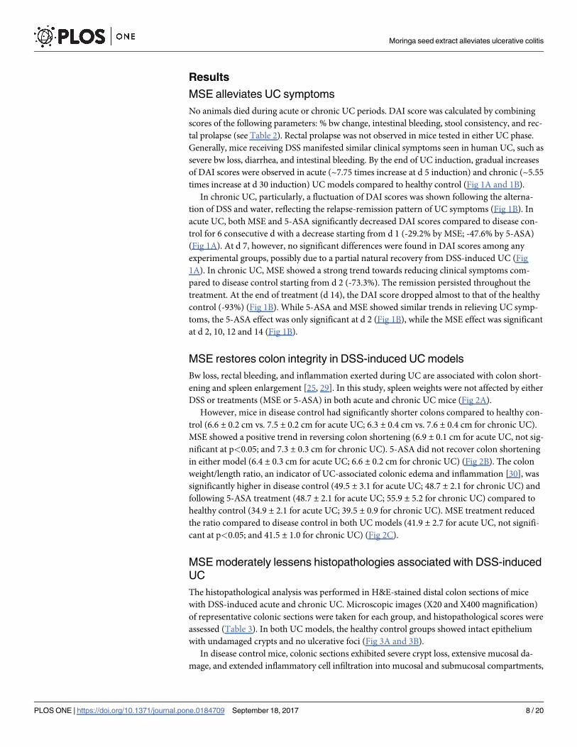
Disease activity index (DAI) assessment
DAI was scored following the criteria described in Table 2 in order to validate the progression
of UC in both models. The DAI score was calculated as a sum of the scores for % bw change,
intestinal bleeding, stool consistency, and rectal prolapse. % bw change was calculated as: [(bw
each d–initial bw) / initial bw] x 100. To score intestinal bleeding, bloody stool was confirmed
by either visible observation or the fecal occult bleeding test (FOBT) using a commercially
available kit (Sure-Vue™; Thermo Fisher Scientific, Inc., Waltham, MA).
Tissue isolation, measurements, and preparations
At the time of sacrifice in acute and chronic UC experiments, the spleen and colon were dis-
sected from each mouse and measured within 5–10 min postmortem. Individual spleen tissues
were weighed (mg). The colons, connected to the cecum, were isolated by separating them
from the ileocecal junction of the small intestine and the distal rectum of the anus, and thor-
oughly flushed with cold PBS (pH 7.4; supplemented with 1% antibiotics) using a ball tip dos-
ing needle attached to a 10 ml syringe to remove feces and blood. After pushing out remaining
colon contents with a blunt forceps, the colons were straightened and measured in length
(cm). The colons were then separated from cecum and weighed. Subsequent to these measure-
ments, the colons were opened by longitudinal incision, and washed three times in cold PBS
(pH 7.4; supplemented with 1% antibiotics) for a complete removal of fecal matter, from
which only middle-to-distal colon segments were used for further analyses. These colon seg-
ments were further cut into 3 pieces longitudinally, each of which was individually processed
for organ culture, quantification of gene expression, and histopathological assessments.
Ex vivo colon organ culture
Approximately 1/3 of washed middle-to-distal colon segments were kept in cryogenic tubes
filled with cold RPMI 1640 medium (supplemented with 1% antibiotics) on ice until delivery
to the cell culture facility. On culturing, each colon specimen was transferred into a separate
well of a 48-well microtiter plate containing 1 ml/well of RPMI 1640 medium (supplemented
with 10% FBS and 1% antibiotics), and incubated at 37˚C with 5% CO2 for 18 h. Supernatants
were collected, centrifuged at 2,500 rpm for 10 min at 4˚C, and stored at -80˚C until analyses
of colonic pro-inflammatory biomarkers.
Table 2. Disease activity index (DAI) for clinical evaluation of DSS-induced UC in micea.
Score % bw change Intestinal bleeding (FOBT)b Stool consistency Rectal prolapse
0 Gain, loss < 1% No color change Normal None
1 Loss 1 to <5% - - -
2 Loss 5 to <10% Color change Loose Observed prolapse
3 Loss 10 to <15% - - -
4 Loss� 15% Gross bleeding Diarrhea Extensive prolapse
a Modified from Dey et al., 2010 [26]b Fecal occult bleeding test
https://doi.org/10.1371/journal.pone.0184709.t002
Moringa seed extract alleviates ulcerative colitis
PLOS ONE | https://doi.org/10.1371/journal.pone.0184709 September 18, 2017 5 / 20

Histopathological evaluation of UC
Another 1/3 of middle-to-distal colon segments were subjected to histopathological assess-
ment. The colonic sections were fixed in 4% paraformaldehyde overnight at 4˚C, which were
then washed with PBS (pH 7.4), dehydrated through ethanol gradients, and paraffin embed-
ded. The embedded blocks were sectioned (5 μm), and the samples were then stained with
hematoxylin and eosin (H&E). Subsequently, all colonic sections were scored in an investiga-
tor-blinded manner relating to the severity of colonic damage and inflammation using light
microscopy with a Retiga 1300 CCD camera (Q-imaging; Burnaby, BC, Canada) and an
Eclipse E800 microscope (Nikon Instruments Inc., Lewisville, TX) with QC-Capture imaging
software under X20 and X400 magnification and air for 20x/0.75 N.A. The severity of UC was
evaluated using the histopathological scoring system with total score range from 0 to 11
(Table 3), according to the matrices previously defined by Laroui et al. (2012) [27].
Quantification of colonic pro-inflammatory biomarkers
From the supernatant of ex vivo colon cultures, pro-inflammatory chemokine, keratinocyte-
derived chemokine (KC), and pro-inflammatory cytokine, TNF-α, were quantified using the
mouse CXCL1/KC DuoSet ELISA kit and the mouse TNF-αQuantikine ELISA kit, respectively.
Myeloperoxidase (MPO), a marker for neutrophil infiltration in colonic epithelial tissues, was also
measured using the mouse MPO DuoSet ELISA kit. All ELISA kits were purchased from R&D
Systems, Inc. (Minneapolis, MN), and utilized according to manufacturer’s protocols. NO pro-
duction was analyzed in duplicates using the Griess Reagent System (Promega Corp., Madison,
WI) following the manufacturer’s protocol. Nitrite levels (μM) in individual samples were deter-
mined at 520 nm by comparison with a standard curve of nitrite (0.1 M NaNO2; 0 to 100 μM).
Throughout these analyses, absorbance was read using a Synergy HT™ microplate reader (BioTek
Instruments, Inc., Winooski, VT), and individual concentrations obtained from each colon sam-
ple were normalized by the measured colon tissue weight (g).
Fecal collection and quantification of fecal lipocalin-2 (Lcn-2)
At the end of treatment periods of acute and chronic UC experiments, fecal samples were col-
lected by taking individual mice out of their cages and collecting 7 fecal pellets per mouse in
sterile cryogenic vials, which were snap-frozen on dry ice immediately, and stored at −80˚C
until further processing. Quantification of fecal Lcn-2 was performed as described by Chassa-
ing et al. (2012) [28] with slight modifications. Briefly, 100 mg of frozen fecal samples were
reconstituted in 1 ml of PBS (pH. 7.4) supplemented with 0.1% (v/v) Tween 20, which were
homogenized for 3 min using a 1600 MiniG1 homogenizer (SPEX SamplePrep, Inc., Metu-
chen, NJ) to obtain a homogenous fecal suspension. The suspension was centrifuged at 12,000
Table 3. Histopathological scoring systema.
Score Severity of inflammation Crypt damage Ulceration
0 Rare inflammatory cells in the lamina propria Intact crypt 0 foci of ulceration
1 Increased numbers of granulocytes in the lamina propria Loss of basal 1/3 of crypt 1–2 foci of ulceration
2 Confluent inflammatory cells extended to submucosa Loss of basal 2/3 of crypt 3–4 foci of ulceration
3 Transmural extension of the inflammatory infiltration Loss of entire crypt Confluent or extensive ulceration
4 - Change in epithelial surface caused by erosion -
5 - Confluent erosion -
a Laroui et al., 2012 [27]
https://doi.org/10.1371/journal.pone.0184709.t003
Moringa seed extract alleviates ulcerative colitis
PLOS ONE | https://doi.org/10.1371/journal.pone.0184709 September 18, 2017 6 / 20
rpm at 4˚C for 10 min and the clear supernatant was used to quantify Lcn-2 levels using the
mouse Lcn-2 DuoSet ELISA kit (R&D Systems, Minneapolis, MN) following manufacturer’s
instructions. The level of fecal Lcn-2 was expressed as Lcn-2 (ng)/feces (g).
Total RNA isolation, purification, and cDNA synthesis
Individual colon tissue samples (~50 mg) were homogenized for 3 min with 2.8 mm stainless steel
beads and Qiazol reagent (Qiagen Inc., Valencia, CA) using a 1600 MiniG1 homogenizer (SPEX
SamplePrep, Inc., Metuchen, NJ). From the colon homogenates, total RNA was isolated and
purified using an RNeasy1 Plus Universal mini kit (Qiagen Inc., Valencia, CA) according to
manufacturer’s protocols. The purity and concentration of total RNA was measured spectrophoto-
metrically at 260 nm using the NanoDrop1 ND-1000 (NanoDrop Technologoes, Inc., Wilming-
ton, DE), and only samples with sufficient purity (A260/280� 2.0) were subjected to the next step.
cDNAs were synthesized from 5 μg RNA (per 25 μl reaction) using a High-Capacity cDNA
Reverse Transcription kit (Applied Biosystems, Foster City, CA) with RNase inhibitor (Applied
Biosystems, Foster City, CA), according to the manufacturer’s protocol. The thermal cycler pro-
gram was set on a Mastercycler1 epgradient (Eppendorf North America, Hauppauge, NY) as fol-
lows: 10 min at 25˚C, 60 min at 37˚C, 60 min at 37˚C, 5 sec at 85˚C, and final hold at 4˚C.
Quantitative real-time PCR (qPCR)
The synthesized cDNAs were diluted 50-fold or 100-fold, appropriately, and 2.0 μl of the dilu-
tion was used for qPCR with 5.0 μl of TaqMan1 Fast Universal PCR Master Mix (2x, No
AmpErase1 UNG; Applied Biosystems, Foster City, CA), 0.5 μl of each primer, and 2.5 μl of
nuclease-free H2O (Thermo Fisher Scientific, Inc., Waltham, MA) to a final reaction volume
of 10.0 μl. Quantitative PCR amplifications were performed on a QuantStudio 31 Real-Time
PCR System (Applied Biosystems, Foster City, CA) with the following thermal cycler profile:
one cycle for 20 sec at 95˚C, followed by 40 cycles of two-step procedure consisting of denatur-
ation for 1 sec at 95˚C and annealing extension for 20 sec at 60˚C. Inventoried TaqMan primer
sets (Thermo Fisher Scientific, Inc., Waltham, MA) were used for analyses of IL-1β (Mm0043
4228_m1), IL-6 (Mm00446190_m1), TNF-α (Mm00443258_m1), iNOS (Mm00440502_m1),
zonula occludens-1 (ZO-1; Mm00493699_m1), claudin-1 (Mm00500912_m1), heme oxyge-
nase 1 (HO1; Mm00516005_m1), NQO1 (Mm01253561_m1), and glutathione S transferase pi
1 (GSTP1; Mm04213618_gH), using either hydroxymethylbilane synthase (HMBS; Mm01143
545_m1) or glyceraldehyde-3-phosphate dehydrogenase (GAPDH; Mm99999915_g1) as en-
dogenous controls. All experimental samples were run in duplicates. For individual qPCR
experiments, means of duplicate samples were taken, and relative quantification of target gene
expression was normalized with respect to the average Ct value of the housekeeping gene,
either HMBS or GAPDH, accordingly. Data were analyzed according to the comparative Ct
method (2-ΔΔCt), where healthy control (water/vehicle) served as a calibrator and was assigned
a value of 1.0. Consequently, all data are expressed as relative fold change between healthy con-
trol and other experimental groups.
Statistical analysis
All statistical analyses were performed using SPSS statistical software package (version 11.5;
SPSS Inc., Chicago, IL). Data were tested for normality of distribution (Shapiro-Wilk). Log
transformations were applied to data that were not normally distributed. Statistical signifi-
cance among different groups was evaluated by one-way analysis of variance (ANOVA), fol-
lowed by a post-hoc test: Duncan’s test or Tukey’s test. Data are expressed as mean ± SEM. A p
value of<0.05 was considered statistically significant.
Moringa seed extract alleviates ulcerative colitis
PLOS ONE | https://doi.org/10.1371/journal.pone.0184709 September 18, 2017 7 / 20
Results
MSE alleviates UC symptoms
No animals died during acute or chronic UC periods. DAI score was calculated by combining
scores of the following parameters: % bw change, intestinal bleeding, stool consistency, and rec-
tal prolapse (see Table 2). Rectal prolapse was not observed in mice tested in either UC phase.
Generally, mice receiving DSS manifested similar clinical symptoms seen in human UC, such as
severe bw loss, diarrhea, and intestinal bleeding. By the end of UC induction, gradual increases
of DAI scores were observed in acute (~7.75 times increase at d 5 induction) and chronic (~5.55
times increase at d 30 induction) UC models compared to healthy control (Fig 1A and 1B).
In chronic UC, particularly, a fluctuation of DAI scores was shown following the alterna-
tion of DSS and water, reflecting the relapse-remission pattern of UC symptoms (Fig 1B). In
acute UC, both MSE and 5-ASA significantly decreased DAI scores compared to disease con-
trol for 6 consecutive d with a decrease starting from d 1 (-29.2% by MSE; -47.6% by 5-ASA)
(Fig 1A). At d 7, however, no significant differences were found in DAI scores among any
experimental groups, possibly due to a partial natural recovery from DSS-induced UC (Fig
1A). In chronic UC, MSE showed a strong trend towards reducing clinical symptoms com-
pared to disease control starting from d 2 (-73.3%). The remission persisted throughout the
treatment. At the end of treatment (d 14), the DAI score dropped almost to that of the healthy
control (-93%) (Fig 1B). While 5-ASA and MSE showed similar trends in relieving UC symp-
toms, the 5-ASA effect was only significant at d 2 (Fig 1B), while the MSE effect was significant
at d 2, 10, 12 and 14 (Fig 1B).
MSE restores colon integrity in DSS-induced UC models
Bw loss, rectal bleeding, and inflammation exerted during UC are associated with colon short-
ening and spleen enlargement [25, 29]. In this study, spleen weights were not affected by either
DSS or treatments (MSE or 5-ASA) in both acute and chronic UC mice (Fig 2A).
However, mice in disease control had significantly shorter colons compared to healthy con-
trol (6.6 ± 0.2 cm vs. 7.5 ± 0.2 cm for acute UC; 6.3 ± 0.4 cm vs. 7.6 ± 0.4 cm for chronic UC).
MSE showed a positive trend in reversing colon shortening (6.9 ± 0.1 cm for acute UC, not sig-
nificant at p<0.05; and 7.3 ± 0.3 cm for chronic UC). 5-ASA did not recover colon shortening
in either model (6.4 ± 0.3 cm for acute UC; 6.6 ± 0.2 cm for chronic UC) (Fig 2B). The colon
weight/length ratio, an indicator of UC-associated colonic edema and inflammation [30], was
significantly higher in disease control (49.5 ± 3.1 for acute UC; 48.7 ± 2.1 for chronic UC) and
following 5-ASA treatment (48.7 ± 2.1 for acute UC; 55.9 ± 5.2 for chronic UC) compared to
healthy control (34.9 ± 2.1 for acute UC; 39.5 ± 0.9 for chronic UC). MSE treatment reduced
the ratio compared to disease control in both UC models (41.9 ± 2.7 for acute UC, not signifi-
cant at p<0.05; and 41.5 ± 1.0 for chronic UC) (Fig 2C).
MSE moderately lessens histopathologies associated with DSS-induced
UC
The histopathological analysis was performed in H&E-stained distal colon sections of mice
with DSS-induced acute and chronic UC. Microscopic images (X20 and X400 magnification)
of representative colonic sections were taken for each group, and histopathological scores were
assessed (Table 3). In both UC models, the healthy control groups showed intact epithelium
with undamaged crypts and no ulcerative foci (Fig 3A and 3B).
In disease control mice, colonic sections exhibited severe crypt loss, extensive mucosal da-
mage, and extended inflammatory cell infiltration into mucosal and submucosal compartments,
Moringa seed extract alleviates ulcerative colitis
PLOS ONE | https://doi.org/10.1371/journal.pone.0184709 September 18, 2017 8 / 20

as is commonly reported in the DSS-induced UC model. Extensive erosion and submucosal
edema were also presented in the distal colon as the indication of an inflammatory phenotype
in this group (Fig 3A and 3B). On the contrary, treatment with either MSE or 5-ASA resulted in
less severe inflammation (relatively less extensive epithelial erosion) and development of a few
regenerating foci compared to disease control in acute UC (Fig 3A). In chronic UC, neither
MSE nor 5-ASA treatments measurably rescued chronic inflammation, as extensive mucosal
damage and confluent ulcerative erosion were similarly observed in treated and disease control
groups (Fig 3B). In the assessment of histopathological scores, all mice in DSS-treated groups
showed a marked increase of the scores, which was significantly reduced by 5-ASA. MSE
showed a trend in reducing histopathological scores in acute UC, which was not significant at
p<0.05 (Fig 3C).
Fig 1. Effects of MSE on DAI scores in DSS-induced UC. (A) Acute UC (n = 8/group): data are shown at
daily intervals throughout UC induction and treatment periods. (B) Chronic UC (n = 6/group): data are shown
at 3-d intervals during UC induction and 2-d intervals during the treatment period. Experimental designs and
disease induction procedures are defined in Table 1 and DAI scoring criteria in Table 2. Data are expressed
as mean ± SEM. *p<0.05, **p<0.01, and ***p<0.001 compared to disease control (DSS/vehicle).
5-aminosalicylic acid (5-ASA); disease activity index (DAI); dextran sulfate sodium (DSS); moringa seed
extract (MSE); ulcerative colitis (UC).
https://doi.org/10.1371/journal.pone.0184709.g001
Moringa seed extract alleviates ulcerative colitis
PLOS ONE | https://doi.org/10.1371/journal.pone.0184709 September 18, 2017 9 / 20

Fig 2. Effects of MSE on spleen and colon in DSS-induced UC. (A) Spleen weight (mg), (B) colon length
(cm), and (C) colon weight/length ratio (mg/cm) were measured at necropsy of acute UC (n = 8/group) and
chronic UC (n = 6/group) experiments. Experimental designs and disease induction procedures are defined in
Table 1. Data are expressed as mean ± SEM. Different letters (a, b, c) indicate significant differences between
groups at p<0.05. 5-aminosalicylic acid (5-ASA); dextran sulfate sodium (DSS); moringa seed extract (MSE);
ulcerative colitis (UC).
https://doi.org/10.1371/journal.pone.0184709.g002
Moringa seed extract alleviates ulcerative colitis
PLOS ONE | https://doi.org/10.1371/journal.pone.0184709 September 18, 2017 10 / 20

Fig 3. Effects of MSE on colon histopathology in DSS-induced UC. Representative histological observations of H&E-
stained colonic sections of (A) acute UC mice (n = 8/group), and (B) chronic UC mice (n = 6/group). Experimental designs
and disease induction procedures are defined in Table 1. (C) Associated colonic histopathological scores calculated as sum
of scores of each parameter consisting of histopathological scoring system as described in Table 3. Histological images
were acquired by X20 and X400 magnification, respectively (Scale bar = 25 μm). Data are expressed as mean ± SEM.
Different letters (a, b, c) indicate significant differences between groups at p<0.05. 5-aminosalicylic acid (5-ASA); dextran
sulfate sodium (DSS); hematoxylin and eosin (H&E); moringa seed extract (MSE); ulcerative colitis (UC).
https://doi.org/10.1371/journal.pone.0184709.g003
Moringa seed extract alleviates ulcerative colitis
PLOS ONE | https://doi.org/10.1371/journal.pone.0184709 September 18, 2017 11 / 20
MSE decreases secretion of colonic pro-inflammatory biomarkers and
fecal Lcn-2 levels
Cytokines and chemokines are important mediators of the mucosal inflammation and
immune responses during UC. In acute UC, compared to disease control, colonic production
of KC, TNF-α, and NO were decreased by MSE (not significantly at p<0.05), whereas 5-ASA
significantly decreased levels of KC (-48.1%) and NO (-27.4%) (Fig 4A–4C).
The levels of MPO were significantly lowered by both MSE (-35.7%) and 5-ASA (-57.0%)
in the acute UC model (Fig 4D). In chronic UC, reductions in these markers were more
obvious, showing that, compared to disease control, colonic productions of all tested pro-
inflammatory markers were significantly lowered by MSE (-2.3% for KC; -26.4% for TNF-α;
-41.8% for NO; -67.1% for MPO). The effect of 5-ASA was negligible in chronic UC, only
showing a significant reduction in MPO (-51.9%) (Fig 4A–4D). Mice receiving DSS showed
a dramatic increase of the fecal Lcn-2 level in both acute and chronic UC, which, in acute
UC, was significantly reduced by MSE (-7.7%), but not by 5-ASA, compared to disease con-
trol (Fig 4E).
MSE downregulates colonic expression of pro-inflammatory cytokines
and iNOS
Pathological process of UC is closely related to the increased level of pro-inflammatory cyto-
kines that can also activate iNOS. We examined the effect of MSE on colonic expression of
pro-inflammatory cytokines including IL-1β, IL-6, and TNF-α, as well as iNOS. In acute UC,
compared to healthy control, all these pro-inflammatory marker genes were upregulated by
DSS (up to ~150 times for iNOS). However, in the acute model, the expression of these pro-
inflammatory marker genes was, in most cases, markedly lowered by MSE (-73.2% for IL-1β;
-97.1% for IL-6; -43.4% for TNF-α, not significant at p<0.05; -73.2% for iNOS) compared to
disease control (Fig 5A–5D).
5-ASA, similar to MSE, downregulated the expression of the pro-inflammatory genes,
except for TNF-α (Fig 5A–5D). In chronic UC, neither MSE nor 5-ASA showed significant
effects on pro-inflammatory marker genes (Fig 5A–5D).
MSE elevates colonic expression of tight-junction proteins and
Nrf2-mediated phase II detoxifying enzymes
UC is associated with increased intestinal permeability and decreased tight-junction protein
expression, such as claudin-1 and ZO-1, in the inflamed mucosa [30]. DSS normally sup-
presses claudin-1 gene expression; however, MSE increased claudin-1 expression in both UC
models and the effect was highly significant in the acute model (558%) (Fig 5E). A similar
result was observed for ZO-1, showing decreased gene expression by DSS, which was upre-
gulated by MSE, but not by 5-ASA, in acute (148%; not significant at p<0.05) and chronic
UC (148%) compared to disease control (Fig 5F). Gene expression of phase II detoxifying
enzymes, including GSTP1, NQO1, and HO1, are directly regulated by the Nrf2 transcrip-
tion factor. Nrf2 can also modulate NF-κB-dependent pro-inflammatory signals [19]. In
chronic UC, MSE led to upregulated gene expression of GSTP1 (160%), NQO1 (144%), and
HO1 (128%), compared to disease control; however, only GSTP1 upregulation was signifi-
cant (Fig 5G–5I). In addition, the expression of NQO1 and HO1 genes was significantly
upregulated by MSE compared to healthy control (Fig 5H and 5I). In acute UC, DSS showed
a trend towards reducing the Nrf2-regulated gene expression that was not affected by MSE
or 5-ASA (Fig 5G–5I).
Moringa seed extract alleviates ulcerative colitis
PLOS ONE | https://doi.org/10.1371/journal.pone.0184709 September 18, 2017 12 / 20

Discussion
This study aimed to investigate the efficacy and mechanisms of action of MIC-1-enriched
MSE in alleviating UC symptoms. Data indicate that oral administration of MSE at 150 mg/kg
bw mitigated UC symptoms and associated biomarkers in both DSS-induced acute and
chronic UC mouse models by modulating inflammatory and antioxidant response systems.
Acute UC is characterized by rapid onset of severe clinical symptoms within a relatively
short induction period, whereas chronic UC represents less severe and more sustained
Fig 4. Effects of MSE on colonic pro-inflammatory biomarkers and fecal Lcn-2 levels in DSS-induced UC. Analyses of (A) KC,
(B) TNF-α, (C) NO, and (D) MPO were performed in colon culture supernatants of acute UC (n = 8/group) and chronic UC (n = 6/group)
mice. All concentrations were normalized to 1 g of the colon tissue weight. Fecal levels of (E) Lcn-2 were normalized to 1 g of the fecal
weight. Experimental designs and disease induction procedures are defined in Table 1. Data are expressed as mean ±SEM. Different
letters (a, b, c) indicate significant differences between groups at p<0.05. 5-aminosalicylic acid (5-ASA); dextran sulfate sodium (DSS);
keratinocyte-derived cytokine (KC); lipocalin-2 (Lcn-2); myeloperoxidase (MPO); moringa seed extract (MSE); nitric oxide (NO); tumor
necrosis factor-α (TNF-α); ulcerative colitis (UC).
https://doi.org/10.1371/journal.pone.0184709.g004
Moringa seed extract alleviates ulcerative colitis
PLOS ONE | https://doi.org/10.1371/journal.pone.0184709 September 18, 2017 13 / 20

Fig 5. Effects of MSE on colonic expression of pro-inflammatory markers, tight-junction proteins, and Nrf2-mediated
enzymes in DSS-induced UC. Gene expression of (A) IL-1β, (B) IL-6, (C) TNF-α, (D) iNOS, (E) claudin-1, (F) ZO-1, (G) GSTP1,
(H) NQO1, and (I) HO1 was analyzed by qPCR in colon tissues of acute UC (n = 8/group) and chronic UC (n = 6/group) mice. All
data are expressed as a relative fold change compared to healthy control (value = 1.0). Experimental designs and disease
Moringa seed extract alleviates ulcerative colitis
PLOS ONE | https://doi.org/10.1371/journal.pone.0184709 September 18, 2017 14 / 20

inflammation developed through a slower but long-lasting induction period with possible flare-
up and remission symptomatic cycles. DSS disrupts the integrity of the mucosal barriers [20],
leading to increased permeability of luminal toxic substances into the lamina propria and sub-
mucosal compartments, resulting in secondary inflammation in the intestine [25]. In our study,
two DSS-induced UC models were successfully established, with more severe UC symptoms ob-
served in the acute than chronic UC model, reflected by higher DAI scores (~7x vs. ~5x higher
DAI score in acute vs. chronic UC, respectively) compared to DSS-untreated mice. Oral deliv-
ery of MSE (150 mg/kg bw for 1 or 2 wks) promptly reduced the severity of UC symptoms in
both UC models, including improvements in bw loss, stool consistency, and intestinal bleeding.
MSE improved the integrity of colon tissues by correcting colon shortening, likely due to loss in
crypt structure, and colon thickness, which is due to edema formation [31]. MSE also reduced
the severity of colonic inflammation developed during UC, diminished epithelial erosion and
regenerated a few foci in colonic tissues in acute UC. However, MSE failed to attenuate these
histopathological signs of UC in the chronic UC model. This is, nevertheless, consistent with a
previous report that complete healing of bowel ulceration may not be concomitant with clinical
remission of UC symptoms, and with recommendations from previous clinical studies to mea-
sure mucosal restitution after 8 wks of treatments [32]. 5-ASA, an anti-inflammatory drug for
treatment of mild-to-moderate severity of UC in humans, was used as a positive control with a
recommended dose of 50 mg/kg bw [32]. 5-ASA produced a comparable DAI score to that of
MSE in acute UC, but was less efficacious than MSE in chronic UC. Moreover, the impact of
5-ASA on colonic histopathological signs of UC was only evident in acute UC, and could not
improve aberrant characteristics of colon tissues, like colon shortening, caused by DSS. Our
observations in chronic UC mice corroborate previous findings that 5-ASA, albeit widely used
as a first-line therapy for UC, is largely ineffective in chronic UC [33].
Our findings are consistent with published reports on powerful anti-inflammatory effects
of various isothiocyanates [12, 15, 16] and previous observations that orally administered
PEITC (~75 mg/kg bw) or sulforaphane (25 mg/kg bw), isothiocyanates from Brassica vegeta-
bles that have the same pharmacophore (N = C = S) as MIC-1, could also relieve acute and
chronic symptoms of DSS-induced UC in mice [14, 26]. Our companion paper published in
this journal [24] demonstrates that MIC-1 is the main anti-inflammatory component of MSE.
MIC-1 was more effective than curcumin (one of the most potent anti-inflammatory phyto-
chemicals [34]) in reducing the production of NO and expression of iNOS, IL-1β, and IL-6 in
LPS-stimulated RAW macrophages. Additionally, MIC-1 significantly upregulated the expres-
sion of Nrf2-mediated phase II detoxification genes (GSTP1, NQO1, and HO1) in these cells
more effectively than curcumin. MSE was more potent than curcuminoid-enriched turmeric
extract in reducing carrageenan-induced paw edema in rats [24]. Taken together with our cur-
rent observations, the available data suggests that MIC-1 is the bioactive component of MSE
responsible for its therapeutic effects in UC.
Imbalanced production of cytokines is a part of UC pathophysiology. These cytokines form
a complex network that mediates mucosal/submucosal inflammation and influences the integ-
rity of epithelium [35, 36]. An increase of pro-inflammatory cytokines, including IL-1β, IL-6,
TNF-α, and interferon-γ (IFN-γ), is associated with disease activity and development of all UC
induction procedures are defined in Table 1. Data are expressed as mean ±SEM. Different letters (a, b) indicate significant
differences between groups at p<0.05. 5-aminosalicylic acid (5-ASA); dextran sulfate sodium (DSS); glyceraldehyde-3-phosphate
dehydrogenase (GAPDH); glutathione-S-transferase pi 1 (GSTP1); hydroxymethylbilane synthase (HMBS); heme oxygenase 1
(HO1); interleukin-1β (IL-1β); interleukin-6 (IL-6); inducible nitric oxide synthase (iNOS); moringa seed extract (MSE); NAD(P)H
quinone oxidoreductase 1 (NQO1); tumor necrosis factor-α (TNF-α); ulcerative colitis (UC), zonula occludens-1 (ZO-1).
https://doi.org/10.1371/journal.pone.0184709.g005
Moringa seed extract alleviates ulcerative colitis
PLOS ONE | https://doi.org/10.1371/journal.pone.0184709 September 18, 2017 15 / 20

[37–39]. Meanwhile, KC serves as a chemoattractant for neutrophils [40], whereas MPO is a
direct indicator of neutrophil infiltration into the colon and serves as a surrogate marker of
UC-related inflammation [23]. Administration of anti-KC antibodies reduced neutrophil traf-
ficking into the colons of mice with DSS-induced UC [41], indicating a key role of KC in
colonic recruitment of neutrophils. Lcn-2, also referred to as neutrophil gelatinase-associated
lipocalin (NGAL) or siderocalin, is a bacteriostatic protein mainly released from neutrophils at
sites of inflammation [42], and its level in serum, colon, and fecal samples is elevated in multi-
ple models of UC and in human IBD [43, 44]. iNOS-generated NO is a signaling molecule in
inflammation, infection, and cancer [45]. iNOS knockout mice exhibited a substantial reduc-
tion of DSS-induced colonic injury compared to wildtype [45]. A positive correlation between
NO and increased pro-inflammatory cytokines (TNF-α, IL-6, IL-17, IL-12, and IFN-γ) is
described in IBD patients [46]. We also observed increased production and/or gene expression
of pro-inflammatory markers (IL-1, IL-6, TNF-α, iNOS, NO, KC, MPO, and Lcn-2) in both
UC models. MSE treatment generally reduced these markers, although the effect of MSE dif-
fered in acute vs. chronic UC models and in protein vs. gene expression levels. The main rea-
son of the latter observation (TNF-α and NO/iNOS) might be due to the different colon
segments from each animal that were used for each analysis. Since the extent of colonic inflam-
mation would not be identical among different segments of the colon in UC mice, some incon-
sistency could occur between protein and gene expression levels. Similarly, a previous study
showed that PEITC downregulated colonic IL-1β gene expression in DSS-induced UC mice
[26]. These results are consistent with the well-documented anti-inflammatory effects of vari-
ous isothiocyanates, which are mediated, at least in part, by Nrf2 activation [46].
Tight-junction proteins, such as ZO-1, occludins, and claudins, form physiologically active
barriers in intestinal epithelial cells that can be disrupted in IBD [30, 47, 48]. For example, the
loss of ZO-1 protein was observed in DSS-induced UC mice [30]. Our data showed that, while
DSS treatment led to some downregulation of claudin-1 and ZO-1 genes, MSE restored or
upregulated these genes, consistent with its observed therapeutic effects in UC.
Oxidative stress is both a consequence of UC [49] and one of its contributing factors [50].
Our data suggest that MSE possibly protects against chronic colonic inflammation by lowering
oxidative stress through upregulation of antioxidant/phase II detoxifying enzymes, in particu-
lar, GSTP1. GSTP1 is the most abundant isoform of glutathione S transferase (GST) in the
colon [49], catalyzing the conjugation of xenobiotics to glutathione [51], and DSS-induced UC
mice exhibited downregulation of GST gene expression [51]. We suggest that the upregulation
of antioxidant enzymes observed in our study might be the result of MIC-1-mediated activa-
tion of Keap1/Nrf2/ARE (antioxidant response element) metabolic pathway [19, 52], similar
to that caused by sulforaphane in DSS-induced UC [14]. Studies have shown that various iso-
thiocyanates, such as sulforaphane, PEITC and benzyl isothiocyanate, induce ARE genes by
activating the nuclear translocation of Nrf2 [52, 53], which subsequently reduces oxidative
stress and inflammation [13, 19, 54]. Nrf2 knockout attenuated the anti-inflammatory effects
of PEITC and curcumin in macrophages ex vivo [55]. Therefore, MIC-1 and other isothiocya-
nates are likely responsible for interactions with the Keap1/Nrf2 complex and further down-
stream processes [56]. Our study did not measure the direct effect of MIC-1 or MSE on Nrf2.
However, changes in the expression of the downstream ARE genes regulated by Nrf2, such as
GSTP1, support the role of Nrf2 in mediating the observed effects.
In conclusion, MSE, enriched with the MIC-1, its main bioactive, ameliorated pathological
events associated with acute and chronic UC. Anti-inflammatory and antioxidant activities of
MSE are likely associated with Nrf2-mediated anti-inflammatory/antioxidant signaling path-
way. This study supports the therapeutic potential of MSE for prevention and treatment of UC
and should be further explored in human studies.
Moringa seed extract alleviates ulcerative colitis
PLOS ONE | https://doi.org/10.1371/journal.pone.0184709 September 18, 2017 16 / 20

Acknowledgments
The authors want to thank Peter Kuhn, Sara Faghani, and Hetal Kalariya for their technical
assistance.
Author Contributions
Conceptualization: Youjin Kim, Carrie Waterman, Ilya Raskin.
Data curation: Youjin Kim.
Formal analysis: Youjin Kim.
Funding acquisition: Ilya Raskin.
Investigation: Youjin Kim, Alex G. Wu, Brittany L. Graf, Michael P. Verzi.
Methodology: Youjin Kim, Alex G. Wu, Michael P. Verzi.
Project administration: Youjin Kim, Ilya Raskin.
Resources: Asha Jaja-Chimedza.
Software: Youjin Kim, Michael P. Verzi.
Supervision: Youjin Kim.
Validation: Youjin Kim.
Visualization: Alex G. Wu, Michael P. Verzi.
Writing – original draft: Youjin Kim.
Writing – review & editing: Youjin Kim, Alex G. Wu, Asha Jaja-Chimedza, Brittany L. Graf,
Carrie Waterman, Michael P. Verzi, Ilya Raskin.
References1. Mikhailov TA, Furner SE. Breastfeeding and genetic factors in the etiology of inflammatory bowel dis-
ease in children. World J Gastroenterol. 2009; 15: 270–279. https://doi.org/10.3748/wjg.15.270 PMID:
19140226
2. Jones DT, Osterman MT, Bewtra M, Lewis JD. Passive smoking and inflammatory bowel disease: a
meta-analysis. Am J Gastroenterol. 2008; 103: 2382–2393. https://doi.org/10.1111/j.1572-0241.2008.
01999.x PMID: 18844625
3. Khor B, Gardet A, Xavier RJ. Genetics and pathogenesis of inflammatory bowel disease. Nature. 2011;
474: 307–317. https://doi.org/10.1038/nature10209 PMID: 21677747
4. Rutgeerts P, Vermeire S, Van Assche G. Biological therapies for inflammatory bowel diseases. Gastro-
enterology. 2009; 136: 1182–1197. https://doi.org/10.1053/j.gastro.2009.02.001 PMID: 19249397
5. Baumgart DC, Sandborn WJ. Inflammatory bowel disease: clinical aspects and established and evolv-
ing therapies. Lancet. 2007; 369: 1641–1657. https://doi.org/10.1016/S0140-6736(07)60751-X PMID:
17499606
6. Saxena A, Kaur K, Hegde S, Kalekhan FM, Baliga MS, Fayad R. Dietary agents and phytochemicals in
the prevention and treatment of experimental ulcerative colitis. J Tradit Complement Med. 2014; 4:
203–217. https://doi.org/10.4103/2225-4110.139111 PMID: 25379461
7. Lean QY, Eri RD, Fitton JH, Patel RP, Gueven N. Fucoidan Extracts Ameliorate Acute Colitis. PLoS
One. 2015; 10: e0128453. https://doi.org/10.1371/journal.pone.0128453 PMID: 26083103
8. Leone A, Spada A, Battezzati A, Schiraldi A, Aristil J, Bertoli S. Cultivation, Genetic, Ethnopharmacol-
ogy, Phytochemistry and Pharmacology of Moringa oleifera Leaves: An Overview. Int J Mol Sci. 2015;
16: 12791–12835. https://doi.org/10.3390/ijms160612791 PMID: 26057747
9. Bennett RN, Mellon FA, Foidl N, Pratt JH, Dupont MS, Perkins L, et al. Profiling glucosinolates and phe-
nolics in vegetative and reproductive tissues of the multi-purpose trees Moringa oleiferaL. (Horseradish
Tree) and Moringa stenopetalaL. J Agr Food Chem. 2003; 51: 3546–3553.
Moringa seed extract alleviates ulcerative colitis
PLOS ONE | https://doi.org/10.1371/journal.pone.0184709 September 18, 2017 17 / 20

10. Mbikay M. Therapeutic potential of Moringa oleifera leaves in chronic hyperglycemia and dyslipidemia:
A review. Front Pharmacol. 2012; 3: 1–12. https://doi.org/10.3389/fphar.2012.00001
11. Fahey JW. Moringa oleifera: A review of the medical evidence for its nutritional, therapeutic, and pro-
phylactic properties. Trees for Life J. 2005; 1: 5.
12. Waterman C, Cheng DM, Rojas-Silva P, Poulev A, Dreifus J, Lila MA, et al. Stable, water extractable
isothiocyanates from Moringa oleifera leaves attenuate inflammation in vitro. Phytochemistry. 2014;
103: 114–122. https://doi.org/10.1016/j.phytochem.2014.03.028 PMID: 24731259
13. Hu R, Xu C, Shen G, Jain MR, Khor TO, Gopalkrishnan A, et al. Gene expression profiles induced by
cancer chemopreventive isothiocyanate sulforaphane in the liver of C57Bl/6J mice and C57Bl/6J/Nrf2
(-/-) mice. Cancer Letters. 2006; 243: 170–192. https://doi.org/10.1016/j.canlet.2005.11.050 PMID:
16516379
14. Wagner AE, Will O, Sturm C, Lipinski S, Rosenstiel P, Rimbach G. DSS-induced acute colitis in C57BL/
6 mice is mitigated by sulforaphane pre-treatment. J Nutr Biochem. 2013; 24: 2085–2091. https://doi.
org/10.1016/j.jnutbio.2013.07.009 PMID: 24231100
15. Brunelli D, Tavecchio M, Falcioni C, Frapolli R, Erba E, Iori R, et al. The isothiocyanate produced from
glucomoringin inhibits NF-kB and reduces myeloma growth in nude mice in vivo. Biochem. Pharmacol.
2010; 79: 1141–1148. https://doi.org/10.1016/j.bcp.2009.12.008 PMID: 20006591
16. Cheenpracha S, Park E-J, Yoshida WY, Barit C, Wall M, Pezzuto JM, et al. Potential anti-inflammatory
phenolic glycosides from the medicinal plant Moringa oleifera fruits. Bioorgan Med Chem. 2010; 18:
6598–6602.
17. Waterman C, Rojas-Silva P, Tumer TB, Kuhn P, Richard AJ, Wicks S, et al. Isothiocyanates from Mor-
inga oleifera reduce weight gain, insulin resistance and hepatic gluconeogenesis in mice. Mol Nutr Food
Res. 2014; 59: 1013–1024.
18. Tumer TB, Rojas-Silva P, Poulev A, Raskin I, Waterman C. Direct and indirect antioxidant activity of
polyphenol- and isothiocyanate-enriched fractions from Moringa oleifera. Food Chem. 2015; 63: 1505–
1513.
19. Li W, Khor TO, Xu C, Shen G, Jeong WS, Yu S, et al. Activation of Nrf2-antioxidant signaling attenuates
NF-kB-inflammatory response and elicits apoptosis. Biochem Pharmacol. 2008; 76: 1485–1489.
https://doi.org/10.1016/j.bcp.2008.07.017 PMID: 18694732
20. Wirtz S, Neufert C, Weigmann B, Neurath MF. Chemically induced mouse models of intestinal inflam-
mation. Nat Protoc. 2007; 2: 541–546. https://doi.org/10.1038/nprot.2007.41 PMID: 17406617
21. Elson CO, Sartor RB, Tennyson GS, Riddell RH. Experimental models of inflammatory bowel disease.
Gastroenterology. 1995; 109: 1344–1367. PMID: 7557106
22. Perse M, Cerar A. Dextran sodium sulphate colitis mouse model: traps and tricks. J Biomed Biotechnol.
2012; 2012: 718617. https://doi.org/10.1155/2012/718617 PMID: 22665990
23. Kim JJ, Shajib MS, Manocha MM, Khan WI. Investigating intestinal inflammation in DSS-induced model
of IBD. J Vis Exp. 2012; 60: 3678.
24. Jaja-Chimedza A, Graf BL, Simmler C, Kim Y, Kuhn P, Pauli GF, et al. Biochemical characterization
and anti-inflammatory properties of an isothiocyanate-enriched moringa (Moringa oleifera) seed extract.
PLoS One. 2017; 12: e0182658. https://doi.org/10.1371/journal.pone.0182658 PMID: 28792522
25. Chassaing B, Aitken JD, Malleshappa M, Vijay-Kumar M. Dextran sulfate sodium (DSS)-induced colitis
in mice. Curr Protoc Immunol. 2014; 104: Unit 15.25.
26. Dey M, Kuhn P, Ribnicky D, Premkumar V, Reuhl K, Raskin I. Dietary phenethylisothiocyanate attenu-
ates bowel inflammation in mice. BMC Chem Biol. 2010; 10: 4. https://doi.org/10.1186/1472-6769-10-4
PMID: 20423518
27. Laroui H, Ingersoll SA, Liu HC, Baker MT, Ayyadurai S, Charania MA, et al. Dextran sodium sulfate
(DSS) induces colitis in mice by forming nano-lipocomplexes with medium-chain-length fatty acids in
the colon. PLoS One. 2012; 7: e32084. https://doi.org/10.1371/journal.pone.0032084 PMID: 22427817
28. Chassaing B, Srinivasan G, Delgado MA, Young AN, Gewirtz AT, Vijay-Kumar M. Fecal lipocalin 2, a
sensitive and broadly dynamic non-invasive biomarker for intestinal inflammation. PLoS One. 2012; 7:
e44328. https://doi.org/10.1371/journal.pone.0044328 PMID: 22957064
29. Hendrickson BA, Gokhale R, Cho JH. Clinical aspects and pathophysiology of inflammatory bowel disease.
Clin Microbiol Rev. 2002; 15: 79–94. https://doi.org/10.1128/CMR.15.1.79-94.2002 PMID: 11781268
30. Poritz LS, Garver KI, Green C, Fitzpatrick L, Ruggiero F, Koltun WA. Loss of the tight junction protein
ZO-1 in dextran sulfate sodium induced colitis. J Surg Res. 2007; 140: 12–19. https://doi.org/10.1016/j.
jss.2006.07.050 PMID: 17418867
31. Islam MS, Murata T, Fujisawa M, Nagasaka R, Ushio H, Bari AM, et al. Anti-inflammatory effects of phy-
tosteryl ferulates in colitis induced by dextran sulphate sodium in mice. Br J Pharmacol. 2008; 154:
812–824. https://doi.org/10.1038/bjp.2008.137 PMID: 18536734
Moringa seed extract alleviates ulcerative colitis
PLOS ONE | https://doi.org/10.1371/journal.pone.0184709 September 18, 2017 18 / 20

32. Murthy S, Flanigan A, Coppola D, Buelow R. RDP58, a locally active TNF inhibitor, is effective in the
dextran sulphate mouse model of chronic colitis. Inflamm Res. 2002; 51: 522–531. PMID: 12540016
33. Fiorucci S, Orlandi S, Mencarelli A, Caliendo G, Santagada V, Distrutti E, et al. Enhanced activity of a
hydrogen sulphide-releasing derivative of mesalamine (ATB-429) in a mouse model of colitis. Br J Phar-
macol. 2007; 150: 996–1002. https://doi.org/10.1038/sj.bjp.0707193 PMID: 17339831
34. Jurenka JS. Anti-inflammatory properties of curcumin, a major constituent of Curcuma longa: a review
of preclinical and clinical research. Altern Med Rev. 2009; 14: 141–153. PMID: 19594223
35. Maillard MH, Snapper SB. Cytokines and chemokines in mucosal homeostasis. In: Targan SR, Shana-
han F, Karp LC, editors. Inflammatory Bowel Disease. Wiley-Blackwell; 2010. pp. 119–156.
36. Neurath MF. Cytokines in inflammatory bowel disease. Nat Rev Immunol. 2014; 14: 329–342. https://
doi.org/10.1038/nri3661 PMID: 24751956
37. Muzes G, Molnar B, Tulassay Z, Sipos F. Changes of the cytokine profile in inflammatory bowel dis-
eases. World J Gastroenterol. 2012; 18: 5848–5861. https://doi.org/10.3748/wjg.v18.i41.5848 PMID:
23139600
38. Nikolaus S, Schreiber S. Diagnostics of inflammatory bowel disease. Gastroenterology. 2007;
133:1670–1689. https://doi.org/10.1053/j.gastro.2007.09.001 PMID: 17983810
39. Schulze HA, Hasler R, Mah N, Lu T, Nikolaus S, Costello CM, et al. From model cell line to in vivo gene
expression: disease-related intestinal gene expression in IBD. Genes Immun. 2008; 9: 240–248.
https://doi.org/10.1038/gene.2008.11 PMID: 18340362
40. Frink M, Hsieh YC, Hsieh CH, Pape HC, Choudhry MA, Schwacha MG, et al. Keratinocyte-derived che-
mokine plays a critical role in the induction of systemic inflammation and tissue damage after trauma-
hemorrhage. Shock. 2007; 28: 576–581. PMID: 18084824
41. Murphy CT, Moloney G, Hall LJ, Quinlan A, Faivre E, Casey P, et al. Use of bioluminescence imaging to
track neutrophil migration and its inhibition in experimental colitis. Clin Exp Immunol. 2010; 162: 188–
196. https://doi.org/10.1111/j.1365-2249.2010.04234.x PMID: 20718784
42. Stallhofer J, Friedrich M, Konrad-Zerna A, Wetzke M, Lohse P, Glas J, et al. Lipocalin-2 Is a Disease
Activity Marker in Inflammatory Bowel Disease Regulated by IL-17A, IL-22, and TNF-α and Modulated
by IL23R Genotype Status. Inflamm Bowel Dis. 2015; 21: 2327–2340. https://doi.org/10.1097/MIB.
0000000000000515 PMID: 26263469
43. Vijay-Kumar M, Sanders CJ, Taylor RT, Kumar A, Aitken JD, Sitaraman AV, et al. Deletion of TLR5
results in spontaneous colitis in mice. J Clin Invest. 2007; 117: 3909–3921. https://doi.org/10.1172/
JCI33084 PMID: 18008007
44. Chassaing B, Srinivasan G, Delgado MA, Young AN, Gewirtz AT, Vijay-Kumar M. Fecal lipocalin 2, a
sensitive and broadly dynamic non-invasive biomarker for intestinal inflammation. PLoS One. 2012; 7:
e44328. https://doi.org/10.1371/journal.pone.0044328 PMID: 22957064
45. Lirk P, Hoffmann G, Rieder J. Inducible nitric oxide synthase—time for reappraisal. Curr Drug Targets
Inflamm Allergy. 2002; 1: 89–108. PMID: 14561209
46. Fuentes F, Paredes-Gonzalez X, Kong AT. Dietary Glucosinolates Sulforaphane, Phenethyl Isothiocya-
nate, Indole-3-Carbinol/3,3’-Diindolylmethane: Anti-Oxidative Stress/Inflammation, Nrf2, Epigenetics/
Epigenomics and In Vivo Cancer Chemopreventive Efficacy. Curr Pharmacol Rep. 2015; 1: 179–196.
https://doi.org/10.1007/s40495-015-0017-y PMID: 26457242
47. Gassler N, Rohr C, Schneider A, Kartenbeck J, Bach A, Obermuller N, et al. Inflammatory bowel dis-
ease is associated with changes of enterocytic junctions. Am J Physiol Gastrointest Liver Physiol. 2001;
281: G216–228. PMID: 11408275
48. Hollander D, Vadheim CM, Brettholz E, Petersen GM, Delahunty T, Rotter JI. Increased intestinal per-
meability in patients with Crohn’s disease and their relatives. A possible etiologic factor. Ann Intern
Med. 1986; 105: 883–885. PMID: 3777713
49. Choi K, Chen J, Mitra S, Sarna SK. Impaired integrity of DNA after recovery from inflammation causes
persistent dysfunction of colonic smooth muscle. Gastroenterology. 2011; 141: 1293–1301. https://doi.
org/10.1053/j.gastro.2011.06.074 PMID: 21745450
50. Theiss AL, Idell RD, Srinivasan S, Klapproth JM, Jones DP, Merlin D, et al. Prohibitin protects against
oxidative stress in intestinal epithelial cells. Faseb J. 2007; 21: 197–206. https://doi.org/10.1096/fj.06-
6801com PMID: 17135366
51. Edalat M, Mannervik B, Axelsson L. Selective expression of detoxifying glutathione transferases in
mouse colon: effect of experimental colitis and the presence of bacteria. Histochem Cell Biol. 2004;
122: 151–159. https://doi.org/10.1007/s00418-004-0688-7 PMID: 15309552
52. Boddupalli S, Mein JR, Lakkanna S, James DR. Induction of Phase 2 Antioxidant Enzymes by Broccoli
Sulforaphane: Perspectives in Maintaining the Antioxidant Activity of Vitamins A, C, and E. Front Genet.
2012; 3: 7. https://doi.org/10.3389/fgene.2012.00007 PMID: 22303412
Moringa seed extract alleviates ulcerative colitis
PLOS ONE | https://doi.org/10.1371/journal.pone.0184709 September 18, 2017 19 / 20

53. Ho E, Karimi GK, Liu CC, Bhindi R, Figtree GA. Biological markers of oxidative stress: Applications to
cardiovascular research and practice. Redox Biol. 2013; 1: 483–491. https://doi.org/10.1016/j.redox.
2013.07.006 PMID: 24251116
54. Lu M, Ji J, Jiang Z, You Q. The Keap1-Nrf2-ARE pathway as a potential preventive and therapeutic tar-
get: an update. Med Res Rev. 2016; 00: 1–40.
55. Boyanapalli SS, Paredes-Gonzalez X, Fuentes F, Zhang C, Guo Y, Pung D, et al. Nrf2 knockout attenu-
ates the anti-inflammatory effects of phenethyl isothiocyanate and curcumin. Chem Res Toxicol. 2014;
27: 2036–2043. https://doi.org/10.1021/tx500234h PMID: 25387343
56. Cheung KL, Kong AN. Molecular targets of dietary phenethyl isothiocyanate and sulforaphane for can-
cer chemoprevention. AAPS. 2010; 12: 87–97.
Moringa seed extract alleviates ulcerative colitis
PLOS ONE | https://doi.org/10.1371/journal.pone.0184709 September 18, 2017 20 / 20