isolation of an rna-directed rna …molecules that control the synthesis of nucleic acids and their...
TRANSCRIPT

The Plant Cell, Vol. 10, 2087–2101, December 1998, www.plantcell.org © 1998 American Society of Plant Physiologists
Isolation of an RNA-Directed RNA Polymerase–Specific cDNA Clone from Tomato
Winfried Schiebel,
a,1
Thierry Pélissier,
a,1
Leonhard Riedel,
b
Sabine Thalmeir,
a
Rosemarie Schiebel,
a
Dirk Kempe,
a
Friedrich Lottspeich,
c
Heinz L. Sänger,
a
and Michael Wassenegger
a,2
a
Max-Planck-Institut für Biochemie, Abteilung Viroidforschung, D-82152 Martinsried, Germany
b
Botanisches Institut, Biologie I der RWTH Aachen, Worringer Weg 1, Germany
c
Max-Planck-Institut für Biochemie, Abteilung Proteinchemie, D-82152 Martinsried, Germany
A 3600-bp RNA-directed RNA polymerase (RdRP)–specific cDNA comprising an open reading frame (ORF) of 1114amino acids was isolated from tomato. The putative protein encoded by this ORF does not share homology with anycharacterized proteins. Antibodies that were raised against synthetic peptides whose sequences have been deducedfrom the ORF were shown to specifically detect the 127-kD tomato RdRP protein. The immunoresponse to the antibod-ies correlated with the enzymatic activity profile of the RdRP after chromatography on Q-, poly(A)–, and poly(U)–Sepharose, hydroxyapatite, and Sephadex G-200 columns. DNA gel blot analysis revealed a single copy of the
RdRP
gene in tomato. RdRP homologs from petunia, Arabidopsis, tobacco, and wheat were identified by using polymerasechain reaction. A sequence comparison indicated that sequences homologous to RdRP are also present in the yeast
Schizosaccharomyces pombe
and in the nematode
Caenorhabditis elegans.
The previously described induction ofRdRP activity upon viroid infection is shown to be correlated with an increased steady state level of the correspondingmRNA. The possible involvement of this heretofore functionally elusive plant RNA polymerase in homology-dependentgene silencing is discussed.
INTRODUCTION
RNA-directed RNA polymerase (RdRP) from healthy tomatoleaf tissue seems to represent a plant-specific and henceexceptional nucleic acid–synthesizing enzyme because higherplants are the only eukaryotes in which the presence of a cellu-lar RdRP has been unambiguously demonstrated to date (fordiscussion, see Schiebel et al., 1993a, 1993b). RdRP activityhas been detected in Chinese cabbage (Astier-Manifacier andCornuet, 1971), cauliflower (Astier-Manifacier and Cornuet,1978), tobacco (Duda et al., 1973; Duda, 1979; Takanamiand Fraenkel-Conrat, 1982), tomato (Boege and Sänger, 1980),cowpea (Dorssers et al., 1982), and cucumber (Khan et al.,1986), but only the RdRP from tomato leaf tissue has beenisolated and characterized with respect to its physicochemi-cal (Schiebel et al., 1993a) and in vitro catalytic (Schiebel etal., 1993b) properties. These cellular RdRPs should not bemistaken for RNA-dependent RNA polymerases (EC 2.7.7.48),which become detectable when bacteria and eukaryotesare infected with RNA viruses. RNA-dependent RNA poly-merases mediate viral RNA replication and are thereforemuch more appropriately called virus RNA replicases.
Despite all of these studies, the origin and the actual bio-logical function(s) of plant-encoded RdRP have remainedunresolved and are enigmatic because its cognate tem-plate(s) and in vivo transcription products remain unknown.Nevertheless, we surmised (Schiebel et al., 1993b) that inthe cell, RdRP might be of paramount importance because ittranscribes from corresponding RNA sequences small RNAmolecules that control the synthesis of nucleic acids andtheir translation into proteins.
Studies on the induction of a highly specific antiviral statein transgenic plants led to a hypothesis that cellular RdRPcould play a role in post-transcriptional gene silencing(Lindbo et al., 1993). Post-transcriptional gene silencing wasthought to involve an RNA-dependent process that de-grades (trans)gene-specific mRNA in the cytoplasm (re-viewed in Baulcombe, 1996; Depicker and Van Montagu,1997; Stam et al., 1997a; Wassenegger and Pélissier, 1998).Numerous examples have been reported in which the intro-duction of a transgene that encodes part or the entire se-quence of a host gene can lead to cosuppression of all ofthe transgene and homologous host gene copies (Napoli etal., 1990; Smith et al., 1990; van der Krol et al., 1990; deCarvalho et al., 1992; Dorlhac de Borne et al., 1994; vanBlokland et al., 1994; de Carvalho Niebel et al., 1995;Vaucheret et al., 1995). Post-transcriptional inactivation of
1
These authors contributed equally to this work.
2
To whom correspondence should be addressed. E-mail [email protected]; fax 49-89-8578-2937.

2088 The Plant Cell
foreign transgenes that are not homologous to host geneshas also been reported (Hobbs et al., 1990, 1993; Dehioand Schell, 1994; Ingelbrecht et al., 1994; Elmayan andVaucheret, 1996). In addition, several examples of plant re-sistance against infectious RNA viruses that display homol-ogy to a post-transcriptionally silenced transgene appearedto be mediated by the same cytoplasmic mechanism that isresponsible for the disappearance of the (trans)gene mRNA(Lindbo et al., 1993; Smith et al., 1994; English et al., 1996;Sijen et al., 1996).
To account for the sequence specificity of post-transcrip-tional gene silencing, it has been suggested that the degra-dation mechanism could be specifically mediated via shortcomplementary RNAs (cRNAs) synthesized from the trans-gene RNA by a cellular RdRP (Dougherty and Parks, 1995).Interaction between sense RNA molecules and cRNAs wouldlead to the formation of double-stranded RNA (dsRNA) struc-tures. The recognition of the duplex structure by dsRNA-specific RNases could then represent the initial step inmRNA degradation.
During the past few years, the phenomenon of homology-dependent gene silencing has been extensively studied, andseveral attempts have been made to combine all observa-tions into working models (Dougherty and Parks, 1995;Meyer, 1995; Baulcombe, 1996; Baulcombe and English,1996; Prins and Goldbach, 1996; Sänger et al., 1996; Sijenet al., 1996; Depicker and Van Montagu, 1997; Stam et al.,1997a; Wassenegger and Pélissier, 1998). Although thesemodels differ in some major aspects, a central role for plantRdRP-produced cRNAs in the degradation mechanism is in-trinsic to all of these models. This is remarkable becauseconvincing evidence for the existence of such RdRP-syn-thesized cRNAs is still missing.
The most promising approach to obtain direct experimen-tal evidence for an RdRP-mediated RNA degradation pro-cess would be the availability of the RdRP itself as well asthe cloning of its cDNA. By using an active enzyme, onecould analyze RdRP substrate specificity in vitro. Transfor-mation experiments with RdRP-specific sense and anti-sense cDNA constructs might result in plants expressingincreased and decreased enzyme activities, respectively.Substantial changes in the occurrence of post-transcrip-tional gene silencing in such lines, as compared with plantsdisplaying wild-type RdRP expression, could provide evi-dence for the crucial role of this enzyme in gene silencing.
In this study, we report the foundation on which these fu-ture studies can be built, namely, the isolation of a full-length cDNA that encodes the 127-kD tomato RdRP. Thecomplete cDNA sequence of the
RdRP
gene, its genomicorganization in tomato, and evidence for the presence of theRdRP sequences in four additional higher plants are pre-sented. On the basis of our sequence data, we discuss thelikely possibility that there are RdRP homologs in non-plantspecies, such as in the yeast
Schizosaccharomyces pombe
and in the nematode
Caenorhabditis elegans.
Experimentalevidence for a correlation between the cDNA-encoded pro-
tein (C-RdRP) and the tomato RdRP (T-RdRP) is providedand is based on immunodetection analysis of the tomatoleaf enzyme by using antibodies that were raised againstcDNA-specific peptides. Finally, an improved preparationprocedure for the isolation of active T-RdRP from leaf tissueis described.
RESULTS
Purification of RdRP for Microsequencing
We have observed that RdRP activity is increased not onlyin virus-infected plants, but also in viroid-infected tomato(Schiebel et al., 1993a). However, because viroids do notencode proteins, no viral RNA replicases were detected inviroid-infected plants. Consequently, any RdRP activity de-tectable in these plants was due solely to the host-encodedenzyme. This situation facilitated the isolation of the T-RdRP.Its activity was induced up to fivefold, and for each purifica-tion step, the recovered activity could be clearly assigned tothe tomato enzyme. Nevertheless, to yield sufficient amountsof protein for microsequencing, we had to process 450 gof viroid-infected apical tomato leaves, essentially as de-scribed by Schiebel et al. (1993a) (see Methods). In our pre-vious study, we reported that RdRP fractions that had beeneluted from a DEAE–Sepharose column were subjected toaffinity chromatography on dsDNA–cellulose (Schiebel et al.,1993a). However, in later experiments, we failed to obtainhighly active enzyme preparations when newly prepared orcommercially available dsDNA–cellulose preparations wereused. Therefore, the dsDNA–cellulose chromatography andthe subsequent MonoQ purification step were substitutedwith two rounds of poly(U)–Sepharose chromatography.After these steps,
z
20 units (50 pmol) of partially purifiedT-RdRP protein were collected; after SDS-PAGE, the 127-kDprotein was used for the microsequencing procedure (de-scribed in Methods).
Isolation and Cloning of RdRP-Specific Sequences
Endoproteolytic digestion of the gel-excised 127-kD pro-tein(s) resulted in
z
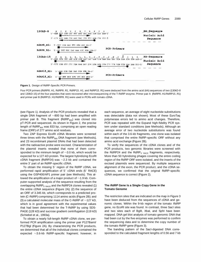
50 reversed-phase HPLC–specific peaks.Most of these contained a mixture of several protein degra-dation products. Nevertheless, microsequencing of pep-tides of the T-RdRP resulted in four amino acid sequencesthat could be used to design degenerate primers. The se-quence of two of the four peptides and the sequence of fourof the corresponding synthetic oligonucleotides that hadbeen designed for polymerase chain reaction (PCR) experi-ments are presented in Figure 1.
Because the relative positions of the sequenced peptideswithin the T-RdRP were not known, we performed PCR withtomato cDNA by using primer pairs A and B, respectively
Cellular
RdRP
Genes 2089
(see Figure 1). Analysis of the PCR products revealed that asingle DNA fragment of
z
800 bp had been amplified withprimer pair B. This fragment (RdRP
800
) was cloned intopT-PCR and sequenced. As shown in Figure 2, the preciselength of RdRP
800
was 833 bp, comprising an open readingframe (ORF) of 277 amino acid residues.
Two ZAP Express EcoRI cDNA libraries were screenedthree times with the RdRP
800
DNA fragment (see Methods),and 23 recombinant plasmid DNAs that had been detectedwith the radioactive probe were excised. Characterization ofthe plasmid inserts revealed that none of them corre-sponded to the mininum length of
z
3.0 kb, which would beexpected for a 127-kD protein. The largest hybridizing EcoRIcDNA fragment (RdRP24) was
z
2.3 kb and contained theentire 3
9
part of an RdRP-specific cDNA.To obtain the missing 5
9
region of the RdRP cDNA, weperformed rapid amplification of 5
9
cDNA ends (5
9
RACE)using the GSP400/AP1 primer pair (see Methods). This al-lowed the amplification of a major product of
z
1.9 kb. Com-puter-supported analysis of the sequence resulting from theoverlapping RdRP
5
9
RACE
and the RdRP24 clones revealed (1)the entire cDNA sequence (Figure 2A); (2) the sequence ofan ORF of 3.345 kb, which corresponds to a predicted pro-tein (C-RdRP) comprising 1114 amino acids (Figure 2B); and(3) a calculated molecular mass of the C-RdRP of
z
127 kD,which is in good agreement with the experimental valuesthat had been determined for the T-RdRP by using SDS-PAGE (128 kD) and sucrose gradient centrifugation (119 kD)(Schiebel et al., 1993a).
To obtain a nearly full-length RdRP cDNA clone, we per-formed PCR amplification using the primer pair P127BamI/P127Bgl (Figure 2A). By characterizing the PCR products,we determined that all of the individual clones contained theexpected
z
3.6-kb RdRP-specific fragment; however, in
each sequence, an average of eight nucleotide substitutionswas detectable (data not shown). Most of these EuroTaqpolymerase errors led to amino acid changes. Therefore,PCR was repeated with the Expand high-fidelity PCR sys-tem under standard conditions (see Methods). Although anaverage error of two nucleotide substitutions was foundwithin each of the 3.6-kb fragments, one clone was isolatedthat comprised the entire RdRP-specific ORF without anyamino acid exchange (Figure 2B).
To verify the sequences of the cDNA clones and of thePCR products, two genomic libraries were screened withthe RdRP24 and the RdRP
5
9
RACE
fragments, respectively.More than 50 hybridizing phages covering the entire codingregion of the RdRP ORF were isolated, and the inserts of theexcised plasmids were sequenced. By multiple sequencealignment of the exon, the PCR product, and the cDNA se-quences, we confirmed that the original RdRP-specificcDNA sequence is correct (Figure 2).
The
RdRP
Gene Is a Single-Copy Gene in theTomato Genome
The restriction sites that are indicated on the map in Figure 3have been deduced from the sequences of cDNA and ge-nomic clones. Within the 9-kb region of the tomato
RdRP
gene, no EcoRI site was found. In contrast, three SacI sitesand two sites each of BglII, XbaI, and SphI have beenmapped. DNA gel blot analysis of tomato genomic DNA thathad been cut by the five enzymes was performed to confirmthe sequencing data and to determine the copy number ofthe tomato
RdRP
gene (Figure 3).The banding pattern of the SacI-digested DNA corre-
sponded to the calculated fragment lengths of 0.56 and 7 kb
Figure 1. Design of RdRP-Specific PCR Primers.
Four PCR primers (RdRP82H1, RdRP82R1, RdRP152H1, and RdRP152R1) were deduced from the amino acid (AA) sequences of two (130kD-8and 130kD-15) of the four peptides that were recovered after microsequencing of the T-RdRP enzyme. Primer pair A (RdRP82H1/RdRP152R1)and primer pair B (RdRP152H1/RdRP82R1) were used in PCRs with tomato cDNA.

2090 The Plant Cell
(Figure 3, lane 1). The length of the latter fragment confirmsthe existence and the lengths of introns 1 to 4 that have beendetected on genomic clones. Interestingly, intron 1 of
z
1.3kb is located 65 bp upstream of the AUG in the 5
9
untrans-lated region. The two additional fragments of 5.5 and 9 kbrepresent the 5
9
and 3
9
border fragments of the
RdRP
gene.The length of the smaller BglII fragment (Figure 3, lane 2) isidentical to the length of 1.4 kb that was determined by se-quence analysis. The 5
9
BglII site is located in the 480-bp-long intron 4, whereas the 3
9
BglII site was found in theputative
RdRP
gene terminator. The XbaI-restricted DNA re-sulted in three hybridizing fragments (Figure 3, lane 4). The630-bp fragment comprises the junction between the 5
9
trans-lated region and the 2.4-kb intron 2. The two other bandsagain represent the 5
9
and 3
9
border fragments, respectively.According to the BglII-XbaI double digest (Figure 3, lane
3), the 7.5- and 2.4-kb XbaI fragments (Figure 3, lane 4)could be assigned to the 3
9
and 5
9
borders, respectively. Asexpected from the sequencing data, the entire tomato
RdRP
gene could be released by EcoRI (Figure 3, lane 5). Becausethe two SphI sites that are located within the
RdRP
gene areseparated by 11 bp, only two bands are visible in the EcoRI-SphI double digest (Figure 3, lane 6). Differences in the in-tensity of the hybridizing fragments are based on the factthat the
a
-
32
P-dCTP–labeled DNA probe comprised theRdRP-specific cDNA but not the intron sequences. Thus,fragments containing large parts of the introns (see Figure 3,map) gave relatively weak signals.
Identification of RdRP Homologs in DifferentPlant Species
HindIII-restricted human genomic DNA and that from po-tato, Arabidopsis, tobacco, and the two tomato cultivarsRentita and St. Pierre were analyzed by DNA gel blot hybrid-ization by using a 1.7-kb 3
9
-specific probe (Figure 4). Com-pared with the identical banding patterns of the two tomatoDNAs, a HindIII-specific restriction fragment length polymor-phism was found for the strongly hybridizing potato DNAand for the tobacco DNA. The barely visible signals of thetobacco fragments became more distinct on an autoradio-graph that was exposed for 72 hr (Figure 4, lane Tobacco*);however, in neither the human nor Arabidopsis DNA was ahybridizing fragment detectable on the 72-hr-exposed auto-radiograph (data not shown).
To find conserved sequences within the
RdRP
genes ofdifferent plant species, we performed PCR amplification
Figure 2.
Structure of the Tomato
RdRP
Gene and the RdRP Protein.
(A)
Schematic representation of the physical structure of the tomato
RdRP
gene. The diagram represents the
RdRP
transcript, with fourintrons indicated by lines angled into V’s. The initiation (ATG) andtermination (TGA) codons are indicated. The locations of theRdRP15
2
H1 and RdRP8
2
R1 primers (Figure 1, primer pair B) areshown. A continuous cDNA clone was isolated after PCR with to-mato cDNA by using the P127BamI and P127Bgl primers (see Meth-ods). The nucleotide sequence of the C-RdRP cDNA has theaccession number Y10403 in the EMBL and GenBank nucleotidesequence databases.
(B)
Predicted amino acid sequence of the RdRP cDNA. The mi-crosequenced peptides are numbered (1 to 4), and their respectiveamino acid sequences are printed in boldface. The peptide se-quences printed in italic were not used for primer design. The first
RdRP-specific PCR product (RdRP
800
) that was amplified withprimer pair B (see
[A]
) is underlined. The stop codon is marked withan asterisk.

Cellular
RdRP
Genes 2091
with tomato (as a control) and tobacco genomic DNA by us-ing five C-RdRP–specific primer pairs (sequences not shown).The PCR products were hybridized using the entire tomato
RdRP
sequence as a probe. One tobacco-specific productof
z
500 bp clearly hybridized with the tomato-specificprobe. Characterization of this cloned fragment (RdgTb
500
)revealed a nucleic acid sequence identity of
z
91.6% withthe tomato
RdRP
coding region (data not shown). To verifythat the mismatches were not due to EuroTaq polymeraseor sequencing errors, we repeated PCR amplification; how-ever, tobacco cDNA was used Fas a template. The forwardprimer was deduced from the RdgTb
500
sequence, and ap(dT)
15
oligomer served as reverse primer. By analyzing thecloned
z
950-bp PCR product (RdcTb
950
), we confirmed theRdgTb
500
sequence. The nucleic acid sequence identity be-
tween RdcTb
950
and the tomato cDNA was 89.7% within the810-bp-long part of the coding region. In the 3
9
untranslatedregion, identity decreased to 55.6% (data not shown). Forthe protein, a similarity of 96.7% and an identity of 88.2%were determined for the 270 amino acids.
Based on these results, two forward and two reverseprimers corresponding to conserved regions of the tomatoand the tobacco cDNA were designed. Using these primers,we amplified a 540-bp intron-free PCR product (RdgPt
500
)from genomic DNA of petunia. Sequence analysis revealed90.81% nucleic acid sequence identity between RdgPt
500
and the tomato cDNA and 90.63% identity between the pe-tunia-specific DNA and the RdcTb
950
. For the protein, simi-larity of 99.45% and identity of 87.3% were determinedbetween the petunia and the tomato sequences, whereassimilarity of 98.34% and identity of 88.96% were calculatedfor the 181 amino acids of the petunia and tobacco clones.
Alignment of the tomato-, tobacco-, and petunia-specificsequences revealed several highly conserved amino acid re-gions. Two forward and two reverse degenerate primerswere used in PCRs with Arabidopsis and with wheat genomic
Figure 3. DNA Gel Blot Analysis of Total Genomic Tomato DNA.
On the physical map of the RdRP gene at top, the four introns are in-dicated by black boxes. The initiation (ATG) and termination (TGA)codons are indicated. The positions of sites recognized by the re-striction endonucleases used in the gel blot (bottom) are shown byvertical bars in the schematic (middle). The lengths of hybridizingDNA fragments are presented in kilobases to the left of the gel. TheDNA gel blot was obtained with total genomic tomato DNA that washybridized with the entire a-32P-dCTP–labeled RdRP-specific cDNAand autoradiographed for 16 hr.
Figure 4. DNA Gel Blot Analysis of Total Genomic DNA from Differ-ent Plant Species and Humans.
The DNA gel blot was obtained with HindIII-restricted total genomicDNAs from the indicated organisms that were hybridized with a 1.7-kba-32P-dCTP–labeled 39-specific RdRP cDNA probe and autoradio-graphed for 16 hr. The filter was reexposed for 72 hr to increase thesignal strength of the tobacco-specific fragments (lane Tobacco*).On the overexposed autoradiograph, no hybridizing fragments werevisible in the human and Arabidopsis lanes. Thus, only the Tobacco*lane was included. l-PstI, phage lambda DNA restricted with PstI.

2092 The Plant Cell
DNAs. Amplified products were characterized, and au-totranslation of the nucleic acid sequence indicated thatboth Arabidopsis and wheat also contained a C-RdRPhomolog. The different RdRP-specific sequences amplifiedfrom Arabidopsis, wheat, petunia, and tobacco overlappedacross a 93–amino acid region. Sequence alignment of thisC-RdRP region with that of the other plant species is pre-sented in Figure 5. This analysis reveals a high level of iden-tity between the different plant sequences and the predictedC-RdRP amino acid sequence, ranging from 72% for the Ar-abidopsis sequence to
z
88% for the petunia and tobaccosequences (see also above). The identity between the to-mato and the Arabidopsis as well as between the tomatoand the wheat coding sequences is
z
70%. This degree ofsequence divergence would explain the failure to detect theArabidopsis
RdRP
gene on the DNA gel blot (Figure 4) withthe tomato-specific probe.
Association of T-RdRP Activity with the cDNA-Encoded 127-kD Protein
To show directly that the C-RdRP and the T-RdRP are iden-tical, we introduced RdRP cDNA constucts into
Escherichiacoli
to express a functional enzyme. Unfortunately, it turnedout that the
E. coli
–synthesized C-RdRP was deposited asan insoluble and inactive protein in bacterial inclusion bodies(data not shown). Refolding of the denatured C-RdRP alsofailed to recover enzyme activity.
Thus, to demonstrate the identity of the T-RdRP with theC-RdRP, we examined whether T-RdRP activity was con-sistently associated with the C-RdRP protein assayed withantisera to C-RdRP peptides (see Methods) during the fivepurification steps. As source material, we used the 30,000
g
pellet of a homogenate that was extracted from 220 g of vi-roid-infected apical tomato leaves. The pellet, containingonly the minor portion of total T-RdRP, was used becauseit was shown to be less contaminated by terminal nucleoti-dyl transferase(s) (TNTase[s]) than was the 30,000
g
super-natant. TNTase activity distorts RdRP activity values thatare determined by the standard assay (Schiebel et al.,1993a).
The relative enrichment of T-RdRP protein and the associ-ation of increasing specific RdRP activity with the degree ofpurity during purification are documented in Figure 6. Thesolubilized pellet (Figure 6A, lane 1) was applied usingQ-Sepharose Fast Flow chromatography (Figure 6A, lane 2),which was followed by a Q-Sepharose high-performancepurification step (Figure 6A, lane 3). These two anion ex-change chromatography steps removed the majority of pro-tein, including the TNTase(s). The most effective increase inspecific activity was achieved by the polynucleotide affinitymedia poly(A)– and poly(U)–Sepharose. The T-RdRP frac-tions from these two columns contained low amounts ofother proteins (Figure 6A, lanes 4 and 5). The purest enzymecould be eluted from a hydroxyapatite column, with only asingle band visible on the silver-stained gel (Figure 6A, lane6). However, hydroxyapatite chromatography was not per-formed for large-scale preparation of active T-RdRP. Forreasons unknown, we recovered only about half of the en-zyme activity that was applied to this column.
The identity of the T-RdRP with the C-RdRP was demon-strated by an immunoblot (Figure 6B) that was comparableto the silver-stained gel. Only one protein of
z
127 kD wasdetectable by the C-RdRP–specific antibody A
P431
(seeMethods). In addition, the intensity of the immunoresponseincreased with respect to the rise in the specific RdRP activ-ity (Figure 6B). The additional signals on the protein gel blot(Figure 6B, lanes 1 to 3) that correspond to proteins of
z
80and 50 kD, respectively, were also detectable with preim-mune serum (data not shown), pointing to contamination ofthe A
P431
immune serum with non-C-RdRP–specific antibod-ies. Enzyme activity of the hydroxyapatite eluates was lowerthan expected from the signal strength of the silver-stainedgel and the protein gel blot. However, the eluate in the sixthlane (Figures 6A and 6B) is presented to show the homoge-neity of the preparation.
Coincidence of the C-RdRP with the activity of the T-RdRPis further demonstrated in Figures 7A and 7B by immuno-blots showing eluted fractions from poly(U)–Sepharose andhydroxyapatite chromatography. On the gels that are de-picted in Figure 6, the highest RdRP activity–containingfractions that were eluted from each of the five columnshave been electrophoresed. In contrast, Figures 7A and 7Breflect parts of elution profiles. The increase and decrease of
Figure 5. Partial Comparison of the C-RdRP Amino Acid Sequencewith That of Tobacco, Petunia, Arabidopsis, and Wheat.
Putative RdRP sequences from other plant species were amplifiedby PCR by using different combinations of RdRP-specific primers(see the text). The deduced amino acid sequences from tobacco(Tb.), petunia (Pt.), Arabidopsis (Ara.), and wheat (Wh.) RdRP-spe-cific PCR products were aligned with the predicted tomato (Tom.)RdRP amino acid sequence. Identical amino acid residues areshaded black. The sequence alignment is given from positions 805to 897, according to the C-RdRP amino acid sequence.

Cellular
RdRP
Genes 2093
enzyme activity during the T-RdRP elution from both col-umns were in perfect accordance with the immunoresponseto A
P431
. The intensity of coloration of the protein gel blot isproportional to the T-RdRP activity values.
Finally, the association of the C-RdRP with the RdRP ac-tivity is documented by the anomalous gel filtration behaviorof T-RdRP on Superdex 200 that is illustrated in Figure 8. Aneluate from the hydroxyapatite chromatography was supple-mented with rabbit IgG (150 kD) and BSA (66 kD). The proteinmixture was chromatographed, and a comparison of theUV
280
values with the RdRP activity profile of the eluted frac-tions led us to believe that the T-RdRP is smaller than theBSA protein. According to size, the T-RdRP was expectedto be eluted very soon after the IgG marker protein andclearly before a 66-kD protein. However, elution volumes of1.35 mL for IgG, 1.50 mL for BSA, and 1.58 mL for theT-RdRP were determined (Figure 8). After SDS gel analysisof the UV
280-absorbing material, we found that the promi-
nent protein of the RdRP activity–containing fraction was127 kD (data not shown). The identity of this 127-kD proteinwith the C-RdRP was demonstrated with AP431 (Figure 8, in-set gel).
Induction of T-RdRP Activity upon Viroid Infection Is Correlated with an Increased Steady State Level of RdRP mRNA
Upon RNA virus and viroid infection, plant-encoded RdRPsincrease both in activity and in amount (Astier-Manifacierand Cornuet, 1971; Duda et al., 1973; Takanami and Fraenkel-Conrat, 1982; van der Meer et al., 1984; Schiebel et al.,1993a). RNA gel blot analysis was performed with total RNAisolated from potato spindle tuber viroid (PSTVd)–infectedand viroid-free tomato plants. Hybridization with an a-32P-dCTP–labeled RdRP-specific DNA probe revealed a three-to fivefold increase in the amount of steady state RdRPmRNA in the two PSTVd-infected tomato cultivars Rutgersand Basket Pak (Figure 9; cf. lanes 1 and 3 with lanes 2 and4). The increased RdRP activity of approximately threefold,which was previously reported for systemically PSTVd-infected tomato leaves (Schiebel et al., 1993a), is in perfectagreement with the results obtained by the RNA gel blotanalysis. This indicates that in viroid-infected tomato plants,RdRP induction is due to an enhancement of transcriptionand/or mRNA stability.
Figure 6. SDS-PAGE Analysis of the Purification Procedure ofT-RdRP from the 30,000g Pellet of a Leaf Homogenate.
(A) Silver-stained protein pattern of RdRP preparations.(B) Protein gel blot corresponding to (A) and visualizing the T-RdRPby the C-RdRP–specific antibody AP431.The positions of molecular mass markers are indicated in kilodaltonsat left. The specific RdRP activity that was determined for each frac-tion is given in units (U) per milliliter. The volume of samples loadedonto the gels was 1 mL per lane. Lane 1, combined extracts from the30,000g pellets (Ex); lane 2, Q-Sepharose Fast Flow eluate (FF); lane3, Q-Sepharose high-performance eluate (HP); lane 4, poly(A)–Sepharose eluate (PA); lane 5, poly(U)–Sepharose eluate (PU); andlane 6, hydroxyapatite eluate (HA). The arrows at right denote the127- kD band of the RdRP, and those at left indicate the positions ofmolecular weight markers in kD. Experimental details are given inMethods.
Figure 7. RdRP Activity Coincides with an AP341-Detectable Tomato-Specific 127-kD Protein in Eluates from Poly(U)–Sepharose andfrom Hydroxyapatite.
(A) Protein gel blot analysis of Poly(U)–Sepharose eluates with theC-RdRP–specific antibody AP431. The staining intensity of the 127-kDband strongly correlates with the activity of RdRP. Samples werefrom 100-mL fractions of poly(U)–Sepharose chromatograph eluates(see Figure 6). U, units.(B) Protein gel blot analysis as shown in (A), but samples were fromthe hydroxyapatite eluate fractions (see Figure 6).

2094 The Plant Cell
In Vitro Analysis of the T-RdRP
If plant-specific RdRPs are the key enzymes in post-tran-scriptional gene silencing processes, then they should becapable of synthesizing cRNAs of z50 to 130 nucleotidesfrom RNA templates. To examine the in vitro T-RdRP–syn-thesized products, we modified the RdRP activity standardassay (see Methods). Total tomato RNA was used as tem-plate, and the products were analyzed by using PAGE.
RNA fragments of .100 nucleotides were produced whenthe assay was performed with 1 mg of template RNA (Figure10, lane 2). A decrease in the template (0.1 mg) also resultedin a decrease in products. On the other hand, increasedamounts of total RNA (5 mg) had little effect on productamount and quality (Figure 10, lanes 1 to 3). The capabilityof the T-RdRP to also use DNA as template appeared to be
very low. The standard assay was performed with total ge-nomic tomato DNA as well as with linearized plasmid DNA,but only a few products were detectable after PAGE (Figure10, lanes 5 to 7). To demonstrate that the products con-sisted of newly synthesized RNA and were not due to aT-RdRP–specific terminal transferase activity (Schiebel etal., 1993b), the nucleotide triphosphates were left out in onereaction. Because no product was visible, it can be con-cluded that the T-RdRP is not able to initiate the addition ofat least a-32P-UTP (see Methods) to the template RNA (Fig-ure 10, lane 8). The identity of RNA as the T-RdRP–synthe-sized product was further investigated by treating thereaction mix with RNase A before PAGE (Figure 10, lane 9).In addition, it was shown that the template is destroyed byRNase A but not by DNase treatment (data not shown), indi-cating that RNA is the real substrate of the T-RdRP.
DISCUSSION
Analysis and characterization of the in vitro and in vivo prop-erties of plant RdRPs require suitable amounts of pure en-zyme. To obtain increased RdRP activity, researchers havealmost always used RdRP purification protocols that beginwith RNA virus–infected plants. But despite many attemptsto clarify the nature of the host-encoded RdRP (reviewed in
Figure 8. Delayed Coelution of T-RdRP from a Superdex 200 Column.
Eluate from the hydroxyapatite chromatography representing z0.75units of T-RdRP in 15 mL of 0.16 M NaPi in buffer F was supple-mented with 2 mg each of rabbit IgG (Sigma) and BSA (Serva 11924)and loaded onto a precalibrated Superdex 200 column in the Smartsystem. The column was equilibrated at 58C with a buffer of 0.15 MNaCl, 10 mM Tris-HCl, pH 8.0, and 1.5 mM DTT. Elution volumeswere 1.35 mL for IgG, 1.505 mL for BSA, and 1.58 mL for RdRP.Fractions containing enzyme activity were subjected to protein gelblot analysis with the C-RdRP–specific antibody AP431. The immuno-blot shows a single band of 127 kD and the maximum of staining in-tensity in fraction 17. Neither RdRP activity nor a 127-kD proteinwas detectable in fraction 13, in which a standard protein of this sizeshould elute. AU, absorbance units; U, units.
Figure 9. RNA Gel Blot Analysis of Total RNA from Viroid-Infectedand Viroid-Free Tomato Plants.
The steady state level of RdRP-specific mRNA was examined in vi-roid-infected (1) and in viroid-free (2) tomato plants. The sameamounts (15 mg per lane) of total leaf RNA from infected as well asfrom noninfected tomato cultivars Rutgers and Basket Pak were hy-bridized with a 1.7-kb a-32P-dCTP–labeled 39-specific RdRP cDNAprobe and autoradiographed for 16 hr.

Cellular RdRP Genes 2095
Fraenkel-Conrat, 1986), the activity of the isolated en-zyme(s) could not be clearly assigned to the plant- and vi-rus-encoded RdRP (Khan et al., 1986). Thus, from cowpeamosaic virus (CPMV)–infected plant tissue, for example, twoRdRP activities were obtained, one of which was assumedto represent the host-encoded RdRP, whereas the otherwas considered to be specific for CPMV-infected leaves(Dorssers et al., 1983). But apart from our studies with theT-RdRP (Schiebel et al., 1993a, 1993b), no detailed charac-terization of an RdRP from other plant species has been re-ported so far.
Microsequencing of the Enzyme and Isolation ofIts cDNA
Endoproteolytic digestion of the gel-excised 127-kD pro-tein(s) resulted in z50 reversed-phase HPLC–specific peaks.Most of these contained a mixture of several protein degra-dation products. Nevertheless, four peptide sequences wereobtained from which PCR primers were designed. By usinga combination of two of these primers, the RdRP800 frag-ment was amplified, which finally allowed isolation of the en-tire C-RdRP cDNA. The fact that the peptide sequencesdeduced from the other two primer sequences could be foundwithin the protein sequence of the C-RdRP substantiatedthat the isolated cDNA is coding for the microsequencedprotein. Because an active C-RdRP protein from E. coli cellswas not expressed, we analyzed indirectly the identity of theC-RdRP with the help of antibodies that had been raisedagainst synthetic C-RdRP–specific peptides. The four anti-body preparations specifically detected a 127-kD tomatoprotein in an aliquot of the protein fraction that was appliedduring the microsequencing procedure. As a control, themost sensitive AP431 immune serum, which has been chosenfor further experiments, was demonstrated also to bind tothe E. coli–expressed C-RdRP (data not shown). It is note-worthy that immunoprecipitation experiments with AP431 ledto inhibition of T-RdRP activity, but splitting off the antibodyfrom the protein did not restore activity (data not shown).
Because the AP431 immunoresponse perfectly correlatedwith the enzyme activity during all enzyme purification steps,we assumed that the C-RdRP and the T-RdRP are identical.However, the remote possibility exists that at least two pro-teins might have comigrated and become excised togetherfrom the SDS gel and also applied together to the microse-quencing procedure. If so, this would mean that the contam-inating protein shares those physical properties that haverendered possible the purification of the T-RdRP up to a sin-gle band in a silver-stained SDS gel. In addition, the obser-vation that the AP431 immunoresponse also correlated withthe unusual behavior of the T-RdRP in the Superdex 200 gelfiltration precluded the presence of a second protein. All ofthese data strongly suggest that the microsequenced pro-tein is the actual T-RdRP and that the isolated cDNA repre-sents its genomic transcript.
Cellular RdRPs in Other Plant Species
The availability of a full-length tomato RdRP cDNA facili-tated the search for the presence of this enzyme in otherplant species. The existence of highly conserved RdRPgenes in potato, tobacco, wheat, Arabidopsis, and petuniacould be clearly shown by DNA gel blot hybridization and/orPCR amplification experiments. The failure to detect a cor-responding sequence in Arabidopsis by DNA gel blot hy-bridization of genomic DNA against the tomato-specificprobe demonstrated the limitation of this procedure. The
Figure 10. Analysis of in Vitro–Synthesized T-RdRP Products byUsing PAGE.
RdRP activity standard assay samples were separated on a 10%polyacrylamide gel to analyze the amount and quality of the RdRP-synthesized products. Substrate saturation was examined by using0.1, 1.0, and 5.0 mg of total tomato (TOM) RNA as template (lanes 1to 3), and the suitability of DNA to serve as template was analyzedby performing the standard assay with 1.0 and 5.0 mg of total to-mato DNA as well as with 1.0 mg of linearized plasmid DNA (PUC)(lanes 5 to 7). As a control, the assay was performed in the absence(2) of nucleotide triphosphates (NTPs [2], lane 8) and in the ab-sence of enzyme (Enzy [2], lane 10). The identity of the RNA natureof the RdRP-synthesized products was confirmed by treating thesample with RNase A (RNas [1], lane 9) before PAGE. The lengths ofthe in vitro–radiolabeled RNA products used as size markers (M,lane 4) are presented in nucleotides to the left of the gel.

2096 The Plant Cell
fact that an RdRP-specific fragment from Arabidopsis couldbe amplified by PCR has shown that the similarity betweenthe T-RdRP and the RdRP of Arabidopsis was too low forcross-hybridization under the applied conditions. It shouldbe mentioned that an expressed sequence tag from Arabi-dopsis (EMBL accession number B61313) of z0.55 kbperfectly matched our Arabidopsis PCR product. Also, of di-rect relevance to our findings are the studies of a putativecowpea-specific RdRP cDNA fragment (sequence accordingto S. Rudd, Norwich, UK). This sequence shows 73% nucle-otide sequence identity and 95% amino acid sequence sim-ilarity with the T-RdRP cDNA.
Cellular RdRPs in Other Organisms and Systems
Our search of RdRP sequences in other organisms and sys-tems has shown that ORFs of S. pombe (EMBL accessionnumber Z98533) and C. elegans (EMBL accession numberZ48334) most likely encode an RdRP homolog. Both of theamino acid sequences contain several regions that are iden-tical or highly similar to that of the plant RdRPs. However,no evidence for a human RdRP has as yet been found. Inter-estingly, plant RdRPs evidently do not share any similarity tothe known virus RNA replicases, including in their commonGDD sequence motif (Jablonski et al., 1993; Routhier andBruenn, 1998). Future PCR experiments with degenerateprimers deduced from peptides that are conserved in thetomato, tobacco, petunia, Arabidopsis, wheat, yeast, andnematode amino acid sequences will help to extend ourknowledge on the still largely enigmatic cellular RdRP enzyme.
Possible Functions of the Cellular RdRP
The involvement of a plant-encoded RdRP in RNA-mediatedgene silencing and in RNA-mediated virus resistance is oneof the most crucial points of the current models for homol-ogy-dependent gene silencing (reviewed in Wasseneggerand Pélissier, 1998). In a series of experiments on trans-gene-induced silencing, it has been suggested that RNAmost likely mediates post-transcriptional gene silencing. Byusing grafting experiments, it has been demonstrated thatthe effector of post-transcriptional gene silencing can betransmitted systemically. Transgene-specific silencing wasunidirectionally transmitted from silenced stocks to nonsi-lenced scions (Palauqui et al., 1997). It was further observedthat post-transcriptional cosuppression of the tobacco ni-trate reductase (Nia) genes and the Nia2 transgene is depen-dent on the transcriptionally active state of the transgene(Vaucheret et al., 1997). Similar results were obtained by an-alyzing the frequency and degree of cosuppression bysense chalcone synthase transgenes in petunia. The findingthat cosuppression was dependent on transgene promoterstrength and was reduced by premature nonsense codons in
the transgene coding sequence led to the conclusion thattransgene transcription is necessary for induction of post-transcriptional gene silencing by single-copy transgenes(Que et al., 1997).
If this is true, how can a primary transcript be enabled todecrease the steady state mRNA of the correspondinggene? It is generally assumed that short cRNA moleculesare transcribed from sense transcripts by a cellular RdRP.Subsequently, the cRNAs could specifically target mRNAdegradation. However, as of yet, there is no proof of such amechanism. Moreover, almost no alternatives have beenpresented by which this highly specific cytoplasmic RNA/RNA interaction–dependent process could be replaced. Nev-ertheless, it should be noted that RdRP-independent mech-anisms of RNA degradation have been proposed (Cameronand Jennings, 1991; Metzlaff et al., 1997) that involve inter-molecular interactions between sense transcripts and recog-nition of paired complementary sequences by dsRNA-specificRNases.
When we examined the existing models, we found thatthe T-RdRP could meet all of the requirements of the pro-posed cRNA-involving mechanism. The analysis of the cata-lytic in vitro properties of T-RdRP revealed that it acceptssingle-stranded RNAs as templates and, regardless ofwhether these RNAs are primed or unprimed, cRNAs aresynthesized (Schiebel et al., 1993b). Although these experi-ments were performed predominantly with 12- to 14-nucle-otide-long synthetic RNA or DNA oligomers, T-RdRP–produced cRNAs can have a length of .100 nucleotides.This was shown by using an in vitro transcript of the P35S(1) strand (data not shown) and total tomato leaf RNA. Inter-estingly, for post-transcriptional gene silencing, a cRNA sizeranging between 10 and 75 nucleotides was proposed to besufficient to target RNA degradation efficiently (Doughertyand Parks, 1995). In addition, homology of 60 to 130 bp be-tween an inactivating transgene and a target sequence hasbeen shown to be sufficient to trigger post-transcriptionalgene silencing and RNA-mediated virus resistance (Cogoniet al., 1996; Sijen et al., 1996; Pang et al., 1997).
According to the threshold hypothesis originally proposedby Lindbo et al. (1993), overexpressed mRNA accumulatingin the cytoplasm above an aberrant level could be used by ahost RdRP for the production of the cRNAs, which would acti-vate the silencing process (Lindbo et al., 1993; Doughertyand Parks, 1995). This hypothesis can explain silencing bysingle-copy, highly transcribed transgenes but does notreadily explain those examples in which gene silencing istriggered with inverted repeat transgenes driven by weakpromoters (Que et al., 1997; Stam et al., 1997b). Other stud-ies also reported examples of silenced and nonsilencedtransgenic lines that display no detectable difference in thetransgene transcription rate (Mueller et al., 1995; English etal., 1996). These results strengthen the emerging view thatnot only quantitative but also qualitative features (e.g., “ab-erration” in the structure and/or the location of a transcript)of the (trans)gene mRNAs could define these molecules as

Cellular RdRP Genes 2097
preferred templates for the host RdRP, resulting in the acti-vation of the post-transcriptional gene silencing process(English et al., 1996; reviewed in Baulcombe, 1996; Sängeret al., 1996; Depicker and Van Montagu, 1997; Stam et al.,1997a; Wassenegger and Pélissier, 1998).
In both the threshold and the aberrant RNA models, it wassuggested that the involvement of RdRP-produced cRNAsaccounts for the high sequence specificity of gene silencing.Therefore, this might indicate that there are at least twoways to trigger the RdRP-dependent degradation mecha-nism. It is also possible that a threshold level of aberrantRNA is required in all cases to initiate the silencing process.In many transformation experiments, it was observed thatsingle copies rarely gave rise to silencing, whereas invertedrepeats of the transgene at a single locus frequently initiatedsilencing (Hobbs et al., 1993; van Blokland et al., 1994;English et al., 1996; Que et al., 1997; Stam et al., 1997b).This might reflect the potentiality of inverted repeats to pro-duce more aberrant RNA than do single-copy transgenes(Que et al., 1997) and/or that aberrant RNA produced by in-verted repeats represents a more efficient substrate for theRdRP than the one produced by single-copy transgenes(Wassenegger and Pélissier, 1998). Future in vitro analysisof T-RdRP substrate specificity might allow us to definesome sequence and/or structural motifs that could tag RNAmolecules as efficient templates for the RdRP.
Interestingly, in viroid-infected tomato plants, enhancedRdRP activity correlates with an increased steady state levelof the RdRP mRNA. Viroids are small, pathogenic, circularRNA molecules that display a stable, rodlike structure (i.e., ahighly double-stranded form). They are capable of autono-mous replication in the nucleus of the host cell, where theycan accumulate up to 5 3 104 copies (Harders et al., 1989).Thus, if the viroid RNA can be considered as a kind of aber-rant RNA, then this result might indicate that RdRP gene ex-pression and consequently RdRP activity could be, at leastto some extent, regulated by nuclear concentrations of ab-errant RNAs. On the other hand, one can speculate that cel-lular RdRPs are involved in a defense mechanism that istargeted against overexpressed foreign RNAs.
Future Perspectives
Even if the T-RdRP and the C-RdRP represent identical pro-teins, the actual biological function of plant-encoded RdRPshas to be examined. The refined T-RdRP purification proce-dure will provide sufficient amounts of pure enzyme to ana-lyze RdRP substrate specificity. The availability of the full-lengthtomato RdRP cDNA will facilitate the analysis of its possibleinvolvement in homology-dependent gene-silencing phe-nomena. Thus, experiments allowing overexpression as wellas downregulation of the enzyme in transgenic plants couldhelp to determine whether RdRPs play some role in normalplant gene regulation or whether they are enzymes of a plantdefense mechanism (Jorgensen et al., 1998).
METHODS
Isolation of RNA-Directed RNA Polymerase for Microsequencing
RNA-directed RNA polymerase (RdRP) was isolated from apicalleaves (1 g per plant) of viroid-infected tomato plants (Lycopersiconesculentum cv Rentita), essentially as described previously (Schiebelet al., 1993a). DEAE–Sepharose was used as fast flow quality (Phar-macia Biotechnology), and the double-stranded DNA (dsDNA)–cellulose was substituted by poly(U)–Sepharose 4B (PharmaciaBiotechnology). RdRP eluted from this latter column (3 mL of gel vol-ume) in a 20-mL linear gradient of 0 to 1.5 M NaCl in buffer A (20 mMTris-HCl, pH 8.0, 0.5 mM EDTA, 1.5 mM dithioerythritol [DTE], and0.012% Tween 20) at z1 M salt. The enzyme was concentrated by asecond round of poly(U)–Sepharose chromatography by using anHR5 column with an 0.4-mL gel volume in the Smart system (Phar-macia Biotechnology). Pooled RdRP was precipitated with trichloro-acetic acid in the presence of deoxycholate and insulin (Bensadounand Weinstein, 1976). The pellet was washed with absolute ethanolat 2208C, and the proteins were subsequently subjected to SDS-PAGE (see below).
Isolation of Enzymatically Active RdRP
Q-Sepharose Chromatography
The extracts obtained from the 30,000g pellet (Schiebel et al., 1993a)by centrifugation (146,000g for 50 min in a Beckman-type 50.2 rotor;Beckman Instruments, Inc., Fullerton, CA) were loaded with a flowrate of 30 cm/hr onto a 70-mL column of Q-Sepharose Fast Flow(Pharmacia Biotechnology) equilibrated with buffer B (25 mM Tris–acetate, pH 8.2, 1 mM EDTA, 20% glycerol, and 3 mM 2-mercapto-ethanol). In a linear 195-mL gradient, ammonium acetate was raisedto 0.7 M in buffer B. RdRP eluted at z370 mM salt. The pool was di-luted with two volumes of buffer B and further purified on Q-Seph-arose high-performance 16 ⁄10 (Pharmacia Biotechnology) with a58-mL salt gradient in buffer B.
Poly(A)–Sepharose Chromatography
The RdRP pool was diluted with 1 volume of buffer C (50 mM Tris-HCl, pH 7.5, 15% glycerol, 1.5 mM DTE, and 0.012% Tween 20) andloaded onto a 4-mL poly(A)–Sepharose 4B C10/10 column (Pharma-cia Biotechnology). After raising NaCl to 0.1 M in buffer C, a 2-mLgradient from 0.1 to 0.3 M salt with a flow rate of 30 cm/hr was ap-plied. RdRP eluted between 0.15 and 0.3 M salt.
Poly(U)–Sepharose Chromatography
RdRP was diluted with 1 volume of buffer C and chromatographedon 0.2 mL of poly(U)–Sepharose 4B by using an HR5 column and theSmart system (Pharmacia Biotechnology). A linear 1-mL gradientfrom 0.35 to 0.7 M NaCl in buffer C was applied with a flow rate of 30cm/hr. RdRP eluted at z0.6 M salt.

2098 The Plant Cell
Hydroxyapatite Chromatography
RdRP was diluted with 2 volumes of buffer D (25 mM Tris–acetate,pH 8.2, 15% glycerol, 1.5 mM DTE, and 0.012% Tween 20) andchromatographed on 0.2-mL hydroxyapatite (ceramic hydroxyapa-tite type I, 40 mm; Bio-Rad) in the Smart system. The column wasprewashed with 5 mL of 1 mM MgCl2 (Gorbunoff, 1985). A concen-tration of 60 mM sodium phosphate, pH 6.8, in buffer D was kept for20 gel volumes and increased in a linear 2.2-mL gradient to a 1-mLplateau of 180 mM sodium phosphate. The RdRP eluted at z150 mM.
Superdex 200 Gel Filtration
Gel filtration was performed with a Superdex 200 PC 3.2/30 column(Pharmacia Biotechnology) by using the Smart system. The columnwas equilibrated at 58C with a buffer of 0.15 M NaCl, 10 mM Tris-HCl,pH 8.0, and 1.5 mM DTE. Rabbit IgG (Eurogentec, Seraing, Belgium)and BSA (Serva 11924; Boehringer Ingelheim Bioproducts, Heidel-berg, Germany) were used as reference proteins.
Standard Assay for RdRP Activity
Enzyme activity was measured as described by Schiebel et al.(1993a) with the following modifications. The final volume of 25 mL inprelubricated 1.7-mL test tubes (Sorenson Bioscience, Inc., SaltLake City, UT) contained 0.02 mM a-32P-UTP (adjusted to 0.5 Ci/mmol) and was supplemented with 0.01% Tween 20. The reactionwas stopped on ice by the addition of 15 mL of 13 mM UTP. Afteradding 10 mL of water, the tube contents were spotted on Whatman(Maidstone, UK) 3MM paper strips.
Protein Determination
Protein concentration was determined by using a microassay modi-fication (Peterson, 1983) of the Coomassie Brilliant Blue R 250 dyebinding method (Bradford, 1976) with 100-mL cuvettes. The concen-tration of purified RdRP in silver-stained polyacrylamide–SDS gelswas estimated as described by Schiebel et al. (1993a).
SDS-PAGE and Protein Gel Blotting
Protein samples were prepared by boiling for 3 min in a standard re-ducing buffer and separated on an electrophoresis unit (Phast-System; Pharmacia Biotechnology) by using 8 to 25% Phastgels.Proteins were visualized by silver staining (Merril et al., 1981) by us-ing a protocol for semiautomated staining (Fabri et al., 1993). Stan-dard proteins (Pharmacia Biotechnology; high molecular weightmarker SDS calibration kit) were supplemented with carbonic anhy-drase (29 kD) and b-lactoglobulin (17 kD) (Sigma).
Immunoblots were obtained from Phastgels by semidry electro-phoretic transfer onto nitrocellulose membrane (Protran 0.45 mm;Schleicher & Schuell). Blotted antigens were detected by an ampli-fied alkaline phosphatase assay kit (Bio-Rad) by using affinity-puri-fied rabbit antibody (1:500 dilution) raised against peptide P431 (seebelow) (Eurogentec). Biotinylated standard proteins were from Sigma(B2787).
Protein Microsequencing
The precipitated protein was prepared for SDS-PAGE, applied onto a7% polyacrylamide gel (Laemmli, 1970), and stained with Coomassieblue (Sambrook et al., 1989). The 127-kD band was excised, andprotein cleavage was performed in the gel according to Eckerskornand Lottspeich (1989), except that instead of trypsin, endoproteaseLysC (Boehringer Mannheim) with an enzyme/protein ratio of 1:10(w/w) was used. The peptides separated by reversed-phase HPLCwere sequenced using a 492A amino acid sequencer (Applied Bio-systems, Foster City, CA), according to the manufacturer’s instruc-tions.
Plant Material
Tobacco (Nicotiana tabacum cv Petit Havana SR1), petunia (Petuniahybrida V26), wheat (Triticum aestivum), and Arabidopsis (Arabidop-sis thaliana ecotype Columbia) were used to search for tomato RdRP(T-RdRP) homologs.
Tomato RNA Isolation, cDNA Synthesis, and PolymeraseChain Reaction
For mRNA isolation, we harvested 20 g of young leaves from potatospindle tuber viroid (PSTVd)–infected tomato plants (L. esculentumcv Rutgers) that were grown in the greenhouse under standard con-ditions. The plant material was quickly frozen in liquid nitrogen, andtotal RNA was extracted as described by Logemann et al. (1987).From 5 mg of total RNA, poly(A)1 RNA was isolated by using Poly-A-Tract mRNA isolation system I (Promega), according to the manufac-turer’s instructions. cDNA synthesis was performed by using 1 mg ofpurified poly(A)1 RNA as template (cDNA synthesis kit; BoehringerMannheim), according to the manufacturer’s instructions. The reac-tion was stopped by phenol extraction, and 1 mL of the sample wasdiluted 1:100 with Tris–EDTA buffer.
A first polymerase chain reaction (PCR) was performed with 1 mL ofthe diluted cDNA by using primer pairs A (RdRP28H1-RdRP215R1;0.1 nmol each) and B (RdRP215H1-RdRP28R1; 0.1 nmol each), re-spectively. Amplifications were assayed in a 100-mL reaction mixturecontaining 10 mL of 10 3 assay buffer (Eurogentec), 10 mL of deoxynu-cleotide triphosphates (2 nmol/mL), and 1 mL of EuroTaq polymerase(4 units per mL; Eurogentec). Thirty cycles of program 1 at 958C for 1min, 558C for 1 min, and 728C for 1 min were started. The PCR prod-ucts were cloned into the T/A-type PCR cloning vector pT-PCR(Wassenegger et al., 1994). Sequences of the primers are as follows:RdRP28H1, 59-TAYCCNGAYTTYATGGAYAA-39; RdRP215H1, 59-AAR-GCNCARGARGCNYTN GA-39; RdRP28R1, 59-GGYTTRTCCATRAAR-TCNGGRTA-39; and RdRP215R1, 59-ATNTTNGTRTTYTCNCCNGG39.
A second PCR was performed with program 2 at 948C for 1 minand 30 cycles at 948C for 30 sec, 608C for 30 sec, and 688C for 4 minwith 1 mL of 1:100 diluted cDNA (see above) by using the forwardprimer P127BamI and the reverse primer P127Bgl. The sequencesfor these two primers are 59-CTTCACCAGGGATCCACTCATCAC-TCCCCTCAAG-39 for P127BamI and 59-GCAGCTTCATGCAGATCT-AAAGACAAAAGGTAGTC-39 for P127Bgl.
PCR, using these two primers, was repeated with the Expandhigh-fidelity PCR system (Boehringer Mannheim). The amplificationwas in a total volume of 100 mL containing 1 mL of 1:100 dilutedcDNA, 10 mL of the 10 3 assay buffer (adjusted to 15 mM Mg21), 1

Cellular RdRP Genes 2099
mM of each primer, 0.2 mM of each deoxynucleotide triphosphate,and 3.5 units of the enzyme. The sample was processed as de-scribed above (program 2).
Rapid Ampification of 59 cDNA Ends
The rapid ampification of 59 cDNA ends (59 RACE) was performed byusing the RdRP gene–specific reverse primer GSP400. Using theMarathon cDNA amplification kit (Clontech, Palo Alto, CA), we li-gated an adapter to both ends of the double-stranded cDNA. Subse-quently, PCR amplification was performed with a 1:100 dilution of thecDNA (1 mL per reaction) by using the adapter sequence–specificAP1 oligonucleotide as a forward primer and GPS400 as a reverseprimer. Their sequences are 59-CATAACGAATCTGGAAAGCAG-ATGG-39 for GSP400 and 59-CCATCCTAATACGACTCACTATAG-GGC-39 for AP1. The applied thermal cycle parameters are de -scribed (program 1) above.
Screening of cDNA and Genomic Libraries
Two custom ZAP Express EcoRI cDNA libraries (Stratagene, La Jolla,CA) had been established from poly(A)1 RNA, which had been iso-lated from young leaves of the tomato cultivars Rutgers and BasketPak. With five to 10 mg of the purified poly(A)1-enriched RNA, weconstructed cDNA libraries (Stratagene). The size-fractionatedoligo(dT)-primed cDNAs (.500 bp) were ligated via EcoRI adaptersinto the l ZAP phage bearing the pBK-CMV phagemid vector. TheRutgers- and Basket Pak–specific cDNA libraries produced by Strat-agene had a representative size of 1.8 3 106 and 3.5 3 106 plaque-forming units, respectively.
Both libraries were screened by plaque hybridization with a-32P-dCTP–labeled DNA fragments by using a random primed DNA label-ing kit (Boehringer Mannheim). Recombinant plasmid DNAs were ex-cised in vivo from the phages and finally introduced into the XLOLREscherichia coli strain, according to the Stratagene ZAP ExpressEcoRI library instruction manual.
Two representative l ZAP Express EcoRI custom genomic librar-ies (Stratagene) had been established from nuclear DNA that hadbeen isolated from 20 g of young leaves of the tomato cultivars Rut-gers and Basket Pak, according to Bedbrook (1981). The size-frac-tionated (.1 kb) plant DNA was ligated via EcoRI adapters into the lZAP phage bearing the pBK-CMV phagemid vector. The Rutgers-and Basket Pak–specific genomic libraries had a representative sizeof 2.4 3 106 and 8.8 3 105 plaque-forming units, respectively. Bothcontained an estimated background of ,5% nonrecombinant clones.Both libraries were screened, and recombinant plasmid DNAs wereexcised as described above.
DNA Sequencing and Sequence Analysis Software
All sequences were determined on an automatic sequencer (ALFex-press; Pharmacia Biotechnology) by using the Cy5 AutoReadsequencing kit (Pharmacia Biotechnology) and following the manu-facturer’s sequencing procedure.
DNA and amino acid sequences were analyzed by using theDNASIS for Windows program (Pharmacia Biotechnology); homol-ogy searches and sequence alignments were performed by using theBlast X and the GAP programs (version 7.0; Genetics Computer
Group, Madison, WI) and the MPsrch2tpn program (Release 3.0.4DJ.F. Collins).
RNA and DNA Gel Blot Analyses
Total RNA was isolated as described above from PSTVd-infectedand from PSTVd-free tomato plants (cvs Rutgers and Basket Pak).Separation of total RNAs (15 mg per lane) was performed in phos-phate-buffered 1.5% agarose gels. The RNAs were pretreated with 1volume of DMSO mix in a final volume of 50 mL and heat denaturedat 658C for 10 min (Spiesmacher et al., 1985). The RNAs were trans-fered onto noncharged nylon membranes (Qiabrane; Qiagen, Chats-worth, CA) by capillary blotting and hybridized against randomprimed a-32P-dCTP–labeled DNA.
DNA gel blot analysis of endonuclease-restricted genomic DNAwas prepared according to Sambrook et al. (1989). The DNA wastransferred (vacuum blotter; Appligene, Illkirch, France) to a posi-tively charged nylon plus membrane (Qiagen) and finally UV312 nm
cross-linked (0.3 J/cm2). Hybridization of DNA gel blots using ran-dom primed a-32P-dCTP–labeled DNAs was performed as describedby Amasino (1986).
Antibody Production
Four different cDNA-encoded RdRP (C-RdRP)–specific antibodieswere produced (Eurogentec). From the entire amino acid sequenceof the C-RdRP, the following peptides were synthesized for the im-munization of rabbits: P430, SNRVLRNYSEDIDN (comprising aminoacids 377 to 390); P431, ASKTFDRRKDAEAI (comprising amino ac-ids 1007 to 1020); P432, EQYDGYLKGRQPPKSPS (comprisingamino acids 331 to 347); and P433, VFPQKGKRPHNEC (comprisingamino acids 784 to 796). The specific reaction of each antiserum withthe T-RdRP was tested by protein gel blotting (see above). The anti-body against P431 (AP431) was affinity purified (Eurogentec) and usedin further experiments.
Substrate Analysis by PAGE
Samples of the RdRP activity standard assay (see above) were notstopped by adding 15 mL of 13 mM UTP but were mixed with 25 mLof gel loading buffer immediately after incubation. Subsequently, thesamples were separated on a 10% polyacrylamide urea gel underdenaturating conditions (Sambrook et al., 1989).
ACKNOWLEDGMENTS
We thank Dr. Jan Kooter and Dr. Erwin Grill for providing genomicDNA, cDNA, and seeds of Petunia hybrida V26 and genomic DNA ofArabidopsis, respectively. We also thank Stephen Rudd for providingthe unpublished RdRP cDNA sequence data of cowpea (Vigna sinen-sis). Our research was supported by the Deutsche Forschungs-gemeinschaft (Grant Nos. Wa 1019⁄1-1 to 1-4). T.P. was supportedby a fellowship from the Human Frontier Science Program (No. LT-0319/1996-M).

2100 The Plant Cell
Received June 29, 1998; accepted October 5, 1998.
REFERENCES
Amasino, R.M. (1986). Acceleration of nucleic acid hybridizationrate by polyethylene glycol. Anal. Biochem. 152, 304–307.
Astier-Manifacier, S., and Cornuet, P. (1971). RNA-dependentRNA polymerase in Chinese cabbage. Biochim. Biophys. Acta232, 484–493.
Astier-Manifacier, S., and Cornuet, P. (1978). Purification et poidsmoléculaire d’une RNA-polymérase RNA dépendante de Bras-sica oleracea var. Botrytis. C. R. Acad. Sci. Ser. III Sci. Vie 287,1043–1046.
Baulcombe, D.C. (1996). RNA as a target and an initiator of post-transcriptional gene silencing in transgenic plants. Plant Mol. Biol.32, 79–88.
Baulcombe, D.C., and English, J.J. (1996). Ectopic pairing ofhomologous DNA and post-transcriptional gene silencing in trans-genic plants. Curr. Opin. Biotechnol. 7, 173–180.
Bedbrook, J. (1981). A plant nuclear DNA preparation procedure.Plant Mol. Biol. News 2, 24.
Bensadoun, A., and Weinstein, D. (1976). Assay of proteins in thepresence of interfering materials. Anal. Biochem. 70, 241–250.
Boege, F., and Sänger, H.L. (1980). RNA-dependent RNA poly-merase from healthy tomato leaf tissue. FEBS Lett. 121, 91–96.
Bradford, M. (1976). A rapid and sensitive method for the quantita-tion of microgram quantities of protein utilizing the principle ofprotein–dye binding. Anal. Biochem. 72, 248–254.
Cameron, F.H., and Jennings, P.A. (1991). Inhibition of geneexpression by a short sense fragment. Nucleic Acids Res. 19,469–474.
Cogoni, C., Irelan, J.T., Schumacher, M., Schmidhauser, T.J.,Selker, E.U., and Macino, G. (1996). Transgene silencing of theal-1 gene in vegetative cells of Neurospora is mediated by cyto-plasmic effector and does not depend on DNA–DNA interactionsor DNA methylation. EMBO J. 15, 3153–3163.
de Carvalho, F., Gheysen, G., Kushnir, S., Van Montagu, M., andInzé, D. (1992). Suppression of b-1,3-glucanase transgeneexpression in homozygous plants. EMBO J. 11, 2595–2602.
de Carvalho Niebel, F., Frendo, P., Van Montagu, M., andCornelissen, M. (1995). Post-transcriptional cosuppression ofb-1,3-glucanase genes does not affect accumulation of transgenenuclear mRNA. Plant Cell 7, 347–358.
Dehio, C., and Schell, J. (1994). Identification of plant genetic lociinvolved in a post-transcriptional mechanism for meioticallyreversible transgene silencing. Proc. Natl. Acad. Sci. USA 91,5538–5542.
Depicker, A., and Van Montagu, M. (1997). Post-transcriptionalgene silencing in plants. Curr. Opin. Cell Biol. 9, 373–382.
Dorlhac de Borne, F., Vincentz, M., Chupeau, Y., and Vaucheret,H. (1994). Cosuppression of nitrate reductase host genes andtransgenes in transgenic tobacco plants. Mol. Gen. Genet. 243,613–621.
Dorssers, L., Zabel, P., van der Meer, J., and van Kammen, A.(1982). Purification of a host-encoded RNA-dependent RNA poly-
merase from cowpea mosaic virus–infected leaves. Virology 116,236–249.
Dorssers, L., van der Meer, J., van Kammen, A., and Zabel, P.(1983). The cowpea mosaic virus RNA replication complex andthe host-encoded RNA-dependent RNA polymerase–templatecomplex are functionally different. Virology 125, 155–174.
Dougherty, W.G., and Parks, T.D. (1995). Transgenes and genesuppression: Telling us something new? Curr. Opin. Cell Biol. 7,399–405.
Duda, C.T. (1979). Synthesis of double-stranded RNA. Virology 92,180–189.
Duda, C.T., Zaitlin, M., and Siegel, A. (1973). In vitro synthesis ofdouble-stranded RNA by an enzyme system isolated fromtobacco leaves. Biochim. Biophys. Acta 319, 62–71.
Eckerskorn, C., and Lottspeich, F. (1989). Internal amino acidsequence analysis of proteins separated by gel electrophoresisafter tryptic digestion in polyacrylamide matrix. Chromatographia28, 92–94.
Elmayan, T., and Vaucheret, H. (1996). Expression of single copiesof a strongly expressed 35S transgene can be silenced post-tran-scriptionally. Plant J. 9, 787–797.
English, J.J., Mueller, E., and Baulcombe, D.C. (1996). Suppres-sion of virus accumulation in transgenic plants exhibiting silencingof nuclear genes. Plant Cell 8, 179–188.
Fabri, L., Maruta, H., Muramatsu, H., Muramatsu, T., Simpson,R.J., Burgess, A.W., and Nice, E.C. (1993). Structural characteri-sation of native and recombinant forms of the neurotrophic cyto-kine MK. J. Chromatogr. 646, 213–225.
Fraenkel-Conrat, H. (1986). RNA-directed RNA polymerases ofplants. Crit. Rev. Plant Sci. 4, 213–226.
Gorbunoff, M.J. (1985). Protein chromatography on hydroxyapatitecolumns. Methods Enzymol. 117, 370–380.
Harders, J., Lukács, N., Robert-Nicoud, M., Jovin, T.M., andRiesner, D. (1989). Imaging of viroids in nuclei from tomato leaftissue by in situ hybridization and confocal laser scanning. EMBOJ. 8, 3941–3949.
Hobbs, S.L.A., Kpodar, P., and DeLong, C.M.O. (1990). Transgenecopy number can be positively or negatively associated withtransgene expression. Plant Mol. Biol. 15, 851–864.
Hobbs, S.L.A., Warkentin, T.D., and DeLong, C.M.O. (1993).Transgene copy number can be positively associated with trans-gene expression. Plant Mol. Biol. 21, 17–26.
Ingelbrecht, I., van Houdt, H., van Montagu, M.C., and Depicker,A. (1994). Posttranscriptional silencing of reporter transgenes intobacco correlates with DNA methylation. Proc. Natl. Acad. Sci.USA 91, 10502–10506.
Jablonski, S.A., Luo, M., and Morrow, C.D. (1993). Enzymaticactivity of poliovirus RNA polymerase mutants with single aminoacid changes in the conserved YGDD amino acid motif. J. Virol.65, 4565–4572.
Jorgensen, R.A., Atkinson, R.G., Forster, R.L.S., and Lucas, W.J.(1998). An RNA-based information superhighway in plants. Sci-ence 279, 1486–1487.
Khan, Z.A., Hiriyanna, K.T., Chavez, F., and Fraenkel-Conrat, H.(1986). RNA-directed RNA polymerase from healthy and from virus-infected cucumber. Proc. Natl. Acad. Sci. USA 83, 2383–2386.

Cellular RdRP Genes 2101
Laemmli, U.K. (1970). Cleavage of structural proteins during theassembly of the head of bacteriophage T4. Nature 227, 680–685.
Lindbo, J.A., Silva-Rosales, L., Proebsting, W.M., and Dougherty,W.G. (1993). Induction of a highly specific antiviral state in trans-genic plants: Implications for regulation of gene expression andvirus resistance. Plant Cell 5, 1749–1759.
Logemann, J., Schell, J., and Willmitzer, L. (1987). Improvedmethod for the isolation of RNA from plant tissues. Anal. Bio-chem. 163, 16–20.
Merril, C.R., Goldman, D., Sedman, S.A., and Ebert, M.H. (1981).Ultrasensitive stain for protein in polyacrylamide gels showsregional variation in cerebrospinal fluid proteins. Science 211,1437–1438.
Metzlaff, M., O’Dell, M., Cluster, P.D., and Flavell, R.B. (1997).RNA-mediated RNA degradation and chalcone synthase A silenc-ing in petunia. Cell 88, 845–854.
Meyer, P. (1995). Understanding and controlling transgene expres-sion. Trends Biotechnol. 13, 332–337.
Mueller, E., Gilbert, J.E., Davenport, G., Brigneti, G., andBaulcombe, D.C. (1995). Homology-dependent resistance:Transgenic virus resistance in plants related to homology-depen-dent gene silencing. Plant J. 7, 1001–1013.
Napoli, C., Lemieux, C., and Jorgensen, R. (1990). Introduction ofa chimeric chalcone synthase gene into petunia results in revers-ible co-suppression of homologous genes in trans. Plant Cell 2,279–289.
Palauqui, J.-C., Elmayan, T., Pollien, J.-M., and Vaucheret, H.(1997). Systemic acquired silencing: Transgene-specific post-transcriptional silencing is transmitted by grafting from silencedstocks to non-silenced scions. EMBO J. 16, 4738–4745.
Pang, S.-Z., Jan, F.-J., and Gonsalves, D. (1997). Nontarget DNAsequences reduce the transgene length necessary for RNA-medi-ated tospovirus resistance in transgenic plants. Proc. Natl. Acad.Sci. USA 94, 8261–8266.
Peterson, G.L. (1983). Determination of total protein. MethodsEnzymol. 91, 95–119.
Prins, M., and Goldbach, R. (1996). RNA-mediated virus resistancein transgenic plants. Arch. Virol. 141, 2259–2276.
Que, Q., Wang, H.-Y., English, J.J., and Jorgensen, R.A. (1997).The frequency and degree of cosuppression by sense chalconesynthase transgenes are dependent on transgene promoterstrength and are reduced by premature nonsense codons in thetransgene coding sequence. Plant Cell 9, 1357–1368.
Routhier, E., and Bruenn, J.A. (1998). Functions of conservedmotifs in the RNA-dependent RNA polymerase of a yeast double-stranded RNA virus. J. Virol. 72, 4427–4429.
Sambrook, J., Fritsch, E.F., and Maniatis, T. (1989). MolecularCloning: A Laboratory Manual, 2nd ed. (Cold Spring Harbor, NY:Cold Spring Harbor Laboratory Press).
Sänger, H.L., Schiebel, W., Riedel, L., Pélissier, T., andWassenegger, M. (1996). The possible links between RNA-directed DNA methylation (RdDM), sense and antisense RNA,gene silencing, symptom-induction upon microbial infections andRNA-directed RNA polymerase (RdRP). In Biology of Plant–Microbe Interactions, G. Stacey, B. Mullin, and P.M. Gresshoff, eds(St. Paul, MN: American Phytopathological Society), pp. 533–540.
Schiebel, W., Haas, B., Marinkovic, S., Klanner, A., and Sänger,H.L. (1993a). RNA-directed RNA polymerase from tomatoleaves. I. Purification and physical properties. J. Biol. Chem. 263,11851–11857.
Schiebel, W., Haas, B., Marinkovic, S., Klanner, A., and Sänger,H.L. (1993b). RNA-directed RNA polymerase from tomato leaves.II. Catalytic in vitro properties. J. Biol. Chem. 263, 11858–11867.
Sijen, T., Wellink, J., Hiriart, J.-B., and van Kammen, A. (1996).RNA-mediated virus resistance: Role of repeated transgenes anddelineation of targeted regions. Plant Cell 8, 2277–2294.
Smith, C.J.S., Watson, C.F., Bird, C.R., Ray, J., Schuch, W., andGrierson, D. (1990). Expression of a truncated tomato polygalac-turonase gene inhibits expression of the endogenous gene intransgenic plants. Mol. Gen. Genet. 224, 447–481.
Smith, H.A., Swaney, S.L., Parks, T.D., Wernsman, E.A., andDougherty, W.G. (1994). Transgenic plant virus resistance medi-ated by untranslatable sense RNAs: Expression, regulation, andfate of nonessential RNAs. Plant Cell 6, 1441–1453.
Spiesmacher, E., Mühlbach, H.-P., Tabler, M., and Sänger, H.L.(1985). Oligomeric forms of potato spindle tuber viroid (PSTV) andits complementary RNA are present in nuclei isolated from viroid-infected potato cells. Biosci. Rep. 5, 251–265.
Stam, M., Mol, J.N.M., and Kooter, J.M. (1997a). The silence ofgenes in transgenic plants. Ann. Bot. 79, 3–12.
Stam, M., de Bruin, R., Kenter, S., van der Hoorn, R.A.L., vanBlokland, R., Mol, J.N.M., and Kooter, J.M. (1997b). Post-tran-scriptional silencing of chalcone synthase in Petunia by invertedtransgene repeats. Plant J. 12, 63–82.
Takanami, Y., and Fraenkel-Conrat, H. (1982). Comparative stud-ies on RNA-dependent RNA polymerases in cucumber mosaicvirus–infected cucumber, tobacco, and uninfected tobacco. Bio-chemistry 21, 3161–3167.
van Blokland, R., van der Geest, N., Mol, J.N.M., and Kooter,J.M. (1994). Transgene-mediated suppression of chalcone syn-thase expression in Petunia hybrida results from an increase inRNA turnover. Plant J. 6, 861–877.
van der Krol, A.R., Mur, L.A., Beld, M., Mol, J.N.M., and Stuitje,A.R. (1990). Flavonoid genes in petunia: Addition of a limitednumber of gene copies may lead to a suppression of geneexpression. Plant Cell 2, 291–299.
van der Meer, J., Dorssers, L., van Kammen, A., and Zabel, P.(1984). The RNA-dependent RNA polymerase of cowpea is notinvolved in cowpea mosaic virus RNA replication: Immunologicalevidence. Virology 132, 413–425.
Vaucheret, H., Palauqui, J.-C., Elmayan, T., and Moffatt, B. (1995).Molecular and genetic analysis of nitrite reductase cosuppressionin transgenic tobacco plants. Mol. Gen. Genet. 248, 311–317.
Vaucheret, H., Nussaume, L., Palauqui, J.-C., Quilléré, I., andElmayan, T. (1997). A transcriptionally active state is required forpost-transcriptional silencing (cosuppression) of nitrate reductasehost genes and transgenes. Plant Cell 9, 1495–1504.
Wassenegger, M., and Pélissier, T. (1998). A model for RNA-medi-ated gene silencing in higher plants. Plant Mol. Biol. 37, 349–362.
Wassenegger, M., Heimes, S., and Sänger, H.L. (1994). An infec-tious viroid RNA replicon evolved from an in vitro–generated non-infectious viroid deletion mutant via a complementary deletion invivo. EMBO J. 13, 6172–6177.

DOI 10.1105/tpc.10.12.2087 1998;10;2087-2101Plant Cell
Kempe, Friedrich Lottspeich, Heinz L. Sänger and Michael WasseneggerWinfried Schiebel, Thierry Pélissier, Leonhard Riedel, Sabine Thalmeir, Rosemarie Schiebel, Dirk
Specific cDNA Clone from Tomato−Isolation of an RNA-Directed RNA Polymerase
This information is current as of February 28, 2020
References /content/10/12/2087.full.html#ref-list-1
This article cites 59 articles, 19 of which can be accessed free at:
Permissions https://www.copyright.com/ccc/openurl.do?sid=pd_hw1532298X&issn=1532298X&WT.mc_id=pd_hw1532298X
eTOCs http://www.plantcell.org/cgi/alerts/ctmain
Sign up for eTOCs at:
CiteTrack Alerts http://www.plantcell.org/cgi/alerts/ctmain
Sign up for CiteTrack Alerts at:
Subscription Information http://www.aspb.org/publications/subscriptions.cfm
is available at:Plant Physiology and The Plant CellSubscription Information for
ADVANCING THE SCIENCE OF PLANT BIOLOGY © American Society of Plant Biologists