isolation of actinomycetes and fungi from mouldy hay using a sedimentation chamber
TRANSCRIPT
Journal of Applied Bacteriology 1976, 41. 3 15-3 19
Isolation of Actinomycetes and Fungi from Mouldy Hay using a Sedimentation Chamber
J. LACEY AND J. DUTKIEWICZ*
Rothamsted Experimental Station, Harpenden, Herts. AL5 2JQ
Received 10 January 1976 and accepted 26 May 1976
1. Actinomycetes and fungi causing farmers’ lung and infections in man and animals were isolated preferentially using an Andersen sampler after shaking spores from mouldy hay in a sedimentation chamber.
2. Actinomycetes were isolated after 2 h sedimentation and Aspergillus fumigatus and Absidiu spp. after 30 min.
THE THERMOPHILIC actinomycetes responsible for farmers’ lung disease are better isolated by suspending their spores in air and using an Andersen sampler (Andersen 1958) than by dilution plating (Gregory & Lacey 1963; Lacey & Dutkiewicz 1976). However, a wind tunnel is rarely available in laboratories to assess the health risk from mouldy fodders. A sedimentation chamber (Gregory & Henden 1976) offered a possible, easily constructed, alternative that would enable the advantages of the Andersen sampler to be utilized more widely. This paper describes the method used to isolate actinornycetes and fungi from hay, with particular reference to Micropoly- spora faeni Cross, Maciver & Lacey ; Thermoactinomvces vulgaris Tsiklinsky, Aspergillus fumigatus Fres. and Ahsidia spp.
Materials and Methods
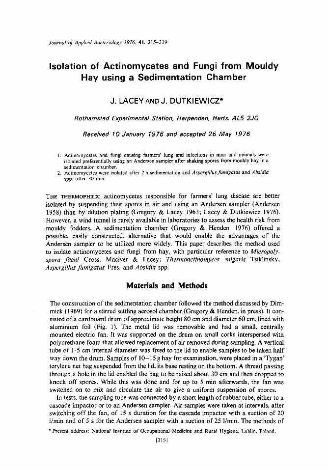
The construction of the sedimentation chamber followed the method discussed by Dim- mick (1 969) for a stirred settling aerosol chamber (Gregory & Henden, in press). It con- sisted of a cardboard drum of approximate height 80 cm and diameter 60 cm, lined with aluminium foil (Fig. 1). The metal lid was removable and had a small, centrally mounted electric fan. It was supported on the drum on small corks interspersed with polyurethane foam that allowed replacement of air removed during sampling. A vertical tube of 1.5 cm internal diameter was fixed to the lid to enable samples to be taken half way down the drum. Samples of 10-15 g hay for examination, were placed in a ‘Tygan’ terylene net bag suspended from the lid, its base resting on the bottom. A thread passing through a hole in the lid enabled the bag to be raised about 30 cm and then dropped to knock off spores. While this was done and for up to 5 min afterwards, the fan was switched on to mix and circulate the air to give a uniform suspension of spores.
In tests, the sampling tube was connected by a short length of rubber tube, either to a cascade impactor or to an Andersen sampler. Air samples were taken at intervals, after switching off the fan, of 15 s duration for the cascade impactor with a suction of 20 I/min and of 5 s for the Andersen sampler with a suction of 25 l/min. The methods of * Present address: National Institute of Occupational Medicine and Rural Hygiene, Lublin. Poland.
[3151
316 1. LACEY AND J. DUTKIEWICZ
C T bE
Fig. I . Diagram of the sedimentation chamber A, sample bag; B, fan; C, sampling tube; D. thread for raising sample bag; E, lid supported on corks and polyurethane foam.
operating, incubating, counting and classifying spores or colonies have been described previously (Gregory & Lacey 1963; Lacey 1971; Lacey & Dutkiewicz 1976). Malt ex- tract with penicillin and streptomycin was used to isolate fungi and half-strength nutrient or tryptone soya agars + 0.2% casein hydrolysate (Oxoid) with actidione to isolate actinomycetes (Lacey & Dutkiewicz 1976).
Results and Discussion
Release of spores from samples
The release of spores from hay samples was caused by dropping the sample bag against the bottom of the chamber from about 30 cm height different numbers of times. Samples were taken with the cascade impactor after the fan had been operating for 1 min. Spores were inevitably released as the hay was placed in the chamber, but subse- quently there was a linear relationship in the range tested between the number of times the sample was dropped and the spore concentration. The regression calculated from the means of two samples was significant (P < 0.05) giving the equation B = 35.8A + 259.9 where A was the number of drops and B the concentration of spores expressed as lo6 spores/m3 air. Four drops gave a satisfactory number of air-borne spores and was selected as standard.
Sedimentation rate
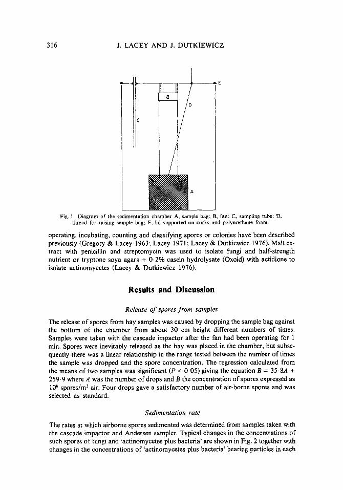
The rates at which airborne spores sedimented was determined from samples taken with the cascade impactor and Andersen sampler. Typical changes in the concentrations of such spores of fungi and ‘actinomycetes plus bacteria’ are shown in Fig. 2 together with changes in the concentrations of ‘actinomycetes plus bacteria’ bearing particles in each

ISOLATION OF ACTINOMYCETES AND FUNGI 317
Fig. 2. Sedimentation of spores of fungi and ‘actinomycetes plus bacteria’ in a sedimentation chamber, showing the decline of total numbers of each and of the four size grades represented by deposits on the four stages of a cascade impactor.
of the four size ranges separated in the cascade impactor (approximately > 6 pm, 2-20 pm, 1-7 pm and 3 pm). As expected from their larger size, fungal spores sedimented more rapidly than ‘actinomycetes plus bacteria’ and diminished to one tenth of their original concentration in 45 min. ‘Actinomycetes plus bacteria’ took 189 min to decline to one tenth. However, particles bearing ‘actinomycetes plus bacteria’ in the four grades differed greatly, reducing to one tenth of their original concentration in: 13.2, 62.2, 127.4 and 274.3 min, respectively, corresponding to sedimentation rates of 0.233, 0.049,0.024 and 0.01 1 cm/s or aerodynamic sizes equivalent to unit density spheres of 8.8, 4.0, 2.8 and 1.9 pm diameter (Dimmick 1969).
Andersen sampler counts were used to study the sedimentation rates of individual fungi, actinomycetes and bacteria. Results for the most common organisms calculated from total colony counts are shown in Table 1. A. fumigatus sedimented most slowly of the fungi, while bacteria sedimented faster than actinomycetes, probably because they were more often dispersed in clumps. The actinomycetes, despite the similarity in their spore sizes, behaved in a slightly different manner in the sedimentation chamber. M. faeni sedimented more rapidly than others, but numbers of T. vulgaris changed little during 3 h sedimentation. The differences in sedimentation rates were reflected in the distribution of colonies between the six stages of the Andersen sampler. Most M. faeni colonies occurred on stages three or four, (described by Andersen, 1958, as retaining particles 2-6 pm diameter) Saccharomonospora viridis (Schuurmans, Olson & San Clemente) Nonomura & Ohara, on stage five (1-2 pm) and T. vulgaris on stages five and six (<1 - 2 pm).

3 18 J. LACEY AND J. DUTKIEWICZ
TABLE 1
Time taken for spores of di@erent organisms to decline to one tenth of their original concentration in a sedimentation chamber
Time to decline Equivalent to I/lOth original Sedimentation aerodynamic
concentration rate diameter (rnin) (cm/s) (,urn)
~ _____ ~ _ _ _ _ ~~~
Aspergillus fumigatus 104.6 0.029 Periicillium spp. 99.6 0.03 I A bsidia spp. 84.1 0.037 .4spergil/ris glaircus group 64.5 0.048 Humicola lanugiriosa 3.19 0.096 Thermoaciinomjres idgaris 4545.5 0.001 Micropoli,sporafaetii 282.6 0.01 1 Saccharomonosporu viridis 299.9 0.010 Nocurdio sp. 234,5 0.013 Bacteria 146.9 0.02 1
Estimated from the total colony count on all stages of an Andersen sampler. sedimentation and equivalent aerodynamic size are shown for each organism.
~
3.11 3.21 3.51 4.00 5.66 0.58 1.91 1.83 2.08 2.65
The rate of
Since actinomycetes sediment more slowly than bacteria and A . fumigatus than other fungi. the proportion of their spores in suspension in the chamber may be increased by prolonging sedimentation. This decreases the likelihood of actinomycetes being overgrown by spreading bacteria, but to be practicable as a method of assessing mouldy hays, sedimentation times cannot be too long. Sedimentation times of 30 min for isolating A . fumigutus and 2 h for M . faeni and other thermophilic actinomycetes have been satisfactory although the latter could perhaps be shortened to 1 h, at the expense of growing more bacteria which could interfere with growth of M. faeni.
Assessment of health hazard
The health hazard presented by a given hay depends on the types of organisms present and their abundance. Gregory et al. (1963) classified hays on their total spore content. This cannot be done with the sedimentation chamber, but arbitrary criteria adopted for classifying colonization of hays by individual species when examined by the standard wind tunnel/Andersen sampler method may be adapted (Lacey & Dutkiewicz 1976). By these criteria, hays lightly colonized by a species yielded up to 25 colonies on the six Andersen sampler plates, those moderately colonized yielded 25-250 colonies and those heavily colonized more than 250 and sometimes more than 1000 colonies.
Direct comparison of wind tunnel and sedimentation chamber results are difficult because of sample variability, but the most important pathogenic fungi, A . fumigatus and Ahsidia spp., were isolated from the sedimentation chamber in similar numbers to the wind tunnel after 30 min settling then incubating plates at 40 OC and actinomycetes after 2 h settling with incubation at 5 5 OC. Similar criteria could, thus, be used for both methods. About 60% more actinomycetes were grown than by wind tunnel/Andersen sampler isolation i f settling was restricted to only 1 h and criteria for these conditions would have to be adjusted accordingly.

ISOLATION OF ACTINOMYCETES AND FUNGI 319
References
ANDERSEN, A. A. 1958 New sampler for the collection, sizing and enumeration of viable air- borne particles. Journal of Bacteriology 76, 47 1 4 3 4 .
DIMMICK, R. L. 1969 Stirred-settling aerosols and stirred-aerosol chambers. In An Introduction to Experimental Aerobiology ed. Dimmick, R. L. & Akers, A. B. New York: Wiley-Inter- science.
GREGORY, P. H. & HENDEN, D. R. 1976 Terminal velocity of basidiospores of the giant puffball (Lycoperdon giganteum). Transactions of the British Mycological Society 67 (in press).
GREGORY, P. H. & LACEY, M. E. 1963 Mycological examination of dust from mouldy hay associated with farmers’ lung disease. Journal of General Microbiology 30, 75-88.
GREGORY, P. H., LACEY, M. E., FESTENSTEIN, G. N. & SKINNER, F. A. 1963 Microbial and biochemical changes during the moulding of hay. Journal of General Microbiology 33,
LACEY, J. 1971 The microbiology of moist barley storage in unsealed silos. Annals of Applied
LACEY, J. & DUTKIEWICZ, J. 1976 Methods for examining the microflora of mouldy hay. Jour-
147-174.
Biology 69, 187-212.
nal of Applied Bacteriology 41, 13-27.