isolation ofkendallasmith.com/pdf/podack_cohn_1985.pdfinhibition by zncl2 was determined in the...
TRANSCRIPT
Proc. Nati. Acad. Sci. USAVol. 82, pp. 8629-8633, December 1985Immunology
Isolation and biochemical and functional characterization ofperforin 1 from cytolytic T-cell granules
(T lymphocytes/cell-mediated cytolysis/polymerization/transmembrane channels/patch-clamp)
ECKHARD R. PODACK*, JOHN DING-E YOUNGt, AND ZANVIL A. COHNt*Department of Microbiology and Immunology, New York Medical College, Valhalla, NY 10595; and tLaboratory of Cellular Physiology and Immunology,The Rockefeller University, New York, NY 10021
Contributed by Zanvil A. Cohn, August 14, 1985
ABSTRACT The Ca2 -dependent cytolytic activity of iso-lated T-lymphocyte granules was purified to apparent homo-geneity by high-salt extraction, gel filtration, and ion-exchangechromatography. The lytic activity resided in a 72- to 75-kDaprotein of cytolytic granules. Incubation of the isolated proteinwith erythrocytes in the presence of Ca2+ ions resulted inhemolysis and the formation of membrane lesions of 160 A indiameter, corresponding in size and morphology to membranelesions formed on target cells by cloned, intact natural killer(NK) and cytolytic T lymphocytes. Hence, the 75-kDa granuleprotein is identified as monomeric perforin 1 (P1), postulatedpreviously from the analysis of membrane lesions formedduring NK and T-cell-mediated cytolysis. Pl-mediatedhemolysis is Ca2+-dependent and is inhibited by Zn2' ions.Lysis is accompanied by the polymerization ofP1 to membrane-associated tubular complexes (poly-Pl) that form largetransmembrane pores. P1 causes a rapid membrane depolar-ization of J774 cells in the presence of Ca2+. Purified P1 alsoinduces transmembrane monovalent and divalent ion flowacross lipid vesicles only in the presence of Ca2 . Whole-cellpatch-clamp recordings of S49 lymphoma cells show a P1-dependent inward membrane current flow in the presence butnot in the absence of Ca2'. The current increase can bedissected as a summation of discrete current events, indicativeof formation of functional channels by polymerization of P1.
Target cell lysis, mediated by natural killer (NK) andcytotoxic T lymphocytes (CTL), is partially caused by theassembly of tubular transmembrane complexes on targetmembranes, designated polyperforin 1 (poly-P1) and 2 (poly-P2) (1-6). It has been postulated that the monomeric precur-sors of polyperforins are contained in the cytoplasmic gran-ules ofclonedNK and CTL (2, 3, 5). This view was supportedby the demonstration that isolated granules from cloned CTLand froni a NK-like tumor line mediated Ca2+-dependentcytolysis, assemble poly-P1 and poly-P2 on target mem-branes, and cause Ca2+-dependent conductance increaseacross planar bilayer membranes (4-8).
Isolated T-lymphocyte granules contain a number of pro-tein bands identifiable by NaDodSO4/polyacrylamide gelelectrophoresis (for review, see ref. 9). Here, we report onthe purification and functional characterization of the mono-meric granule protein responsible for the formation of the160-A-wide polyperforin complex.
MATERIALS AND METHODSCells. CTLL2 cells were grown in Iscove's modified
Dulbecco's minimum essential medium (DMEM) as de-scribed (5). Five milliliters of packed cells was usually
harvested from 6-liter cultures containing 10-15% rat spleencell concanavalin A supernatant (5).
Preparation of Granules. Granules were prepared from 5 mlofpacked CTLL2 cells that were washed in Ca2+-free Hanks'balanced salt solution by Percoll density-gradient centrifu-gation as described (5). After fractionation of the Percollgradients, granule-containing fractions, as identified by he-molytic assays (5), were pooled and used for the purificationof P1.
Purification of P1. All reactions were carried out at 4°Cunless stated otherwise. Granules isolated from Percollgradients and containing Percoll were mixed with an equalvolume of 1 M NaKHPO4 buffer (pH 7.4) containing 10 mMbenzamidine HCl, 1 mM EDTA, and 3 mM NaN3.- Afteraddition of 2 mM phenylmethylsulfonyl fluoride, the mixturewas allowed to stand on ice for 30 min. Addition of highphosphate concentrations to Percoll suspensions causesprecipitation of Percoll as a white precipitate, which can beremoved easily by centrifugation for 30 min at 20,000 rpm inthe Sorvall centrifuge. The clear supernatant was harvestedand the pellet was discarded. The supernatant was applieddirectly to a 2.5 x 90 cm column containing Sephacryl S-300(Pharmacia) equilibrated with 0.1 M NaKHPO4, pH 7.4/0.5M NaCl/1 mM benzamidine HCl/1 mM EDTA/3 mM NaN3.The sample was eluted at a flow rate of 25 ml/hr and 15-minfractions were collected. The fractions were assayed forprotein and hemolytic activities. Hemolytically active frac-tions were pooled and dialyzed against 10 mM NaKHPO4buffer, pH 7.4/1 mM EDTA/3 mM NaN3.The dialyzed pool was applied to the Mono Q column of the
fast protein liquid chromatography system by using the 50-mlsuperloop (Pharmacia). The superloop and the Mono Qcolumn were kept in ice. The flow rate was 1 ml/min and,after sample application, the chromatogram was developedwith an automatic gradient programer by using buffers A (10mM NaKHPO4, pH 7.4/1 mM EDTA/3 mM NaN3) and B(same as buffer A with 2 M NaCl) and the followingparameters: equilibration done with buffer A; after sampleapplication, column washing with buffer A for 5 min; lineargradient for 35 min to final 50% A:50% B, followed by bufferB for 5 min. The column effluent was monitored by a flow cellat 280 nm and collected in 1-ml fractions on ice. Thehemolytic activity of each fraction was determined.
Hemolytic Assay for P1. Sheep erythrocytes, washed andresuspended to 2-4 x 10⁹ per ml in 0.1-ml aliquots with 0.15M NaCl/10 mM Tris HCl, pH 7.2 (Tris/NaCl buffer), con-taining 5 mM CaC12, were incubated with 2- to 20-,ul columnfractions for 20 min at 37°C. Lysis was determined by dilutionof samples to 2 ml with Tris/NaCl, followed by centrifugationand spectrophotometric quantitation of hemoglobin release
Abbreviations: AP, membrane potential(s); P1, perforin 1; Ph4P',tetraphenylphosphonium+; NK, natural killer; CTL, cytoxic Tlymphocyte(s).
8629
The publication costs of this article were defrayed in part by page chargepayment. This article must therefore be hereby marked "advertisement"in accordance with 18 U.S.C. §1734 solely to indicate this fact.

Proc. Natl. Acad. Sci. USA 82 (1985)
in the supernatant at 541 nm. Z units of hemolysis are definedas in ref. 5.Metal Dependence of P1. Sheep erythrocytes were washed
in Tris/NaCi, to which CaCl2 was added to the desiredconcentration. P1 was then added to the cell suspension,followed by incubation as above. Inhibition by ZnCl2 wasdetermined in the presence of 0.5mM CaCl2 in a similarmanner.Membrane Potential (Al) Measurements. The At of J774
macrophages was assayed by the uptake of [3H]tetraphenyl-phosphonium ion ([3H]Ph4P+) as described (10-12), with thefollowing modifications. Cells were plated overnight oncoverslips (4 x 10⁴ per ml, 1 ml per coverslip) in culturemedium (DMEM/5% fetal bovine serum) prior to washingthree times with low-K+ buffer (118mM NaCl/5mM KCl/1.3mM CaCl2/0.8mM MgSO4/5.5mM glucose/20mM Hepes/9mM Na2CO3, pH 7.4) and incubation with 20,uM [3H]Ph4P+for 30 min at 37°C. P1 was then incubated with the cells at37°C for the indicated periods, at which time the medium wasaspirated and coverslips were washed rapidly by dipping fourtimes into separate beakers containing ice-cold phosphate-buffered saline (P1/NaCl) and 1 mg of bovine serum albuminper ml. Cells were then lysed in 0.5 ml of 0.05% Triton X-100in H20 and 0.25-ml aliquots were used for radioactivitydeterminations (12). The values of [3H]Ph4P+ accumulated inthe cells bathed in high-K+ buffer (as in refs. 10-12) wereused as background and were subtracted from values ob-tained with cells bathed in low-K+ buffer. Conversion ofcorrected radioactivity into intracellular probe concentra-tions and AT was done as described (10-12). Experimentswith P1 were done in duplicate.Formation of Lipid Vesicles and Ion-Flow Measurements. A
mixture of phosphatidylethanolamine/phosphatidylcholine/cholesterol at 3:2:1 (wt/wt), 25mM P3-D-octyl glucoside inHepes buffer (10 mM, pH 7.0, adjusted with Tris base)containing 0.05 M sodium isethionate, and 0.165 M sucrose(buffer C) was sonicated to clarity. Vesicles were formed bya one-step dilution with buffer C (1:100). Vesicles formed thisway were collected by centrifugation at 100,000 x g for 90min and resuspended to 10 mg/ml. The uptake of [3H]Ph4P+into lipid vesicles was quantitated by filtration (13). Briefly,the reaction was initiated by diluting 0.1 ml of lipid vesiclesinto 0.9 ml of buffer D (0.25 M sucrose/10mM Hepes, pH 7.0)containing 50,M [3H]Ph4P+ with P1(700 ng/ml) or controlcolumn buffer. At intervals, aliquots of the reaction mixturewere filtered and washed, and radioactivity remaining on thefilter was determined (13).
Patch-Clamp Recording. S49.1 T cells, maintained in cul-ture as described (14), were washed and resuspended intolow-K+ buffer, with or without Ca2 . Whole-cell recordingsfrom these cells were obtained essentially as described (15).The cell dish was placed on the stage of a Nomarski opticsmicroscope. Pipettes were fire-polished and used whenresistance values ranged between 2 and 6 Mfl. The pipettesolution consisted of high-K+ buffer, without MgSO4 andcontaining 1.3 mM EGTA. High-resistance seals (>20 GQ1)formed spontaneously on touching the cell surface with thepipette; whole-cell recording was obtained by application ofstrong negative pressure through the pipette. Currents werelow-pass-filtered at 2 kHz and recorded on floppy disks in a4094-2 Nicolet digital oscilloscope at sampling rates of 0.1-2ms per point and simultaneously registered on a Gould 2200S dual-pen recorder. Data were analyzed directly by using asoftware package available from Nicolet or transferred to aIBM PC for further analysis. Reagents were introduced intothe cell medium by means of another micropipette (tipdiameter, 4 um) fitted onto a syringe, at 100 um from cell.Other Assays. Protein determination (16), NaDod-
S04/polyacrylamide gel electrophoresis (17), and electronmicroscopy were done as described (2).
u,0)LA0
c
4-
CL
Lu
CA
Ea)I
50 100Fraction
FIG. 1. Gel filtration of phosphate extract of granules. o, Hemo-lytic activity; -, protein. The position of albumin marker is indicatedby an arrow.
RESULTS
Purification of P1. Solubilization of the cytolytic activityfrom isolated CTLL2 granules was effected by 0.5 M phos-phate at pH 7.4 in the presence of protease inhibitors. Thisprocedure was more effective than extractions with NaCl,KC1, or guanidine HCO (not shown). High phosphate, inaddition, caused precipitation of Percoll, allowing its removalby centrifugation at 20,000 rpm. The soluble supernatantcontaining the cytolytic activity was then fractionated by gelfiltration on Sephacryl S-300 (Fig. 1). Three major proteinpeaks eluted from the gel filtration column. Cytolytic activitywas found in the trailing end of the center peak, eluting withan apparent molecular mass of 70-80 kDa. The activefractions were combined and, after dialysis, subjected toion-exchange chromatography on Mono Q by using the fastprotein liquid chromatography system. Fig. 2 shows theelution profile of protein and hemolytic activity from thiscolumn. Activity elutes in a single symmetrical peak con-taining only trace amounts of protein. Analysis of the pooledmaterial by NaDodSO4/polyacrylamide slab gel electropho-resis is shown in the Fig. 2 Inset in comparison to markerproteins. The active fractions contain a single protein bandmigrating with an apparent molecular mass of 75 kDa underreducing conditions (Fig. 2 Inset) and 58 kDA undernonreducing conditions (not shown). The mobility of thisprotein on NaDodSO4 gels corresponds to the K1 band ofisolated granules (5).
(1)0ta,
C1 i! -ri|~~-OrI_IusF ~ ~~-1.!A;--~~ ~ ~ ~ ~ ~~-
i0 20Fraction
30 40
0.4
0.2
0~
co-Soon)
02JEL
FIG. 2. Anion-exchange chromatography of pool from gel filtra-tion. The fast liquid protein chromatography system and a Mono Qcolumn were used. o, Hemolytic activity; -, protein. The gradientranges from 0 to 2 M NaCl.
8630 Immunology: Podack et A
Proc. Natl. Acad. Sci. USA 82 (1985) 8631
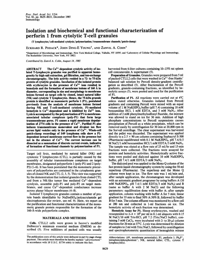
Table 1. Purification of P1Specific
Volume, Protein, activity, Purification,Fraction ml mg Z/mg fold
Granules 15 45 80 1Perforin 4 0.16 2187 27.3Z units are as defined in ref. 5.
Table 1 shows the comparison of the hemolytic activity ofgranules with that of the isolated protein. The specificactivity increased 27-fold during the purification procedure,with the recovery of 160 yg of apparently homogeneousprotein from 45 mg of isolated granules.
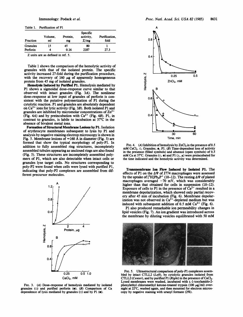
Hemolysis Induced by Purified P1. Hemolysis mediated byP1 shows a sigmoidal dose-response curve similar to thatobserved with intact granules (Fig. 3A). The nonlineardose-response at low input of granules of perforin is con-sistent with the putative polymerization of P1 during thecytolytic reaction. P1 and granules are absolutely dependenton Ca2" ions for lytic activity (Fig. 3B). Both isolated P1 andgranules are inhibited by micromolar concentrations of Zn2+(Fig. 4A) and by preincubation with Ca2+ (Fig. 4B). P1, incontrast to granules, is labile to incubation at 370C in theabsence of bivalent metal ions.Formation of Structural Membrane Lesions by P1. Isolation
of erythrocyte membranes subsequent to lysis by P1 andanalysis by negative staining electron microscopy is shown inFig. 5. Membrane lesions of -160 A in diameter (Fig. 5) areformed that show the typical morphology of poly-Pl. Inaddition to fully assembled ring structures, incompletelyassembled tubules appearing as unclosed rings are also found(Fig. 5). These structures are incompletely assembled poly-mers of P1, which are also detectable when intact cells orgranules lyse target cells. No structures corresponding topoly-P2 were found when cells were lysed with purified P1,indicating that poly-P2 complexes are assembled from dif-ferent precursor molecules.
A1.2 A
0.8 / 0
0.4 ]
N 2 4Protein, ug
E BI
0.8
0.47-
0.25 0.5 1.0CaCI2, mM
FIG. 3. (A) Dose-response of hemolysis mediated by isolatedgranules (o) and purified perforin (e). (B) Comparison of Cadependence of lysis mediated by granules (o) and by P1 (o).
N
at
E0Ir
A
0.5
ZnCI2, mM
30Time, min
FIG. 4. (A) Inhibition ofhemolysis by ZnCl2 in the presence of0.5mM CaCl2. o, Granules; e, P1. (B) Time-dependent loss of activityin the presence (filled symbols) and absence (open symbols) of 0.5mM Ca at 37°C. Granules (o, *) and P1 (A, A) were preincubated forthe time indicated and the hemolytic activity was determined.
Transmembrane Ion Flow Induced by Isolated P1. Theeffects of P1 on the A, of J774 macrophages were assessedby the uptake of [3H]Ph4P+ (10-12). The resting AT ofplatedmacrophages averaged -70 mV, which was considerablyhigher than that obtained for cells in suspension (10-12).Exposure of cells to P1 in the presence of Ca2" resulted in amembrane depolarization, which showed only partial recov-ery after 45 min of incubation (Fig. 6). Membrane depolar-ization was not observed in Ca2"-depleted medium but wasinduced with subsequent addition of 0.5 mM Ca2" (Fig. 6).P1 also produced remarkable ion permeability changes in
lipid vesicles (Fig. 7). An ion gradient was introduced acrossthe membrane by diluting vesicles equilibrated with 50 mM
FiG. 5. Ultrastructural comparison ofpoly-Pl complexes assem-bled by intact CTLL2 (Left), by cytolytic granules isolated fromCTLL2 (Center), and by purified P1 (Right) in the presence of CaCl2.Lysed membranes were washed, incubated with L-1-tosylamido-2-phenylethyl chloromethyl ketone-treated trypsin (100 g/mIl) over-night at 22°C, washed again, and then mounted for electron micros-copy by negative staining with uranyl formate (2%).
Immunology: Podack et al.

Proc. Natl. Acad. Sci. USA 82 (1985)
-100I-80k
E-60
-40
-20k
\I
L-A
-_ _ L,
0 A\
~~~~~~~~~\"
0~~~~~0\
10 20 30 40Time, min
FIG. 6. Effect of P1 on J774 macrophage At. J774 macrophagesin low-K+ buffer, loaded with [3H]Ph4P', were treated with 2 jug ofP1 per ml at time 0 (o) or control Tris/NaCl buffer (0). P1 inTris/NaCl was diluted 1:100 into cell medium. Cells washed andincubated in Ca2+-free low-K+ buffer (A) were treated with 2 ug ofP1 per ml at time 0, followed by addition of CaCl2 to a finalconcentration of 0.5 mM (arrow). Experiments carried out induplicate at 370C.
sodium isothionate into equiosmotic solutions of sucrosecontaining [3H]Ph4P+. Since Na' ions permeate more rapidlythrough the poly-P1 channel than the larger anions (isothio-nate), a transient At is produced by unequal accumulation ofnegative charges inside the vesicles in the presence ofpolymerizing P1 (Fig. 7). However, the vesicle At wasdissipated after a few minutes (Fig. 7), indicating thatP1-treated vesicles were also made leaky to isothionate,albeit at a slower rate. Experiments with other electrolytesshowed that in the presence of P1, lipid membranes becamepermeable to Ca2+, Mg2+, EGTA2-, and Tris- (not shown).Pi-treated-vesicles also became permeable to Lucifer yellow(molecular weight, 457) and sucrose (molecular weight, 342)(not shown). In all instances, the effect of P1 on trans-membrane ion flow was absolutely Ca2+-dependent (Fig. 7).Patch-Clamp Recording. Whole-cell recording of S49-1
cells revealed resting At of -55 ± 6 mV (31 cells), whichwere measured as the current-free steady-state potentials.Resting At was largely dependent on the day ofculture. Onlycells at a density of 2 x 10⁴ in culture were used fordetermination of resting At. Very low leakage currents wereobserved at voltages around the resting potentials; the inputresistance was often >10 Gfl. Following application of P1(0.5 ug/ml), current increased in the inward direction, whichoccurred in a progressive manner, without any decay, re-sulting frequently in loss of the seal (Fig. 8A). The current
30 .E
E 20a.
. 10 jji-C)
3 9 15Time, min
FIG. 7. Transmembrane ion flow in lipid vesicles induced by P1.Vesicles equilibrated with 50mM sodium isothionate were diluted attime 0 into sucrose solution containing [3H]Ph4P' or P1 (1 j.g/ml),with (e) or without (A) 1 mM CaCl2. o, Control vesicles diluted intosucrose solution, with [3H]Ph4P' and 1 mM CaCl2. Data pointsrepresent the average of triplicate experiments performed at 370C.
increase could be dissociated as a summation of discretecurrent steps of 20-80 pA per unit (Fig. 8B), indicative ofincorporation of single P1 channels into the cell membrane.The current effect produced by P1 was dependent on thepresence of Ca2W (Fig. 8C). Single-channel recordings andkinetic analysis of channel openings were similarly obtainedwith membrane patches; results of these experiments will bedescribed elsewhere. Single-channel fluctuations were rarelyseen at resting potential levels (-55 mV); they became morefrequent only at transmembrane potentials of >100 mV (notshown).
DISCUSSIONThis communication describes the isolation and character-ization of a cytolytic protein from a murine, cytolytic T-lymphocyte clone. According to its putative function, thisprotein was named P1 (3) because it was thought to perforatemembranes during polymerization on target cell surfaces.Evidence presented in Figs. 4 and 5 demonstrates that P1, infact, produces structural and functional lesions consistentwith this hypothesis. P1 is a 72- to 75-kDa protein thatundergoes rapid Ca2+-dependent polymerization and forms atubular complex that functions as a large and voltage-independent transmembrane channel when inserted intomembranes.The activity of isolated P1 resembles closely the functional
activity of cytolytic granules (see Figs. 3 and 4), including itsability to lyse a variety of tumor cells (not shown). Aninteresting difference is the lability of P1 at 370C in the
FIG. 8. Whole-cell currents recorded from S49.1 cells. Cells were clamped at -60 mV during P1 application. P1 (to 0.1 ug/ml) was addedfrom a second micropipette at a distance of about 100 Am from the cell (arrowheads point to beginning of perfusion). (A) Cells bathed in low-K+buffer. (Scales per box: vertical, 200 pA; horizontal, 5 s.) (B) Horizontal expansion of A, 4x. (C) Cell bathed in Ca2+-free low-K+ buffer.Arrowhead points to addition of P1, as in A. Ca2+ was added from a third pipette to a final concentration of 0.5 mM (arrow). Same scale asin A. Downward deflections represent inward currents.
8632 Immunology: Podack et al.

Proc. Natl. Acad. Sci. USA 82 (1985) 8633
absence of metal ions and the higher sensitivity of granules toCa2+ and Zn2+. Possibly other granule proteins stabilize P1and enhance the effect of metal ions inducing P1 (polymer-ization.
Isolated P1 increases the ion permeability of bilayer mem-branes (Figs. 6-8). The increase in transmembrane ioniccurrent represents the functional counterpart of structuralmembrane ring-like lesions by P1. As measured by whole-cellpatch-clamp recordings, P1 induces voltage-insensitive ionchannels on target membranes (Fig. 8). The functionalactivity of P1 on membranes depends absolutely on thepresence of Ca2', an observation which provides furthersupport for the Ca2'-dependent polymerization of P1 totransmembrane ring-like tubular structures.
It is worthwhile to compare the structure and function ofmurine P1 to the human ninth component ofcomplement (C9)(for review, see ref. 18). Both proteins polymerize to tubularcomplexes that cause membranolysis upon membrane inser-tion (19-21). C9 and P1 are inactivated by Zn2+ ions atmicromolar concentrations, a reaction that is accompaniedby rapid polymerization of C9 (ref. 22; unpublished data).Whereas C9 polymerization under physiological conditionsrequires the assembly of the C5b-8 complex as C9 receptor(23, 24), P1 polymerization is not dependent on additionalproteins and requires only low concentrations of Ca2+ (Figs.3 and 5). This difference is important because C5b-8 directsC9 polymerization to the target cell, thus preventing indis-criminate C9 polymerization. In the case of P1, directionalityis achieved by compartmentalization of the cytolytic T celland segregation of P1 in the granules, which, after specifictarget conjugation, are released onto the target membrane.Despite these differences, C9 and P1 show remarkablebiochemical, structural, and functional homology, suggestingthat they have arisen from an ancestral cytolytic protein andbecame specialized in humoral or cell-mediated cytolyticfunctions.
Note Added in Proof. While this work was in press, Masson andTschopp (25) reported on purification of a similar pore-formingprotein/P1 from cytolytic T-cell granules.
The excellent technical assistance of K. Penichet, R. Manganiello,S. S. Ko, and M. A. DiNome is heartily acknowledged. Discussionswith A. Steinacker and A. Mauro on the patch-clamp system andC. F. Nathan were also most helpful. This work was supported byPublic Health Service Grants CA39201 and A121999 and AmericanCancer Society Grant IM396 to E.R.P., a fellowship from The JaneCoffin Childs Memorial Fund for Medical Research and a Cancer
Research Institute grant to J.D.-EY., and Grants CA30198 andAI070127 from the National Institutes of Health to Z.A.C.
1. Dourmashkin, R. R., Deteix, P., Simone, C. B. & Henkart,P. A. (1980) Clin. Exp. Immunol. 42, 554-560.
2. Podack, E. R. & Dennert, G. (1983) Nature (London) 302,442-445.
3. Dennert, G. & Podack, E. R. (1983) J. Exp. Med. 157,1483-1495.
4. Millard, P. J., Henkart, M. P., Reynolds, C. W. & Henkart,P. A. (1984) J. Immunol. 132, 3197-3204.
5. Podack, E. R. & Konigsberg, P. J. (1984) J. Exp. Med. 160,695-710.
6. Young, J. D.-E, Nathan, C. F., Podack, E. R., Palladino,M. A. & Cohn, Z. A. (1985) Proc. Natl. Acad. Sci. USA 82, inpress.
7. Blumenthal, R., Millard, P. J., Henkart, M. P., Reynolds,C. W. & Henkart, P. A. (1984) Proc. Natl. Acad. Sci. USA 81,5551-5555.
8. Henkart, P. A., Millard, P. J., Reynolds, C. W. & Henkart,M. P. (1984) J. Exp. Med. 160, 75-93.
9. Podack, E. R. (1985) Immunol. Today 6, 21-27.10. Young, J. D.-E, Young, T. M., Lu, L. P., Unkeless, J. C. &
Cohn, Z. A. (1982) J. Exp. Med. 156, 1677-1690.11. Young, J. D.-E, Unkeless, J. C., Kaback, H. R. & Cohn,
Z. A. (1983) Proc. Natl. Acad. Sci. USA 80, 1636-1640.12. Sung, S.-S. J., Young, J. D.-E, Origlio, A. M., Heiple, J. M.,
Kaback, H. R. & Silverstein, S. C. (1985) J. Biol. Chem., inpress.
13. Young, J. D.-E, Black, M., Mauro, A. & Cohn, Z. A. (1983)Proc. Natl. Acad. Sci. USA 80, 3831-3835.
14. Mellman, I. S. & Unkeless, J. C. (1980) J. Exp. Med. 152,1048-1069.
15. Hamill, 0. P., Marty, A., Neher, E., Sakmann, B. &Sigworth, F. J. (1981) Pflugers Arch. 391, 85-100.
16. Bradford, M. (1976) Anal. Biochem. 72, 248-254.17. Laemmli, U. K. (1970) Nature (London) 227, 680-685.18. Podack, E. R. & Tschopp, J. (1984) Mol. Immunol. 21,
589-603.19. Podack, E. R. & Tschopp, J. (1982) Proc. Natl. Acad. Sci.
USA 79, 574-578.20. Tschopp, J., Muller-Eberhard, H. J. & Podack, E. R. (1982)
Nature (London) 298, 534-538.21. Young, J. D.-E, Cohn, Z. A. & Podack, E. R. (1985) J. Cell.
Biochem., in press.22. Tschopp, J. (1984) J. Biol. Chem. 259, 10569-10573.23. Podack, E. R., Tschopp, J. & Muller-Eberhard, H. J. (1982) J.
Exp. Med. 156, 268-282.24. Tschopp, J., Podack, E. R. & Muller-Eberhard, H. J. (1985) J.
Immunol. 134, 495-505.25. Masson, D. & Tschopp, J. (1985) J. Biol. Chem. 260,
9069-9072.
Immunology: Podack et al.