isolation and characterization of a novel thraustochytrid
TRANSCRIPT

Instructions for use
Title Isolation and Characterization of a Novel Thraustochytrid-like Microorganism that Efficiently ProducesDocosahexaenoic Acid
Author(s) Perveen, Zakia; Ando, Hitomi; Ueno, Akio; Ito, Yukiya; Yamamoto, Yusuke; Yamada, Yohko; Takagi, Tomoko;Kaneko, Takako; Kogame, Kazuhiro; Okuyama, Hidetoshi
Citation Biotechnology Letters, 28(3), 197-202https://doi.org/10.1007/s10529-005-5335-4
Issue Date 2006-02
Doc URL http://hdl.handle.net/2115/5782
Rights The original publication is available at www.springerlink.com
Type article (author version)
File Information BG28-3.pdf
Hokkaido University Collection of Scholarly and Academic Papers : HUSCAP

Isolation and characterization of a novel thraustochytrid–like microorganism that
efficiently produces docosahexaenoic acid
Zakia Perveen1, Hitomi Ando1, Akio Ueno1, Yukiya Ito2, Yusuke Yamamoto2, Yohko
Yamada3, Tomoko Takagi4, Takako Kaneko3, Kazuhiro Kogame5 & Hidetoshi
Okuyama1
1Laboratory of Environmental Molecular Biology, Graduate School of Environmental
Earth Science, Hokkaido University, Kita-ku, Sapporo 060-0810, Japan
2ROM Co. Ltd., Chuo-ku, Sapporo 064-0804, Japan
3Department of Chemical and Biological Sciences, Faculty of Science, Japan Women's
University, Bunkyo-ku, Tokyo 112-8681, Japan
4Division of Biology, Department of Life Sciences, Graduate School of Arts and
Sciences, The University of Tokyo, Meguro-ku, Tokyo 153-8902, Japan
5Division of Biological Sciences, Graduate School of Science, Hokkaido University,
Kita-ku, Sapporo 060-0810, Japan
*Author for correspondence: (Fax: +81-11-706-2347, E-mail: [email protected])
1
Key words: docosahexaenoic acid (DHA), Labyrinthulomycota, polyunsaturated fatty
acid (PUFA),
Abstract
A thraustochytrid−like microorganism (strain 12B) was isolated from the mangrove area
of Okinawa, Japan. On basis of its ectoplasmic net structure and biflagellate zoospores
we determined strain 12B as a novel member of the phylum Labyrinthulomycota in the
kingdom Protoctista. When grown on glucose/seawater at 28 ºC, it had a lipid content of
57.8% with DHA of 43.1% of the total fatty acids. It had a growth rate of 0.38 h¯1. The
DHA production rate of 2.8 ± 0.7 g l−1day−1 is the highest value reported for any
microorganism.
Introduction
2

Interest in n-3 polyunsaturated fatty acids (n-3 PUFAs) began some 30 years ago and it is
now known that n-3 PUFAs are important for human nutrition. n-3 PUFAs such as
docosahexaenoic acid (DHA; 22:6n-3) are the building structures of membrane
phospholipids of nervous, visual, and reproductive tissues cells and have physiologically
important functions in humans (reviewed by Kroes et al. 2003, Gill & Valivety 1997) and
animals (Bezard et al. 1994). Marine fish-oil is the major source of DHA and is often used in
pharmaceuticals and/or food supplements. Oils that are extracted from cultured phototrophic
microalgae have also been marketed in recent years (Lewis et al. 1999, Sijtsma & Swaaf
2004). However, fish and micro-algal oils have complex PUFA profiles which make the cost
of preparation of highly pure individual PUFA oils very high. Oils produced by fermentation
of thraustochytrids and Crypthecodinium cohnii as a dinoflagellate which are another source
of PUFAs, preferably contain one specific PUFA especially DHA (Sijtsma & Swaaf 2004)
and are now commercially available as poultry feed and as a feed for aquaculture. Moreover,
the use of DHA-containing oils in adult nutritional supplement will start in near future
(Ratledge 2004) and those from C. cohnii are used extensively in infant formulas (Kyle et al.
1995).
Thraustochytrids are a marine microheterotroph and are commonly isolated from
3
various samples such as seawater, estuarine water, sediments and fallen leaves of mangrove
forests (Lewis et al. 1999). There is large variation in biomass, lipid, and DHA yields of
thraustochytrid strains, and the yields depend on the strains used and their cultivation
conditions (Singh & Ward 1996, Vazhappilly & Chen 1998, Aki et al. 2003, Sijtsma &
Swaaf 2004). Schizochytrium limacinum SR21 produced 4.2 g DHA l−1 in 5 days in
shake-flask cultures (Yokochi et al. 1998). The commercially used C. cohnii produced 11.7 g
DHA l−1 after 9.2 days in fed-batch culture (Swaaf et al. 2003). To our knowledge S.
limacinum SR21 is in general the best DHA-producing strain. It is therefore unfortunate,
however, that S. limacinum SR21 has never been commercially utilized as a source of
DHA-containing oils.
The main focus of the present study is to isolate microorganisms of which the DHA
productivity is superior to that of S. limacinum SR21. Such microorganisms are expected to
be of interest as a source organism for the commercial production of DHA.
Materials and methods
Microorganisms and cultivation
4
To isolate DHA-producing microorganisms, fallen leaves from the mangrove area in
Okinawa Prefecture in Japan were collected. These leaves were directly placed on agar
plates comprising of 0.5% (w/v) glucose, 0.1% (w/v) yeast extract, 0.1% (w/v) peptone and
50% (v/v) seawater, 1% (w/v) agar, and streptomycin and penicillin G (0.3 g l−1 each)
(designated as By+ medium) and incubated at 28 °C for several days. Colonies were taken
from plate and re-grown in 50 ml flasks with By+ medium. After cultivation for 2–3 days at
28 °C a small portion of the liquid cultures was spread on agar plates of By+ medium and
cultivated for several days. Pure isolates were obtained by the streak plate technique. The
liquid cultures were then centrifuged at 14,000 × g for 20 min and cells were directly
transmethylation (see below) after washing twice with 1% (v/v) NaCl and once with distilled
water. Natural seawater was utilized after filtration throughout this work.
Optimization of the cultivation of strain 12B
For optimization strain 12B was cultured in basal F medium consisting of 5% (w/v) glucose,
1% (w/v) yeast extract, 1% (w/v) peptone and 50% (v/v) seawater. The pH of the basal F
medium, which was originally around 6, was not adjusted unless otherwise stated. Strain
5

12B was pre-cultured at 28 °C with shaking at 180 rpm for 1 day and then approx. 0.1 ml of
the pre-culture was transferred to 10 ml fresh medium in a 50 ml flask to give an OD value
of 0.1 at 600 nm. Thus, the inoculum size was 1% (v/v) or less. Cultivation was performed
for 2–3 days under the same conditions. For optimizing biomass, total fatty acid (TFA), and
DHA yields glucose and seawater concentrations in medium F were changed but the pH was
not adjusted. When necessary, pH of medium F was adjusted with NaOH or HCl. The
biomass (cells) from each culture was harvested and washed twice with 1% (v/v) NaCl, and
finally washed with distilled water. The weight of pellet was measured after each pellet was
lyophilized for more than 1 day. The growth of isolates in liquid medium was measured at
OD600.
Lipid extraction and fatty acid analysis
Total lipids were extracted from lyophilized cells by the method of Bligh & Dyer (1959) and
weighed. Ten mg of total lipid and dry cells were directly transmethylated with aqueous 2 M
hydrochloric acid in methanol at 100 ºC for 1 h (Gaver & Sweeley 1965, Takakuwa et al.
2002). As an internal standard, heneicosanoic acid (21:0) was added to the reaction mixture.
Resultant fatty acid methyl esters were extracted with n-hexane and then analyzed by gas
6

chromatography and gas chromatography/mass spectrometry as described previously
(Orikasa et al. 2004). The yield of lipid was expressed as the amount of TFA or total lipid
per liter of culture.
Morphological observation of strain 12B
The strain was morphologically studied by light microscopy and transmission electron
microscopy.
Results and discussion
Isolation of DHA−producing microorganisms
Only one DHA−producing microorganism out of 26 was obtained from leaf samples. It was
a thraustochytrid–like microorganism (see below). Since this strain, named as 12B, had a
high content of DHA up to 40% of the total fatty acids, it was utilized for further
characterization.
7

Microscopic observations of strain 12B
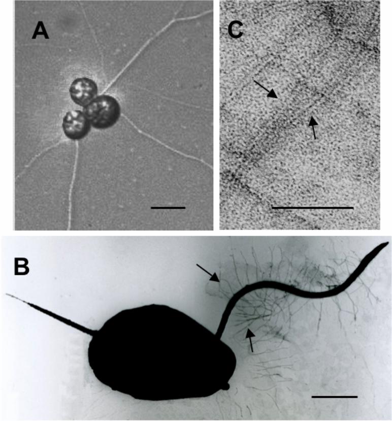
Figure 1A shows a cluster of vegetative cells of strain 12B. The cells formed ectoplasmic net
elements. The heterokont biflagellate zoospore of strain 12B was clearly observed and the
anterior long flagellum possessed tubular hairs (Figures 1B and C). From these results strain
12B should be classified into the class Labyrinthulea of the phylum Labyrinthulomycota in
the kingdom Protoctista (Porter 1989).
Effects of growth temperature on DHA yield of strain 12B
Strain 12B had the ability to grow at range temperatures varying between 15.0 ºC–32.9 ºC.
The yield of biomass (g l-1) slightly increased with increasing temperature (Figure 2A). In
contrast, however, the amount of TFA (g l-1) decreased with increased temperature. The
profile of fatty acids scarcely changed in a range between 25 °C–30 °C. The highest yield of
3.7 g DHA l-1 was obtained at 28 °C.
8
Effects of glucose concentration on DHA yield of strain 12B
When cultivated at 28 °C for 2 and 3 days strain 12B maximally produced 4.6 and 6.8 g
DHA l−1, respectively, with 8% (v/w) glucose (Table 1). The content of DHA varied between
40–48%, depending on the concentration of glucose and the duration of cultivation.
Effects of seawater concentration on DHA yield of strain 12B
The biomass yield at 25% (v/v) and 50% (v/v) seawater was approx. 24 g l-1 and it was
significantly decreased to approx. 8 g l-1 at 0% seawater (Figure 2B). The proportion of
DHA was 41–42% at 25–100% (v/v) seawater and it was decreased to 35% in medium
containing no seawater. The maximal yield of 3.2 g DHA l−1 was obtained at 50% seawater.
Effects of pH on DHA yield of strain 12B
Strain 12B was cultivated at 28 °C for 2 days in media containing 8% (w/v) glucose, 1%
(w/v) yeast extract, 1% (w/v) peptone, and 50% (v/v) of seawater of which the pH was
adjusted to a range between 2–9. The strain could completely not grow at pH 2 and 9. The
9
levels of biomass increased with increasing pH and maximized at pH 8. The fatty acid
composition of dry cell from the culture (pH 8) is given in Table 2. The maximal yield of 5.6
g DHA l−1 was obtained (Figure 2C).
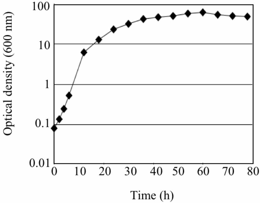
Growth rate and lipid content of strain 12B under optimal conditions
When grown under the optimal conditions in F medium strain 12B reached the stationary
phase within 2 days (Figure 3). The maximum growth rate of strain 12B was 0.38 h−1 (13.2
doublings day-1). This growth rate is notably much higher than those (less than 10 doublings
day-1) of some ATTC Schizochytrium and Thraustochytrium strains and DHA-producing
microorganisms screened and isolated by Barclay (1992).
The content of total lipid was 12.4 ± 0.4 g l-1 and the total lipid was 57.8 ± 5.5%
(w/w) of the biomass after 2 days. The proportion of DHA in total lipid was 43.1 ± 0.3%.
When the DHA yield of 5.6 g l-1 after 2 days was reached, the DHA production rate a day of
strain 12B under optimal conditions was thus calculated as 2.8 g l−1 day-1. This value is to
our knowledge the highest ever reported for flask-cultured DHA-producing microorganisms
(Sijtsma & Swaaf 2004).
Some microorganisms grow less efficiently in flasks than in bioreactors (e.g.
10

fermenters) where aeration and agitation speed triggers the growth of cells (see Yaguchi et.
al. 1997 and Yokochi et al. 1998). One of the most notable characteristics of strain 12B is
that it has a very high growth rate. Optimizing cultivation conditions for strain 12B using
bioreactors would probably result in even higher yields of biomass and DHA per time.
Another characteristic of this strain is that its fast growth-rate is made possible by a
relatively small inoculum, which would be of great benefit for industrial exploitation of this
strain.
Acknowledgement
We gratefully acknowledge the participation of members of the laboratory of electron
microscopy, Japan Women's University.
References
Aki T, Hachida K, Yoshinaga K, Katai Y, Yamasaki T, Kawamoto S, kakizono T, Yamaoka T,
11
Shigeta S, Suzuki O, Ono K (2003) Thraustochytrid as a potential source of carotenoids. J.
Am. Oil. Chem. Soc. 80: 789−794.
Barclay WR (1992) Process for the heterotrophic production of microbial products with
high concentrations of omega-3 highly unsaturated fatty acids. United States Patent
5,130,242.
Bezard J, Blond JP, Bernard A, Clouet P (1994) The metabolism and availability of essential
fatty acids in animal and human tissues. Reprod. Nutr. Dev. 34: 539−568.
Bligh EG, Dyer WJ (1959) A rapid method of total lipid extraction and purification. Can. J.
Biochem. Physiol. 37: 911−917.
Gaver RC, Sweeley CC (1965) Methods for methanolysis of sphingolipids and direct
determination of long-chain bases by gas chromatography. J. Am. Oil. Chem. Soc. 42:
294−298.
Gill I, Valivety R (1997) Polyunsaturated fatty acids, part 1: Occurrence, biological activities
and applications. Trends Biotechnol. 15: 401−409.
Haschemeyer RH, Meysers RJ (1972) Negative staining. In: Hayat MA, ed. Principles and
Techniques of Electron Microscopy. Vol. 2, New York: Van Nostrand Reinhold Co.,
114−117.
Kroes R, Schaefer EJ, Squire RA, Williams GM (2003) A review of the safety of
12
DHA45−oil. Food Chem. Toxicol. 41: 1433−1446.
Kyle DJ, Reeb SE, Sicotte VJ (1995) Infant formula and baby food containing
docosahexaenoic acid obtained from dinoflagellates. United States Patent 5,397,591.
Lewis TE, Nichols PD, McMeekin TA (1999) The biotechnological potential of
thraustochytrids. Mar. Biotechnol. 1: 580−587.
Orikasa Y, Yamada A, Yu R, Ito Y, Nishida T, Yumoto I, Watanabe K, Okuyama H
(2004) Characterization of the eicosapentaenoic acid biosynthesis gene cluster from
Shewanella sp. strain SCRC-2738. Cell Mol. Biol. 50: 625–630.
Porter, D (1989) Phylum Labyrinthulomycota. In: Margulis L, eds. Handbook of Protoctists.
Boston: Jones & Bartlett Publishers, pp. 388−398.
Ratledge C (2004) Fatty acid biosynthesis in microorganisms being used for single cell oil
production. Biochimie 86: 807−815.
Sijtsma L, Swaaf ME de (2004) Biotechnological production and applications of the ω−3
polyunsaturated fatty acid docosahexaenoic acid. Appl. Microbiol. Biotechnol. 64:
146−153.
Singh A, Ward OP (1996) Production of high yields of docosahexaenoic acid by
Thraustochytrium roseum ATCC 28210. J. Indust. Microbiol. 16: 370-373.
Swaaf ME de, Rijk TC de, Meer P van der, Eggink G, Sijtsma L (2003) Analysis of
13
docosahexaenoic acid biosynthesis in Crypthecodinium cohnii by 13C labeling and
desaturase inhibitor experiments. J. Biotechnol. 103: 21-29.
Takakuwa N, Kinoshita M, Oda Y, Ohnishi M (2002) Existence of cerebroside in
Saccharomyces kluyveri and its related species. FEMS Yeast Res. 2: 533-538.
Yaguchi T, Tanaka S, Yokochi T, Nakahara T, Higashihara T (1997) Production of high
yields of docosahexaenoic acid by Schizochytrium limacinum strain SR21. J. Am. Oil.
Chem. Soc. 74: 1431−1434.
Yokochi T, Honda D, Higashihara T, Nakahara T (1998) Optimization of
docosahexaenoic acid production by Schizochytrium limacinum SR21. Appl.
Microbiol. Biotechnol. 49: 72−76.
Vazhappilly R, Chen F (1998) Heterotrophic production potential of omega-3
polyunsaturated fatty acids by microalgae and algae-like microorganisms. Botanica
Marina 41: 553-558.
14

Table 1. Effects of glucose concentration on DHA yield of strain 12B.
15

Table 2. Fatty acid composition of strain 12B grown under optimal conditions.
16
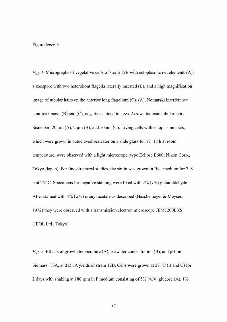
Figure legends
Fig. 1. Micrographs of vegetative cells of strain 12B with ectoplasmic net elements (A),
a zoospore with two heterokont flagella laterally inserted (B), and a high magnification
image of tubular hairs on the anterior long flagellum (C). (A), Nomarski interference
contrast image. (B) and (C), negative stained images. Arrows indicate tubular hairs.
Scale bar; 20 μm (A), 2 μm (B), and 50 nm (C). Living cells with ectoplasmic nets,
which were grown in autoclaved seawater on a slide glass for 17−18 h at room
temperature, were observed with a light microscope (type Eclipse E600; Nikon Corp.,
Tokyo, Japan). For fine-structural studies, the strain was grown in By+ medium for 7–8
h at 25 ˚C. Specimens for negative staining were fixed with 2% (v/v) glutaraldehyde.
After stained with 4% (w/v) uranyl acetate as described (Haschemeyer & Meysers
1972) they were observed with a transmission electron microscope JEM1200EXS
(JEOL Ltd., Tokyo).
Fig. 2. Effects of growth temperature (A), seawater concentration (B), and pH on
biomass, TFA, and DHA yields of strain 12B. Cells were grown at 28 °C (B and C) for
2 days with shaking at 180 rpm in F medium consisting of 5% (w/v) glucose (A), 1%
17

(w/v) yeast extract, 1% (w/v) peptone, and 50% (v/v) seawater (A and C). In (A) and
(B) pH was not adjusted. In (B) and (C) glucose concentration was 8%. Hatched and
dotted bars represent dry biomass and TFA, respectively. Solid line shows DHA yield.
Values represent the averages of triplicate determinations.
Fig. 3. Growth profile of strain 12B under optimal conditions.
18

Table 1. Effects of glucose concentration on DHA yield of strain 12B.
Time
(day)
Glucose
(% [w/v])
Dry biomass
(g l-1)
TFAa
(g l-1)
DHA content
of TFA (%)
DHA yield
(g l-1)
5 21.2±2.0b 9.8±0.3 40.1±1.1 3.9±0.2
8 22.5±0.6 11.4±0.9 40.1±1.2 4.6±0.3
10 20.5±0.2 8.3±0.7 44.6±1.8 3.7±0.2
12 17.4±3.1 7.4±2.6 44.3±2.4 3.2±1.0
2
15 15.4±1.1 5.0±0.9 42.9±1.0 2.1±0.4
5 20.1±0.5 7.6±0.2 43.4±1.7 3.3±0.1
8 30.9±0.5 16.0±1.9 42.4±3.8 6.8±0.2
10 27.5±1.5 13.7±0.5 42.4±2.9 5.8±0.6
12 27.6±1.0 13.6±0.7 43.9±1.3 6.0±0.4
3
15 24.3±0.4 12.3±1.2 47.5±0.4 5.9±0.6
aTotal fatty acids
bValues represent the averages of triplicate determinations.
15

Table 2. Fatty acid composition of strain 12B grown under optimal conditions.
Fatty acid, % (w/w)
14:0a 15:0 16:0 17:0 18:0 20:0 20:4 20:5n-3 22:5n-6 22:6n-3 Othersb
2.5±0.2c 5.2±1.8 34.5±1.4 1.5±0.3 1.3±0.3 1.0±0.5 0.6±0.4 0.9±0.2 8.7±0.2 40.1±0.1 3.4±1.9
aFor fatty acid, the numbers before and after colon indicate those of carbon atom and double bond, respectively.
bOthers include 12:0, 13:0, 18:1, 18:2, 18:3, and unknowns.
cValues represent the averages of triplicate determinations.
16