is it really that difficult? hopefully, our lab exercise ... · bacterial photosynthesis - lh2 and...
TRANSCRIPT

Membrane proteins – extraction, purification and crystallization.
Andy Freer - Glasgow University, Scotland
Is it really that difficult?
Bacterial photosynthesis - LH2 and core complex.
Hopefully, our lab exercise will give you a little confidence.

Inspiration

"The message is that there are known knowns - there are thingsthat we know that we know.
There are known unknowns - that is to say, there are thingsthat we now know we don't know.
But there are also unknown unknowns - there are things wedo not know we don't know.
And each year we discover a few more of those unknown unknowns."

Bacterial photosynthesis – probably first reactions taking place on earth
These organisms still found today in polluted streams and pools
Although still complicated, they are a little easier to work with than green plants.
It’s now 20 years since Michel and Deisenhofer solved the first membrane protein structure – a bacterial photosynthetic reaction centre.
11 years ago we solved the other part of the photosynthetic apparatus –the light harvesting complex or LH2 and only 3 years ago we solved the largestpart of the photo-jigsaw with the structure of the core (LH1-RC) complex.
This is the start of our membrane story – which, for me, has evolved into other areas.After all, more than 30% of the genome is making membrane proteins!

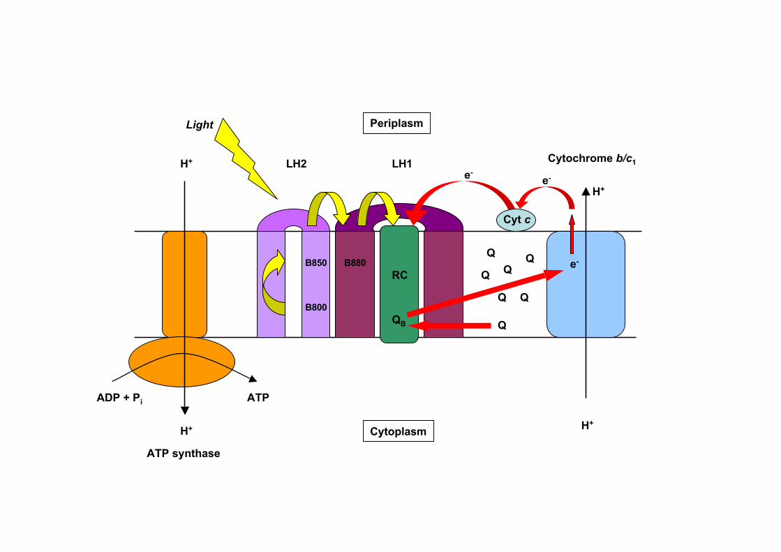
ATP synthase
ATPADP + Pi
H+
H+
H+
H+
LH2 LH1
RC
Light
Cyt c
Q
Q
Q
Q Q
Qe-
e-e-
B800
B850 B880
Cytochrome b/c1
Cytoplasm
Periplasm
QB
Q

LH2, LH1-RC protocol
Open cells
Solubilise membranes
Sucrose gradient
Anionic exchange / Gel filtration
Concentration and
detergent exchange.

LH2
LH1/RC
High light Low light

Detergent exchange
Extraction in LDAO – but crystallization in BOG.Several ways to do this – discuss later.
CrystallizationSitting drop vapour diffusion using 4 mM benzamidine HCl as small amphiphile.(phosphate / ammonium sulphate system)
Initial crystallization took at least 6 weeks – now we hope to do it in 2-3 days.
Things we learned – the hard way.

Peripheral light-harvesting antenna (LH2 complex) fromRh. Acidophila, McDermott et al. (1995) Nature 374, 517.

Proteins

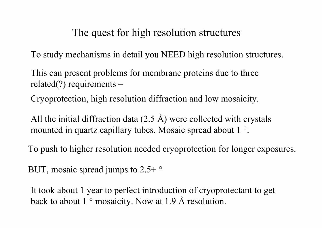
The quest for high resolution structures
To study mechanisms in detail you NEED high resolution structures.
This can present problems for membrane proteins due to threerelated(?) requirements –
Cryoprotection, high resolution diffraction and low mosaicity.
All the initial diffraction data (2.5 Å) were collected with crystals mounted in quartz capillary tubes. Mosaic spread about 1 °.
To push to higher resolution needed cryoprotection for longer exposures.
BUT, mosaic spread jumps to 2.5+ °
It took about 1 year to perfect introduction of cryoprotectant to get back to about 1 ° mosaicity. Now at 1.9 Å resolution.

Cryocooling
• Our experience with cryocooling showed twoproblems:
• 1. Loss of resolution.• 2. Large increase in mosaic spread to 2.5 ° or
more.• Both these problems were eventually overcome by
using stepwise addition of cryoprotectant usingdialysis buttons.
• Major variation from structure to structure.• ***BUT…..always do initial diffraction
experiment with your crystal mounted in acapillary.
ATP synthase
ATPADP + Pi
H+
H+
H+
H+
LH2 LH1
RC
Light
Cyt c
Q
Q
Q
Q Q
Qe-
e-e-
B800
B850 B880
Cytochrome b/c1
Cytoplasm
Periplasm
QB
Q
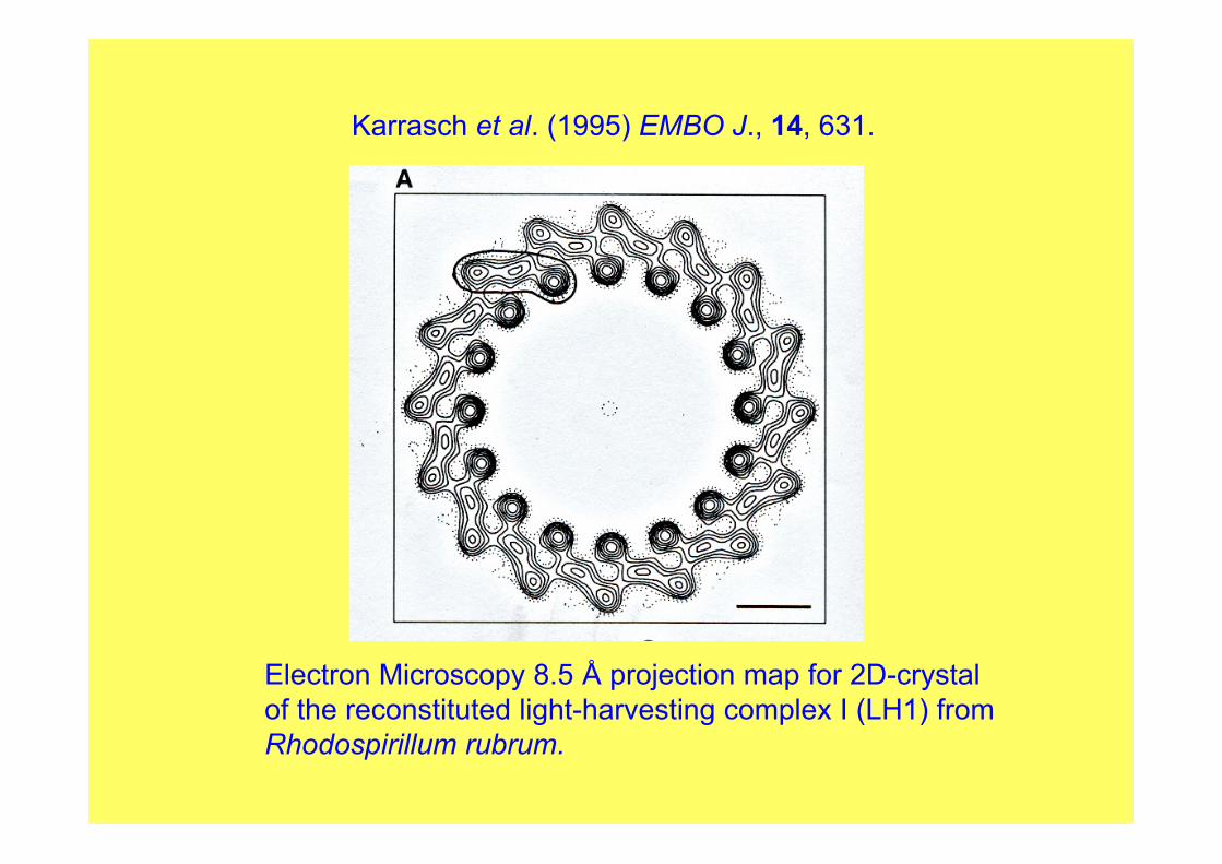
Karrasch et al. (1995) EMBO J., 14, 631.
Electron Microscopy 8.5 Å projection map for 2D-crystalof the reconstituted light-harvesting complex I (LH1) fromRhodospirillum rubrum.

Words of caution
LH1 from 2-D electron crystallography was very well done.
As with LH2 advantage was made of non-crystallographic symmetry since the 16- __ polypeptides were identical.
We used the LH1 model to “build” our photosynthetic unit and all looks good apart from one tiny problem.
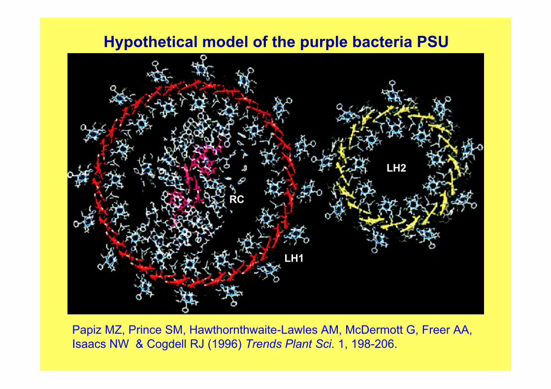
Hypothetical model of the purple bacteria PSU
Papiz MZ, Prince SM, Hawthornthwaite-Lawles AM, McDermott G, Freer AA,Isaacs NW & Cogdell RJ (1996) Trends Plant Sci. 1, 198-206.
LH1
RC
LH2

ATP synthase
ATPADP + Pi
H+
H+
H+
H+
LH2 LH1
RC
Light
Cyt c
Q
Q
Q
Q Q
Qe-
e-e-
B800
B850 B880
Cytochrome b/c1
Cytoplasm
Periplasm
QB
Q


1995 – lets look at crystallizing the “core” complex.
What – are you mad? YES!
1997-8 first core crystals taken to ESRF – diffraction to ~ 7 Å
Thought good start – make speedy progress……
Five years later, yes five years!, we get ~4.0Å
How are we going to get an answer?
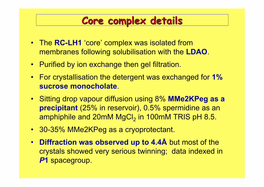
Core complex detailsCore complex details
• The RC-LH1 ‘core’ complex was isolated frommembranes following solubilisation with the LDAO.
• Purified by ion exchange then gel filtration.
• For crystallisation the detergent was exchanged for 1%sucrose monocholate.
• Sitting drop vapour diffusion using 8% MMe2KPeg as aprecipitant (25% in reservoir), 0.5% spermidine as anamphiphile and 20mM MgCl2 in 100mM TRIS pH 8.5.
• 30-35% MMe2KPeg as a cryoprotectant.
• Diffraction was observed up to 4.4Å but most of thecrystals showed very serious twinning; data indexed inP1 spacegroup.


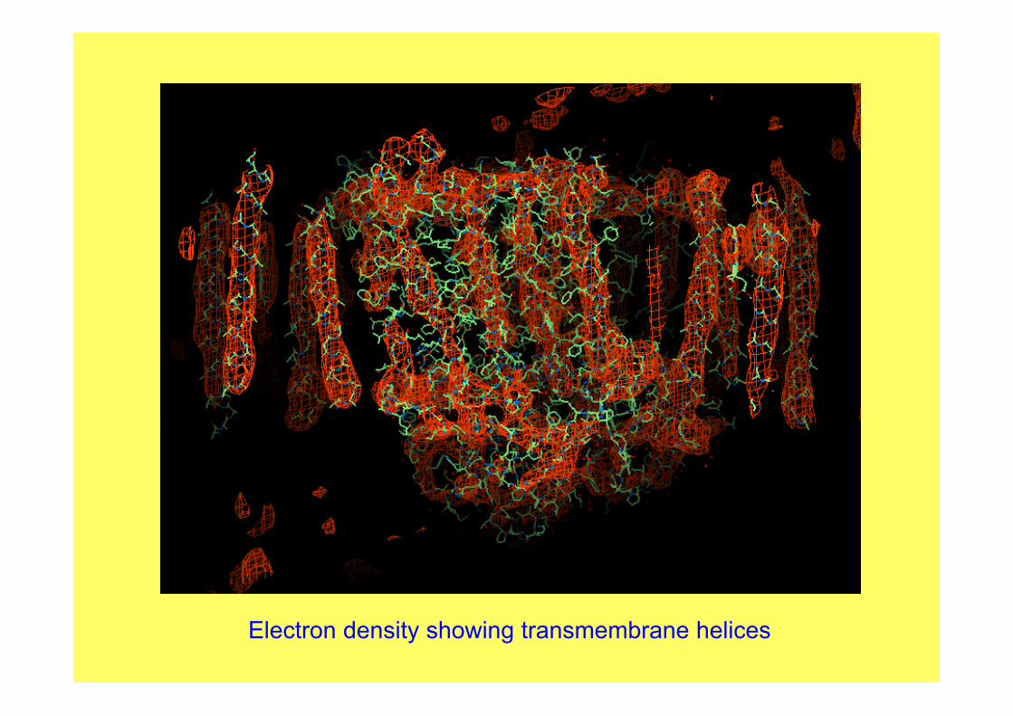
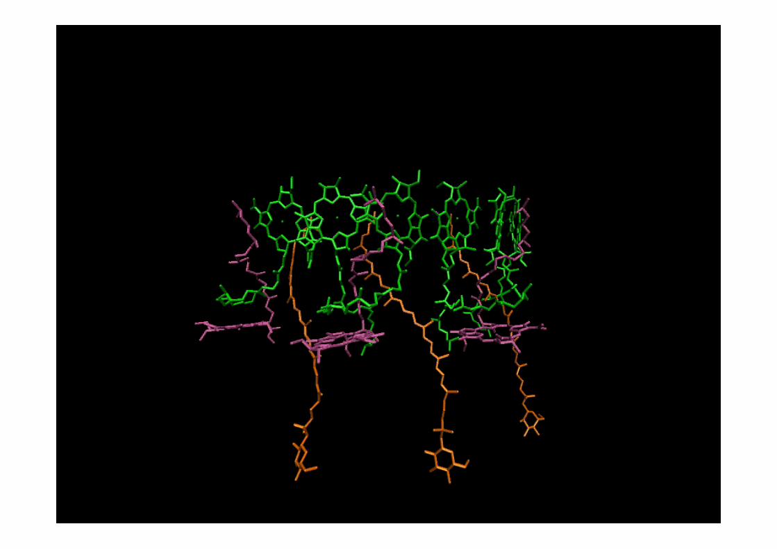
Electron density showing transmembrane helices
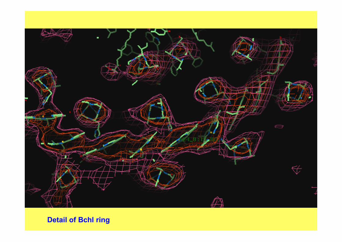
Detail of Bchl ring
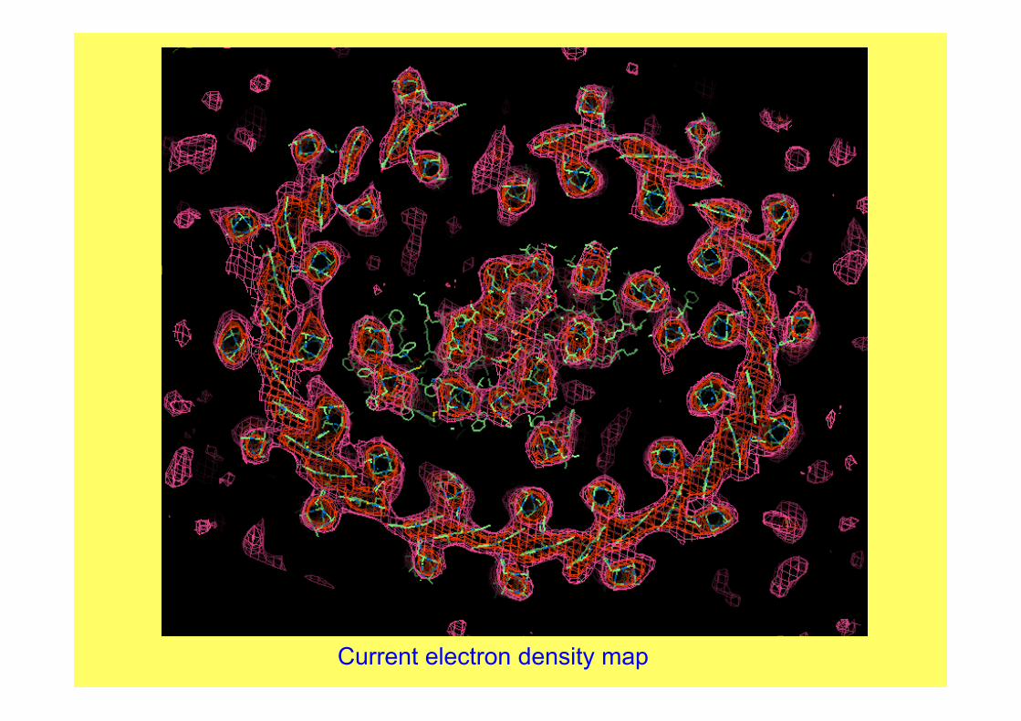
Current electron density map
Having seen what can be achieved in the membrane field – how do we go about it?
Here are some of the techniques used in membrane work- some of the do’s and don’ts

Extraction and purification strategy
M.P. extraction and purification similar to that for W-S.Ps.
But need a bit of lateral thinking!
Important that you think about the environment in whichthe M.P. is found.
Hydrophobic, packed against lipid bilayer.
Need to somehow preserve this local environment.
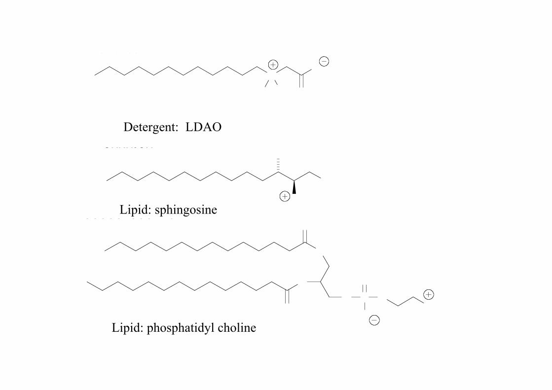
Enter the detergent molecule!
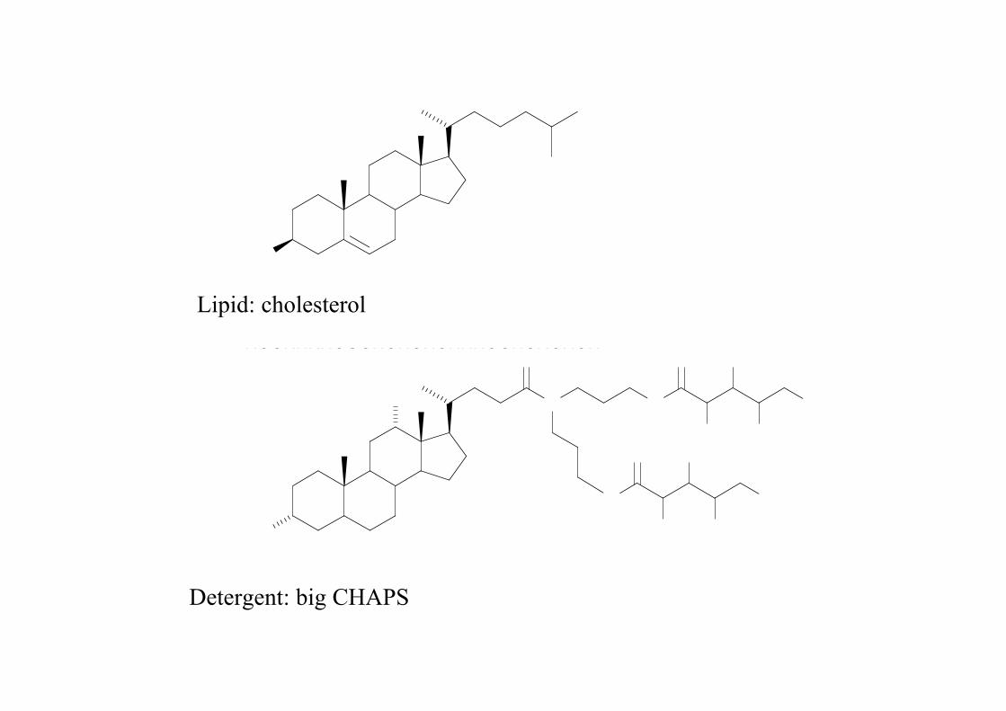
In many ways detergents mimic the lipid
NOMeMeO
OHNH3OH
OOOOOPOOONMe2
Detergent: LDAO
Lipid: sphingosine
Lipid: phosphatidyl choline
HO
HOOHNNHOOOHOHOHOHNHOOHOHOHOH
Lipid: cholesterol
Detergent: big CHAPS

So, how do we know what the detergent is doing? How do we know how it interacts with the protein molecule?
In an X-ray diffraction experiment the bulk detergent molecules are too mobile to have their positions determined.(unless they’re bound as single molecules within the protein structure)
It is, however, possible to image the bulk detergent by contrast neutron diffraction.
We did this for LH2 a couple of years ago by substituting normal detergentwith deuterated detergent.
3 contrast data sets were collected at the ILL – neutrons are very, very slow compared to X-rays. 6 hours/X-ray data set : 4 weeks/ neutron data set!But, worth it!
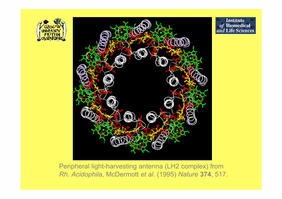
Peripheral light-harvesting antenna (LH2 complex) fromRh. Acidophila, McDermott et al. (1995) Nature 374, 517.

(b) Plan view: looking down on the LH2 complex.
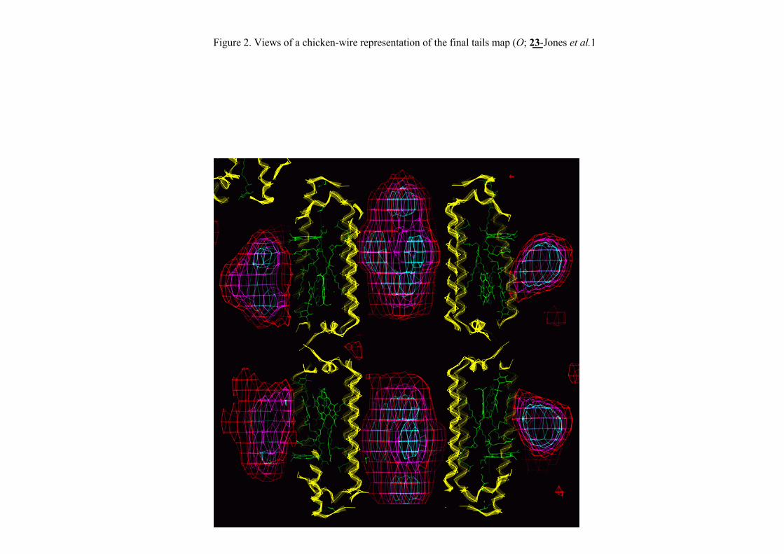
Figure 2. Views of a chicken-wire representation of the final tails map (O; 23-Jones et al.1991); the red contour level encloses detergent tail volume corresponding to 16%,magenta 8% and cyan 2.5% of the cell volume. Polypeptides are represented by yellowribbons, pigment molecules are outlined in green. (a) Elevation showing a cross sectionof the LH2 complex

Detergent characteristics
• Which detergent should I use?
• The range is pretty big!
• Some are neutral, some zwitterionic.
• Essentially trial and error to find compatibility.
• Does it solubilize my protein, or do I get a largepellet?
• Do you want to do pre-solubilization?
• Learn all you can about the detergents youchoose…………...

The concentration above which the detergentforms micelles.
The most important characteristic is:critical micelle concentration, CMC
Large variation in CMC from detergent to detergent.
e.g. C-HEGA-8 has CMC of 227 mM, whereas HDM as a CMC of 0.0006 mM.
CMC will also change with the presence of salt.Detergent alone will rarely solubilize membrane.
Other characteristics: temperature sensitivity - cloud points etc.

Practical need to know CMC?
Solubilization usually requires great excess ofdetergent, well above the CMC.
Think about a pre-solubilization strategy.
Need control of detergent concentration close to CMC.
After solubilization you should return to a normal
working detergent concentration close to its CMC.
Several reasons for this……..

• Detergent molecules above CMC form very largemicelles with very large molecular masses.
• Think of the consequences!
• Difficult to dialyze out detergent.
• Concentrating the protein problematic as you over-concentrate the detergent molecules.
• Try to keep correct CMC by dilution or gel filtrationchromatography.
• High % of detergent….your gels will suck-big time!

DETERGENT
Detergent is now present - is this the correct detergent
for crystallization?
Need to exchange detergent?
Several ways to do this:• Bind protein to ion exchanger and wash off old
detergent and elute in new (best)
• Use a final gel filtration step with different detergent
in eluting buffer.
• Exchange detergent using concentrators…but great
care needed because of micelle concentration (quickest)
How do I know if it’s correct?

0.0
50.0
100.0% Buffer B
1 2 3 4 5 6 7 8 9 10 11 12 13 14 15 16 17 18 19 20 21 22 23 24 25
-0.10
0.00
0.10
0.20
0.30
0.40
0.50
0.60
0.70
0.80
0.90
1.00
-50.0
0.0
50.0
100.0
150.0
200.0
250.0
300.0
350.0
400.0
450.0
500.0
00:00:00 00:10:00 00:20:00 00:30:00 00:40:00
Fractions
Hr:Min:Sec mS/cmAU
0.0
50.0
100.0% Buffer B
1 2 3 4 5 6 7 8 9 10 11 12 13 14 15 16 17 18 19 20 21 22 23 24 25
-0.10
0.00
0.10
0.20
0.30
0.40
0.50
0.60
0.70
0.80
0.90
1.00
-50.0
0.0
50.0
100.0
150.0
200.0
250.0
300.0
350.0
400.0
450.0
500.0
00:00:00 00:10:00 00:20:00 00:30:00 00:40:00
Fractions
Hr:Min:Sec mS/cmAU
Detergent exchange on S-200 gel filtration from LDAO into (a) BOG and (b) DDM
(a) (b)
20060405 LH1 RC 002:10_UV1_280nm 20060405 LH1 RC 002:10_UV2_376nm 20060405 LH1 RC 002:10_UV3_590nm
0
1000
2000
3000
4000
5000
mAU
0.0 5.0 10.0 15.0 20.0 ml
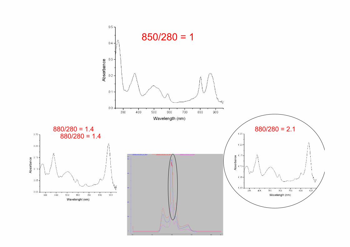
850/280 = 1
880/280 = 2.1880/280 = 1.4880/280 = 1.4

Acknowledgements
Many, many people…..
LH2 and Core complex:Alex Roszak, Steve Prince, Mads Gabrielsen + +

….and finally...
But, most importantly, it has to be fun!
It takes an inordinate amount of time, the
patience of a saint, the ability to handle many
disappointments, the understanding of a good
woman (or man), many, many beers, …….