ip-10-mediated t cell homing promotes cerebral ... t cell homing promotes cerebral inflammation over...
TRANSCRIPT

IP-10-Mediated T Cell Homing Promotes CerebralInflammation over Splenic Immunity to Malaria InfectionCatherine Q. Nie1,2, Nicholas J. Bernard1, M. Ursula Norman3, Fiona H. Amante4, Rachel J. Lundie1,
Brendan S. Crabb5, William R. Heath6, Christian R. Engwerda4, Michael J. Hickey3, Louis Schofield1,
Diana S. Hansen1*
1 The Walter and Eliza Hall Institute of Medical Research, Parkville, Victoria, Australia, 2 Department of Medical Biology, The University of Melbourne, Parkville, Victoria,
Australia, 3 Centre for Inflammatory Diseases, Monash University, Department of Medicine, Monash Medical Centre, Clayton, Victoria, Australia, 4 Queensland Institute of
Medical Research, Herston, Queensland, Australia, 5 Burnet Institute, Melbourne, Victoria, Australia, 6 Department of Microbiology and Immunology, The University of
Melbourne, Parkville, Victoria, Australia
Abstract
Plasmodium falciparum malaria causes 660 million clinical cases with over 2 million deaths each year. Acquired hostimmunity limits the clinical impact of malaria infection and provides protection against parasite replication. Experimentalevidence indicates that cell-mediated immune responses also result in detrimental inflammation and contribute to severedisease induction. In both humans and mice, the spleen is a crucial organ involved in blood stage malaria clearance, whileorgan-specific disease appears to be associated with sequestration of parasitized erythrocytes in vascular beds andsubsequent recruitment of inflammatory leukocytes. Using a rodent model of cerebral malaria, we have previously foundthat the majority of T lymphocytes in intravascular infiltrates of cerebral malaria-affected mice express the chemokinereceptor CXCR3. Here we investigated the effect of IP-10 blockade in the development of experimental cerebral malaria andthe induction of splenic anti-parasite immunity. We found that specific neutralization of IP-10 over the course of infectionand genetic deletion of this chemokine in knockout mice reduces cerebral intravascular inflammation and is sufficient toprotect P. berghei ANKA-infected mice from fatality. Furthermore, our results demonstrate that lack of IP-10 during infectionsignificantly reduces peripheral parasitemia. The increased resistance to infection observed in the absence of IP-10-mediated cell trafficking was associated with retention and subsequent expansion of parasite-specific T cells in spleens ofinfected animals, which appears to be advantageous for the control of parasite burden. Thus, our results demonstrate thatmodulating homing of cellular immune responses to malaria is critical for reaching a balance between protective immunityand immunopathogenesis.
Citation: Nie CQ, Bernard NJ, Norman MU, Amante FH, Lundie RJ, et al. (2009) IP-10-Mediated T Cell Homing Promotes Cerebral Inflammation over SplenicImmunity to Malaria Infection. PLoS Pathog 5(4): e1000369. doi:10.1371/journal.ppat.1000369
Editor: Eleanor M. Riley, London School of Hygiene and Tropical Medicine, United Kingdom
Received October 24, 2008; Accepted March 6, 2009; Published April 3, 2009
Copyright: � 2009 Nie et al. This is an open-access article distributed under the terms of the Creative Commons Attribution License, which permits unrestricteduse, distribution, and reproduction in any medium, provided the original author and source are credited.
Funding: Supported by the NHMRC Project grant 356239, NHMRC Program Grant 215201, NHMRC IRIISS grant 361646 and Victorian State Government OIS grant.LS, BSC and WRH are International Research Scholars of the Howard Hughes Medical Institute.
Competing Interests: The authors have declared that no competing interests exist.
* E-mail: [email protected]
Introduction
Malaria is one of the most serious infectious diseases in humans,
infecting 5–10% of the world’s population. The most severe
complication caused by Plasmodium falciparum infection is cerebral
malaria (CM), which is responsible for about 2.5 million deaths
each year [1]. This neurological syndrome is characterized by the
occurrence of seizures and coma [2]. Although the precise
mechanism leading to cerebral disease is not fully understood, it
has been suggested that sequestration of parasitised red blood cell
(pRBC) in brain blood vessels induces blood flow obstruction
resulting in hypoxia, haemorrhage and pathology [3].
The analysis of brain infiltrates predisposing to CM in humans
is limited as it can only be deduced from post-mortem samples.
Much useful evidence contributing to the understanding of disease
has been provided by experimental infection with P. berghei ANKA.
This rodent infection has many features in common with human
disease and is thus a good model for some important aspects of
clinical malaria [4]. A large body of work in this and other rodent
models of CM demonstrated that immune responses elicited
during infection play a role in the control of parasitemia but can
also result in detrimental inflammation and contribute to disease
induction [5,6]. Current views support the idea that CM is caused
by the combined effect of sequestration of pRBC and a strong
inflammatory response mediated by cytokines such as TNF-a [7],
LT-a [8], IFN-c [9] and effector cells such as CD4+ [10] and
CD8+ T cells [11,12], NKT cells [13] and NK cells [14]. Since it is
known that these cells produce cytokines that up-regulate the
expression of adhesion molecules like ICAM-1, involved in the
recognition of parasitic proteins expressed on pRBC, it has been
proposed that this systemic inflammatory cascade exacerbates
parasite sequestration. However, emerging evidence in human
malaria and animal models [11,12,15,16] revealed the presence of
leukocytes in brain blood vessels during infection, suggesting that
intravascular infiltration of these cells might result in local
inflammation and could also contribute to disease induction.
Both CD4+ and CD8+ T cells have been found in brain blood
vessels of CM-affected mice [11,12]. Brain-sequestered cytotoxic
PLoS Pathogens | www.plospathogens.org 1 April 2009 | Volume 5 | Issue 4 | e1000369
CD8+ T cells have been shown to mediate CM via a perforin-
dependent mechanism [11]. Recent work indicates that CD8+ T
cells specific for parasite-expressed antigens are amongst those
recruited to the brain during infection and are capable of
mediating lethal disease [17]. Like T cells, NK cells have been
found in brain blood vessels of malaria infected mice and appear
to be abundant at early stages of infection [14].
The chemokine pathways responsible for leukocyte recruitment
to the brain in CM have not been completely characterized. Mice
deficient in CC chemokine receptor 5 (CCR5) have been reported
to be either 80% resistant to P. berghei ANKA-mediated CM [18]
or to display a delayed onset of cerebral disease symptoms [11]. In
a previous study, we found that the majority of NK cells and T
cells in brains of malaria-infected mice express CXC chemokine
receptor 3 (CXCR3) suggesting that trafficking through this
pathway is strongly associated with lymphocyte recruitment
leading to cerebral disease [14]. Moreover, splenic T cells from
CM susceptible but not resistant mice were found to up-regulate
expression of CXCR3 and to acquire the capacity to migrate in
response to CXCR3 chemokines during malaria infection,
indicating that CXCR3 expression correlates with disease severity
[19]. In agreement, it has been recently found that 70–80% of
CXCR32/2 mice are resistant to P. berghei-mediated CM [20,21].
CXCR3 recognizes 3 ligands: MIG, IP-10 and I-TAC.
Although all these chemokines are induced by IFN-c, experimen-
tal evidence suggests that they play non-redundant roles in
leukocyte homing [22]. These chemokines have been shown to
recruit NK cells and TH1 cells in several inflammatory conditions
and in addition to their chemotactic activity they have been shown
to participate in the induction of effector immune responses. IP-10
and MIG have been found to stimulate T cell proliferation and
IFN-c production in response to alloantigen or to exogenous
antigen implying a role in TH1 polarization [23,24].
The role of CXCR3 chemokines in malaria has not been
extensively investigated. Although it has been shown that IP-10
and MIG are up-regulated in the brain in response to infection
[19,21], their precise role in disease induction remains elusive. It
has been recently shown that MIG2/2 and IP-102/2 mice are
partially resistant to P. berghei ANKA infection [20]. However,
whether the increased survival rates to malaria infection in the
absence of these chemokines result from reduced leukocyte
recruitment to the brain and/or a differential induction of
immune response to infection has not been examined. Moreover,
experimental evidence indicates that the same cell-mediated
immune responses involved in severe disease induction are also
required for the control of infection [5,6] and whether leukocyte
trafficking blockade has an impact on the development of malaria-
specific immunity and the control of parasite burden remains
unknown. The precise understanding of these processes and their
implications are highly relevant in assessing the feasibility of anti-
leukocyte trafficking therapies to reduce organ-specific inflamma-
tion and fatalities associated with CM. Since recent reports
identified IP-10 as a biomarker associated with mortality in P.
falciparum-mediated CM [25,26], in this study we thoroughly
investigated the role of this chemokine during experimental CM
and in the induction of parasite-specific immunity. We found that
specific neutralization of IP-10 during infection reduces intravas-
cular inflammation in brains of P. berghei ANKA-infected mice and
is sufficient to protect from fatality. Furthermore, our data reveal
that inhibition of IP-10-mediated leukocyte trafficking also results
in retention of parasite-specific T cells in the spleen, which favors
induction of protective immunity and facilitates control of parasite
burden.
Results
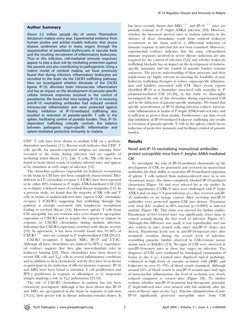
Novel anti-IP-10 neutralizing monoclonal antibodiesprotect susceptible mice from P. berghei ANKA-mediatedCM
To investigate the role of IP-10-mediated chemotaxis on the
development of CM, we generated and screened rat monoclonal
antibodies for their ability to neutralize IP-10-mediated migration
of splenic T cells isolated from malaria-infected mice in in vitro
chemotaxis assays. Six clones completely blocked IP-10-mediated
chemotaxis (Figure 1A) and were selected for in vivo studies. In
these experiments, C57BL/6 mice were challenged with P. berghei
ANKA and on days 3–9 post-infection (p.i.) they received anti-IP-
10 antibodies or an isotype control. Mice receiving anti-IP-10
antibodies were protected against CM (not shown). Treatment
with clone 8A7 resulted in 80% survival (p = 0.0002) in infected
animals (Figure 1B). This clone was selected for further studies.
Parasitemia of 8A7-treated mice was significantly lower than in
control animals during the first week of infection (Figure 1C).
Although this difference was small, it was reproducible and was
also evident in mice treated with other anti-IP-10 clones (not
shown). Parasitemia levels rose in anti-IP-10-injected mice after
treatment cessation during the second week of infection,
resembling parasitic burden observed in CM-resistant mouse
strains such as BALB/c [13]. No signs of CM were observed in
anti-IP-10-treated mice at these late stages on infection. The
diagnoses of CM were confirmed by histological examination of
brains at day 6 p.i. Control mice displayed typical pathology,
evidenced as high levels of vascular occlusion with pRBC and
leukocytes in over 65–70% of blood vessels examined. Although
around 50% of blood vessels in anti-IP-10 treated mice had signs
of intravascular inflammation, the level of occlusion was clearly
reduced compared to control mice (Figure 1D). To further
evaluate whether anti-IP-10 treatment had therapeutic potential,
P. berghei-infected mice were treated with this antibody after the
onset of disease signs on day 5 p.i. This late administration of anti-
IP-10 significantly protected susceptible mice from CM
Author Summary
About 2.5 million people die of severe Plasmodiumfalciparum malaria every year. Experimental evidence fromhuman studies and animal models indicates that severedisease syndromes arise in many organs through thesequestration of parasitized erythrocytes in vascular bedsand the resulting recruitment of inflammatory leukocytes.Thus in this infection, cell-mediated immune responsesappear to play a dual role by mediating protection againstthe parasite and also contributing to pathogenesis. Using arodent model of cerebral malaria, we have previouslyfound that during infection, inflammatory leukocytes arerecruited to the brain via the CXCR3 trafficking pathway.Here we investigated whether blockade of the CXCR3ligand, IP-10, alleviates brain intravascular inflammationand has an impact on the development of parasite-specificcellular immune responses involved in the control ofparasitemia. We found that mice lacking IP-10 or receivinganti-IP-10 neutralizing antibodies had reduced cerebralintravascular inflammation and were protected againstfatality. Inhibition of IP-10-mediated trafficking alsoresulted in retention of parasite-specific T cells in thespleen, facilitating control of parasite burden. Thus, IP-10-dependent trafficking critically controls the balancebetween pathogenic organ-specific inflammation andspleen-mediated protective immunity to malaria.
IP-10 Regulates Disease and Immunity in Malaria
PLoS Pathogens | www.plospathogens.org 2 April 2009 | Volume 5 | Issue 4 | e1000369
(p = 0.0062), resulting in 50% survival of infected animals
(Figure 1E). Since CXCR3 chemokines share around 30% of
sequence homology with each other, 8A7 specificity was tested in
chemotaxis assays. 8A7 completely blocked IP-10-mediated
chemotaxis but was unable to prevent migration of splenic T cells
from malaria-infected mice in response to MIG or I-TAC
(Figure 1F). Thus specific neutralization of IP-10 alone during
infection is sufficient to protect susceptible mice from CM.
IP-10 neutralization alleviates intravascular infiltrationand does not affect parasite sequestration
To more accurately assess organ-specific inflammation after
anti-IP-10 treatment, C57BL/6 mice were challenged with P.
berghei ANKA, treated with 8A7 or isotype control antibody and
their brain blood vessels were examined on day 5 p.i. by intravital
microscopy. To that end, mice were anaesthetized, injected with
rhodamine 6G to label circulating cells and pial microcirculation
Figure 1. Anti-IP-10 neutralization protects susceptible mice from P. berghei ANKA-mediated CM. (A) Anti-IP-10 hybridoma supernatantswere incubated with IP-10. Transwell inserts containing splenic T cells from malaria-infected mice were then added to the wells and chemotaxis wasassessed. Bars represent chemotaxis indices. C57BL/6 mice were infected with P. berghei-ANKA and treated with anti-IP-10 or isotype control on days3–9 p.i. (B) or on days 5–9 p.i. (E). Survival was monitored daily. (C) Parasitemia was determined by Giemsa-stained blood smears. Each pointrepresents mean parasitemia6SD, ***p,0.0005 (Mann-Whitney test). Data is representative of 3 infections. (D) Histological examination of brainsfrom P. berghei ANKA-infected anti-IP-10 and isotype control-treated mice. (F) Anti-IP-10 monoclonal antibody 8A7 was incubated with IP-10, MIG orI-TAC. Transwell inserts containing splenic T cells from malaria-infected mice were added to wells and chemotaxis was determined. Bars representmeans of 3 samples6SEM.doi:10.1371/journal.ppat.1000369.g001
IP-10 Regulates Disease and Immunity in Malaria
PLoS Pathogens | www.plospathogens.org 3 April 2009 | Volume 5 | Issue 4 | e1000369

was observed. In contrast to naı̈ve mice (Video S1), in which
leukocyte rolling and adhesion were rarely observed, large
numbers of rolling and adherent cells were observed in blood
vessels of malaria-infected mice (Figure 2A and Video S2). Anti-
IP-10 treatment reduced the number of rolling and adherent cells
by 60% compared to isotype control-injected animals (Figure 2B,C
and Video S3), indicating that this treatment alleviates intravas-
cular infiltration during infection. Since rhodamine 6G not only
labels circulating leukocytes but also pRBC [27], we sought to
assess the possibility that the reduced levels of occlusion in anti-IP-
10 treated mice reflected a reduction in parasite sequestration. To
that end, C57BL/6 mice were infected with a transgenic P. berghei
ANKA line that constitutively expresses luciferase [28]. Following
luciferin injection on day 6 p.i., mice were sacrificed and
bioluminescent parasites were observed in the brain. No significant
differences were found in bioluminescence levels emerging from
parasites in brains of anti-IP-10 and isotype control-treated mice
(Figure 2D,E). Together these results suggest that IP-10 neutral-
ization does not affect parasite sequestration but instead alleviates
vascular inflammation by reducing recruitment of inflammatory
leukocyte to the brain of infected mice. To test this idea, brain-
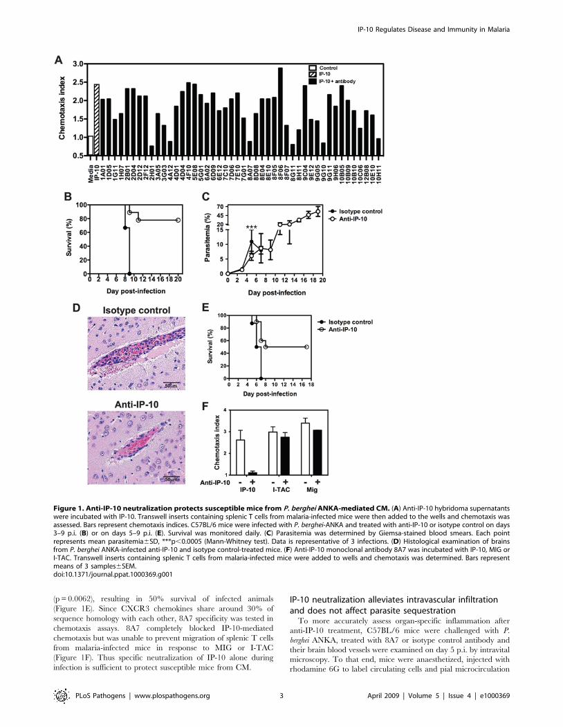
infiltrating leukocytes were purified from anti-IP-10 treated and
control mice at day 6 p.i., stained with fluorescent antibodies and
analysed by flow cytometry. The percentage of NK cells, CD4+
and CD8+ T cells recruited to the brain during infection was
similar in anti-IP-10-treated and control mice (Figure 3A).
However, the total number of sequestered leukocytes was
significantly reduced in anti-IP-10-treated mice compared to
controls (control: 226133640014; anti-IP-10: 94933613897;
p,0.05, Mann-Whitney test). Consistently, the absolute number
of CD4+ and CD8+ T cells was reduced by 50% in anti-IP-10-
treated animals compared to controls (Figure 3C–E). No
significant differences were found in the number of NK cells in
brains of anti-IP-10-treated or control mice (Figure 3B). Analysis
of chemokine receptor usage of brain-infiltrating T cells showed
that the majority of abT cells present in brain blood vessels of
malaria-infected control mice expressed CXCR3 but not CCR5
(Figure 3F). Similarly, over 70% of abT cells recovered from
brains of anti-IP-10-treated mice expressed this receptor
(Figure 3F), suggesting that 8A7 blocking effect of IP-10 may not
be absolute or that in the absence of IP-10 other CXCR3
chemokines can recruit leukocytes to the brain during infection.
Anti-IP-10 treatment does not alter the intrinsic capacityof T cells to migrate in response to chemotactic stimuli
To investigate whether anti-IP-10 treatment had an effect on T
cell activation during malaria, we first examined the percentage of
activated CD4+ and CD8+ T cells expressing CD69 and CD25 in
anti-IP-10 and isotype-control-treated mice after P. berghei ANKA
infection. Figures 4A and B show that the expression of CD25 and
CD69 on splenic CD4+ and CD8+ T cells increased in response to
malaria infection. No differences were found in the percentage of
activated T cells from anti-IP-10-treated and control mice. In
another set of experiments, we sought to determine whether anti-
IP-10-treament altered the intrinsic capacity of cells to migrate in
response to chemotactic stimuli. To that end, splenic T cells were
isolated from anti-IP-10 and isotype-control-treated mice at day 5
p.i. with P. berghei ANKA and their ability to migrate in response to
IP-10 was evaluated in an in vitro chemotaxis assay. As expected, T
cells from isotype control-treated malaria-infected mice readily
migrated in response to IP-10 compared to cells from naı̈ve mice
(Figure 4C). No significant differences were found between the
chemotactic response of T cells isolated from control and anti-IP-
10-treated mice (Figure 4C). The migratory properties of T cells
were also analysed in vivo. For that, P. berghei ANKA-infected
C57BL/6 Ly5.1+ mice were treated with anti-IP-10 or isotype
control antibodies. Splenic Ly5.1+ T cells were isolated on day 6
p.i., adoptively transferred into malaria-infected (day 4 p.i)
C57BL/6 Ly5.2+ recipients and 2 days later, their ability migrate
Figure 2. IP-10 neutralization alleviates brain intravascular infiltration. P. berghei ANKA-infected mice were treated with anti-IP-10 orisotype control antibodies. (A) Pial microcirculation was examined by intravital microscopy on day 5 p.i.. The number of rolling (B) and adherent (C)cells was determined. Bars represent means of 4–5 mice6SEM, *p,0.05, **p,0.01 (Mann-Whitney test). C57BL/6 mice were infected with luciferase-expressing P. berghei ANKA and then treated with anti-IP-10 or isotype control antibodies. (D) Brain-sequestered parasites were visualized on day 6 p.i1 h after luciferin injection. (E) Parasite-associated bioluminescence was determined. Bars represent means of 5 samples6SD.doi:10.1371/journal.ppat.1000369.g002
IP-10 Regulates Disease and Immunity in Malaria
PLoS Pathogens | www.plospathogens.org 4 April 2009 | Volume 5 | Issue 4 | e1000369

IP-10 Regulates Disease and Immunity in Malaria
PLoS Pathogens | www.plospathogens.org 5 April 2009 | Volume 5 | Issue 4 | e1000369
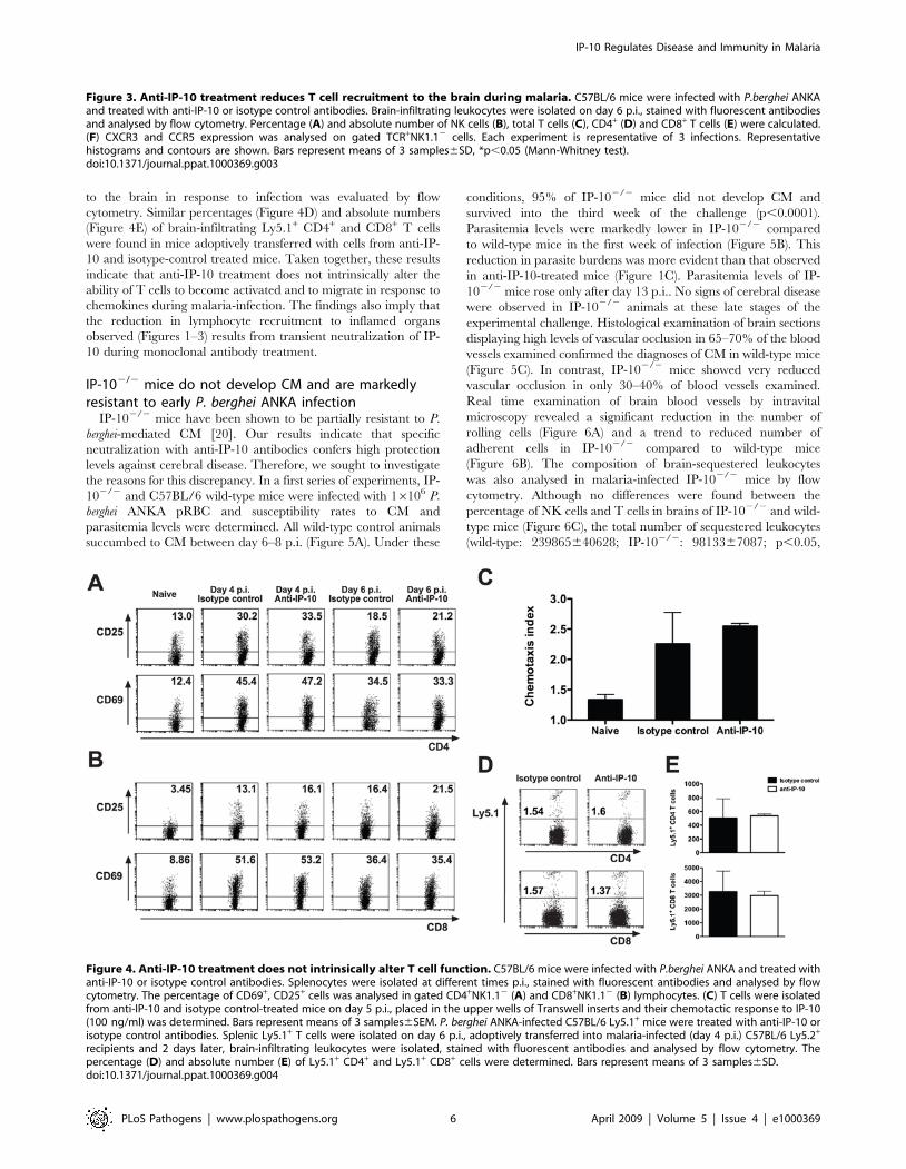
to the brain in response to infection was evaluated by flow
cytometry. Similar percentages (Figure 4D) and absolute numbers
(Figure 4E) of brain-infiltrating Ly5.1+ CD4+ and CD8+ T cells
were found in mice adoptively transferred with cells from anti-IP-
10 and isotype-control treated mice. Taken together, these results
indicate that anti-IP-10 treatment does not intrinsically alter the
ability of T cells to become activated and to migrate in response to
chemokines during malaria-infection. The findings also imply that
the reduction in lymphocyte recruitment to inflamed organs
observed (Figures 1–3) results from transient neutralization of IP-
10 during monoclonal antibody treatment.
IP-102/2 mice do not develop CM and are markedlyresistant to early P. berghei ANKA infection
IP-102/2 mice have been shown to be partially resistant to P.
berghei-mediated CM [20]. Our results indicate that specific
neutralization with anti-IP-10 antibodies confers high protection
levels against cerebral disease. Therefore, we sought to investigate
the reasons for this discrepancy. In a first series of experiments, IP-
102/2 and C57BL/6 wild-type mice were infected with 16106 P.
berghei ANKA pRBC and susceptibility rates to CM and
parasitemia levels were determined. All wild-type control animals
succumbed to CM between day 6–8 p.i. (Figure 5A). Under these
conditions, 95% of IP-102/2 mice did not develop CM and
survived into the third week of the challenge (p,0.0001).
Parasitemia levels were markedly lower in IP-102/2 compared
to wild-type mice in the first week of infection (Figure 5B). This
reduction in parasite burdens was more evident than that observed
in anti-IP-10-treated mice (Figure 1C). Parasitemia levels of IP-
102/2 mice rose only after day 13 p.i.. No signs of cerebral disease
were observed in IP-102/2 animals at these late stages of the
experimental challenge. Histological examination of brain sections
displaying high levels of vascular occlusion in 65–70% of the blood
vessels examined confirmed the diagnoses of CM in wild-type mice
(Figure 5C). In contrast, IP-102/2 mice showed very reduced
vascular occlusion in only 30–40% of blood vessels examined.
Real time examination of brain blood vessels by intravital
microscopy revealed a significant reduction in the number of
rolling cells (Figure 6A) and a trend to reduced number of
adherent cells in IP-102/2 compared to wild-type mice
(Figure 6B). The composition of brain-sequestered leukocytes
was also analysed in malaria-infected IP-102/2 mice by flow
cytometry. Although no differences were found between the
percentage of NK cells and T cells in brains of IP-102/2 and wild-
type mice (Figure 6C), the total number of sequestered leukocytes
(wild-type: 239865640628; IP-102/2: 9813367087; p,0.05,
Figure 3. Anti-IP-10 treatment reduces T cell recruitment to the brain during malaria. C57BL/6 mice were infected with P.berghei ANKAand treated with anti-IP-10 or isotype control antibodies. Brain-infiltrating leukocytes were isolated on day 6 p.i., stained with fluorescent antibodiesand analysed by flow cytometry. Percentage (A) and absolute number of NK cells (B), total T cells (C), CD4+ (D) and CD8+ T cells (E) were calculated.(F) CXCR3 and CCR5 expression was analysed on gated TCR+NK1.12 cells. Each experiment is representative of 3 infections. Representativehistograms and contours are shown. Bars represent means of 3 samples6SD, *p,0.05 (Mann-Whitney test).doi:10.1371/journal.ppat.1000369.g003
Figure 4. Anti-IP-10 treatment does not intrinsically alter T cell function. C57BL/6 mice were infected with P.berghei ANKA and treated withanti-IP-10 or isotype control antibodies. Splenocytes were isolated at different times p.i., stained with fluorescent antibodies and analysed by flowcytometry. The percentage of CD69+, CD25+ cells was analysed in gated CD4+NK1.12 (A) and CD8+NK1.12 (B) lymphocytes. (C) T cells were isolatedfrom anti-IP-10 and isotype control-treated mice on day 5 p.i., placed in the upper wells of Transwell inserts and their chemotactic response to IP-10(100 ng/ml) was determined. Bars represent means of 3 samples6SEM. P. berghei ANKA-infected C57BL/6 Ly5.1+ mice were treated with anti-IP-10 orisotype control antibodies. Splenic Ly5.1+ T cells were isolated on day 6 p.i., adoptively transferred into malaria-infected (day 4 p.i.) C57BL/6 Ly5.2+
recipients and 2 days later, brain-infiltrating leukocytes were isolated, stained with fluorescent antibodies and analysed by flow cytometry. Thepercentage (D) and absolute number (E) of Ly5.1+ CD4+ and Ly5.1+ CD8+ cells were determined. Bars represent means of 3 samples6SD.doi:10.1371/journal.ppat.1000369.g004
IP-10 Regulates Disease and Immunity in Malaria
PLoS Pathogens | www.plospathogens.org 6 April 2009 | Volume 5 | Issue 4 | e1000369

Mann-Whitney test) as well as CD4+ and CD8+ T cells recovered
from brains of IP-102/2 mice was significantly lower than in wild-
type animals (Figure 6D–G). To determine whether the reduced
parasite growth rates observed in IP-102/2 mice had an impact on
pRBC sequestration, wild-type and IP-102/2 mice were infected
with luciferase-expressing P. berghei parasites and bioluminescence
in the brain was determined as described in Figure 2. Unlike in
anti-IP-10-treated mice (Figure 2), parasite-associated biolumines-
cence was reduced by 80% in brains of IP-102/2 mice compared
to wild-type controls (Figure 6H,I). Since in human malaria
cytoadhesion of infected erythrocytes to the vascular endothelium
is known to be mediated by ICAM-1 [29], the expression of this
adhesion molecule was examined in brain blood vessels of P.
berghei-infected wild-type and IP-102/2 mice by immunohisto-
chemistry (Figure 6J). No significant differences were found in the
number of blood vessels expressing ICAM-1 between IP-102/2
mice and wild-type controls (Figure 6K). Taken together, these
results indicate that genetic deletion of IP-10 protects from CM by
reducing recruitment of inflammatory leukocytes and by prevent-
ing pRBC sequestration in the brain.
Lack of IP-10 enhances splenic anti-P. berghei immuneresponses
The fact that lack of IP-10 during malaria reduced parasite
burden suggested that the absence of this chemokine has a
beneficial effect for the development of parasite-specific responses
involved in the control of infection. Interestingly, in addition to its
chemotactic activity, IP-10 has been shown to stimulate and not
inhibit the induction of several effector responses. To address this
paradox, the induction of immune responses to malaria was
investigated in anti-IP-10-treated and IP-102/2 mice. Since the
spleen is a key organ involved in the initiation of immune
responses to blood-stage malaria [30], splenic parasite-specific
responses were examined. In a first set of experiments, CD4+ T
cells from anti-IP-10-treated, IP-102/2 and control mice were
isolated at day 5 p.i. and proliferative responses to parasite lysate
as well as IFN-c and IL-4 production were determined. A small
but significant increase in parasite-specific proliferation was
observed in CD4+ T cells of anti-IP-10-treated animals compared
to controls (Figure 7A). These responses as well as IFN-cproduction were significantly more pronounced in CD4+ T cells
from IP-102/2 mice (Figure 7A,B). IL-4 production was virtually
absent in T cells from malaria-infected C57BL/6 mice (not
shown). No significant differences were found in proliferative
responses or cytokine production to anti-CD3 antibody across
experimental groups (not shown). The frequency and absolute
number of IFN-c producing cells was also evaluated in gated
CD4+ and CD8+ T cells by flow cytometry. Similar frequencies of
IFN-c+ CD4+ and CD8+ cells were found among splenocytes of
malaria-infected anti-IP-10-treated and control mice (Figure 7C).
These percentages were somewhat higher in IP-102/2 mice. A
small increase in the absolute number of IFN-c-producing CD4+
and CD8+ T cells was found in anti-IP-10-treated compared to
naı̈ve mice (Figure 7D,E). Like proliferation, genetic deletion of IP-
10 resulted in a more dramatic effect, as the total number of CD4+
and CD8+ IFN-c-secreting cells in IP-102/2 mice was around 3
times higher than in wild-type animals (Figure 7D,E). To examine
whether IP-10 deletion also affected systemic IFN-c responses to
malaria, serum IFN-c levels were determined in wild-type and IP-
102/2 mice at day 5 p.i. Interestingly, serum IFN-c content was
significantly lower in malaria-infected IP-102/2 mice compared to
wild-type control animals (Figure S1). Together these results
suggest that lack of IP-10 enhances splenic but not systemic
immune responses to malaria.
Figure 5. IP-102/2 mice do not develop CM and are markedly resistant to malaria infection. Wild-type and IP-102/2 mice were infectedwith P. berghei ANKA. (A) Survival was monitored daily. (B) Parasitemia was determined by Giemsa-stained blood smears. Each point representsmean6SD, ***p,0.0001, *p,0.05 (Mann-Whitney test). Pooled data from 2 infections (n = 20) is shown. (C) Histological examination of brains from P.berghei-infected wild-type and IP-102/2 mice.doi:10.1371/journal.ppat.1000369.g005
IP-10 Regulates Disease and Immunity in Malaria
PLoS Pathogens | www.plospathogens.org 7 April 2009 | Volume 5 | Issue 4 | e1000369

Figure 6. Intravascular infiltration and parasite sequestration are reduced in brains of malaria-infected IP-102/2 mice. Numbers ofrolling (A) and adherent (B) cells in brain blood vessels of wild-type and IP-102/2 mice were determined by intravital microscopy on day 5 p.i. with P.berghei ANKA. Bars represent means of 4–6 mice6SEM, *p,0.05 (Mann-Whitney test). Brain-infiltrating leukocytes were isolated on day 6 p.i., stainedwith fluorescent antibodies and analysed by flow cytometry. Percentage (C) and total number of NK cells (D), total T cells (E), CD4+ (F) and CD8+ Tcells (G) were calculated. Bars represent means of 3 samples6SD, *p,0.05 (Mann-Whitney test). Wild-type and IP-102/2 mice were infected withluciferase-expressing P. berghei ANKA. (H) Brain-sequestered parasites were visualized 1 h after luciferin injection. (I) Parasite-associatedbioluminescence was recorded. Bars represent means of 4–5 samples6SD, **p,0.01. (J) ICAM-1 staining was performed on brain sections of wild-type and IP-102/2 mice prepared on day 6 p.i. (K) The number of ICAM-1 positive vessels was determined. Bars represent means of 4 samples6SD.doi:10.1371/journal.ppat.1000369.g006
IP-10 Regulates Disease and Immunity in Malaria
PLoS Pathogens | www.plospathogens.org 8 April 2009 | Volume 5 | Issue 4 | e1000369
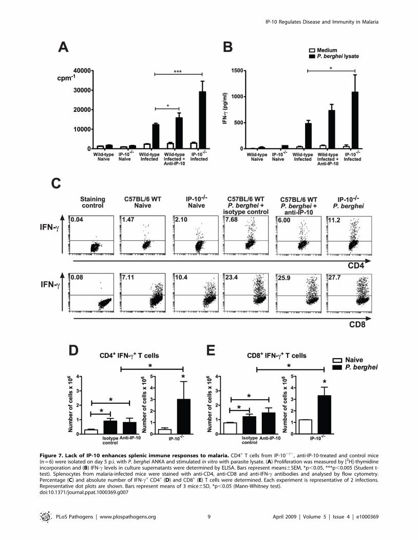
Figure 7. Lack of IP-10 enhances splenic immune responses to malaria. CD4+ T cells from IP-102/2, anti-IP-10-treated and control mice(n = 6) were isolated on day 5 p.i. with P. berghei ANKA and stimulated in vitro with parasite lysate. (A) Proliferation was measured by [3H]-thymidineincorporation and (B) IFN-c levels in culture supernatants were determined by ELISA. Bars represent means6SEM, *p,0.05, ***p,0.005 (Student t-test). Splenocytes from malaria-infected mice were stained with anti-CD4, anti-CD8 and anti-IFN-c antibodies and analysed by flow cytometry.Percentage (C) and absolute number of IFN-c+ CD4+ (D) and CD8+ (E) T cells were determined. Each experiment is representative of 2 infections.Representative dot plots are shown. Bars represent means of 3 mice6SD, *p,0.05 (Mann-Whitney test).doi:10.1371/journal.ppat.1000369.g007
IP-10 Regulates Disease and Immunity in Malaria
PLoS Pathogens | www.plospathogens.org 9 April 2009 | Volume 5 | Issue 4 | e1000369

Inhibition of IP-10-mediated trafficking favors retentionof parasite-specific T cells in spleens of malaria-infectedmice
It has been proposed that the spleen is the source of activated
inflammatory cells that migrate to the site of parasite sequestration
in target organs during severe malaria [31]. In that context, we
postulated that the increased number of splenic IFN-c-producing
T cells observed during malaria when IP-10 mediated chemotaxis
was impaired reflected a retention of CXCR3+ T cells in spleens of
infected animals. To address this hypothesis, we first examined the
expression levels and total numbers of CXCR3+ T cells in spleens
of anti-IP-10-treated, IP-102/2 and control animals at day 5 p.i.
with P. berghei ANKA. Both CD4+ and CD8+ T cells from C57BL/
6 mice up-regulated CXCR3 expression levels during infection
(Figure 8A). This response was of similar magnitude in anti-IP-10-
treated animals and it was somewhat higher in IP-102/2 mice
(Figure 8A). In spite of no evident splenomegaly, a 2-3-fold
increase in the number of CXCR3+ CD4+ and CD8+ T cells was
observed in IP-102/2 mice compared to controls (Figure 8B,C).
Thus, in the absence of IP-10-dependent trafficking, CXCR3+ T
cells accumulate in spleens of malaria-infected animals.
To determine whether parasite-specific T cells were retained in
the spleen when IP-10 is impaired, we used transgenic parasites
expressing green fluorescent protein (GFP) alone (PbG) or fused to
T cell epitopes including the MHC I and MHC II-restricted
epitopes from OVA (PbTG) [17]. These parasites allowed us to
track the response of T cells specific for parasite-expressed OVA
by using transgenic T cells from OT-I and OT-II mice. These
transgenic T cells recognise the encoded MHC I and MHC II-
restricted OVA epitopes, respectively. To enumerate specific T
cells during infection, OT-I or OT-II cells from Ly5.1+ mice were
transferred into congenic C57BL/6 (Ly5.2+) mice 2 days before
infection with PbTG or PbG, and transgenic T cells numbers were
enumerated in the spleen on day 5 p.i. (Figure 8D,E). The number
of OT-I and OT-II cells increased in spleens of PbTG-infected
mice relative to naı̈ve and PbG-infected mice, indicating parasite-
specific T cell proliferation in response to infection. Importantly,
the number of both OT-I (Figure 8D) and OT-II (Figure 8E) cells
were significantly increased in IP-102/2 mice relative to wild-type
controls, a finding that was reflected in anti-IP-10 treated mice,
though only reaching significance for OT-II cells (Figure 8E). The
increase in specific T cell numbers in IP-102/2 mice was not due
to increased proliferation of these T cells, since transfer of CFSE-
labelled transgenic OT-I and OT-II cells under similar conditions
revealed identical proliferative profiles in wild-type, anti-IP-10
treated and IP-102/2 mice (Figure 8F,G). These results indicate
that inhibition of IP-10-mediated trafficking in malaria favors
retention of parasite-specific T cells in the spleens of infected mice.
These results also suggest that splenic accumulation of parasite-
specific T cells might be responsible for the reduced parasitemia
levels observed when IP-10-mediated chemotaxis is impaired
(Figures 1C and 5B).
To support this view and to determine whether a particular T
cell subset was required for the improved control of parasite
burden that takes place when IP-10-mediated chemotaxis is
prevented, we investigated the effect that trafficking inhibition has
on parasitemia levels of mice lacking CD4+ or CD8+ T cells. To
that end, P. berghei ANKA infected C57BL/6 wild-type, MHC II2/2
and b2-microglobulin2/2 mice were injected with anti-IP-10 or
isotype control antibodies, and parasitemia levels were determined
at different times p.i. Similar to wild-type mice, anti-IP-10-
treatment significantly reduced parasitemia in b2-microglobulin2/2
mice (Figure 9A). In contrast, IP-10 neutralization did not
facilitate control of parasitemia in infected MHC II2/2 mice. Thus
these results suggest that CD4+ T cell-mediated responses are
required for the increased control of parasite burdens that occurs
during malaria infection when IP-10-meditated chemotaxis is
inhibited.
Figure 7 indicates that genetic deletion of IP-10 (and to lesser
extent anti-IP-10 treatment) results in retention of IFN-c+ cells in
the spleen of malaria-infected animals. To examine whether the
increased frequency of IFN-c producing cells contributes to the
efficient control of parasitemia occurring in the absence of IP-10-
mediated trafficking, IP-102/2 mice were infected with P. berghei
ANKA, treated with neutralizing anti-IFN-c antibodies or an
isotype control and parasitemia levels were determined. Parasit-
emia levels of C57BL/6 wild-type control mice were significantly
higher than those from both anti-IFN-c or istoype control-treated
IP-102/2 mice on days 5 and 7 p.i. (Figure 9B). IFN-c did not
appear to play a major role in the control of parasite burden in this
model as neutralization with antibodies over the course of
infection did not significantly change parasitemia levels of IP-
102/2 mice compared to isotype control-treated animals.
Discussion
This study provides evidence that IP-10 has a critical role in the
development of CM pathogenesis. Neutralization with specific
antibodies or genetic deletion in IP-102/2 mice protected malaria-
infected mice from cerebral disease and fatalities although by non-
identical mechanisms. Passive transfer of anti-IP-10 antibodies did
not intrinsically alter lymphocyte activation or function and thus
protected from CM mainly by reducing recruitment of inflam-
matory leukocytes to brain blood vessels, whereas genetic deletion
of IP-10 in knockout mice not only alleviated intravascular
inflammation but also reduced pRBC sequestration in the brain
and peripheral parasitemia. The increased resistance to infection
observed in the absence of IP-10-mediated trafficking was
associated with retention of parasite-specific T cells in the spleen.
Thus our results support the notion that disruption of IP-10-
dependent trafficking not only reduces recruitment of pathogenic
cells to target sites but also facilitates control of parasite burden by
favoring accumulation of effector cells in secondary lymphoid
organs.
IP-10 has been shown to play an important role in lymphocyte
recruitment in several inflammatory conditions. Similar to our
results, antibody neutralization studies reported decreased T cell
recruitment into the CNS in experimental autoimmune enceph-
alomyelitis [32] and during infection with the neurotropic mouse
hepatitis virus (MHV) [33]. In the present study, antibody
treatment as well as infection of IP-102/2 mice resulted in a
partial reduction in leukocyte recruitment to the brain, which was
sufficient to alleviate disease and prevent fatalities. Interestingly,
the majority of brain-sequestered T cells found in anti-IP-10-
treated mice expressed CXCR3, suggesting that in the absence of
IP-10 other CXCR3 ligands are able to mediate trafficking to the
brain during infection but at levels that are not sufficient to induce
severe disease. A role for MIG in this process cannot be excluded
since high levels of this chemokine have been found in brains of
malaria-infected animals [19,21] and MIG2/2 mice were
reported to be partially resistant to P. berghei ANKA infection [20].
In contrast to the high protection levels against CM in IP-102/2
mice detected here, a previous study [20], in which mice were
challenged with higher parasite doses resulted in 60% protection
from disease. In general, P. berghei ANKA infection results in
consistent CM induction when mice are infected with doses
ranging from 16105–16106 pRBC. It is possible that even in CM-
susceptible animals, infection with higher parasitic inocula may
IP-10 Regulates Disease and Immunity in Malaria
PLoS Pathogens | www.plospathogens.org 10 April 2009 | Volume 5 | Issue 4 | e1000369

IP-10 Regulates Disease and Immunity in Malaria
PLoS Pathogens | www.plospathogens.org 11 April 2009 | Volume 5 | Issue 4 | e1000369

accelerate parasite growth rates resulting in higher parasitemia
and concomitant haemolytic anemia (unpublished observations).
Under these conditions, fatalities may arise from the combination
of different overlapping disease syndromes. Thus inconsistencies
among studies might reflect the development of other malaria-
associated syndromes contributing to the increased fatality rates of
IP-102/2 mice inoculated with higher parasite doses. However,
with the current evidence these differences remain difficult to
interpret, as neither parasitemia nor other pathological endpoints
in IP-102/2 mice were previously investigated.
In addition to its chemotactic activity, IP-10 has been shown to
participate in the induction of immune responses. Therefore, we
initially reasoned that lack of IP-10 during malaria could inhibit
the induction of inflammatory lymphocytes that then migrate to
target organs. Instead, we found that despite similar in vivo
proliferation rates, there were increased numbers of parasite-
specific T cells in spleens of anti-IP-10-treated and IP-102/2 mice
after malaria challenge compared to controls, indicating that
trafficking inhibition resulted in retention of CXCR3+ T cells in
this organ. These effects were more evident in the knockout
animals, where frequencies of IFN-c producing cells were also
increased, suggesting that splenic T cell retention facilitates further
activation and expansion. IP-10 blockade has been shown to
inhibit the induction of splenic effector immune responses in
tumor models [34], MHV [23] and Toxoplasma gondii infection
[35]. In the latter study, IP-10 neutralization was shown to inhibit
the influx (and subsequent expansion) of antigen-specific CD4+
and CD8+ T cells into infected-spleens, which resulted in impaired
parasite clearance. Presumably, these cells entered the blood-
stream after activation in lymph nodes and found their way into
the inflamed tissue. Unlike T. gondii, malaria asexual stages are
blood-borne parasites. Thus the spleen, which is a crucial organ
involved in blood filtration constitutes a key site in the initiation of
immune responses to the parasite. The importance of the spleen in
the immunity to malaria is highlighted by the fact that
splenectomy impairs parasite clearance in both humans [36] and
mice [37,38]. Moreover, it has been suggested that the spleen is
the organ of initial induction of inflammatory cells that then
Figure 9. CD4+ T cells contribute to the control of parasite burden. (A) Wild-type, b2-microglobulin2/2 and MHC-II2/2 mice (n = 5–10) wereinfected with P. berghei ANKA and then treated with anti-IP-10 or isotype control antibodies. Parasitemia was determined at different days p.i.. Scatterplots represent mean parasitemia6SD, *p,0.05, **p,0.01 (Mann-Whitney test). (B) IP-102/2 mice were infected with P. berghei ANKA. Mice weretreated with anti-IFN-c or isotype control antibodies every second day starting on day 1 p.i. Untreated C57BL/6 mice were included. Each pointrepresents mean parasitemia6SD, *p,0.05 between C57BL/6 and both anti-IFN-c or isotype control-treated IP-102/2 mice, (Mann Whitney test).doi:10.1371/journal.ppat.1000369.g009
Figure 8. Inhibition of IP-10-mediated trafficking favors splenic accumulation of parasite-specific T cells. Splenocytes from malaria-infected IP-102/2, anti-IP-10-treated or control mice were purified and stained with anti-CD4, anti-CD8 and anti-CXCR3 antibodies for analysis by flowcytometry. Percentage (A) and absolute number of CXCR3+CD4+ (B) and CD8+ (C) T cells were calculated. Bars represent means of 3 mice6SD,*p,0.05, **p,0.01 (Mann-Whitney test). Each experiment is representative of 3 infections. Wild-type and IP-102/2 mice were adoptively transferredwith OT-I CD8+ and OT-II CD4+ T cells (Ly5.1+) 2 days before challenge with OVA-expressing PbTG or control PbG parasites. Wild-type mice weretreated with anti-IP-10 or isotype control antibodies. On day 5 p.i., splenocytes were stained with anti-CD4, anti-CD8 and anti-Ly5.1 antibodies andanalysed by flow cytometry. Absolute numbers of OT-I CD8+ (D) and OT-II CD4+ T (E) cells were determined. Bars represent means of 5–9samples6SD, *p,0.05 (Mann-Whitney test). CFSE-labelled CD8+ and CD4+ T cells from Ly5.1+ OT-I and OT-II mice respectively, were adoptivelytransferred into Ly5.2+ recipients. Two days later, mice were infected with PbTG or PbG parasites. Splenocytes were harvested on day 5 p.i. and CFSEstaining was assessed on gated Ly5.1+ CD8+ (F) or Ly5.1+ CD4+ (G) cells by flow cytometry. Representative histograms are shown.doi:10.1371/journal.ppat.1000369.g008
IP-10 Regulates Disease and Immunity in Malaria
PLoS Pathogens | www.plospathogens.org 12 April 2009 | Volume 5 | Issue 4 | e1000369

migrate to the site of parasite sequestration in target organs, as
splenectomized mice do not develop P. berghei-mediated CM [39].
Our results here are consistent with such hypothesis and suggest
that the effect that IP-10 has on the induction of adaptive
responses varies depending on the nature of the microbial
stimulus, pathogen tropisms and the tissue of origin of the
acquired response to infection.
Although lack of IP-10 increased frequencies of splenic IFN-cproducing T cells, serum levels of this cytokine during malaria
infection were not increased but reduced in IP-10 deficient
animals compared to wild-type controls. Further work is required
to confirm whether the low IFN-c content in sera reflects reduced
frequencies of circulating cells secreting this cytokine as IFN-c+
CXCR3+ T cells appear to accumulate in responding lymphoid
organs when IP-10-mediated trafficking is inhibited.
CD4+ T cells have been shown to play a central role in the
development of protective immunity against blood stage malaria
both in humans [40] and rodent models [41]. They produce
cytokines that enhance macrophage phagocytic activity and are
essential to provide help for antibody production. Consistently, our
findings indicate that the enhanced control of parasite burden that
occurs when IP-10-mediated trafficking is impaired requires CD4+
T cells. Interestingly, experimental evidence indicates the same
TH1 responses that contribute to the control of infection can
induce severe disease and pathology [6]. Our results confirm and
extend these observations demonstrating that 1) modulating the
homing of cellular immune responses to the spleen is critical for
reaching a balance between protective immunity and pathogenesis
and 2) inflammatory processes that occur during infection are not
only detrimental for their involvement in severe disease but can
also compromise the induction of anti-parasite immunity by
inducing T cell migration away from the spleen.
The precise mechanism by which CD4+ T cells accumulating in
the spleen in the absence of IP-10-mediated trafficking control
parasite burden needs to be further investigated. Although these
cells appeared to produce IFN-c, in vivo neutralization of this
cytokine did not limit the ability of IP-102/2 mice to efficiently
control parasitemia. Our findings here are in agreement with
previous observations [9] in CBA/Ca mice, supporting the notion
that IFN-c does not play a major role in the control of parasite
growth in the P. berghei ANKA rodent model of CM. It remains to
be determined whether other TH1 cytokines such as TNF-acontribute to the splenic control of parasitemia in IP-10 deficient
mice as it has been observed in previous investigations using P.
berghei ANKA parasites [6].
Throughout this study, splenic T cell accumulation and the
associated control of parasitemia was more evident in IP-102/2
mice than in anti-IP-10 treated animals. Although it is reasonable
to postulate that the inhibition of pRBC sequestration in IP-102/2
mice reflects high levels of resistance to infection observed in these
animals, we reasoned that it was also possible that constitutive lack
of IP-10 could alter the adhesive properties of the brain
microvasculature, resulting in reduced pRBC cytoadhesion. In
fact, IP-102/2 mice have impaired IFN-c production in the brain
during MHV infection [23] and this cytokine is known to up-
regulate expression levels of adhesion molecules such as ICAM-1
and VCAM-1 in brain endothelial cells [42]. In the current study,
although genetic deletion of IP-10 substantially reduced systemic
IFN-c levels, it did not affect ICAM-1 expression on the brain
microvasculature during infection. However, whether reduced
expression of other adhesins inhibits sequestration of P. berghei
pRBC is still unclear as the precise mechanisms responsible for
cytoadhesion in rodent malaria have not been established.
Recent studies have identified IP-10 as a biomarker associated
with increased risk of P. falciparum-mediated CM mortality [25,26].
In addition to pRBC sequestration, emerging data has provided
evidence for intravascular leukocyte infiltration in human CM,
implying its association with disease induction [15,16,43]. Further
work is required to determine whether these inflammatory cells are
recruited to the brain via an IP-10-dependent mechanism as it
occurs in mice. Nevertheless, this study shows that it is possible to
prevent fatalities by administration of anti-IP-10 antibodies during
the course of infection, providing proof of concept for the
therapeutic potential of anti-leukocyte trafficking strategies as
adjunctive therapy to improve treatment outcomes of CM.
Materials and Methods
Ethics statementAll experiments carried out were approved by the Walter and
Eliza Hall Institute of Medical Research Animal Ethics Committee
and in compliance with the Committee’s requirements.
Mice and infectionsEight to 12 week-old C57BL/6, IA-b2/2 (F10 generation) [44],
b2-microglobulin2/2 (F11 generation) or IP-102/2 (F9 generation)
mice (The Jackson laboratory, ME) were used. A polyclonal line of
P. berghei ANKA was used in the study. Parasites were maintained
as stabilates in liquid nitrogen. Groups of 10–15 mice were
injected intraperitoneally (i.p.) with 16106 freshly passaged P.
berghei ANKA pRBC. In some experiments, mice were injected
intravenously (i.v.) with 200 mg of anti-IP-10 monoclonal antibod-
ies or an isotype control from day 3–9 p.i. or from day 5–9 p.i.
Alternatively, mice were injected i.p. with 0.5 mg of anti-IFN-c(clone HB170) or an isotype control every second day starting on
day 1 p.i. Parasitemia was assessed by counting 10 microscope
fields from Giemsa-stained smears of tail blood prepared every 2–3
days. Mortality was checked daily. Mice were judged as
developing CM if they displayed neurological signs such as ataxia,
loss of reflex and hemiplegia, and died between days 6 to 10 p.i.
with low parasitemia. All experiments complied with the Walter &
Eliza Hall Institute Animal Ethics Committee requirements.
Production of anti-IP-10 monoclonal antibodiesWistar rats were immunized with recombinant mouse IP-10 in
complete Freund’s adjuvant and then boosted twice with antigen
in incomplete Freund’s adjuvant. Splenocytes from immunized
rats were fused to P3X63 myeloma cells lines using standard
polyethylene glycol 150 protocols. Positive clones were detected by
ELISA and then subcloned twice by limiting dilution. Selected
clones were cultured in Hybridoma Serum Free Medium using
Cellmax/Miniperm Bioreactors. Monoclonal antibodies were then
purified by Protein A chromatography.
Chemotaxis assaysT cells were purified from splenocytes of malaria-infected mice
with a negative isolation kit (Invitrogen Dynal, Oslo, Norway)
following the manufacturer’s instructions. For migration studies
Transwell inserts (Corning Costar, Acton, MA) containing T cells
(56105/100 ml) were placed in duplicates or triplicates in 24-well
plate wells with mouse recombinant IP-10, MIG (200 ng/ml) or I-
TAC (400 ng/ml) (all chemokines from Peprotech, Rocky Hill,
NJ). In some experiments, anti-IP-10 antibodies were added to the
wells and incubated for 30 min before addition of cells. The cells
were incubated for 4 h at 37uC with 5% CO2. Cells in the lower
chambers were collected and counted in a Neubauer hemocy-
tometer. A chemotaxis index was calculated by dividing the
IP-10 Regulates Disease and Immunity in Malaria
PLoS Pathogens | www.plospathogens.org 13 April 2009 | Volume 5 | Issue 4 | e1000369

number of cells migrating in response to chemokines by the
number of cells migrating in wells with medium alone.
Brain histologyFor histological analysis of cerebral pathology, brains from P.
berghei-infected mice were taken into 10% neutral-buffered
formalin, sectioned (5 mm) and stained with Haematoxylin/Eosin.
Slides were coded and scored blind for histological evidence of
cerebral syndrome.
Intravital microscopyMice were prepared for cerebral intravital microscopy exactly as
described previously [45]. Briefly, animals were anaesthetized by
i.p. injection of 150 mg/kg ketamine hydrochloride (Caringbah,
NSW, Australia) and 10 mg/kg xylazine (Bayer Pharmaceuticals,
Pymble, NSW, Australia) and maintained at 37uC using a heating
pad. The animal’s head was held in a stereotaxic board and the skull
was exposed by a skin incision and the periosteum over the parietal
bone was removed. A craniotomy was performed in the right
parietal bone and a stainless steel superfusion chamber was applied
to the skull with bone wax and Loctite 406 rapid adhesive (Loctite
Australia, Caringbah, NSW, Australia). The chamber was filled
with artificial CSF then the bone cap and the underlying dural
membrane were removed to expose the pial vessels. The chamber
was then sealed using a coverslip held in place with vacuum grease
and CSF infusion maintained at 0.3 ml/min. The pial microvas-
culature was observed using an intravital microscope (Axioplan 2
Imaging; Carl Zeiss, Australia) with a 640 water immersion
objective lens (Achroplan X40/0.80 NA, Carl Zeiss). Images were
visualized using a SIT video camera (Dage-MTI VE-1000; Sci Tech
Pty. Ltd.). Leukocytes were detected by i.v. injection of 50 ml of
0.05% rhodamine 6G (Sigma Co, Australia). Three-five postcap-
illary venules (25–50 mm in diameter) were examined and average
data generated for each animal. Leukocyte rolling flux and adhesion
were calculated as previously described [45].
In Vivo bioluminescence imagingMice were infected (16105 pRBC, i.v.) with a transgenic P.
berghei ANKA line expressing luciferase and GFP under the control
of the elongation factor 1-a promoter [28]. On day 6 p.i, mice
were sacrificed and brains were removed after perfusion.
Luciferase-expressing pRBCs were visualized in the brain with
an I-CCD photon-counting video camera and in vivo imaging
system (IVIS 100; Xenogen, Alameda, CA). Bioluminescence
generated by luciferase transgenic parasites in brain tissue was
measured according to the manufacturer’s instructions using the
same regions of measurement for all samples being compared.
Purification and analysis of brain-sequestered leukocytesBrain-sequestered leukocytes were purified on day 6 p.i with P.
berghei ANKA as described before [14]. Briefly, euthanized mice
were perfused to remove circulating leukocytes. Brains were then
removed, crushed in RPMI medium and pushed through a cell
mesh. The tissue extract was centrifuged at 2006g for 10 min and
the pellet was dissolved in RPMI containing 0.05% Collagenase D
(Worthington, Lakewood, NJ) and 2 U/ml DNAase I (Sigma).
After 1 h incubation at 22uC, the mixture was filtered through a
cell strainer, seeded on a 35% Percoll (Amersham Bioscience,
Uppsala, Sweden) cushion and centrifuged at 4006g for 20 min at
22uC. The pellet was collected and erythrocytes were lysed with
Tris-NH4Cl Buffer. After washing, recovered cells were incubated
with anti-CD16 antibody, washed and stained with PE-anti-
NK1.1 (PK136), APC-anti-TCR (H57-597), FITC-anti-CD4
(L3T4) and PerCP-Cy5.5-anti-CD8 (53-6.7). Alternatively, cells
were stained with FITC-anti-NK1.1, APC-anti-TCR, PE-anti-
CXCR3 (220803) (R&D Systems) and biotinylated anti-CCR5
(C34-3448). The cells were then washed and incubated with a
Streptavidin-PerCP-Cy5.5 conjugate (all antibodies from BD
Pharmingen, San Diego, CA, except otherwise indicated). After
washing, cells were resuspended in PBS and analysed by flow
cytometry.
Adoptive transfer of Ly5.1+ lymphocytesC57BL/6 Ly5.1+ mice were infected with P. berghei-ANKA
(16106 pRBC i.p.) and then treated with anti-IP-10 or isotype
control antibodies. Mice were euthanized on day 6 p.i. and splenic
T cells were purified with a negative isolation kit (Invitrogen
Dynal, Oslo, Norway) following the manufacturer’s instructions.
Ly5.1+ T cells were then adoptively transferred into malaria-
infected (day 4 p.i.) C57BL/6 Ly5.2+ recipient animals. Two days
later, brains were harvested and infiltrating leukocytes were
incubated with anti-CD16 antibody, washed and stained with PE-
anti-Ly5.1, PerCPCy5.5-anti-CD8 and APC-anti-CD4 for detec-
tion of adoptively transferred T cells. After washing, cells were
resuspended in PBS and analysed by flow cytometry.
Flow cytometrySpleen cells were incubated with anti-CD16 antibody, washed
and then stained with FITC-anti-NK1, APC-anti-CD4, PerCP-
Cy5.5-anti-CD8 and PE-anti-CXCR3 for 1 h on ice. For analysis
of T cell activation, splenocytes were stained with APC-anti-CD4
or PerCP-Cy5.5-anti-CD8 together with PE-anti-NK1.1 and
either FITC-anti-CD69 (H1.2F3) or FITC-anti-CD25 (7D4). For
intracellular cytokine staining experiments, splenocytes were
incubated with PE-anti-NK1.1, APC- anti-CD4 or PerCP-
Cy5.5- anti-CD8 for 1 h. After washing, cells were fixed and
permeabilized with Citofix/Citoperm (BD Pharmingen, San
Diego, CA), and incubated with either FITC-anti-IFN-c or an
isotype-matched antibody. The cells were then washed twice,
resuspended in PBS and analysed in a FACSCalibur cytofluo-
rometer (BD Biosciences, NJ). Viable cells were gated by forward
and side scatter.
ImmunohistochemistryICAM-1 staining was conducted on acetone-fixed brain sections
as described before [46]. Briefly, anti-ICAM antibodies were
detected with appropriate secondary detection reagents and
horseradish peroxidase according to the manufacturer’s instruc-
tions (Vector Laboratories, Peterborough, UK). Sections were
dehydrated and mounted before microscopic examination. These
sections were then used to count ICAM-1-positive vessels in 25
consecutive microscopic fields at 4006magnification.
Proliferation assaysCD4+ T cells were purified from splenocytes of P. berghei
ANKA-infected mice by negative selection with Dynabeads
following the manufacturer’s instructions (Invitrogen Dynal,
Norway). CD4+ cells suspended in complete RPMI-1640 medium,
5% FCS, were seeded in 96-well plates (56105 cells/ml). Naı̈ve
syngeneic splenocytes irradiated to 3000 rads were added as
antigen presenting cells at a density of 26106 cells/ml. Cells were
then stimulated in triplicate for 3 days with P. berghei ANKA lysate
(20 mg/ml) or anti-CD3 (5 mg/ml). Cells cultured in medium
alone were used as background controls. [Methyl-3H]-thymidine
(2 mCi/well, 5 Ci/mmol, Amersham UK) was added 16 h before
harvest and radioactivity was measured in a betaplate counter.
IP-10 Regulates Disease and Immunity in Malaria
PLoS Pathogens | www.plospathogens.org 14 April 2009 | Volume 5 | Issue 4 | e1000369

P. berghei ANKA lysate preparationBlood collected from P. berghei ANKA infected mice were
diluted 1:2 in RPMI-1640 medium and passed through a
Whatman CF-11 cellulose column. The erythrocytes were eluted
by washing the column with 2 volumes of medium. The purified
erythrocytes were centrifuged at 4006g for 5 min and trypsinized
for 10 min at 37uC to remove mouse antibodies bound to cell
membranes. After washing 3 times with RPMI-1640 medium, the
erythrocytes were lysed with PBS-0.05% saponin and centrifuged
at 10000 rpm for 10 min. The pellet was washed and resuspended
in PBS. The parasites were disrupted by 5 cycles of freezing-
thawing and centrifuged for 5 min at 4006g. The supernatant was
collected and its protein content was determined using a protein
assay (BioRad, CA). Lysate aliquots were stored at 220uC until
use.
ELISA for IFN-c detectionNinety-six-well plates were coated with capture antibody (R4-
6A2) by overnight incubation at 4uC in Phosphate Buffer pH 9.0.
Plates were then blocked with 1% BSA for 1 h at 37uC. Culture
supernatants or serum samples were tested in triplicates by
overnight incubation at 4uC. Plates were then incubated for 2 h at
20uC with the biotinylated antibody (XMG1-2) and then for 1 h
with streptavidin-peroxidase conjugate (Pierce, Rockford, IL).
Bound complexes were detected with tetramethyl-benzidine (KBL,
Gaithersburg, MD) and H2O2. Absorbance was read at 450 nm.
Cytokine concentration was calculated using recombinant IFN-cfor the preparation of standard curves.
Adoptive transfer of T cells specific for parasite-expressedantigens
P. berghei transgenic parasites expressing MHC I and MHC II-
restricted epitopes from chicken OVA (OVA257–264, H-2Kb-
restricted; OVA323–339, I-Ab and IAd-restricted) linked to GFP
(PbTG), or GFP alone (PbG) were used for analysis of antigen
specific T cells [17]. Both lines also express a selection cassette
encoding a mutated form of the dihydrofolate reductase synthase
gene of T. gondii that confers resistance to pyrimethamine. Mice
were challenged with 16106 PbTG or PbG pRBC and treated
with pyrimethamine (10 mg/kg) in the drinking water to maintain
transgene expression. OT-I CD8+ and OT-II CD4+ T cells were
purified from TCR transgenic (Ly5.1+) mice by negative selection
as described [17]. In some experiments, purified CD4+ and CD8+
T cells were resuspended at a density of 16107cells/ml in PBS
containing 5 mM CFSE (Molecular Probes, Eugene, OR). Cells
were incubated at 37uC for 10 min and washed 3 times with
complete RPMI medium, 5% FSC. Ly5.2+ mice were injected
with 26106 CD4+ and CD8+ T cells 2 days before challenge with
transgenic parasites. Splenocyte suspensions were prepared on day
5 p.i. and T cells specific for parasite-expressed OVA were
detected by staining with PE-anti-Ly5.1, PerCPCy5.5-anti-CD8 or
APC-anti-CD4. CFSE staining was assessed by flow cytometry on
gated CD4+ or CD8+ Ly5.1+ cells.
Statistical analysisNormal distributions of data sets were assessed using the
Kolmogorov-Smirnov test. A Student’s t-test was used for data
evaluation of data sets with confirmed normal distribution. All
other data sets were evaluated using a Mann-Whitney nonpara-
metric test. Differences in mortality rates of P. berghei infected mice
were assessed by Cox-Mantel logrank analysis.
Supporting Information
Figure S1 Lack of IP-10 reduces systemic IFN-c responses to
malaria.
Found at: doi:10.1371/journal.ppat.1000369.s001 (1.06 MB TIF)
Video S1 Cerebral (pial) postcapillary venule of a naı̈ve control
C57BL/6 mouse. Rhodamine 6G was used to stain interacting
cells.
Found at: doi:10.1371/journal.ppat.1000369.s002 (5.92 MB AVI)
Video S2 Cerebral (pial) postcapillary venule of a P. berghei
ANKA-infected mouse treated with an isotype control antibody.
Rhodamine 6G was used to stain interacting cells and video was
captured on day 5 p.i.
Found at: doi:10.1371/journal.ppat.1000369.s003 (5.07 MB AVI)
Video S3 Cerebral (pial) postcapillary venule of a P. berghei
ANKA-infected mouse treated with anti-IP-10 monoclonal
antibody. Rhodamine 6G was used to stain interacting cells and
video was captured on day 5 p.i.
Found at: doi:10.1371/journal.ppat.1000369.s004 (5.11 MB AVI)
Acknowledgments
We thank Dr. K. Wycherley of the Walter and Eliza Hall Institute
Monoclonal Antibody Services for preparation and maintenance of anti-
IP-10 monoclonal antibodies.
Author Contributions
Conceived and designed the experiments: CQN CRE MJH DSH.
Performed the experiments: CQN NJB MUN FHA RJL DSH. Analyzed
the data: CQN CRE MJH DSH. Contributed reagents/materials/analysis
tools: BSC WRH LS. Wrote the paper: CQN DSH.
References
1. WHO (1992) World malaria situation 1990. Division of Control of Tropical
Diseases. World Health Organization, Geneva. World Health Stat Q 45:
257–266.
2. White NJ, Ho M (1992) The pathophysiology of malaria. Adv Parasitol 31:
83–173.
3. Miller LH, Baruch DI, Marsh K, Doumbo OK (2002) The pathogenic basis of
malaria. Nature 415: 673–679.
4. Schofield L, Grau GE (2005) Immunological processes in malaria pathogenesis.
Nat Rev Immunol 5: 722–735.
5. Hermsen C, van de Wiel T, Mommers E, Sauerwein R, Eling W (1997)
Depletion of CD4+ or CD8+ T-cells prevents Plasmodium berghei induced cerebral
malaria in end-stage disease. Parasitology 114(Pt 1): 7–12.
6. Hirunpetcharat C, Finkelman F, Clark IA, Good MF (1999) Malaria parasite-
specific Th1-like T cells simultaneously reduce parasitemia and promote disease.
Parasite Immunol 21: 319–329.
7. Grau GE, Fajardo LF, Piguet PF, Allet B, Lambert PH, et al. (1987) Tumor
necrosis factor (cachectin) as an essential mediator in murine cerebral malaria.
Science 237: 1210–1212.
8. Engwerda CR, Mynott TL, Sawhney S, De Souza JB, Bickle QD, et al. (2002)
Locally up-regulated lymphotoxin alpha, not systemic tumor necrosis factor
alpha, is the principle mediator of murine cerebral malaria. J Exp Med 195:
1371–1377.
9. Grau GE, Heremans H, Piguet PF, Pointaire P, Lambert PH, et al. (1989)
Monoclonal antibody against interferon c can prevent experimental cerebral
malaria and its associated overproduction of tumor necrosis factor. Proc Natl
Acad Sci USA 86: 5572–5574.
10. Yanez DM, Manning DD, Cooley AJ, Weidanz WP, van der Heyde HC (1996)
Participation of lymphocyte subpopulations in the pathogenesis of experimental
murine cerebral malaria. J Immunol 157: 1620–1624.
11. Nitcheu J, Bonduelle O, Combadiere C, Tefit M, Seilhean D, et al. (2003)
Perforin-dependent brain-infiltrating cytotoxic CD8+ T lymphocytes
mediate experimental cerebral malaria pathogenesis. J Immunol 170:
2221–2228.
12. Belnoue E, Kayibanda M, Vigario AM, Deschemin JC, van Rooijen N, et al.
(2002) On the pathogenic role of brain-sequestered alphabeta CD8+ T cells in
experimental cerebral malaria. J Immunol 169: 6369–6375.
IP-10 Regulates Disease and Immunity in Malaria
PLoS Pathogens | www.plospathogens.org 15 April 2009 | Volume 5 | Issue 4 | e1000369
13. Hansen DS, Siomos MA, Buckingham L, Scalzo AA, Schofield L (2003)
Regulation of murine cerebral malaria pathogenesis by CD1d-restricted NKTcells and the natural killer complex. Immunity 18: 391–402.
14. Hansen DS, Bernard NJ, Nie CQ, Schofield L (2007) NK cells stimulate
recruitment of CXCR3+ T cells to the brain during Plasmodium berghei-mediatedcerebral malaria. J Immunol 178: 5779–5788.
15. Taylor TE, Fu WJ, Carr RA, Whitten RO, Mueller JS, et al. (2004)Differentiating the pathologies of cerebral malaria by postmortem parasite
counts. Nat Med 10: 143–145.
16. Grau GE, Mackenzie CD, Carr RA, Redard M, Pizzolato G, et al. (2003)Platelet accumulation in brain microvessels in fatal pediatric cerebral malaria.
J Infect Dis 187: 461–466.17. Lundie RJ, de Koning-Ward TF, Davey GM, Nie CQ, Hansen DS, et al. (2008)
Blood-stage Plasmodium infection induces CD8+ T lymphocytes to parasite-expressed antigens, largely regulated by CD8alpha+ dendritic cells. Proc Natl
Acad Sci U S A 105: 14509–14514.
18. Belnoue E, Kayibanda M, Deschemin JC, Viguier M, Mack M, et al. (2003)CCR5 deficiency decreases susceptibility to experimental cerebral malaria.
Blood 101: 4253–4259.19. Van den Steen PE, Deroost K, Aelst IV, Geurts N, Martens E, et al. (2008)
CXCR3 determines strain susceptibility to murine cerebral malaria by
mediating T lymphocyte migration toward IFN-gamma-induced chemokines.Eur J Immunol 38: 1082–1095.
20. Campanella GS, Tager AM, El Khoury JK, Thomas SY, Abrazinski TA, et al.(2008) Chemokine receptor CXCR3 and its ligands CXCL9 and CXCL10 are
required for the development of murine cerebral malaria. Proc Natl AcadSci U S A 105: 4814–4819.
21. Miu J, Mitchell AJ, Muller M, Carter SL, Manders PM, et al. (2008) Chemokine
gene expression during fatal murine cerebral malaria and protection due toCXCR3 deficiency. J Immunol 180: 1217–1230.
22. Christen U, McGavern DB, Luster AD, von Herrath MG, Oldstone MB (2003)Among CXCR3 chemokines, IFN-gamma-inducible protein of 10 kDa (CXC
chemokine ligand (CXCL) 10) but not monokine induced by IFN-gamma
(CXCL9) imprints a pattern for the subsequent development of autoimmunedisease. J Immunol 171: 6838–6845.
23. Dufour JH, Dziejman M, Liu MT, Leung JH, Lane TE, et al. (2002) IFN-gamma-inducible protein 10 (IP-10; CXCL10)-deficient mice reveal a role for
IP-10 in effector T cell generation and trafficking. J Immunol 168: 3195–3204.24. Whiting D, Hsieh G, Yun JJ, Banerji A, Yao W, et al. (2004) Chemokine
monokine induced by IFN-gamma/CXC chemokine ligand 9 stimulates T
lymphocyte proliferation and effector cytokine production. J Immunol 172:7417–7424.
25. Armah HB, Wilson NO, Sarfo BY, Powell MD, Bond VC, et al. (2007)Cerebrospinal fluid and serum biomarkers of cerebral malaria mortality in
Ghanaian children. Malar J 6: 147.
26. Jain V, Armah HB, Tongren JE, Ned RM, Wilson NO, et al. (2008) Plasma IP-10, apoptotic and angiogenic factors associated with fatal cerebral malaria in
India. Malar J 7: 83.27. Ho M, Hickey MJ, Murray AG, Andonegui G, Kubes P (2000) Visualization of
Plasmodium falciparum-endothelium interactions in human microvasculature:mimicry of leukocyte recruitment. J Exp Med 192: 1205–1211.
28. Franke-Fayard B, Janse CJ, Cunha-Rodrigues M, Ramesar J, Buscher P, et al.
(2005) Murine malaria parasite sequestration: CD36 is the major receptor, butcerebral pathology is unlinked to sequestration. Proc Natl Acad Sci U S A 102:
11468–11473.
29. Chakravorty SJ, Craig A (2005) The role of ICAM-1 in Plasmodium falciparum
cytoadherence. Eur J Cell Biol 84: 15–27.
30. Engwerda CR, Beattie L, Amante FH (2005) The importance of the spleen in
malaria. Trends Parasitol 21: 75–80.
31. Renia L, Potter SM, Mauduit M, Rosa DS, Kayibanda M, et al. (2006)
Pathogenic T cells in cerebral malaria. Int J Parasitol 36: 547–554.
32. Fife BT, Kennedy KJ, Paniagua MC, Lukacs NW, Kunkel SL, et al. (2001)
CXCL10 (IFN-gamma-inducible protein-10) control of encephalitogenic CD4+
T cell accumulation in the central nervous system during experimental
autoimmune encephalomyelitis. J Immunol 166: 7617–7624.
33. Liu MT, Chen BP, Oertel P, Buchmeier MJ, Armstrong D, et al. (2000) The T
cell chemoattractant IFN-inducible protein 10 is essential in host defense against
viral-induced neurologic disease. J Immunol 165: 2327–2330.
34. Pertl U, Luster AD, Varki NM, Homann D, Gaedicke G, et al. (2001) IFN-
gamma-inducible protein-10 is essential for the generation of a protective tumor-
specific CD8 T cell response induced by single-chain IL-12 gene therapy.
J Immunol 166: 6944–6951.
35. Khan IA, MacLean JA, Lee FS, Casciotti L, DeHaan E, et al. (2000) IP-10 is
critical for effector T cell trafficking and host survival in Toxoplasma gondii
infection. Immunity 12: 483–494.
36. Chotivanich K, Udomsangpetch R, McGready R, Proux S, Newton P, et al.
(2002) Central role of the spleen in malaria parasite clearance. J Infect Dis 185:
1538–1541.
37. Sayles PC, Yanez DM, Wassom DL (1993) Plasmodium yoelii: splenectomy alters
the antibody responses of infected mice. Exp Parasitol 76: 377–384.
38. Yap GS, Stevenson MM (1994) Differential requirements for an intact spleen in
induction and expression of B-cell-dependent immunity to Plasmodium chabaudi
AS. Infect Immun 62: 4219–4225.
39. Hermsen CC, Mommers E, van de Wiel T, Sauerwein RW, Eling WM (1998)
Convulsions due to increased permeability of the blood-brain barrier in
experimental cerebral malaria can be prevented by splenectomy or anti-T cell
treatment. J Infect Dis 178: 1225–1227.
40. Pombo DJ, Lawrence G, Hirunpetcharat C, Rzepczyk C, Bryden M, et al.
(2002) Immunity to malaria after administration of ultra-low doses of red cells
infected with Plasmodium falciparum. Lancet 360: 610–617.
41. Stephens R, Albano FR, Quin S, Pascal BJ, Harrison V, et al. (2005) Malaria-
specific transgenic CD4+ T cells protect immunodeficient mice from lethal
infection and demonstrate requirement for a protective threshold of antibody
production for parasite clearance. Blood 106: 1676–1684.
42. Weiser S, Miu J, Ball HJ, Hunt NH (2007) Interferon-gamma synergises with
tumour necrosis factor and lymphotoxin-alpha to enhance the mRNA and
protein expression of adhesion molecules in mouse brain endothelial cells.
Cytokine 37: 84–91.
43. Armah H, Dodoo AK, Wiredu EK, Stiles JK, Adjei AA, et al. (2005) High-level
cerebellar expression of cytokines and adhesion molecules in fatal, paediatric,
cerebral malaria. Ann Trop Med Parasitol 99: 629–647.
44. Cosgrove D, Gray D, Dierich A, Kaufman J, Lemeur M, et al. (1991) Mice
lacking MHC class II molecules. Cell 66: 1051–1066.
45. Lister KJ, Hickey MJ (2006) Immune complexes alter cerebral microvessel
permeability: roles of complement and leukocyte adhesion. Am J Physiol Heart
Circ Physiol 291: H694–704.
46. Amante FH, Stanley AC, Randall LM, Zhou Y, Haque A, et al. (2007) A role for
natural regulatory T cells in the pathogenesis of experimental cerebral malaria.
Am J Pathol 171: 548–559.
IP-10 Regulates Disease and Immunity in Malaria
PLoS Pathogens | www.plospathogens.org 16 April 2009 | Volume 5 | Issue 4 | e1000369