ionizing radiation – x-ray imaging gerald r. aben, md, facr department of radiology college of...
TRANSCRIPT

Ionizing Radiation – X-Ray Imaging
Gerald R. Aben, MD, FACRDepartment of RadiologyCollege of Osteopathic Medicine

X-Rays
• High energy electromagnetic radiation• Behaves both like a particle (photon) and a
wave• Production of X-Rays
• Free electrons produced at filament of x-ray tube (cathode)
• High Speed movement of electrons• Rapid deceleration of electrons at anode• Emission of a x-ray photon

X-ray Tube Schematic
CathodeAnode – Tungsten Target ElectronBeam
Window
X-rays
Envelope
Collimator

Production of Image
• X-ray pass through tissue to expose detector
• Passage depends on• Tissue characteristics
• Density• Atomic Number• Number of electrons per gram • Thickness

Production of Image1. Differential absorption of X-ray as the
beam passes through the patient2. Unabsorbed X-rays expose the detector
(i.e. film, CR Plate, solid state detector), creating the image (photographic effect)
3. Differential absorption of X-ray by the tissues is the cardinal feature of image formation
4. Special terms used on x-ray reports•Radiopaque, Radiolucent, High
attenuation, Low attenuation, Water density

Standard X-Ray MachineX-Ray Tube
X-Ray Tube
Detector
Detector

Fluoroscopic Imaging Unit
Detector
X-Ray Tube
X-Ray Tube
Detector

Natural Densities
• Natural densities in the body• Bone• Soft tissue and
body fluid• Fat• Lung and air
containing organs
Appearance on the radiographic image White
Black
Shades of Gray
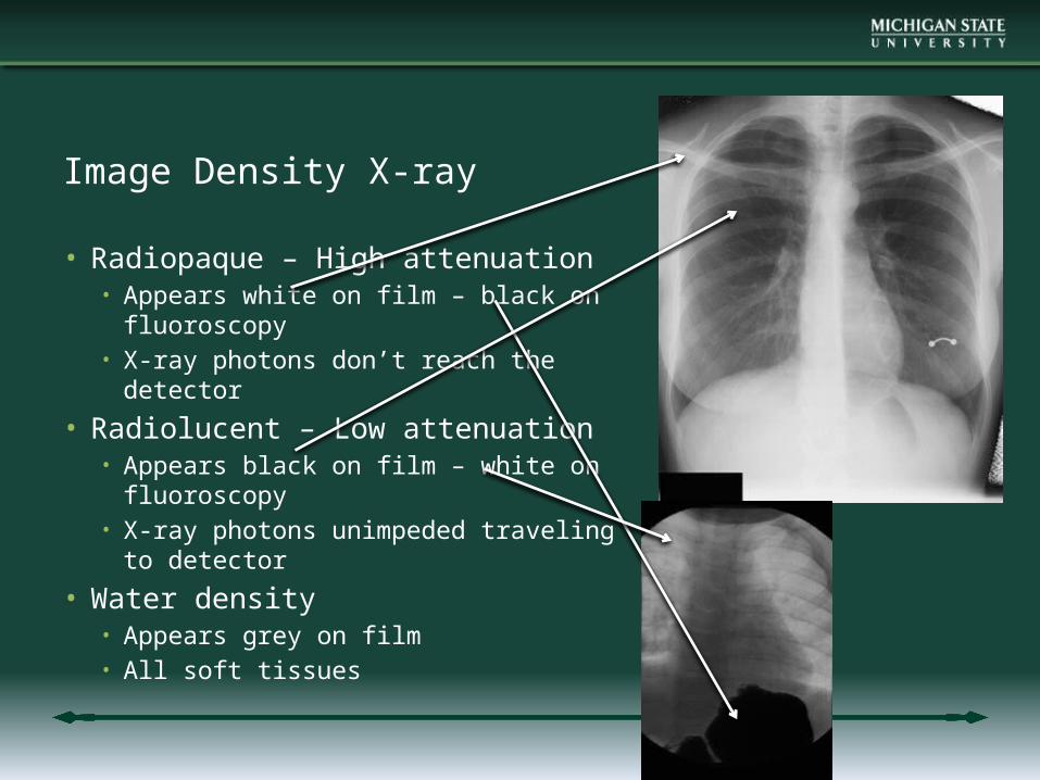
Image Density X-ray
• Radiopaque – High attenuation• Appears white on film – black on
fluoroscopy• X-ray photons don’t reach the
detector
• Radiolucent – Low attenuation• Appears black on film – white on
fluoroscopy• X-ray photons unimpeded traveling
to detector
• Water density• Appears grey on film• All soft tissues

Natural Contrast
• Differential contrast between bone and soft tissues
• Differential contrast between soft tissues and air
• Little difference between various tissue types i.e. fat, muscle, solid organs, blood….

Natural Contrast
• Pathologic processes may cause differences in natural densities that can be visualized on the X-ray; • high density tumor in air filled lung- white• Low density cyst in radio-opaque bone- black
• Pathologic processes of almost the same density as adjoining structures are not visible on X-ray.
• May need to use additional artificial contrast to visualize a density difference

Contrast Agents
• Contrast material (radio-opaque or radio-lucent) administered to see structures or pathologic processes that would not be seen otherwise
• Some useful contrast agents• Barium sulfate in the GI tract• Iodine compounds in the vessels• Carbon dioxide in the vessels or GI tract• Naturally occurring air in the GI tract

Fluoroscopic Room
RadiosensitiveScreen
Video Camera