involvement of aryl hydrocarbon receptor in myelination
TRANSCRIPT

Involvement of Aryl hydrocarbon receptor inmyelination and in human nerve sheath tumorigenesisGhjuvan’Ghjacumu Shackleforda,1, Nirmal Kumar Sampathkumara,1, Mehdi Hichora, Laure Weilla, Delphine Meffrea,Ludmila Juriceka, Ingrid Laurendeaub, Aline Chevalliera, Nicolas Ortonnec, Frédérique Larousseried, Marc Herbine,Ivan Biècheb, Xavier Coumoula, Mathieu Beraneckf, Etienne-Emile Baulieug,2, Frédéric Charbonniera, Eric Pasmantb,and Charbel Massaada,2
aUniversity Paris Descartes, INSERM UMR 1124, Faculty of Basic and Biomedical Sciences, 75270 Paris Cedex 6, France; bEA7331, Université Paris Descartes,Faculté de Pharmacie de Paris, 75270 Paris Cedex 6, France; cDepartment of Pathology, Henri Mondor Hospital, 94010 Créteil, France; dDepartment ofPathology, Cochin Hospital, 75014 Paris, France; eCNRS UMR 7179, Département Ecologie et Gestion de la Biodiversité, Muséum National d’HistoireNaturelle, 75231 Paris Cedex 5, France; fUniversity Paris Descartes, CNRS UMR 8119, Faculty of Basic and Biomedical Sciences, 75270 Paris Cedex 6, France;and gParis-Saclay University, INSERM UMR-1195, 94276 Le Kremlin-Bicêtre Cedex, France
Contributed by Etienne-Emile Baulieu, December 18, 2017 (sent for review September 18, 2017; reviewed by Ruth M. Stassart and Carles Vilarino-Guell)
Aryl hydrocarbon receptor (AHR) is a ligand-activated transcriptionfactor involved in xenobiotic metabolism. Plexiform neurofibromas(PNFs) can transform into malignant peripheral nerve sheath tumors(MPNSTs) that are resistant to existing therapies. These tumors areprimarily composed of Schwann cells. In addition to neurofibroma-tosis type 1 (NF1) gene inactivation, further genetic lesions are re-quired for malignant transformation. We have quantified the mRNAexpression levels of AHR and its associated genes in 38 human sam-ples. We report that AHR and the biosynthetic enzymes of its en-dogenous ligand are overexpressed in human biopsies of PNFs andMPNSTs. We also detect a strong nuclear AHR staining in MPNSTs.The inhibition of AHR by siRNA or antagonists, CH-223191 and tri-methoxyflavone, induces apoptosis in human MPNST cells. SinceAHR dysregulation is observed in these tumors, we investigateAHR involvement in Schwann cell physiology. Hence, we studiedthe role of AHR in myelin structure and myelin gene regulation inAhr−/− mice during myelin development. AHR ablation leads to lo-comotion defects and provokes thinner myelin sheaths around theaxons. We observe a dysregulation of myelin gene expression andmyelin developmental markers in Ahr−/− mice. Interestingly, AHRdoes not directly bind to myelin gene promoters. The inhibition ofAHR in vitro and in vivo increased β-catenin levels and stimulatedthe binding of β-catenin on myelin gene promoters. Taken together,our findings reveal an endogenous role of AHR in peripheral myeli-nation and in peripheral nerve sheath tumors. Finally, we suggest apotential therapeutic approach by targeting AHR in nerve tumors.
AHR | myelin | nerve | MPNST | neurofibroma
Neurofibromatosis type 1 (NF1, MIM162200) is an autosomaldominant neurocutaneous tumor predisposition syndrome
caused by a germ-line heterozygous mutation in the NF1 gene (1).The most common NF1-associated tumors are benign peripheralnerve sheath tumors called neurofibromas, which may be dermal orplexiform (2). Dermal neurofibromas are typically small and growas discrete lesions in the dermis, whereas plexiform neurofibromas(PNFs) can develop internally along the plexus of major peripheralnerves and become quite large. PNFs can undergo transformationto malignant peripheral nerve sheath tumors (MPNSTs) in about10% of NF1 patients (3). MPNSTs are resistant to conventionaltherapies, and their deep-seated position and locally invasive growthhinder complete surgical resection with a high degree of relapse,rendering new treatments highly indispensable. Peripheral nerveSchwann cells (SCs) are the primary pathogenic cells in neurofi-bromas and MPNSTs, as biallelic mutation or loss of the NF1 geneoccurs uniquely in tumor SCs (4). NF1 encodes the RAS-GAP(Rat sarcoma GTPase activating protein) neurofibromin, and RASsignaling is elevated in neurofibroma SCs (5). Neurofibromas maydevelop from SCs or Schwann cell progenitors (SCPs) due to theinactivation of Nf1 at the SCP stage or in adult mice (6, 7).
SCs carry out the myelination of axons in the nerve (8). Myelinis essential for rapid action potential propagation along the axons(9). Peripheral myelin gene expression [Myelin Protein Zero (P0)and Peripheral Myelin Protein-22 (PMP22)] is tightly regulated inSCs (10). KROX20 (11) and the Wnt/β-catenin signaling pathway(12) are known positive regulators of myelin gene expression,whereas SOX2 (13) and LXR (14) exert a negative effect.Little is known of the additional cooperating genetic events
potentially required for full PNFs’ malignant transformation.Previous research from our laboratory and others have shown anactivation of the Wnt signaling pathway during malignant trans-formation of PNFs to MPNSTs (15, 16). Indeed, the Wnt pathwayis a common pathway acting on myelin development as well as ma-lignant transformation. There is growing evidence that overactivationof the AHR (aryl hydrocarbon receptor) signaling pathway canalter Wnt/β-catenin signaling in development, different humancancers, and disease (17–19).AHR is a ligand-activated transcription factor that is best
known for mediating the toxicity and tumor-promoting propertiesof the carcinogen 2,3,7,8-tetrachlorodibenzo-p-dioxin (TCDD),commonly referred to as dioxin. The activation of AHR provokesits heterodimerization with AHR Nuclear Translocator (ARNT).
Significance
Aryl hydrocarbon receptor (AHR) is well known to mediatexenobiotic metabolism in vertebrates. Growing evidence re-veals that AHR seems to have endogenous roles in the devel-opment and functioning of different organs. In our currentstudy, we describe a role of AHR in peripheral myelination andin nerve sheath tumors. We show that the AHR pathway isdysregulated in human biopsies of nerve tumors. The blockadeof AHR provokes cell death in nerve tumors, suggesting atherapeutic avenue in the treatment of this invasive cancer.Furthermore, the inhibition of Ahr in mice provokes locomotordefects and alteration of myelin structure. This work unravelsan endogenous role of Ahr in peripheral myelination and apotential treatment of nerve tumours.
Author contributions: M.B., E.-E.B., F.C., E.P., and C.M. designed research; G.S., N.K.S.,M.H., L.W., D.M., L.J., I.L., A.C., M.H., I.B., M.B., and C.M. performed research; N.O., F.L.,and X.C. contributed new reagents/analytic tools; G.S., N.K.S., L.W., I.B., X.C., M.B., E.-E.B.,F.C., E.P., and C.M. analyzed data; and E.-E.B., E.P., and C.M. wrote the paper.
Reviewers: R.M.S., University Clinic Leipzig; and C.V.-G., University of British Columbia.
The authors declare no conflict of interest.
Published under the PNAS license.1G.S. and N.K.S. contributed equally to this work.2To whom correspondence may be addressed. Email: [email protected] [email protected].
This article contains supporting information online at www.pnas.org/lookup/suppl/doi:10.1073/pnas.1715999115/-/DCSupplemental.
www.pnas.org/cgi/doi/10.1073/pnas.1715999115 PNAS | Published online January 19, 2018 | E1319–E1328
NEU
ROSC
IENCE
PNASPL
US
Dow
nloa
ded
by g
uest
on
Janu
ary
26, 2
022
They bind specific responsive elements called Xenobiotic responsiveelements (XREs), located in the promoter regions of the targetgenes, consequently leading to their transactivation (e.g., Cyto-chrome 1A1). Nevertheless, its endogenous functions begin toemerge, and natural ligands of the AHR, like tryptophan deriv-atives, were identified. The AHR is present in ancient invertebrateorganisms like Caenorhabditis elegans, where it is responsible forthe regulation of GABAergic neuron fate specification (18).Kynurenine is a tryptophan catabolite generated via TryptophanDioxygenase (TDO) and Indoleamin 2,3-Dioxygenases (IDO).By activating AHR, kynurenine promotes tumor-cell survival andmalignancy of glioma (19). Despite the discovery of the cognateligand of AHR, the endogenous role of the AHR remains poorlyexplored, especially in the peripheral nervous system.In this study, we report an up-regulation of AHR signaling in
PNFs and MPNSTs. Furthermore, the inhibition of AHR pro-motes MPNST cell apoptosis. On the other hand, we investigatethe role of AHR in normal SC physiology and myelin generegulation using Ahr−/− mice. These mice have thinner myelinsheaths, dysregulated myelin gene expression, and consequentlocomotor deficiencies. Taken together, our results elicit a roleof the AHR in peripheral myelin gene regulation and a stimulationof AHR activity in malignant transformation of peripheral nervesheath tumors. Our results also suggest that targeting AHR couldbe a promising avenue in the treatment of PNFs and MPNSTs.
ResultsAHR Signaling Pathway Is Activated in Peripheral Nerve SheathTumorigenesis. In a series of 38 tumors, we first analyzed mRNAexpression of AHR, AHRR (AHR Repressor), and ARNT (Table1). AHR was significantly up-regulated in PNFs (sevenfold) and inMPNSTs (ninefold), compared with the dermal neurofibromas.AHRR was significantly down-regulated in PNFs (threefold) andMPNSTs (twofold) compared with the dermal neurofibromas,while ARNT expression was not significantly altered in either thePNFs or the MPNSTs.Several genes implicated in the AHR pathway were also tested,
including kynurenine metabolism (IDO1, IDO2, and TDO2), in-flammation and immune pathways (IL-1β, IL-6, IL-8, CXCL5, andCXCL6), SC markers (S100B and P0), and proliferation (MKI67).IDO1 and TDO2 (but not IDO2) were significantly up-regulated inPNFs and MPNSTs. Concerning the inflammatory pathway, IL-1β,IL-6, IL-8, CXCL5, and CXCL6 mRNA levels showed significantoverexpression in PNFs and MPNSTs, compared with the dermalneurofibromas (Table 1). It is noteworthy that AHR and the en-zymes of its endogenous ligands are remarkably higher in MPNSTsthan in PNFs. The proliferation-associated gene MKI67 was over-expressed in the MPNSTs. SC-specific gene (S100B, P0) expressionwas shown to be significantly down-regulated in MPNSTs.We also performed an immunostaining of AHR in MPNSTs
(Fig. 1). We detected strong nuclear AHR staining in a pro-portion of tumor cells, while intratumoral vessels and fibroblastsin a fibrous septum (taken as internal negative controls) were notstained. Altogether, our data show that the AHR pathway isactivated in human PNFs and MPNSTs.
AHR Inhibition Induced Apoptosis in MPNST Cell Lines. As we ob-served, the AHR pathway is activated in human PNFs andMPNSTs. Subsequently, we hypothesized that inhibiting theAHR could affect MPNST cell survival. We used two AHRantagonists [CH-223191 (CH) and 6,2′,4′-trimethoxyflavone(TMF)] to inhibit AHR activity in STS26T. Treatment with CH(50 μM at 48 h and 72 h) modestly increased cell death andenhanced apoptosis and shifted to the late apoptotic state(double positive for both Annexin and PI cell population) asquantified by Annexin V and PI staining (Fig. 2A, 48 h, and Fig.2B, 72 h and Fig. S1A). CH did not have any effect at 10 μM,whereas TMF exhibited a more potent effect on MPNST cell
death even at 10 μM. TMF (10 μM, 48 h or 72 h) decreased cellviability by ∼50%. After incubation with TMF (10 μM, 72 h),∼20% of the cells underwent apoptosis and 30% were late apo-ptotic. As expected, CH and TMF inhibited the expression of theAHR target gene CYP1A1 (Fig. 2C). To confirm that this is due toAHR inhibition, we silenced AHR by siAHR. AHR silencing alsoinduced apoptosis in STS26T dose-dependently (Fig. 2D). Si-lencing of AHR drastically decreased the mRNA levels of AHRand its target genes CYP1A1 and CYP1B1 (Fig. 2E). As the IL-1βexpression was enhanced in PNFs and MPNSTs (Table 1), ac-cordingly the inhibition of AHR either by siRNA or antagonistsreduced IL-1β expression in STS26T cells (Fig. 2F).We confirmed these results using another MPNST cell line,
90–8. As expected, CH inhibited the expression of the classicalAHR target genes CYP1A1 and CYP1B1 (Fig. S2A). Treatmentwith CH (50 μM) increased the number of dead cells (Fig. S2 Band C) and enhanced apoptosis. We confirmed this result withsiAHR (Fig. S2 D and E). Furthermore, it also reduced the ex-pression of IL-1β (Fig. S2G).
Ahr Ablation in Mice Caused Locomotor Defects and Led to ThinnerMyelin Sheath.Given the fact that PNF and MPNST are complextumors with different cells, we analyzed the expression of AHRand related components in different cells like SCs, mast cells,and endothelial cells. AHR mRNA is predominant in SCs andmay suggest its importance in SC physiology (Table 2). The ob-served activation of the AHR pathway and the down-regulation ofthe myelin gene (P0) and as well as S100B in PNFs and MPNSTscompelled us to investigate further the role of AHR in normal SCs.Henceforth, we analyzed AHR expression in sciatic nerves and SCs(Fig. 3 A–C). AHR is expressed in the nerve (Fig. 3 A and C) and islocalized in the nucleus of an SC as well as the perinuclear com-partment (Fig. 3B).We analyzed the locomotor behavior of Ahr−/−. We measured
stride time using a motorized treadmill (Fig. 3D). No significantdifference was observed with treadmill speed at 10 cm/s. How-ever, when the treadmill speed was increased at 16 cm/s, Ahr−/−
mice showed a slower stride than WT mice. At a higher speed,Ahr−/− mice were not able to maintain the rhythm and thereforecould not be tested.Mice were then subjected to fixed speed rotarod protocol
(14 rpm) for 5 consecutive days where the time to withstand therotating rod was measured (Fig. 3E). The WT mice significantlyimproved their performance over days, indicating an ameliorationof their motor coordination, whereas the time latency of Ahr−/−
mice remained constant. Indeed, WT mice were able to stay onthe rotating bar on day 5 for 207 ± 100 s, while Ahr−/− miceremained only for 12 ± 3 s. Our findings suggest that the Ahr−/−
mice have impaired locomotion and motor coordination. Then, weperformed the static rod test (Fig. 3G). All WT mice stayed 100 son the bar. In contrast, Ahr−/− mice were unsuccessful in staying(Fig. 3H). Finally, motor performance was evaluated using a dy-namic rod test. The Ahr−/− mice were three times slower than theWT mice in crossing the rod (Fig. 3I). The Ahr−/− mice alsoexhibited more faults while crossing the rod (Fig. 3I).We studied myelin structure by electron microscopy. The my-
elin sheath of Ahr−/− mice was significantly thinner than controls(Fig. 3 J and K), and the axonal perimeter was higher in Ahr−/−
mice compared with controls (Fig. 3L). We plotted the g ratios ofsciatic nerve fibers as a function of their respective axonal pe-rimeters (Fig. 3M). We observed reduction in myelin thickness forall axon calibers, but smaller axons were more affected.Western blot (WB) showed a significant increase of P0 and
PMP22 in Ahr−/− sciatic nerves (Fig. 3N and Fig. S5). Consistentwith these results, the expression of the negative regulator ofmyelination, SOX2, was reduced by 40% in Ahr−/− mice (Fig. 3N),while the expression of active β-catenin was enhanced by twofoldand total β-catenin was decreased by 40%. Therefore, the ratio of
E1320 | www.pnas.org/cgi/doi/10.1073/pnas.1715999115 Shackleford et al.
Dow
nloa
ded
by g
uest
on
Janu
ary
26, 2
022

active β-catenin to total β-catenin is increased 3.5-fold in Ahr−/−
mice, suggesting an activation of the Wnt/β-catenin pathway.
Ahr Is Involved in Myelination During Development. To determinewhen these myelin abnormalities arose, we analyzed the ex-pression pattern of AHR during the myelination process at P7,P21, and in 8-wk-old (8WO) mice and compared it to the ex-pression of positive modulators of the myelination process:KROX20 and β-catenin. As depicted in Fig. 4A, AHR expressionwas at the highest level at P7 and then decreased at later timepoints to reach its lowest level in adult mice. KROX20 expres-
sion gradually increased during the myelination process to attainthe highest level at P21 and, as expected, dropped in adult ani-mals. Finally, β-catenin increased at P21, and then its expressionwas brought back to the same levels of P7 and at 8WO.Taking account of this difference in AHR expression during
development, we analyzed the structure of myelin sheaths fromsciatic nerves of P7 mice (Fig. 4B). We observed a mild disor-ganization of myelin sheaths in Ahr−/− mice without affecting theg ratio (Fig. 4C). A moderate increase of the axonal perimeterwas detected in Ahr−/− mice (Fig. 4D). Nevertheless, Ahr−/− micedisplayed fewer myelinated axons (28.88 ± 1.18 for WT vs.20.88 ± 1.23 for Ahr−/−; Fig. 4E). P0 (−50%) and PMP22(−60%) protein expression was decreased in 7-d-old Ahr−/− mice(Fig. 4F). To explore if myelin gene inhibition is due to a dys-regulation in the expression of the major transcriptional regu-lators of SC myelination, we quantified the expression of SOX2,KROX20, total β-catenin, and active β-catenin. The knockout ofAhr did not affect the expression of SOX2. However, we ob-served a decreased KROX20 expression (−60%) that was con-firmed by immunohistochemistry experiments. We observed a25% reduction in the number of KROX20-positive cells in thesciatic nerves as suggested by DAPI (Fig. 4G). Furthermore, WBanalyses showed that active β-catenin expression was diminishedby 30% in Ahr−/− nerves (Fig. 4F). As the amount of total β-cateninwas increased in Ahr−/− mice (+35%), we observed a decrease inthe ratio of active β-catenin to total β-catenin, suggesting animpairment of the Wnt/β-catenin pathway at P7 when Ahr isknocked out. At P21, electron microscopy images revealed thatmyelin sheaths are affected by the deletion of Ahr (Fig. 4H). Theg ratio was decreased, designating thicker myelin sheaths aroundthe axons of Ahr−/− mice (Fig. 4I). The axonal perimeter wasreduced (Fig. 4J) while the numbers of myelinated were not af-fected in Ahr−/− mice (Fig. 4K).
Fig. 1. (A and A′) AHR nuclear staining is detected in MPNST tissues.Formalin-fixed, paraffin-embedded human MPNST samples were stainedwith hematoxylin–eosin–saffron (HES), and AHR was revealed by a specificantibody. (A) A strong nuclear AHR staining was detected in a proportion oftumor cells, while intratumoral vessels (arrows) and fibroblasts in a fibroussepta (*) were not stained, taken as internal negative controls. (A′) A per-oxidase technique, diaminobenzidine chromogen, and hematoxylin coun-terstain were employed. (Scale bar, 100 μm.)
Table 1. Median mRNA levels and ranges of 16 genes in dermal and PNFs and MPNSTs
Dermal neurofibromas,n = 8 PNFs, n = 16 MPNSTs, n = 14
Genes Median Range Median Range Median Range P*
AHR pathwayAHR 1.00 0.13–11.59 6.84 0.01–71.01 8.98 0.57–63.28 P < 0.05AHRR 1.00 0.56–8.44 0.39 0.13–1.60 0.49 0.07–1.80 P < 0.01ARNT 1.00 0.76–3.38 2.12 0.65–5.78 1.71 0.49–7.62 NS
Trp metabolismIDO1 1.00 0.04–10.75 2.98 0.11–41.54 49.38 1.59–681.64 P < 0.01IDO2 1.00 0.21–6.74 0.60 0.07–5.06 0.65 0.01–18.21 NSTDO2 1.00 0.05–101.38 7.21 0.25–77.62 24.77 0.86–2,931.70 P < 0.01
Myelin SCsS100B 1.00 0.64–1.73 0.82 0.10–4.49 0.02 0.01–2.54 P < 0.01MPZ 1.00 0.10–1.79 0.46 0.10–2.54 0.01 0.01–2.24 P < 0.01
InflammationIL-1B 1.00 0.26–4.09 19.44 0.01–809.67 6.15 2.66–644.12 P < 0.05IL-6 1.00 0.50–1.95 19.86 0.18–247.60 25.67 3.06–32.04 P < 0.05IL-8 1.00 0.07–3.15 8.98 0.01–648.32 13.65 0.19–269.89 P < 0.05CXCL5 1.00 0.08–40.19 32.42 0.01–643.00 185.53 0.17–25,864.45 P < 0.01CXCL6 1.00 0.01–5.66 10.86 2.28–1,642.39 24.33 4.60–862.02 P < 0.01
Xenobiotics metabolismCYP1A1 1.00 0.01–1.88 2.16 0.10–18.85 1.50 0.40–4.11 NSCYP1B1 1.00 0.49–5.78 1.01 0.12–9.07 0.69 0.11–4.42 NS
ProliferationMKI67 1.00 0.30–2.82 1.39 0.88–4.77 31.43 13.6–82.9 P < 0.01
AHR pathway is dysregulated in nerve tumors. Total RNAwas isolated from patients having dermal neurfibromas (n = 8), PNFs (n = 16),and MPNSTs (n = 14). qRT-PCR was performed using primers recognizing specifically the genes indicated. Table indicates median andrange mRNA levels. mRNA levels were normalized such that the median value of the dermal neurofibromas was 1. Kruskal Wallis H testwas used to compare MPNSTs vs. PNFs vs. dermal neurofibromas. NS, not significant. The bold values are statistically significant (P < 0.05).*Kruskal Wallis H test: MPNSTs vs. PNFs vs. dermal neurofibromas.
Shackleford et al. PNAS | Published online January 19, 2018 | E1321
NEU
ROSC
IENCE
PNASPL
US
Dow
nloa
ded
by g
uest
on
Janu
ary
26, 2
022

At P21, P0 and PMP22 expression were stimulated by twofoldin Ahr−/− mice. SOX2 expression was still not affected, while theexpression of all of the positive regulators was activated in Ahr−/−
mice: KROX20 (+50%), total β-catenin (+60%), and activeβ-catenin (+35%) (Fig. 4L). Altogether, our data point out acrucial and differential role of Ahr during the myelination processand highlight that Ahr knockout leads to defects in myelin sheaths.
AHR Regulates Myelin Gene Expression in SCs. We transfectedMSC80 cells with either nontargeting siRNA (NT) or siAhr.First, the efficacy of the knockdown was evaluated by qRT-PCR(Fig. S3A) and WB (Fig. S3B). In addition, we assessed thefunctional efficacy of the siAhr on Cyp1A1—luciferase activity
that was reduced drastically (−80%) (Fig. S3C). The knock-down of Ahr increased mRNA levels of P0 and Pmp22 by 50%(Fig. 5A) and the promoter activity of P0 and Pmp22 (Fig. 5B).Taken together, the silencing of Ahr in the SC line or itsknockout in mice led to a dysregulation of myelin gene expression.Then, we examined the mechanism by which the AHR regu-
lates myelin gene expression. We identified by MatInspectorsoftware four potential binding sites for AHR on the level of P0promoter and one putative AHR binding site on the Pmp22promoter (Fig. 5C). Therefore, we tested the AHR binding onP0 and Pmp22 promoters by ChIP. As a positive control, weassayed the binding of the AHR on the level of its target gene,
Fig. 2. AHR inhibition causes apoptosis in the MPNST cell line. (A and B) STS26T cells were treated with either AHR antagonists (CH or TMF) or DMSO (vehicle)for 48 h (A) or 72 h (B). Cells are stained with Annexin and PI to detect cell death by flow cytometry: living cell population (Annexin V−/PI−), apoptotic cells(Annexin V+/PI−), late apoptotic cells (Annexin V+/PI+), and dead cells (Annexin V−/PI+). Results represent the means ± SEM of at least four independentexperiments. *P < 0.05, **P < 0.01, by Bonferroni’s post hoc tests after one-way ANOVA compared with DMSO. (C) The STS26T cell line was treated witheither AHR antagonists (CH or TMF) or DMSO (vehicle). Forty-eight hours later, total RNA was extracted and qRT-PCR was performed using primers recog-nizing CYP1A1 and TBP to normalize the results. They represent the means ± SEM of at least five independent experiments. *P < 0.05 by Student’s t testcompared with DMSO. (D) STS26T cells were transfected with either siAHR (10 nM and 20 nM) or NT; 72 h after medium change, cells were stained withAnnexin and PI to detect cell death by flow cytometry: living cell population (Annexin V−/PI−), apoptotic cells (Annexin V+/PI−), late apoptotic cells (AnnexinV+/PI+), and dead cells (Annexin V−/PI+). Results represent the means ± SEM of at least four independent experiments. *P < 0.05 by Bonferroni’s post hoc testsafter one-way ANOVA compared with control (NT). (E) STS26T cells were transfected with siAHR (20 nM) or NT; 72 h later, total RNA was extracted and qRT-PCR was performed using primers to amplify AHR, CYP1A1, and CYP1B1. Results were normalized to the TBPmRNA level and represent the means ± SEM of atleast five independent experiments. *P < 0.05 by Student’s t test compared with control (NT). (F) STS26T cells were reverse-transfected with siAHR (20 nM) orNT or incubated with CH (50 μM, 72 h), TMF (10 μM, 72 h), or DMSO. Total RNA was extracted and qRT-PCR experiments were performed using primersrecognizing IL-1β and TBP to normalize. Results represent the means ± SEM of at least five independent experiments. *P < 0.05 by Student’s t test comparedwith control (NT).
E1322 | www.pnas.org/cgi/doi/10.1073/pnas.1715999115 Shackleford et al.
Dow
nloa
ded
by g
uest
on
Janu
ary
26, 2
022

Cyp1A1 promoter. As expected, the AHR bound to the Cyp1A1promoter (Fig. 5D). This binding was enhanced by twofold aftertreatment with TCDD (100 nM, 1 h). However, we did not detectAHR interaction with any of the putative binding sites identifiedon Pmp22 (Fig. 5E) and P0 (Fig. 5F), even after the incubation ofMSC80 with TCDD (loading control, Fig. S4A). These datashowed that AHR does not seem to interact directly with myelingene promoters.The other possible mechanism of action of AHR is the cross-
talk with another signaling pathway. We previously showed thatthe activated Wnt/β-catenin pathway enhances the expression ofperipheral myelin genes (12). Furthermore, we demonstrated thatthe knocking out of Ahr in mice altered β-catenin expression in thesciatic nerve (Fig. 3N). Therefore, we addressed the question ofwhether the action of AHR is mediated by the Wnt/β-cateninpathway. After silencing Ahr in MSC80 cells, we observed a 50%increase in the protein levels of active β-catenin and a 30% in-crease of total β-catenin levels (Fig. 5G). Furthermore, to supportthe hypothesis, knockdown of Ahr led to a significant stimulationof mRNA expression levels of Wnt signaling components Lrp6,Dvl2, Dvl3, and Axin2 (Fig. 5H). Then, we assayed the functionalityof the Wnt/β-catenin pathway. The SuperTOP Flash-Luciferaseconstruct, bearing several binding sites for T cell factor/lymphoidenhancer factor (TCF/LEF), was activated by 2.5-fold 48 h aftersiAHR (Fig. 5I). To assess how AHR modifies β-catenin levels inMSC80 cells, we assumed that AHR is able to interact withβ-catenin and stimulate its degradation as described in intestinecancer cells (20). Immunoprecipitation of AHR indicated protein–protein interaction of β-catenin and AHR in MSC80 cells (Fig. 5J).Consequently, we hypothesized that the knockdown of Ahr
increases the recruitment of β-catenin with TCF/LEF complexon the level of myelin gene promoters. We localized LEF/TCF–β-catenin binding sites on P0 and PMP22 promoters (12) (Fig.5K). ChIP assays with β-catenin antibodies showed that Ahr si-lencing increased the recruitment of β-catenin on P0 (Fig. 5Land Fig. S4B, loading control) and Pmp22 promoters (Fig. 5Mand Fig. S4B). Moreover, the binding of β-catenin was also en-hanced after Ahr silencing on the level of a Wnt target gene,Axin2 (Fig. S4C). GAPDH was used as a negative control (Fig.S4D). Collectively, these data demonstrate that AHR exerts itseffect on myelin genes through the Wnt/β-catenin pathway.
DiscussionWe reveal that AHR plays a crucial role in the myelinationprocess of the nerve and is overexpressed in nerve tumors. We
demonstrated that the AHR signaling pathway is activated in aseries of NF1-associated nerve sheath tumors. We further ana-lyzed other genes linked to the AHR and involved in tryptophanmetabolism that lead to the synthesis of kynurenine, an endog-enous ligand of AHR (20–22). The expression of two genesparticipating in the first steps of tryptophan metabolism, namelyIDO1 and TDO2, was significantly increased in PNFs andMPNSTs. IDO1 and TDO2 were found to be remarkably in-creased in malignant form compared with the benign form ofthese tumors, suggesting an increased AHR activity in the ma-lignant transformation. The enzyme IDO1 mediates the first,rate-limiting, step in tryptophan metabolites to kynurenine and isup-regulated under an inflammatory microenvironment (23).Through generation of downstream metabolites, IDO1 enzymeactivity may affect immunity, including specific immunomodu-latory or cytotoxic functions (21). It has been reported thatL-kynurenine activates AHR (19), which positively regulates IDO1expression in immune cells, such as dendritic cells. Our observationshowing a progressive up-regulation of IDO1 and TDO2 in PNFs(threefold and fivefold) and a further stimulation in MPNSTs (50-fold and 25-fold) suggests that kynurenine synthesis leading toAHR activation may play a role in peripheral nerve sheath tumorprogression to malignancy.In addition to the biallelic inactivation of the NF1 gene, sup-
plemental genetic lesions are necessary for malignant progres-sion of PNFs. Alterations of additional genes (CDKN2A/B,TP53, SUZ12, EED) were detected in MPNSTs (3, 24). We havepreviously shown that the Wnt/β-catenin pathway was dysregu-lated in MPNSTs (15), and the present study reveals that theAHR pathway is also activated in these tumors. Furthermore, wehave described an elevation of Wnt5a (15) and AHR expressionin MPNST tumors. Conversely, the knockdown of AHR in MPNSTcells inhibits Wnt5a expression (Fig. S2 H and I). Therefore, ourdata suggest a possible link between AHR and Wnt/β-cateninpathways in MPNSTs.Consequently, we inhibited AHR in MPNST cell lines to alter
their survival. Both AHR antagonists (CH or TMF) and siAHRinduced apoptosis of two MPNST cell lines: 90–8 and STS26T.Again, this observation highlights the importance of AHR in SCmalignancy in addition to NF1. Furthermore, AHR inhibitioncould down-regulate IL-1β, which was elevated in MPNST tu-mors. Previous results suggested that AHR activation coupledwith inflammatory signals can lead to synergistic induction of IL-6 expression in tumor cells (25). We revealed a significant increaseof IL-1β, IL-6, IL-8, CXCL5, and CXCL6 mRNA levels in PNFsand MPNSTs. These results confirm previous in vitro findingsshowing that AHR activation may promote the induction of IL-6 and IL-8 (26, 27) and highlight the implication of AHR in theregulation of an inflammation pathway (28).Previous studies from our laboratories and others have shown
that some signaling pathways that are involved in the myelinationprocess (Wnt/β-catenin, mTOR, ERK) are deregulated inMPNST (12, 29, 30). Therefore, we inquired about the endog-enous role of AHR in normal SC physiology and peripheralnerve myelination. We revealed that AHR is expressed in SCand in the sciatic nerve and that Ahr−/− mice present an alter-ation in locomotion. Additionally, electron microscopy analysesdepicted thinner myelin sheaths. These data unveil the role ofAHR on SC myelinating functions. We performed electron mi-croscopy analyses during the myelination process. They depicteda disorganization of myelin sheaths depending on the develop-mental stage. The first observation was that the myelin thicknesswas not affected at an early stage of myelination (P7). However,the number of myelinated axons was decreased in Ahr−/− mice,suggesting a delay in myelination. Then, we observed thickermyelin sheaths at P21, while adult animals had thinner myelinsheaths around the axons. These observations could be compa-rable to CMT1A patients (duplication of the PMP22 gene) who
Table 2. Relative mRNA expression of eight genes in differenthuman cells
Genes SCs Mast cells Endothelial cell
AHR pathwayAHR 426 11 6AHRR 200 1,342 78ARNT 1,105 492 479
Trp metabolismIDO1 10 NE 2IDO2 NE NE NETDO2 14 3 NE
Myelin markersS100B 66,360 2 6MPZ 24,336 4 5
AHR is predominantly expressed in SCs compared with mast cells and en-dothelial cells. Total RNA was isolated from normal human tissues. The genemRNA levels (calculated as described in Materials and Methods) were basedon the amount of the target message relative to the endogenous control TBPmessage, to normalize the starting amount and quality of total RNA. NE, notexpressed.
Shackleford et al. PNAS | Published online January 19, 2018 | E1323
NEU
ROSC
IENCE
PNASPL
US
Dow
nloa
ded
by g
uest
on
Janu
ary
26, 2
022

Fig. 3. Ahr ablation modifies myelin structure and locomotor behavior in adult mice. (A) Immunohistochemistry was performed on WT sciatic nerve tolocalize AHR (green) and P0 (red). The slides were imaged using a confocal microscope. This experiment was repeated at least three times, and a typicalexperiment is presented here. (Scale bar, 100 μm.) (B) An immunocytochemistry experiment was performed on MSC80 cells. AHR localization is highlighted inred; nuclei were stained with Hoechst (blue). This experiment was repeated at least three times, and a typical experiment is presented here. (Scale bar, 10 μm.)(C) WB showing the expression of AHR in MSC80 cells and in sciatic nerve. (D) In the gait parameter, 8-wk-old WT and Ahr−/− mice were placed on a treadmillat the speed of 10 cm/s and 16 cm/s. Stride time was measured, ***P < 0.001 by Student’s t test when Ahr−/− were compared with control mice. (E) For therotarod test, 8-wk-old WT and Ahr−/− mice were subjected to the rotarod test for 5 d. The time that mice could stay on the rotarod was measured each day. Atleast five animals per group were tested. *P < 0.05, **P < 0.01 using Mann–Whitney test compared with control for each day of the experiment. (F) For therod test, 8-wk-old WT and Ahr−/− mice were placed on the rod to perform either static or dynamic rod tests. Representative pictures of posture of the mice onthe rod are shown. The arrowhead indicates right hind limb where the Ahr−/− mouse slipped (G) For the static rod test, measurement of the time that WT andAhr−/− mice could stay without dropping from the rod. The test was stopped when mice succeeded in staying 100 s. (H) For the dynamic rod test, the averagespeed to cross the rod was measured (cm/s). (I) The number of faults (slips) counted for either WT or Ahr−/−. At least five animals per group were tested. *P < 0.05,**P < 0.01 by Student’s t test when Ahr−/− were compared with WT mice. (J) Sciatic nerves were isolated from eitherWT or Ahr−/− mice (8 wk old). Then, ultrathin(50–90 nm) cross-sections were prepared from Epon embedded nerves. (Scale bar, 2 μm.) (K) Myelin thickness was calculated by g-ratio determination. (L) Axonalperimeter was estimated using electronic microscopy pictures quantified by ImageJ software. At least five animals per genotype were used. (M) The g ratios wereplotted against the axonal perimeter. (N) Adult male WT or Ahr−/− mice were killed, and then their sciatic nerves were dissected (at least n = 6 per group). Totalproteins were extracted, andWBs were performed using antibodies against P0, PMP22, SOX2, KROX20, β-catenin, active β-catenin, and α-tubulin (loading control).Figures represent a typical experiment. WBs were quantified using ImageJ software. Black horizontal line between the lanes in the gels signifies a crop was made.Error bars indicate SEM. *P < 0.05, **P < 0.01 by Student’s t test when Ahr−/− were compared with control mice.
E1324 | www.pnas.org/cgi/doi/10.1073/pnas.1715999115 Shackleford et al.
Dow
nloa
ded
by g
uest
on
Janu
ary
26, 2
022
are hypermyelinated in early childhood and then, 1 y after, ahypomyelination of some fibers is observed; it is caused by de-myelination and remyelination cycles (31). The Ahr constitutiveinvalidation also modified the perimeters of the axons, showing acorrelation between the formation of the myelin sheaths andaxonal development. However, we cannot exclude that couldalso affect neuronal development directly. Prenatal exposure ofTCDD in rats provoked a delay in oligodendrocyte differentia-tion and maturation (32). TCDD could also disrupt the functionsof AHR to provoke neuropathies. In fact, rats intoxicated by
TCDD showed severe defects in myelin structure of tibial andsciatic nerve and reduction in nerve conduction velocity (33, 34).Our results reinforce not only the endogenous role of AHR in anormal myelination process but also the mechanism by which itregulates myelination.P0 and PMP22 myelin proteins are essential for an adequate
structure of myelin sheath. Indeed, their dysregulation or mutationis deleterious for myelin sheath structure, leading to peripheralneuropathies. P0 expression was drastically down-regulated inMPNST, and the knockout of AHR enhanced P0 expression in SCs.
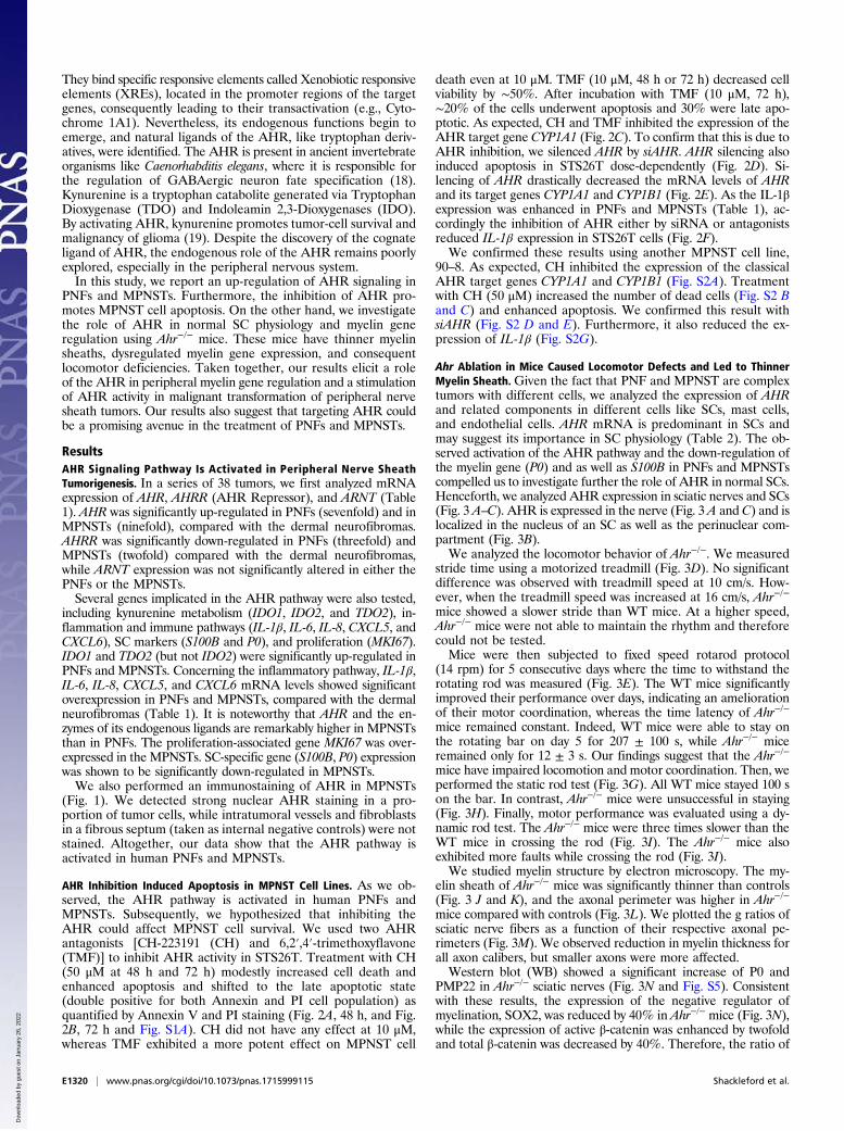
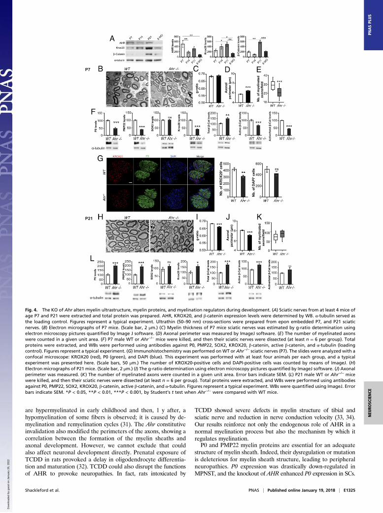
Fig. 4. The KO of Ahr alters myelin ultrastructure, myelin proteins, and myelination regulators during development. (A) Sciatic nerves from at least 4 mice ofage P7 and P21 were extracted and total protein was prepared. AHR, KROX20, and β-catenin expression levels were determined by WB. α-tubulin served asthe loading control. Figures represent a typical experiment. Ultrathin (50–90 nm) cross-sections were prepared from epon embedded P7, and P21 sciaticnerves. (B) Electron micrographs of P7 mice. (Scale bar, 2 μm.) (C) Myelin thickness of P7 mice sciatic nerves was estimated by g-ratio determination usingelectron microscopy pictures quantified by Image J software. (D) Axonal perimeter was measured by ImageJ software. (E) The number of myelinated axonswere counted in a given unit area. (F) P7 male WT or Ahr−/− mice were killed, and then their sciatic nerves were dissected (at least n = 6 per group). Totalproteins were extracted, and WBs were performed using antibodies against P0, PMP22, SOX2, KROX20, β-catenin, active β-catenin, and α-tubulin (loadingcontrol). Figures represent a typical experiment. (G) Immunohistochemistry was performed onWT or Ahr−/− sciatic nerves (P7). The slides were analyzed with aconfocal microscope: KROX20 (red), P0 (green), and DAPI (blue). This experiment was performed with at least four animals per each group, and a typicalexperiment was presented here. (Scale bars, 50 μm.) The number of KROX20-positive cells and DAPI-positive cells was counted by means of ImageJ. (H)Electron micrographs of P21 mice. (Scale bar, 2 μm.) (I) The g-ratio determination using electron microscopy pictures quantified by ImageJ software. (J) Axonalperimeter was measured. (K) The number of myelinated axons were counted in a given unit area. Error bars indicate SEM. (L) P21 male WT or Ahr−/− micewere killed, and then their sciatic nerves were dissected (at least n = 6 per group). Total proteins were extracted, and WBs were performed using antibodiesagainst P0, PMP22, SOX2, KROX20, β-catenin, active β-catenin, and α-tubulin. Figures represent a typical experiment. WBs were quantified using ImageJ. Errorbars indicate SEM. *P < 0.05, **P < 0.01, ***P < 0.001, by Student’s t test when Ahr−/− were compared with WT mice.
Shackleford et al. PNAS | Published online January 19, 2018 | E1325
NEU
ROSC
IENCE
PNASPL
US
Dow
nloa
ded
by g
uest
on
Janu
ary
26, 2
022

The knockout of Ahr severely altered P0 and PMP22 expressionduring myelin development. At P7, when AHR expression is sup-posed to reach its maximal expression, we observed an inhibition ofP0 and PMP22 expression, while starting from P21 until adulthoodthe KO of Ahr increased the expression of myelin genes. Thus, itappears that AHR differentially regulates P0 and PMP22 during de-velopment. The dysregulation of the myelin gene could be attributed
to a modification of developmental markers that modulate P0 andPMP22 expression. For example, at P7, the positive regulators of my-elin gene expression (KROX20 and β-catenin) are down-regulated.This undoubtedly provoked the inhibition of P0 and PMP22. At latertime points (P21 and adult), myelin genes are activated consequentlyto the Ahr ablation. Concomitantly, KROX20 and β-catenin areoverexpressed and SOX2 (negative effector) is inhibited.
Fig. 5. AHR does not bind to myelin gene promoters in MSC80 cells but acts via the Wnt/β-catenin pathway. MSC80 cells were transfected with a siRNAdirected against Ahr (siAHR) or a NT siRNA (NT). (A) Total mRNA was extracted, and P0 and Pmp22 transcripts were quantified by qRT-PCR. All results werenormalized to the 26S mRNA level. (B) MSC80 cells were cotransfected with siAhR or NT and P0-luciferase or PMP22-luciferase constructs. Luciferase activitywas assayed. Results represent the means ± SEM of at least five independent experiments. ***P < 0.001 by Student’s t test compared with control (NT).(C) Putative binding sites of AHR located on the levels of P0 and PMP22 were identified by means of MatInspector software. MSC80 cells were incubated withvehicle (Nonane) or TCDD (100 μM, 24 h). ChIP assays with antibodies against AHR or Mock IgG (nonrelevant negative control) or Histone H3 (positive control)were performed. qRT-PCR was performed with primers recognizing AHR putative sites located on (D) Cyp1A1, an AHR target gene; (E) Pmp22; and (F) P0genes. Results represent the means ± SEM of at least four independent experiments. *P < 0.05 and ***P < 0.001 by Tukey’s post hoc tests after one-wayANOVA compared with control. (G) MSC80 cells were transfected with siRNA directed against Ahr (siAhr) or a nontargeting siRNA (NT) for 48 h. Total proteinswere extracted, and WBs were performed using antibodies against total β-catenin or active β-catenin and α-tubulin as a loading control. Figures represent atypical experiment. WB quantifications were done using ImageJ software. (H) Total RNA was extracted, and qRT-PCR experiments were performed usingprimers recognizing Lrp6, Dvl2, Dvl3, and Axin2. RT-PCR was normalized using 26S RNA. Data represent the mean ± SEM of at least four independent ex-periments. (I) MSC80 cells were transiently transfected with SuperTOP Flash-luciferase (STF-Luciferase) and with either siAHR or NT. Forty-eight hours later,luciferase activity was analyzed. Results represent the means ± SEM of at least six independent experiments performed in duplicate. *P < 0.05, **P < 0.01, byStudent’s t test. (J) Coimmunoprecipitation assays were performed on MSC80 cell extracts using AHR, blotted for β-catenin by WB, and lgG was used as acontrol. (K) The binding sites for TCF/LEF–β-catenin are localized on the promoter level for P0 and Pmp22 as described in ref. 12. MSC80 cells were transientlytransfected with either siAhr or NT for 48 h after; ChIP assays with antibodies against β-catenin or Mock IgG or Histone H3 were peformed. Primers rec-ognizing TCF/LEF–β-catenin binding sites on the levels of P0 (L) and Pmp22 (M) were used. Results represent the means ± SEM of at four independent ex-periments. *P < 0.05, ***P < 0.001 by Tukey’s post hoc tests after one-way ANOVA compared with control.
E1326 | www.pnas.org/cgi/doi/10.1073/pnas.1715999115 Shackleford et al.
Dow
nloa
ded
by g
uest
on
Janu
ary
26, 2
022

We confirmed that the knockdown of Ahr activates the Wnt/β-catenin pathway in the SC line. The expression of the majorcomponents of the canonical Wnt pathway (i.e., Lrp6,Dvl2, β-catenin,and Axin2) and Wnt target promoter (Super-TOP Flash) areenhanced after Ahr silencing. How could AHR stimulate P0 andPMP22 promoter activities? The first hypothesis is that AHRbinds directly on the level of putative XREs present on P0 andPMP22 promoter regions. Unexpectedly, none of these bindingsites recruited AHR even in the presence of the AHR agonistdioxin. Therefore, we postulated that the action of AHR ispossibly mediated by β-catenin because we observed an increaseof β-catenin binding on TCF/LEF binding sites of P0 and Pmp22promoters after Ahr knockdown.AHR has a dual function in regulating gene expression as a
ligand-activated transcription factor and as an E3 ubiquitin ligase.Kawajiri et al. (20) showed that AHR E3 ubiquitin activity has arole in the β-catenin degradation pathway that is independent ofthe adenomatous polyposis coli system in intestine. We assumethat the inhibition of AHRmay reduce the degradation of β-catenin,which is consistent with (i) the direct interaction that we have de-tected between AHR and β-catenin proteins and (ii) the β-cateninincrease observed after Ahr knockdown.In conclusion, our findings unraveled that AHR is involved in
the myelination process. This pathway is overexpressed in nervetumors, which could also serve, along with Wnt/β-catenin pathway,as new markers for this specific type of cancer. As the inhibition ofAHR induced apoptosis of MPNST cells, we can propose that thisapproach is further translatable. For instance, using either nano-particles or adeno-associated virus (35) to deliver siAHR to in-duce apoptosis of these malignant cells could be suggested inadjuvant to existing chemotherapy with reduced doses. This approachcan reduce complications like chemotherapy-induced peripheralneuropathy. Hence, our results indicate that AHR could representa novel and potential therapeutic target for this type of periph-eral nerve sheath tumor that is poorly responsive to existingchemotherapeutics.
Materials and MethodsAnimals. Ahr knockout mice were a gift from Alvaro Puga, Center for Envi-ronmental Genetics, University of Cincinnati, College of Medicine, Cincinnati.Ahr−/− mice and WT controls were maintained on a mixed strain background(C57BL/6j:RJ) and housed in a temperature-controlled room with a 12 h light/dark cycle. All experiments were performed on age-matched male mice. Adultanimals were fed ad libitum with water and Global-diet 2016S from Harlan. Allaspects of animal care were approved by the ethical committee for animalresearch of the University Paris Descartes (CEEA.34).
Treadmill Test: Forced Locomotion. Adult male Ahr−/− (n = 5) and control micewere acclimated to the testing room for 24 h before each test. The forcedlocomotion of the mice was investigated using a commercial motorizedtreadmill. The speed of the treadmill was controlled by a tachymeter. Afterthe habituation session (speed, 6 cm/s), mice were tested at a speed of 10 cm/sand 16 cm/s the next day.
Rod Test: Static Paradigm. The same set of animals were subjected to both thetreadmill test and rod test. The setup consisted of a rod 9.5 mm in diameterand 5 cm in length. After a period of habituation, animals were securelyplaced on the rod. The ability of the animal to remain on the rod withoutfalling was investigated for a maximum period of 100 s.
Rod Test: Dynamic Paradigm.Dark platforms were setup at one end of the rod,where themicewere let for habituation for 10min.Micewere primed towalkfrom a starting point along the bar (70 cm long, 9.5 mmdiameter) fixed 30 cmabove the bench. Once the mice had succeeded in reaching the final desti-nation several times by the priming procedure, the test was recorded in threetrials. We calculated the speed and the number of faults.
Rotarod Test: Forced Paradigm. Balance and motor coordination was testedusing amotorized rotarod LE8200 apparatus (Bioseb). Adultmicewere placedon a rotating treadmill drum (3 cm diameter), and the mice’s latency to fall
during the first 5 min of the trial was noted. The average time of the fall foreach trial was measured. Fixed-speed rotarod has been performed at 14 rpmfor 5 consecutive days for WT mice and Ahr−/− mice (n = 5).
Transmission Electron Microscopy. Electron microscopy was done using fixedsciatic nerves following the protocol described previously (14). Electron mi-croscopy images were used for calculating the g ratio by ImageJ.
Patients and Samples. After clinical examination, the surgical removal ofdermal neurofibromas was proposed in case of esthetical burden. Resectionor removal of PNFs was described previously (ethical authorization IRB-HumanCPP17/79, A0296746) (15).
Expression of Genes Involved in the AHR Signaling Pathway in Patients’Samples. QRT-PCR was carried out as described previously (15).
Immunohistochemistry.Human samples. Immunohistochemistry of patient samples was carried out asdescribed previously (15). Immunostaining of AHR (rabbit polyclonal antibodyclone H211; Santa Cruz) was done at 3 μg/mL after heat antigen retrieval inpH 6.0 citrate buffer. In all samples, we checked for the absence of staining inintratumoral vessels and fibroblasts as for internal negative controls.Mouse samples. Sciatic nerves were dissected and immersed in 4% para-formaldehyde and embedded with Tissue-Tek. Sections (10 μm) were cutusing Cryostat. Primary anitbodies against P0 (Aves Laboratories Inc.), AHR(Enzo), and KROX20 (Thermo Fisher Scientific) were used. Then, the sampleswere incubated at room temperature for 1 h, 30 min with the appropriatesecondary antibody (Alexa Fluor 488 donkey anti-mouse, Cy3-conjugatedAffiniPure Donkey anti-Rabbit, and Cy3-conjugated Affinity Pure Donkeyanti-Chicken). Nuclei were stained with Hoechst 33342 (Pierce Biotechnology).Sections were imaged using a confocal microscope (LSM510; Carl Zeiss).
Cell Culture. The mouse SC line (MSC80) was maintained in DMEM supple-mented with 10% decomplemented FCS (HyClone-Perbio), 1% penicillin, 1%streptomycin (Gibco), and 1% Glutamine. All cultures were grown at 37 °C ina humidified atmosphere of 5% CO2.
The MPNST-derived cell lines STS26T and 90–8 were maintained in ad-vanced RPMI-1640 with 15% heat-inactivated FBS, 2.75‰ Bovine PituitaryExtract, 100 U/mL penicillin, and 100 μg/mL streptomycin (Invitrogen).
Plasmids and Chemicals. p1646 (Cyp1A1-luc) was graciously provided by A.Puga, Cincinnati, OH. siRNAs directed against Ahr were purchased fromQiagen. The Ahr was targeted by four siRNAs recognizing four differentregions of the Ahr transcript. TCDD was purchased from LGC standards andCH-22319 and TMF from Sigma.
Transient Transfections. MSC80 cells (2.5 × 10⁵ cells per well) were grown insix-well plates, transfected using Effecten reagents according to the manu-facturer’s instructions (Qiagen), and luciferase activity was determined usingthe enzymatic method described previously (36). The β-galactosidase activitywas used to normalize the transfection efficiency.
STS26T or 90–8 cells were transfected with either NT siRNA (DHARMACON)or siAHR (DHARMACON) using RNAiMAX (Thermo Fisher Scientific) accordingto the manufacturer’s instruction.
WB. WB assay was performed using the standard techniques. Primary anti-bodies against P0 and PMP22 were purchased from Abcam and Sigma, re-spectively; β-catenin from BD Transduction; active β-catenin from Millipore;α-tubulin from Sigma; SOX2 from Abcam; and AHR from Enzo Life Science(Biomol SA-210).
qPCR for MSC80. Total RNA from cultured MSC80 was extracted using TRIzolReagent (Invitrogen) according to the manufacturer’s instructions. qPCR wasdone as described previously (14). All results were normalized to the 26SmRNA level and calculated using the ΔCt method. The primer sequencesused in real-time qPCR are listed in Table S1.
ChIP. MSC80 cells (107) were fixed with 1% formaldehyde for 10 min. Thecross-linking was quenched with 0.125 M glycine for 10 min. Fixed chromatinwas then isolated and physically sheared by sonication to obtain DNAfragments 300–600 bp in length. Immunoprecipitation was done usingProtein A/G magnetic nanobeads (Dynabeads Life Technologies). The anti-bodies used in ChIP assays were as follows: 5 μg of AHR-specific antibody(Biomol SA-210), 5 μg of β-catenin (BD Transduction #610154), Histone H3
Shackleford et al. PNAS | Published online January 19, 2018 | E1327
NEU
ROSC
IENCE
PNASPL
US
Dow
nloa
ded
by g
uest
on
Janu
ary
26, 2
022

(Cell Signaling 2650), and 5 μg Rabbit IgG (Sigma I5006). qRT-PCRs were doneas mentioned earlier, with the primer pairs listed in Table S1. Then, thepercentage of input was calculated as mentioned in ref. 37.
Co-Immunoprecipitation. MSC80 cell lysates were precleared with protein-A(Dynabeads Life Technologies), and then the total protein was measuredwith BioRad RCDC. The lysate was treated with DNase to digest the genomicDNA. We used 3 μg of AHR antibody (Biomol SA-210) to enrich AHR in 100 μgof total protein by using protein A Dynabeads (Rabbit IgG was used as thenegative control). Finally, the enriched protein samples were blotted withusual standard WB techniques with β-catenin antibody (Biomol SA-210).
Flow Cytometry Analysis. STS26T or 90–8 cells cultured in 12-well plates weretreated with AHR antagonists or DMSO. Cells undergoing apoptosis at theindicated time were identified using the Annexin V-FITC apoptosis detectionkit (Abcam ab14085), following the manufacturer’s instructions. BD FACS-CantoII was used. A total of 10,000 events were recorded at a light scatter toexclude debris and aggregated with a flow rate of less than 200 cells persecond for each assay. The compensations and the settings were adjustedaccording to the assay. Data were analyzed using FlowJo Software.
Statistical Analysis. Unless otherwise specified, means of treatment groupswere compared with one-way analysis of variance (ANOVA). When theANOVA showed that there were significant differences between the groups,Tukey’s test was used to identify the sources of these differences. P ≤0.05 was considered statistically significant. Two group comparisons wereperformed by the Student’s t test. As the mRNA levels did not fit a Gaussiandistribution in human samples, (i) the mRNA levels in each subgroup ofsamples were expressed as their median values and ranges, rather than theirmean values and coefficients of variation, and (ii) comparison between thethree types of nerve sheath tumors was assessed by using the nonparametricKruskal Wallis H test. Differences were considered significant at confidencelevels greater than 95% (P < 0.05).
ACKNOWLEDGMENTS. The authors acknowledge Dr. Alain Schmitt of theCochin Imaging Facility, Jean-Maurice Petit of the Imaging Facility des SaintsPères, and Stephanie Dupuy from the flow cytometry facility. This work wasfunded by INSERM, University Paris Descartes, the Agence National pour laRecherche (ANR), and theAgence Nationale de Sécurité Sanitaire de l’Alimentation,de l’Environnement et du travail (ANSES). G.S., N.K.S., M.H., and A.C. receivedPhD fellowships from the French Ministry of Research (MNRT). L.J. received aPhD fellowship from Region Ile de France.
1. Friedman JM (1999) Epidemiology of neurofibromatosis type 1. Am J Med Genet 89:1–6.
2. Sabbagh A, et al.; Members of the NF France Network (2009) Unravelling the geneticbasis of variable clinical expression in neurofibromatosis 1. Hum Mol Genet 18:2768–2778.
3. Tucker T, Wolkenstein P, Revuz J, Zeller J, Friedman JM (2005) Association betweenbenign and malignant peripheral nerve sheath tumors in NF1. Neurology 65:205–211.
4. Serra E, et al. (1997) Confirmation of a double-hit model for the NF1 gene in benignneurofibromas. Am J Hum Genet 61:512–519.
5. Cichowski K, Jacks T (2001) NF1 tumor suppressor gene function: Narrowing the GAP.Cell 104:593–604.
6. Chen Z, et al. (2014) Cells of origin in the embryonic nerve roots for NF1-associatedplexiform neurofibroma. Cancer Cell 26:695–706.
7. Wu J, et al. (2008) Plexiform and dermal neurofibromas and pigmentation are causedby Nf1 loss in desert hedgehog-expressing cells. Cancer Cell 13:105–116.
8. Garbay B, Heape AM, Sargueil F, Cassagne C (2000) Myelin synthesis in the peripheralnervous system. Prog Neurobiol 61:267–304.
9. Nave K-A, Werner HB (2014) Myelination of the nervous system: Mechanisms andfunctions. Annu Rev Cell Dev Biol 30:503–533.
10. Niemann A, Berger P, Suter U (2006) Pathomechanisms of mutant proteins in Charcot-Marie-Tooth disease. Neuromolecular Med 8:217–242.
11. Topilko P, et al. (1994) Krox-20 controls myelination in the peripheral nervous system.Nature 371:796–799.
12. Tawk M, et al. (2011) Wnt/beta-catenin signaling is an essential and direct driver ofmyelin gene expression and myelinogenesis. J Neurosci 31:3729–3742.
13. Le N, et al. (2005) Analysis of congenital hypomyelinating Egr2Lo/Lo nerves identifiesSox2 as an inhibitor of Schwann cell differentiation and myelination. Proc Natl AcadSci USA 102:2596–2601.
14. Makoukji J, et al. (2011) Interplay between LXR and Wnt/β-catenin signaling inthe negative regulation of peripheral myelin genes by oxysterols. J Neurosci 31:9620–9629.
15. Luscan A, et al. (2014) The activation of the WNT signaling pathway is a hallmark inneurofibromatosis type 1 tumorigenesis. Clin Cancer Res 20:358–371.
16. Watson AL, et al. (2013) Canonical Wnt/β-catenin signaling drives human Schwann celltransformation, progression, and tumor maintenance. Cancer Discov 3:674–689.
17. Long X, et al. (2015) MMP-12-mediated by SARM-TRIF signaling pathway contributesto IFN-γ-independent airway inflammation and AHR post RSV infection in nude mice.Respir Res 16:11.
18. Huang X, Powell-Coffman JA, Jin Y (2004) The AHR-1 aryl hydrocarbon receptor andits co-factor the AHA-1 aryl hydrocarbon receptor nuclear translocator specify GABAergicneuron cell fate in C. elegans. Development 131:819–828.
19. Opitz CA, et al. (2011) An endogenous tumour-promoting ligand of the human arylhydrocarbon receptor. Nature 478:197–203.
20. Kawajiri K, et al. (2009) Aryl hydrocarbon receptor suppresses intestinal carcinogen-esis in ApcMin/+ mice with natural ligands. Proc Natl Acad Sci USA 106:13481–13486.
21. Mezrich JD, et al. (2010) An interaction between kynurenine and the aryl hydrocar-bon receptor can generate regulatory T cells. J Immunol 185:3190–3198.
22. Zhai L, et al. (2015) The role of IDO in brain tumor immunotherapy. J Neurooncol 123:395–403.
23. Litzenburger UM, et al. (2014) Constitutive IDO expression in human cancer is sus-tained by an autocrine signaling loop involving IL-6, STAT3 and the AHR. Oncotarget5:1038–1051.
24. Zhu Y, Ghosh P, Charnay P, Burns DK, Parada LF (2002) Neurofibromas in NF1:Schwann cell origin and role of tumor environment. Science 296:920–922.
25. DiNatale BC, Schroeder JC, Francey LJ, Kusnadi A, Perdew GH (2010) Mechanistic in-sights into the events that lead to synergistic induction of interleukin 6 transcriptionupon activation of the aryl hydrocarbon receptor and inflammatory signaling. J BiolChem 285:24388–24397.
26. Kolasa E, Houlbert N, Balaguer P, Fardel O (2013) AhR- and NF-κB-dependent in-duction of interleukin-6 by co-exposure to the environmental contaminant benzan-thracene and the cytokine tumor necrosis factor-α in human mammary MCF-7 cells.Chem Biol Interact 203:391–400.
27. Vogel CFA, et al. (2014) Cross-talk between aryl hydrocarbon receptor and the in-flammatory response: A role for nuclear factor-κB. J Biol Chem 289:1866–1875.
28. Hollingshead BD, Beischlag TV, Dinatale BC, Ramadoss P, Perdew GH (2008) In-flammatory signaling and aryl hydrocarbon receptor mediate synergistic induction ofinterleukin 6 in MCF-7 cells. Cancer Res 68:3609–3617.
29. Kendall JJ, et al. (2016) CK2 blockade causes MPNST cell apoptosis and promotesdegradation of β-catenin. Oncotarget 7:53191–53203.
30. Brundage ME, et al. (2014) MAF mediates crosstalk between Ras-MAPK and mTORsignaling in NF1. Oncogene 33:5626–5636.
31. Gabreëls-Festen A, Wetering RV (1999) Human nerve pathology caused by differentmutational mechanisms of the PMP22 gene. Ann N Y Acad Sci 883:336–343.
32. Fernández M, et al. (2010) A single prenatal exposure to the endocrine disruptor2,3,7,8-tetrachlorodibenzo-p-dioxin alters developmental myelination and remyeli-nation potential in the rat brain. J Neurochem 115:897–909.
33. Grehl H, Grahmann F, Claus D, Neundörfer B (1993) Histologic evidence for a toxicpolyneuropathy due to exposure to 2,3,7,8-tetrachlorodibenzo-p-dioxin (TCDD) inrats. Acta Neurol Scand 88:354–357.
34. Grahmann F, Claus D, Grehl H, Neundörfer B (1993) Electrophysiologic evidence for atoxic polyneuropathy in rats after exposure to 2,3,7,8-tetrachlorodibenzo-p-dioxin(TCDD). J Neurol Sci 115:71–75.
35. Ali HM, et al. (2014) Effects of silencing the RET/PTC1 oncogene in papillary thyroidcarcinoma by siRNA-squalene nanoparticles with and without fusogenic companionGALA-cholesterol. Thyroid 24:327–338.
36. Massaad C, Garlatti M, Wilson EM, Cadepond F, Barouki R (2000) A natural sequenceconsisting of overlapping glucocorticoid-responsive elements mediates glucocorti-coid, but not androgen, regulation of gene expression. Biochem J 350:123–129.
37. Frank SR, Schroeder M, Fernandez P, Taubert S, Amati B (2001) Binding of c-Myc tochromatin mediates mitogen-induced acetylation of histone H4 and gene activation.Genes Dev 15:2069–2082.
E1328 | www.pnas.org/cgi/doi/10.1073/pnas.1715999115 Shackleford et al.
Dow
nloa
ded
by g
uest
on
Janu
ary
26, 2
022