involvement an extracellular glucan sheath populus ...aem.asm.org/content/57/2/374.full.pdf ·...
TRANSCRIPT
Vol. 57, No. 2APPLIED AND ENVIRONMENTAL MICROBIOLOGY, Feb. 1991, p. 374-3840099-2240/91/020374-11$02.00/0Copyright © 1991, American Society for Microbiology
Involvement of an Extracellular Glucan Sheath during Degradationof Populus Wood by Phanerochaete chrysosporium
KATIA RUEL AND JEAN-PAUL JOSELEAU*Centre de Recherches sur les Macromolecules Vegetales, Centre Nationale de la Recherche Scientifique,
Universite Joseph Fourier, B.P. 53 X, 38041 Grenoble Cedex, France
Received 19 July 1990/Accepted 28 November 1990
Observations by transmission electron microscopy of wood samples of Populus tremula inoculated with thewhite rot fungus Phanerochaete chrysosporium showed that, at certain stages of their growth cycle, hyphae wereencapsulated by a sheath which seems to play an active role in the wood cell wall degradation. Chemical andimmunochemical techniques and '3C nuclear magnetic resonance spectroscopy were applied to demonstrate the0-1,3-1,6-D-glucan nature of the sheath. Double-staining methods revealed the interaction between theextracellular peroxidases involved in lignin degradation and the glucan mucilage. The glucan was also shownto establish a material junction between the fungus and the wood cell wall. It was concluded that, by means ofthese interactions, the sheath provides a transient junction between the hyphae and the wood, thus establishinga point of attachment to the site of the degradation. The association of peroxidases to the glucan matrix is infavor of the role of the sheath as a supporting structure. Furthermore, that the sheath was hydrolyzed duringthe attack demonstrated its active role both in providing the H202 necessary to the action of peroxidases andin providing a mode of transport of the fungal enzymes to their substrates at the surface of the wood cell wall.
Many important groups of fungi, like Basidiomycetes,Ascomycetes, and Oomycetes, contain mixed linked 1-1,3-1,6-D-glucans as part of their cell walls. Depending upon thefungal species and life cycle, the glucan may be found in theextracellular culture medium, where both its production andits degree of branching vary by culture conditions and age (2,3). Glucans may thus be found at three different locations infungal culture: first they may be part of the hyphal walls,they may also be excreted in the culture medium, and finallythey may constitute a sheath thought to be covalently linkedto the wall glucans and chitin (39). The 1-linked glucans aredifferently soluble in alkali or dilute acids. This behavior wastentatively related to structural differences, particularly inthe degree of branching of the 13-1,6-linked glucosyl residuesonto the main 1-1,3-glucan main chain (14). The structuraldiversity presented by the glucans due to their origin andtheir location must be related to the diversity of functionsthat they display, such as their antitumor activity (7, 44) ortheir involvement as elicitors in plant pathogenic fungiinteractions (1). The chemical variations in the primarystructure of the glucans influence the secondary and tertiarystructures of individual chains, molecular assembly, or ag-gregation, providing various physical properties in the solu-bility and gel-forming ability which in turn control thebiological functions of the fungal glucans (37).The wood-degrading white rot fungus Phanerochaete
chrysosporium possesses ,B-1,3-1,6-linked glucans in its cellwalls and as an extracellular sheath but can also excreteglucan in the culture medium (4). The extracellular forms ofthe polysaccharides are not produced at all stages of growthby the fungus and therefore are not always present. Thefungus is generally grown in low-form culture flasks (4), andin these conditions several factors can affect the productionof the glucan. The exopolysaccharide was not producedunder high nitrogen conditions but was excreted when theglucose concentration in the medium fell below a certain
* Corresponding author.
level (16). The chemical features of the primary structure ofthe main extracellular glucan produced by P. chrysosporiumwere shown to consist of a ,B-1,3-linked backbone carryingsingle glucosyl groups attached by ,B-1,6 linkages to the mainchain on approximately every second glucosyl residue (8).The degree of polymerization and the extent of branching ofthe glucan are functions of the fungal growth. It was sug-gested that degradation of the glucan by exo- and endoglu-canases, induced during the secondary metabolism of thefungus when the glucose concentration becomes too low,was related to the formation of hydrogen peroxide (4)necessary for lignin degradation by the fungus. P. chrysos-porium is a white rot that displays a glucan sheath surround-ing living hyphae. The sheath appears to have a complex rolein the support and the transport of depolymerizing enzymesin wood decay (28, 29). It could also serve in the nutrition ofthe fungi. A similar possibility of interaction between theglucan sheath and fungal proteins was described by Dicker-son and Baker (13), who demonstrated that a relativelyunspecific affinity of proteins on the wall or sheath polysac-charides could take place. The easy in vitro desorption of theenzymes from the glucan promotes noncovalent binding.Such an interaction between a ,B-glucosidase and the extra-cellular mucilage was recently described in Coriolus versi-color (16), and it was proposed that the immobilization of theenzyme could be the primary function of the glucan matrix.Our previous results (21) obtained with P. chrysosporium
K3, which has relatively high ligninolytic activity, indicatedthat not only cellulases but also hemicellulases and ligninperoxidases (LiP) form associations with the glucan networkof the sheath of P. chrysosporium (36). Similarly, Daniel etal. (11, 12), by using anti-LiP antibodies on another strain ofP. chrysosporium, mentioned an association of the lignin-degrading enzymes with an extracellular material. Also,Blanchette et al. (5), by using anti-xylanase sera to xylanasesfrom different white and brown rot fungi, showed that theseextracellular enzymes could interact with the surroundinghyphal slime layers. Here, in our present work, the glucannature of the slime was characterized by chemical and
374
on Septem
ber 17, 2018 by guesthttp://aem
.asm.org/
Dow
nloaded from

GLUCAN SHEATH OF P. CHRYSOSPORIUM 375
immunochemical techniques. Antibodies directed againstdifferent enzymes involved in wood degradation and se-creted by P. chrysosporium were used to study their possibleextracellular location on the sheath network.The extent of the interaction between the proteins and the
glucan matrix was demonstrated by a double-staining tech-nique which used immunocytochemical markers of the pro-teins together with a polysaccharide chemical staining.
MATERIALS AND METHODS
Plant material and organism. Wood samples were takenfrom a 20-year-old aspen tree (Populus tremula) harvested inFrance. Wood wafers (4 by 20 by 50 mm) were degraded for6 weeks (at STFI, Stockholm, Sweden) by the wild-typestrain K3 of the white-rot fungus P. chrysosporium asdescribed previously (32).
Preparation of the antisera. Four of the antisera used inthis work were directed against the protein of the enzyme,and one was directed against a polysaccharide component ofthe fungus wall. The anti-LiP antibodies were polyclonalantibodies raised in rabbits. One was a gift from E. Odier(INAPG, France) and was from LiP 9, pl 4.6 (9). Theimmunoglobulins G were from the third injection (incom-plete Freund adjuvant), harvested at week 9.The second anti-LiP antibody was a gift from M. L.
Niku-Paavola from the Technical Research Centre of Fin-land, Espoo, Finland. The enzyme, LiP (L3), was isolatedfrom the fungus Phebia radiata as described previously (27),and the immunoglobulins G were purified. The antiserumreacts with LiP Li and L3. The use of this antiserum forlocalizing LiP from P. chrysosporium was justified by theresults published in the literature (22, 25).The antiserum directed against the crude enzyme extract
was prepared from P. chrysosporium culture filtrate col-lected during the primary phase of metabolism. The culturefiltrate was concentrated by ultrafiltration before being pre-cipitated with ammonium sulfate. The main enzymatic ac-tivities detected by using the Somogyi-Nelson method (40)were cellulases (carboxymethyl cellulases), xylanases, and,B-glucosidase. No ligninolytic activities were detected. Afterdesalting on a UMH 10 membrane (Schleicher & Schuell),the proteins were injected in rabbits (performed in collabo-ration with R. Guinet, Institut Pasteur, Lyon, France). Theimmunoglobulins G were then purified.The antibody directed against the P-1,3-glucan (3-O-,-D-
glucopyranosyl-D-glucose [laminaribiose]) was a gift fromM. Horisberger. It was a polyclonal antibody and was
prepared as described previously (19).Tissue preparation for transmission electron microscopy.
Wood samples were fixed in different mixtures of glutaral-dehyde (GA) and paraformaldehyde (PF) (0.3% GA, 1% PF;0.5% GA, 4% PF; or 2.5% GA, 2% PF, 0.02% picric acid).All solutions were in 0.1 M phosphate buffer, pH 7.2 to 7.4.Samples were dehydrated in ethanol before being embeddedin methacrylate (32) or glycol-methacrylate (41).
Chemical staining. Lignin was stained by fixation in 2.5%KMnO4 solution (23) before being dehydrated and embeddedin methacrylate or glycol-methacrylate. Polysaccharideswere stained on thin sections by using the periodic acid-thiocarbohydrazide-silver proteinate (PATAg) method ofThiery (43) modified by Ruel et al. (35).Immunocytochemical labeling. Antibodies were used as
postembedding markers. Thin sections of decayed woodwere first incubated on a drop of TBS (0.01 M Tris-phos-phate saline buffer [pH 7.4], 0.5 M NaCl)-0.15 M glycine.
After being rinsed in TBS, the thin sections were floated ona drop of 1% TBS-bovine serum albumin (BSA) or 5 to 10%TBS-normal goat serum before being treated with either ofthe antibodies adjusted to the correct dilution. Duration ofcontact with the primary antibody varied from 14 to 24 h.The secondary marker, labeled with gold (10 nm in diam-
eter), was a goat anti-rabbit antiserum or protein A (JanssenPharmaceutics, Beerse, Belgium). It was diluted 1:10 to 1:30in TBS-normal goat serum or TBS containing 0.1% BSA plus0.5% gelatin from fish skin (Sigma).
Controls. The following control experiments were per-formed: (i) substitution of the primary antibody with preim-mune (or nonimmune) rabbit serum, immunoglobulin Gfraction; (ii) treatment of sections with the gold-labeledsecondary antibody alone, omitting the primary antibodystep; (iii) labeling with the antisera preadsorbed with theirrespective antigens.
13C NMR spectroscopy. The P-glucan was isolated from a
crude polysaccharide fraction precipitated with ethanol froma culture filtrate of P. chrysosporium (a gift from A. J.Buchala, Fribourg, Switzerland). The glucan was obtained inpure form by extraction in dimethyl sulfoxide in an autoclaveby the method of Buchala and Leisola (8), yielding a polymerwhich upon acid hydrolysis contained only D-glucose. Fornuclear magnetic resonance (NMR) recording, the freeze-dried glucan was solubilized in dimethyl sulfoxide-d6 (25 mgin 1 ml) at 70°C. The resulting viscous solution was placed inan NMR tube (10 mm in diameter), and the 13C spectrum wasrecorded on a Brucker AM 300 spectrometer, at 75.46 MHz,taking the middle signal of the CH3 of dimethyl sulfoxide at39.5 ppm downfield from tetramethyl silane.
Affinity adsorption of enzymes on extracellular 0-glucan.Freeze-dried purified P-glucan (25 mg) was equilibrated inacetate buffer (5 mM, pH 5.2) and poured into a Pasteurpipette (31). The enzyme mixtures (1 ml) were applied on thecolumn, which was eluted and then washed with 5 ml ofacetate buffer. The protein contents of the applied solutionand the elution solution were measured by the Bradfordmethod (6).
RESULTS
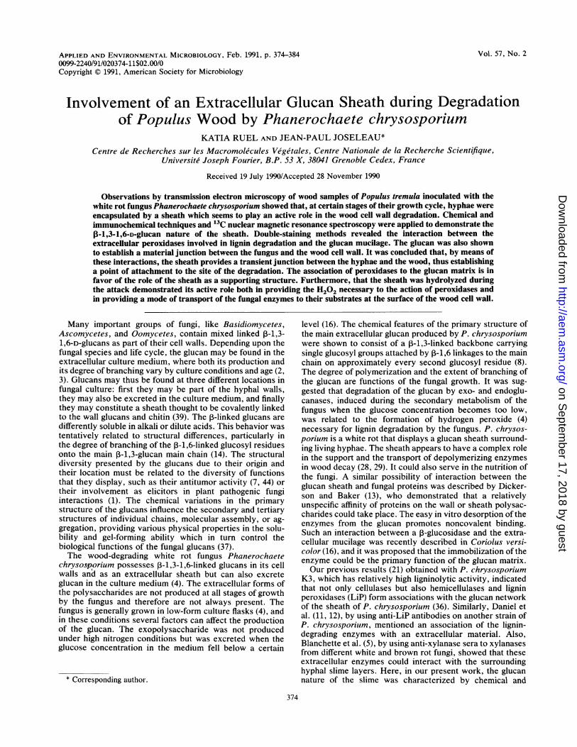
Visualization of the sheath around P. chrysosporium hy-phae. Potassium permanganate is a good contrasting reagentfor polyphenols in transmission electron microscopy and istherefore often used for visualizing lignin and lignin degra-dation products (33) in wood decayed by fungi. In wood cellwalls which essentially consist of polysaccharides and poly-phenols, KMnO4 is specific for lignin. However, otherstructures such as proteins may be contrasted by potassiumpermanganate (18). When thin sections of Populus tremulawere examined after KMnO4 fixation, a strong staining oflignin in wood was observed but the KMnO4 fixation alsoprovided a general contrast of the fungal hyphae. In partic-ular, in some cases, an abundant material became visiblearound hyphae and certain zones of decayed wood (Fig. la).The lack of specificity of the contrasting reagent makes itdifficult to clearly distinguish between mucilage of fungalorigin and lignin degradation or repolymerization products(34) coming from the wood cell wall (Fig. lb). In agreementwith most observations (29), the thick sheath surroundedliving hyphae and accumulated principally at the apex (Fig.la). However, all hyphae with a living cell content were not
always surrounded with a mucilage layer (Fig. lc).A different aspect of the sheath was shown when a general
reagent for contrasting polysaccharides was applied to the
VOL. 57, 1991
on Septem
ber 17, 2018 by guesthttp://aem
.asm.org/
Dow
nloaded from

376 RUEL AND JOSELEAU APPL. ENVIRON. MICROBIOL.
.l
N
on Septem
ber 17, 2018 by guesthttp://aem
.asm.org/
Dow
nloaded from
GLUCAN SHEATH OF P. CHRYSOSPORIUM 377
FIG. 1. Staining of decayed wood and of invading hyphae. Panels a and b were fixed with KMnO4, panels c, d, and e were stained withPATAg, and panel f was stained via immunocytochemical marker. Abbreviations: H, hypha; WCW, wood cell wall; HW, hyphal wall. (a) Thestain underlines the lignin part of the strongly degraded wood cell walls and contrasts with the abundant sheath (arrows) surrounding fungalhyphae and particularly the hyphal tip (arrowhead). (b) The distinction of the limit between fungal sheath and wood degradation products isnot possible with certitude. (c) Hypha in the lumen of a parenchyma cell. Wood cell wall is degraded (arrows), and the hyphal wall is naked.(d) PATAg staining delineates the mesh formed by the glucan. Arrows show the intensively stained fungal wall from which the glucanoriginates. (e) Adhesion of living and dead hyphae via the glucan network of mucilage. (f) The anti-P-1,3-glucan antibody shows the glucanlocalized in the inner part of the hyphal walls and concentrated in the pore separating the hyphal tip from the next cell (arrowhead).
embedded thin sections of decayed wood. The periodicoxidation-based reaction (i.e., PATAg) of Thiery (43), al-though not active on 1->3-linked polysaccharides, gave a
positive reaction with the P-1,3-1,6-D-glucans of P. chryso-sporium. The polysaccharide-specific staining provided de-tailed images of the sheath. The glucan can be seen as a lightand thin mesh anchored in the fungal cell wall from apicalhypha (Fig. ld). The mucilage seems to be mostly associatedwith apical hyphae which have a thick cell wall and may
sometimes expand in such a way that they join together withseveral other hyphae, including dead ones (Fig. le).Another specific staining of P-1,3-glucans was assayed on
Populus tremula samples decayed by P. chrysosporium. Ananti-P-1,3-glucan antibody prepared against 3-0-p-D-glu-copyranosyl-D-glucose (laminaribiose) (19) was applied tothin sections of the wood, and the antigen-antibody complexwas revealed by an immuno-gold labeling technique. Thismarker was specific for unsubstituted linear ,B-1,3-D-glucosylsequences and failed to disclose the P-1,3-1,6-D-glucanpresent in the sheath. However, the wall glucan formed a
specific complex with the antibody (Fig. lf). The goldlabeling was found on all of the hyphal walls, with a
particularly dense marking in the septum localized on thedolipore. This suggests that a particularly high concentration
of linear P-1,3-glucan structures should be present at thisplace.
In all micrographs visualized with either mode of staining,the sheath appeared as an amorphous network different fromthe membranous structures described previously (11). TheKMnO4 and PATAg reactivity of the slime distinguish itfrom the membrane structures often present in myceliumcultures of Sporotrichum pulverulentum (an anamorph of P.chrysosporium) and described previously (15).
Interaction between P. chrysosporium mucilage and thedegraded wood cell walls. The sheath constitutes an exten-sion of the hyphal wall and seems to have, among otherfunctions, a role in establishing contact with the wood cellwall. Hyphal cells, agglutinated in the same mucilage mate-rial, were kept in close proximity with the wood by theinteraction between the sheath and the inner surface of thewood cell wall (Fig. 2a). The unspecific KMnO4 stainingshowed that the hyphal sheath could establish a materialjunction between the fungus and the wood cell wall, thusacting as an adhesive agent between the microorganism andthe wood. The more selective PATAg staining revealed thatthe ,B-glucan network effectively bound to the degradedwood cell wall (Fig. 2b). Such an attachment to cellulosicmaterial was recently demonstrated in the case of Tricho-
VOL. 57, 1991
on Septem
ber 17, 2018 by guesthttp://aem
.asm.org/
Dow
nloaded from

4~~~~~~~~$~~~~~~~~
!~ ~ ~~~_ O:gk 5pm-Q5m;a b
FIG. 2. Interaction between fungal mucilage and wood cell walls. Abbreviations: H, hypha; HS, hyphal sheath; WCW, wood cell wall;ML, compound middle lamella; S2 outer layer of the secondary wall of the wood. (a) Association of the hyphal sheath with the wood cellwall (KMnO4 staining) (b) PATAg staining shows the tight interaction existing between the hyphal sheath and the wood cell wall duringdecay. (c) Residual wood cell walls (arrow) bound to the mucilagenous hyphal sheath (PATAg staining). (d) Loosening of the PATAg-positivenetwork of the fungal sheath (PATAg staining).
378
on Septem
ber 17, 2018 by guesthttp://aem
.asm.org/
Dow
nloaded from

FIG. 3. Interaction of LiP with the fungal sheath. Abbreviations: H, hypha; HS, hyphal sheath; HW, hyphal wall; WCW, wood cell wall.(a) Anti-LiP detects the presence of LiP (arrows) in the mucilagenous part (HS) joining the hyphal wall to the wood cell wall. Note that LiPis also inside the fungal wall (uranyl acetate poststaining). (b) LiP (arrows) is associated with the PATAg-positive mesh of the slime (anti-LiPplus PATAg staining). (c) An abundant slime (HS) surrounds the hypha, but LiP is restricted to the hyphal wall (arrows) (anti-LiP and uranylacetate poststaining). (d) Two morphologically active hyphae; one of them only is excreting LiP (anti-LiP and uranyl acetate poststaining).
379
on Septem
ber 17, 2018 by guesthttp://aem
.asm.org/
Dow
nloaded from
380 RUEL AND JOSELEAU
derma reesei (42). In more advanced stages of degradation,fragments of the wood cell walls could be seen bound to themucilagenous sheath (Fig. 2c). It is remarkable that, onceattached to the wood cell wall, the glucan mesh underwenttwo important changes. The first consisted of a disorganiza-tion of the network, which appeared loosened (Fig. 2d) andin which only a few threads remained. This can be related tothe concomitant thinning of the wood cell wall, whichindicates that an intensive degradation was occurring at thepoint of interaction with the slime. At a more advancedstage, the mucilage eventually disappeared. Another changeundergone by the sheath was that, in zones of active wooddegradation, the slime appeared to be cleaved from thehypha and remained attached to places where the wood cellwall was being attacked.
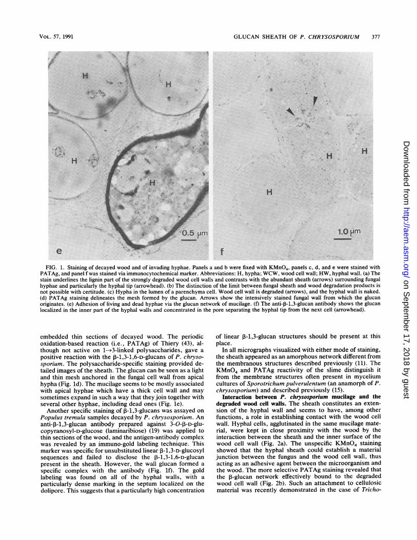
Interaction between 1-glucan sheath matrix and enzymesreleased by P. chrysosporium. Immuno-gold-cytochemicallabeling of the principal types of P. chrysosporium enzymesimplicated in the degradation of the wood cell wall polymersshowed (21, 36) that cellulases and hemicellulases, as well asLiP, could have different localizations in the hyphae. Theymay be intracellular or extracellular, depending on thephysiological state of the fungus. It must be noted that, onthe same section of decayed wood, several stages of decaycould be found. Hyphae at different physiological statescould also be observed (Fig. le) on the same section.Microscopy studies have shown that extracellularly releasedperoxidases can be found at a certain distance from thehyphae in advanced stages of wood decay (11). However, noexplanation was provided for the mechanisms of transport ofthe enzymes from the hypha to the degrading cell wallregion. In the present study, the possible participation of theglucan sheath in the transportation of extracellular glyca-nases and peroxidases was studied. The combination of twomodes of staining, i.e., by chemical reaction for the under-lying structure of the mucilage and by immuno-gold labelingfor the excreted enzymes, demonstrated that the proteinscould be adsorbed on the P-glucan mesh. When an anti-LiPwas applied in combination with uranyl acetate, the unspe-cific uranyl acetate staining showed the mass of mucilagemaking a junction between the hyphae and wood cell wall,and the anti-LiP immuno-gold marker demonstrated both thehyphal wall localization of the LiP and its interaction withthe slime (Fig. 3a). When the periodate-silver marker of theglucan was used with the anti-LiP primary antibody (Fig.3b), gold particles appeared scattered onto the thin silvergrains, thereby delineating the mesh of the glucan sheath.The direct interaction of the enzyme on the P-glucan wasthus evidenced by this double-staining approach. Because ofthis association, it seems that the diffusion of the enzymeswas restricted in distance to the extent of the glucan net-work. However, it must be stressed that the presence of thesheath around hyphae is not necessarily concomitant withthe presence of extracellular enzyme. Figure 3c shows anexample of a hypha in which all of the LiP was concentratedin the wall and none of it was seen associated with theexternal slime. In Fig. 3d, two morphologically identicalhyphae are shown, only one of which is secreting LiP.
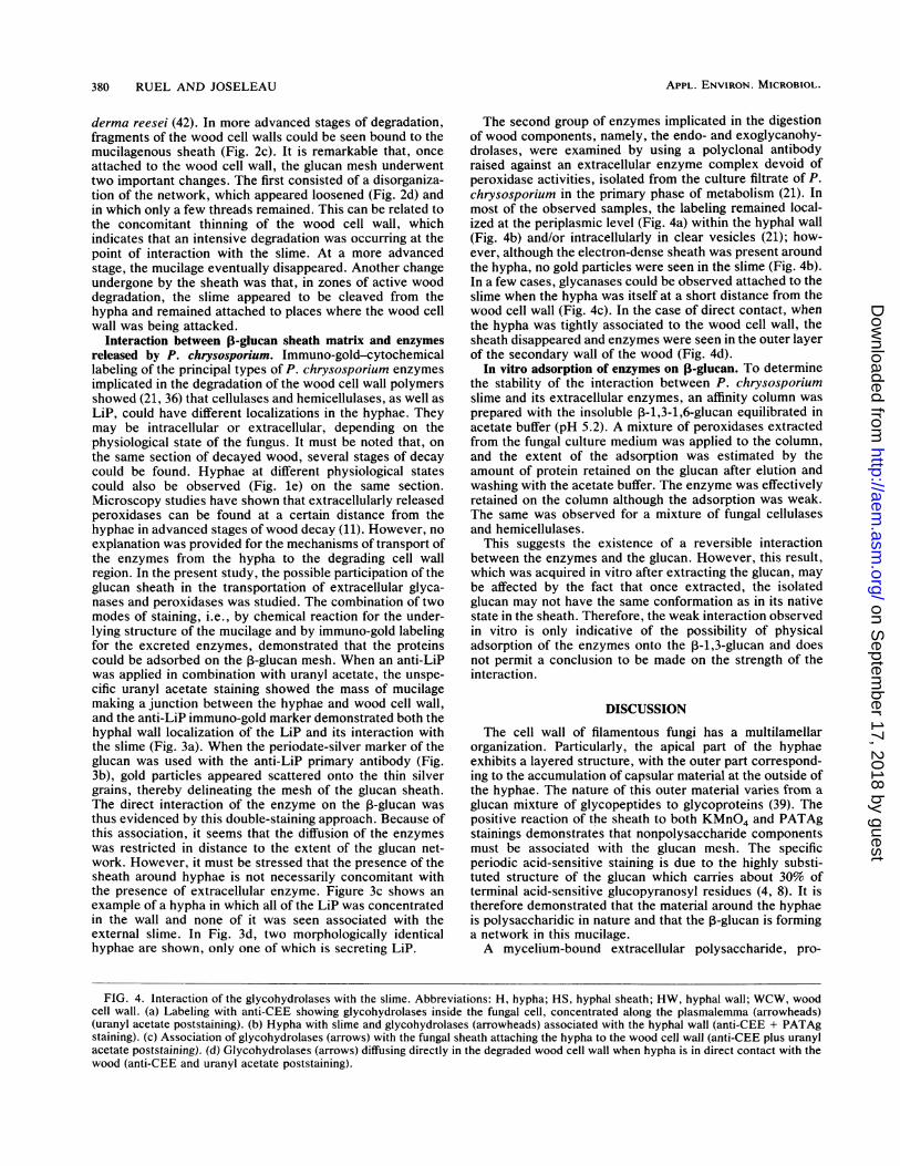
The second group of enzymes implicated in the digestionof wood components, namely, the endo- and exoglycanohy-drolases, were examined by using a polyclonal antibodyraised against an extracellular enzyme complex devoid ofperoxidase activities, isolated from the culture filtrate of P.chrysosporium in the primary phase of metabolism (21). Inmost of the observed samples, the labeling remained local-ized at the periplasmic level (Fig. 4a) within the hyphal wall(Fig. 4b) and/or intracellularly in clear vesicles (21); how-ever, although the electron-dense sheath was present aroundthe hypha, no gold particles were seen in the slime (Fig. 4b).In a few cases, glycanases could be observed attached to theslime when the hypha was itself at a short distance from thewood cell wall (Fig. 4c). In the case of direct contact, whenthe hypha was tightly associated to the wood cell wall, thesheath disappeared and enzymes were seen in the outer layerof the secondary wall of the wood (Fig. 4d).
In vitro adsorption of enzymes on ,8-glucan. To determinethe stability of the interaction between P. chrysosporiumslime and its extracellular enzymes, an affinity column wasprepared with the insoluble P-1,3-1,6-glucan equilibrated inacetate buffer (pH 5.2). A mixture of peroxidases extractedfrom the fungal culture medium was applied to the column,and the extent of the adsorption was estimated by theamount of protein retained on the glucan after elution andwashing with the acetate buffer. The enzyme was effectivelyretained on the column although the adsorption was weak.The same was observed for a mixture of fungal cellulasesand hemicellulases.
This suggests the existence of a reversible interactionbetween the enzymes and the glucan. However, this result,which was acquired in vitro after extracting the glucan, maybe affected by the fact that once extracted, the isolatedglucan may not have the same conformation as in its nativestate in the sheath. Therefore, the weak interaction observedin vitro is only indicative of the possibility of physicaladsorption of the enzymes onto the P-1,3-glucan and doesnot permit a conclusion to be made on the strength of theinteraction.
DISCUSSION
The cell wall of filamentous fungi has a multilamellarorganization. Particularly, the apical part of the hyphaeexhibits a layered structure, with the outer part correspond-ing to the accumulation of capsular material at the outside ofthe hyphae. The nature of this outer material varies from aglucan mixture of glycopeptides to glycoproteins (39). Thepositive reaction of the sheath to both KMnO4 and PATAgstainings demonstrates that nonpolysaccharide componentsmust be associated with the glucan mesh. The specificperiodic acid-sensitive staining is due to the highly substi-tuted structure of the glucan which carries about 30% ofterminal acid-sensitive glucopyranosyl residues (4, 8). It istherefore demonstrated that the material around the hyphaeis polysaccharidic in nature and that the P-glucan is forminga network in this mucilage.A mycelium-bound extracellular polysaccharide, pro-
FIG. 4. Interaction of the glycohydrolases with the slime. Abbreviations: H, hypha; HS, hyphal sheath; HW, hyphal wall; WCW, woodcell wall. (a) Labeling with anti-CEE showing glycohydrolases inside the fungal cell, concentrated along the plasmalemma (arrowheads)(uranyl acetate poststaining). (b) Hypha with slime and glycohydrolases (arrowheads) associated with the hyphal wall (anti-CEE + PATAgstaining). (c) Association of glycohydrolases (arrows) with the fungal sheath attaching the hypha to the wood cell wall (anti-CEE plus uranylacetate poststaining). (d) Glycohydrolases (arrows) diffusing directly in the degraded wood cell wall when hypha is in direct contact with thewood (anti-CEE and uranyl acetate poststaining).
APPL. ENVIRON. MICROBIOL.
on Septem
ber 17, 2018 by guesthttp://aem
.asm.org/
Dow
nloaded from

VOL. 57, 1991 GLUCAN SHEATH OF P. CHRYSOSPORIUM 381
on Septem
ber 17, 2018 by guesthttp://aem
.asm.org/
Dow
nloaded from
382 RUEL AND JOSELEAU
unsubsti tuted
C6substi tuted
I C6
II
C3
I1 Ippm
100 75 50
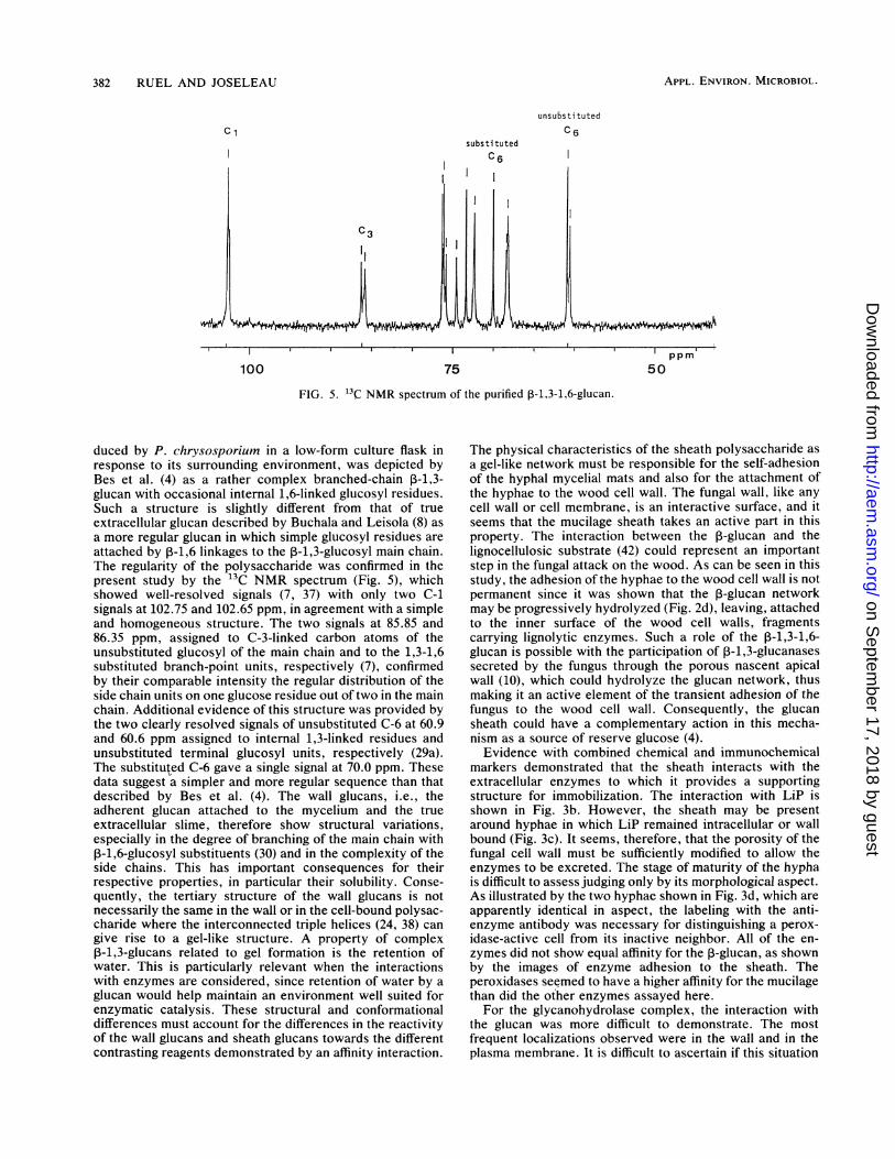
FIG. 5. 13C NMR spectrum of the purified P-1,3-1,6-glucan.
duced by P. chrysosporium in a low-form culture flask inresponse to its surrounding environment, was depicted byBes et al. (4) as a rather complex branched-chain 3-1,3-glucan with occasional internal 1,6-linked glucosyl residues.Such a structure is slightly different from that of trueextracellular glucan described by Buchala and Leisola (8) asa more regular glucan in which simple glucosyl residues are
attached by ,B-1,6 linkages to the P-1,3-glucosyl main chain.The regularity of the polysaccharide was confirmed in thepresent study by the 13C NMR spectrum (Fig. 5), whichshowed well-resolved signals (7, 37) with only two C-1signals at 102.75 and 102.65 ppm, in agreement with a simpleand homogeneous structure. The two signals at 85.85 and86.35 ppm, assigned to C-3-linked carbon atoms of theunsubstituted glucosyl of the main chain and to the 1,3-1,6substituted branch-point units, respectively (7), confirmedby their comparable intensity the regular distribution of theside chain units on one glucose residue out of two in the mainchain. Additional evidence of this structure was provided bythe two clearly resolved signals of unsubstituted C-6 at 60.9and 60.6 ppm assigned to internal 1,3-linked residues andunsubstituted terminal glucosyl units, respectively (29a).The substituted C-6 gave a single signal at 70.0 ppm. Thesedata suggest a simpler and more regular sequence than thatdescribed by Bes et al. (4). The wall glucans, i.e., theadherent glucan attached to the mycelium and the trueextracellular slime, therefore show structural variations,especially in the degree of branching of the main chain with,-1,6-glucosyl substituents (30) and in the complexity of theside chains. This has important consequences for theirrespective properties, in particular their solubility. Conse-quently, the tertiary structure of the wall glucans is notnecessarily the same in the wall or in the cell-bound polysac-charide where the interconnected triple helices (24, 38) can
give rise to a gel-like structure. A property of complex3-1,3-glucans related to gel formation is the retention ofwater. This is particularly relevant when the interactionswith enzymes are considered, since retention of water by a
glucan would help maintain an environment well suited forenzymatic catalysis. These structural and conformationaldifferences must account for the differences in the reactivityof the wall glucans and sheath glucans towards the differentcontrasting reagents demonstrated by an affinity interaction.
The physical characteristics of the sheath polysaccharide asa gel-like network must be responsible for the self-adhesionof the hyphal mycelial mats and also for the attachment ofthe hyphae to the wood cell wall. The fungal wall, like anycell wall or cell membrane, is an interactive surface, and itseems that the mucilage sheath takes an active part in thisproperty. The interaction between the P-glucan and thelignocellulosic substrate (42) could represent an importantstep in the fungal attack on the wood. As can be seen in thisstudy, the adhesion of the hyphae to the wood cell wall is notpermanent since it was shown that the 3-glucan networkmay be progressively hydrolyzed (Fig. 2d), leaving, attachedto the inner surface of the wood cell walls, fragmentscarrying lignolytic enzymes. Such a role of the P-1,3-1,6-glucan is possible with the participation of 3-1,3-glucanasessecreted by the fungus through the porous nascent apicalwall (10), which could hydrolyze the glucan network, thusmaking it an active element of the transient adhesion of thefungus to the wood cell wall. Consequently, the glucansheath could have a complementary action in this mecha-nism as a source of reserve glucose (4).Evidence with combined chemical and immunochemical
markers demonstrated that the sheath interacts with theextracellular enzymes to which it provides a supportingstructure for immobilization. The interaction with LiP isshown in Fig. 3b. However, the sheath may be presentaround hyphae in which LiP remained intracellular or wallbound (Fig. 3c). It seems, therefore, that the porosity of thefungal cell wall must be sufficiently modified to allow theenzymes to be excreted. The stage of maturity of the hyphais difficult to assess judging only by its morphological aspect.As illustrated by the two hyphae shown in Fig. 3d, which areapparently identical in aspect, the labeling with the anti-enzyme antibody was necessary for distinguishing a perox-idase-active cell from its inactive neighbor. All of the en-zymes did not show equal affinity for the ,-glucan, as shownby the images of enzyme adhesion to the sheath. Theperoxidases seemed to have a higher affinity for the mucilagethan did the other enzymes assayed here.For the glycanohydrolase complex, the interaction with
the glucan was more difficult to demonstrate. The mostfrequent localizations observed were in the wall and in theplasma membrane. It is difficult to ascertain if this situation
APPL. ENVIRON. MICROBIOL.
on Septem
ber 17, 2018 by guesthttp://aem
.asm.org/
Dow
nloaded from

GLUCAN SHEATH OF P. CHRYSOSPORIUM 383
is general since the variations in the physiological states offungi are numerous and the release of extracellular enzymescorresponds to an enhanced porosity of the hyphae, which isprobably controlled by autolysis of the wall (10). This mayexplain why hyphae with the same morphological appear-ance seem to behave differently in the excretion of theirenzymes. In cases where the extracellular hydrolases wereseen adsorbed on the sheath, it should be noted that thehypha is situated in close vicinity to the wood cell wall. Itseems that the polysaccharide-degrading enzymes may betransferred to the wood cell wall by a more direct contactthan are the enzymes involved in lignin attack. Severalauthors have shown in vitro interactions between ,-glucansand proteins (13, 16, 26). However, when the LiP extract anda cellulase mixture were put in contact with the extracellularP-glucan in an affinity column, only a little protein adsorp-tion was measured. This means that a weak interactionoccurred between the glucan and the enzymes, in agreementwith a reversible association which is necessary for theenzyme to release from the glucan network of the slime tothe wood cell wall. On the other hand, the in vitro experi-ment does not necessarily reflect the in vivo association ofthe protein on the glucan since, after extraction and isolationby freeze-drying, the resulting conformation of the glucanmight differ from its native tertiary structure. It is wellestablished that native P-1,3-D-glucans which have helicalchain conformations form rather soft gels in aqueous media(17). However, during the process of their isolation whichincludes drying, the polymer undergoes a conformationaltransition and becomes insoluble. As a result, the affinitybetween the enzyme and the isolated glucan packed in thecolumn may have been considerably altered from what itwas in vivo with the glucan in its native conformation. Thepreferential affinity of LiP observed in vivo for the glucanmay be related to the potential source of hydrogen peroxiderepresented by the oxidation of the glucose released byglucan-hydrolyzing enzymes (4). In this process, hydrogenperoxide and the peroxidases would be present at the sametime and in the same location. Such associations betweenlignin-degrading enzymes and the fungal sheath could facil-itate the interaction of lignin products detached from thewood cell wall and the peroxidases (20). Besides providing asource of metabolic glucose and H202, another interestingaspect of the hydrolysis of the I-glucan is related to itstransient existence. The disappearance of the 3-glucan, onceattached to the wood cell wall, results in the release of theadsorbed enzymes at the site of attack of the lignocellulosicwall. Consequently, not only could the glucan matrix servefor the adsorption of the enzymes, but it also could conveythe immobilized proteins to the site of wood degradation bymeans of its hydrolysis by P-1,3-glucanases.
ACKNOWLEDGMENTSWe thank A. J. Buchala for the gift of glucan extract, M. L.
Niku-Paavola for providing the anti-LiP antibody (financed bythe Technology Development Center of Finland), and E. Odier(INAPG, France) for the anti-LiP 9.
REFERENCES1. Anderson-Prouty, A. J., and P. Albersheim. 1975. Host-patho-
gen interactions. VIII. Isolation of a pathogen-synthesizedfraction rich in glucan that elicits a defence response in thepathogen's host. Plant Physiol. 56:286-291.
2. Bartnicki-Garcia, S. 1969. Cell wall chemistry, morphogenesisand taxonomy of fungi. Annu. Rev. Microbiol. 22:87-107.
3. Bartnicki-Garcia, S. 1973. Fundamental aspects of hyphal mor-phogenesis, p. 245-267. In J. M. Ashworth and J. E. Smith
(ed.), Microbial differentiation. Cambridge University Press,Cambridge.
4. Bes, B., B. Pettersson, H. Lennholm, T. Iversen, and K. E.Eriksson. 1987. Synthesis, structure and enzyme degradation ofan extracellular glucan produced in nitrogen-starved cultures ofthe white rot fungus Phanerochaete chrysosporium. Appl. Bio-chem. Biotechnol. 9:310-318.
5. Blanchette, R. A., A. R. Abad, K. R. Cease, R. L. Farrell, R. E.Lovrien, and T. D. Leathers. 1990. Enzyme immunocytochem-istry and ultrastructural localization of cell wall components byenzyme-gold complexes, p. 69-82. In T. K. Kirk and H. M.Chang (ed.), Biotechnology in pulp and paper manufacture,applications and fundamental investigations. Butterworth-Heinemann, Oxford, United Kingdom.
6. Bradford, M. M. 1976. A rapid and sensitive method for thequantitation of microgram quantities of protein utilizing theprinciple of protein-dye binding. Anal. Biochem. 72:248-254.
7. Bruneteau, M., I. Fabre, J. Perret, G. Michel, P. Ricci, J. P.Joseleau, J. Kraus, M. Schneider, W. Blaschhek, and G. Franz.1988. Antitumor active ,-glucans from Phytophtora parasitica.Carbohydr. Res. 175:137-143.
8. Buchala, A. J., and M. Leisola. 1987. Structure of the P-D-glucansecreted by Phanerochaete chrysosporium in continuous cul-ture. Carbohydr. Res. 165:146-149.
9. Buswell, J. A., B. Mollet, and E. Odier. 1985. Ligninolyticenzymes production by Phanerochaete chrysosporium underconditions of nitrogen sufficience. FEMS Microbiol. Lett. 25:295-299.
10. Chang, P. L. Y., and J. R. Trevithick. 1974. How important issecretion of exoenzymes through apical cell walls of fungi.Arch. Microbiol. 19:281-293.
11. Daniel, G., T. Nilsson, and B. Pettersson. 1989. Intra- andextracellular localization of lignin peroxidase during the degra-dation of solid wood fragments by Phanerochaete chryso-sporium by using transmission electron microscopy and immu-no-gold labeling. Appl. Environ. Microbiol. 55:871-881.
12. Daniel, G., B. Pettersson, J. Vole, and T. Nilsson. 1990. Spatialdistribution of lignin and manganese II peroxidase(s) duringdegradation of wood and wood fragments by Phanerochaetechrysosporium as revealed by T.E.M. Immunogold labelling, p.99-110. In T. K. Kirk and H. M. Chang (ed.), Biotechnology inpulp and paper manufacture, applications and fundamentalinvestigations. Butterworth-Heinemann, Oxford, United King-dom.
13. Dickerson, A. G., and R. C. F. Baker. 1979. The binding ofenzymes to fungal 3-glucans. J. Gen. Microbiol. 112:67-75.
14. Fleet, G. H., and H. J. Phaff. 1981. Fungal glucans, structureand metabolism, p. 416-440. In W. Tanner and F. A. Loewus(ed.), Plant carbohydrates II. Extracellular carbohydrates.Springer-Verlag, New York.
15. Foisner, R., K. Messner, H. Stachelberger, and M. Roehr. 1985.Isolation and characterization of extracellular three-lamellarstructure of Sporotrichum pulverulentum. J. Ultrastruct. Res.92:36-46.
16. Gallagher, I. M., and C. S. Evans. 1990. Immunogold-cytochem-ical labelling of 3-glucosidase in the white-rot fungus Coriolusversicolor. Appl. Microbiol. Biotechnol. 32:588-593.
17. Harada, T. 1977. Production, properties, and application ofcurdlan, p. 265-283. In P. A. Sanford and A. Laskin (ed.),Extracellular microbial polysaccharides. American ChemicalSociety, Washington, D.C.
18. Hayat, M. A. 1981. Permanganates, p. 183-193. In M. A. Hayat(ed.), Fixation for electron microscopy. Academic Press, Inc.,New York.
19. Horisberger, M., and M. Rouvet-Vauthey. 1985. Cell-wall archi-tecture of the fission yeast Schizosaccharomyces pombe. Expe-rientia 41:748-750.
20. Janshekar, H., C. Brown, T. Haltmeier, M. Leisola, and A.Fiechter. 1982. Bioalteration of kraft lignin by Phanerochaetechrysosporium. Arch. Microbiol. 132:14-21.
21. Joseleau, J. P., and K. Ruel. 1989. Enzyme excretion duringwood cell wall degradation by Phanerochaete chrysosporium,p.443-453. In N. G. Lewis and M. G. Paice (ed.), Plant cell wall
VOL. 57, 1991
on Septem
ber 17, 2018 by guesthttp://aem
.asm.org/
Dow
nloaded from

384 RUEL AND JOSELEAU
polymers: biogenesis and biodegradation. American ChemicalSociety Symposium Series, 399, Washington, D.C.
22. Kantelinen, A., R. Waidner, M. L. Niku-Paavola, and M. S. A.Leisola. 1988. Comparison of two lignin-degrading fungi: Phle-bia radiata and Phanerochaete chrysosporium. Appl. Micro-biol. Biotechnol. 28:193-198.
23. Kerr, A. I., and D. A. I. Goring. 1975. The ultrastructuralarrangement of the wood cell wall. Cellulose Chem. Technol.9:136-139.
24. Marchessault, R. H., and Y. Deslandes. 1979. Fine structure of13-1--*3-,-glucans: curdlan and paramylon. Carbohydr. Res.75:231-242.
25. Markku, S., V. Bavajas, M. L. Niku-Paavola, and J. K. C.Knowles. 1989. A lignin peroxidase-encoding cDNA from thewhite-rot fungus Phlebia radiata: characterization and expres-sion in Trichoderma reesei. Gene 85:343-351.
26. Mauch, F., and L. A. Stachelin. 1989. Functional implications ofthe subcellular localization of ethylene-induced chitinase andP-1,3-glucanase in bean leaves. Plant Cell 1:447-457.
27. Niku-Paavola, M. L., E. Karhunen, P. Salola, and V. Raunio.1988. Ligninolytic enzymes of the white-rot fungus Phlebiaradiata. Biochem. J. 254:877-884.
28. Palmer, J. G., L. Murmanis, and T. L. Highley. 1983. Visual-ization of hyphal sheath in wood-decay Hymenomycetes. I.Brown-rotters. Mycologia 75:995-1004.
29. Palmer, J. G., L. Murmanis, and T. L. Highley. 1983. Visual-ization of hyphal sheath in wood-decay Hymenomycetes. II.White-rotters. Mycologia 75:1005-1009.
29a.Perret, J., M. Bruneteau, G. Michel, P. Ricchi, M. F. Marais,and J. P. Joseleau. Arch. Microbiol., in press.
30. Pielker, P., P. Stahmann, and H. Sahm. 1990. Increase in glucanformation by Botrytis cinerea. Appl. Microbiol. Biotechnol.33:1-6.
31. Renganathan, V., S. N. Usha, and F. Lindenburg. 1990. Cello-biose-oxidizing enzymes from the lignocellulose-degrading ba-sidiomycete Phanerochaete chrysosporium: interaction withmicrocrystalline cellulose. Appl. Microbiol. Biotechnol. 32:609-613.
32. Ruel, K., F. Barnoud, and K. E. Eriksson. 1981. Micromorpho-logical and ultrastructural aspects of spruce wood degradationby wild-type Sporotrichum pulverulentum and its cellulaselessmutant Cel 44. Holzforschung 35:157-171.
33. Ruel, K., F. Barnoud, and D. A. I. Goring. 1978. Lamellation in
the S2 layer of softwood tracheids as demonstrated by scanningtransmission electron microscopy. I. Wood Sci. Technol. 12:287-291.
34. Ruel, K., M. F. Butez, F. Barnoud, and J. P. Joseleau. 1988.Evidence by electron microscopy of lignin recondensation dur-ing fungal degradation. Cellulose Chem. Technol. 22:287-292.
35. Ruel, K., J. Comtat, and F. Barnoud. 1977. Localisation his-tologique et ultrastructurale des xylanes dans les parois pri-maires des tissus d'Arundo donax. C.R. Acad. Sci. Ser. D284:1421-1424.
36. Ruel, K., E. Odier, and J. P. Joseleau. 1989. Immunocytochem-ical observations of fungal enzymes during degradation of woodcell walls by Phanerochaete chrysosporium (strain K-3), p.83-98. In T. K. Kirk and H. M. Chang (ed.), Biotechnology inpulp and paper manufacture, applications and fundamentalinvestigations. Butterworth-Heinemann, Oxford, United King-dom.
37. Saito, H., Y. Mokoko, and Y. Yoshioka. 1989. Effect of hydra-tion on conformational change or stabilization of (1-*3)-P-D-glucans of various chain lengths in the solid state as studied byhigh resolution solid-state 13C NMR spectroscopy. Macromol-ecules 22:38.
38. Sato, T., T. Norisuye, and H. Fujita. 1981. Melting behaviour ofSchizophyllum commune polysaccharides in mixtures of waterand dimethyl sulfoxide. Carbohydr. Res. 95:195-204.
39. Sietsma, J. H., and J. G. H. Wessels. 1981. Solubility of(1-*3)-P/(1-*6)-0-D-glucan in fungal walls: importance of pre-sumed linkages between glucan and chitin. J. Gen. Microbiol.125:209-212.
40. Somogyi, M. 1952. Notes on sugar determination. J. Biol. Chem.195:19-23.
41. Spaur, C. R., and G. C. Moriarty. 1977. Improvements of glycolmethacrylate. I. Its use as an embedding medium for electronmicroscopic studies. J. Histochem. Cytochem. 25:163-174.
42. Sprey, B. 1990. Adhesion of cellulose to cell walls to Tricho-derma reesei. FEMS Microbiol. Lett. 67:301-306.
43. Thiery, J. P. 1967. Mise en evidence des polysaccharides surcoupes fines en microscopie dlectronique. J. Microscopie 6:987-1018.
44. Whistler, R. L., A. A. Bushway, P. P. Singh, W. Nakahara, andR. Tokuzen. 1976. Noncytotoxic, antitumor polysaccharide.Adv. Carbohydr. Chem. Biochem. 32:235-275.
APPL. ENVIRON. MICROBIOL.
on Septem
ber 17, 2018 by guesthttp://aem
.asm.org/
Dow
nloaded from