in vitro optical quality differences between multifocal apodized diffractive intraocular lenses
TRANSCRIPT

LABORATORY SCIENCE
In vitro optical qualit
y differences betweenmultifocal apodized diffractive intraocular lensesRobert Mont�es-Mic�o, PhD, David Madrid-Costa, PhD, Javier Ruiz-Alcocer, PhD,Teresa Ferrer-Blasco, PhD, �Alvaro M. Pons, PhD
SubmittFinal revAccepte
From theof Valen
SupportMinistercomplem
Correspment, USpain. E
Q
P
928
ed: Augision sd: Dece
Optomcia, Va
ed in pio deentary
ondingniversi-mail:
2013 A
ublished
PURPOSE: To compare the in vitro optical quality of multifocal apodized intraocular lenses (IOLs) atdifferent focal points.
SETTING: University of Valencia, Valencia, Spain.
DESIGN: Experimental study.
METHODS: The Acrysof Restor C3.0 diopter (D) multifocal IOL with 2 main foci (bifocal IOL) andthe Finevision multifocal IOL with 3 main foci (trifocal IOL) were evaluated. The optical quality wasquantified using the modulation transfer function (MTF) at 7 focal points and for 3.0 mm and4.5 mm apertures. The through-focus MTF at 10 focal points of the IOLs was also recorded.
RESULTS: For the 0.0 D and�2.5 D focal points, the bifocal IOL showed the highest MTF values forpupil sizes as well as for the �3.0 D focal point for a 3.0 mm aperture. For the �1.5 D and �3.5 Dfocal points, the trifocal IOL provided better MTF values, whereas for�2.0 D and�4.0 D both IOLsprovided comparable results for both apertures. The through-focus MTF curves showed 3 and 2peaks for the trifocal IOL and the bifocal IOL, respectively. With the bifocal IOL, better peakvalues were obtained at the 0.0 D, �2.0 D, �2.5 D, and �3.0 D focal points, while the trifocalIOL yielded better peak values at the �1.5 D and �3.5 D focal points.
CONCLUSION: The bifocal IOL is likely to provide a greater range of vision from distance to nearthan the trifocal IOL; however, at the intermediate focal point (�1.5 D), the trifocal IOL will probablyyield better optical quality.
Financial Disclosure: No author has a financial or proprietary interest in any material or methodmentioned.
J Cataract Refract Surg 2013; 39:928–936 Q 2013 ASCRS and ESCRS
The impoverishment of near vision and the depen-dence on spectacles affect the presbyopic population’squality of life.1 Multifocal intraocular lenses (IOLs) arenow a widely used solution for presbyopic patients
ust 29, 2012.ubmitted: December 11, 2012.mber 21, 2012.
etryResearchGroup,OpticsDepartment, Universitylencia, Spain.
art by a research grant (SAF2009-13342) from theCiencia e Innovaci�on and a Generalitat Valencianaaction.
author: Robert Mont�es-Mic�o, PhD, Optics Depart-ty of Valencia, C/Dr. Moliner 50, 46100 Valencia,[email protected].
SCRS and ESCRS
by Elsevier Inc.
who wish to be spectacle independent. Different IOLdesigns provide multifocality; namely, refractive, dif-fractive, or accommodative. An experimental study2
showed that diffractive designs yield better resultsfor far and near vision than refractive or accommoda-tive designs. In addition to far and near vision, itshould be kept in mind that due to the lifestyle thatpresbyopic patients lead nowadays, intermediatevision is also crucial; for example, for most tasks per-formed with a computer. It has been shown that loweraddition (add) IOLs improve intermediate vision overthat with higher add IOLs; hence, it has been sug-gested that for presbyopic patients who rely heavilyon intermediate vision, a low-add multifocal IOLshould be implanted.3–6 However, in these bifocalIOLs, the intermediate vision is still penalized com-pared with far and near vision. In fact, the curve asa function of defocus obtained for bifocal IOLs has
0886-3350/$ - see front matter
http://dx.doi.org/10.1016/j.jcrs.2012.12.038

929LABORATORY SCIENCE: OPTICAL QUALITY WITH BIFOCAL AND TRIFOCAL IOLS
been described as a V-pattern with 2 peaks corre-sponding to near and far vision plus a gap in betweenfor intermediate vision.4–9
Recently, aiming to improve intermediate vision,new trifocal designs have been developed. Neverthe-less, to fully characterize these IOLs’ performances,the distribution of the total energy that passes throughthe eye should also be taken into account because thecreation of a third intermediate focus could affect theamount of energy allocated to the main foci of vision(far and near ones). Therefore, in this context an inter-esting dilemma arises:What is the best option for pres-byopic patients: 2 main foci or 3 foci having an energydrop at the far and near focal points?
In an attempt to answer the question above, weperformed a quantitative comparison of the opticalperformance of 2 diffractive IOLs, 1 of each kind. Firstwas the Acrysof Restor C3.0 diopters (D) (AlconLaboratories, Inc.), which is a multifocal IOL with2 main foci whose aim is to provide optimum farand near vision. Second was the Finevision multifocalIOL (PhysIOL SA.), which is a multifocal IOL with3 main foci to improve intermediate vision as well.
MATERIALS AND METHODS
Intraocular Lenses
The Acrysof RestorC3.0 D is a bifocal IOL that comprisesan apodized diffractive region and a refractive region. Theformer is located within the central 3.6 mm optic zone ofthe IOL’s anterior surface. This area consists of 9 concentricsteps of gradually decreasing height, thus producing bifoca-lity from near to far (2 foci). The IOL’s refractive regionsurrounds the apodized diffractive region. This area directslight onto a distant focal point for larger pupil diametersand is therefore devoted to far vision. The IOL has a symmet-ric biconvex design with an anterior aspheric optic aiming toreduce whole-eye spherical aberration. (The IOL has�0.10 mm of negative spherical aberration with a 6.0 mmpupil.) The aspheric optics flatten the edge to reduce thecentral thickness. The IOL’s overall diameter is 13.0 mmand the optic diameter, 6.0 mm. The IOL power rangesfrom C10.0 to C30.0 D and incorporates a C3.0 D nearadd. Moreover, the IOL incorporates ultraviolet (UV) andblue-light filters.
The Finevision IOL has a trifocal design that combines2 apodized bifocal diffractive profiles, yielding 3 foci toenhance intermediate vision without a substantial impacton near or far visual acuity.10 This IOL comprises 2 kinoformpatterns. One pattern is designed with aC3.50 D add as thefirst diffraction order. The second kinoformpattern has a ver-gence ofC1.75 D in the first diffraction order and ofC3.50 Din the second one. The first order is responsible for interme-diate vision and the second for near vision. In addition, dueto the profile’s gradual attenuation across the IOL optic, thisIOL has a continuous change in the distribution of light en-ergy allocated to the near, intermediate, and far foci. It isalso designed so the step height decreases toward the periph-ery and as the pupil size increases (mydriasis), the peripheralsteps of the IOL are incrementally exposed, with increasingamount of light dedicated to distance vision. Conversely,
J CATARACT REFRACT SURG
intermediate and near vision should improve during miosis,which occurs for instance when a patient performs near vi-sual tasks. The lens has a biconvex aspheric optic with a pos-terior asphericity of �0.11 mm for a 6.0 mm pupil. The IOL’soverall diameter is 10.75 mm and the optic diameter,6.15 mm. The IOL power ranges from C10.0 to C30.0 D insteps of 0.50 D. This IOL also incorporates a UV and blue-light filter, which gives the IOL its slightly yellow color.
Optical Quality Assessment
The optical quality of the IOLswas assessed using a PMTFoptical bench (Lambda-X, software version 1.13.6). The de-vice complies with International Standard Organization(ISO) 11979-211 and 11979-912 requirements; that is, it comeswith additional lenses for an aberration-free model corneaand it allows modulation transfer function (MTF) measure-ments for different apertures (2.0 mm, 3.0 mm, 3.75 mm,and 4.50 mm) at various frequencies (through-frequencycurve) and at different focal planes (through-focus curve).Once the set of images is processed, the system producesthe corresponding MTF curves. The test object the instru-ment uses is a transilluminated pattern; that is, edge forMTF measurement and circle for power measurement. Atthe same time, the test object is always at infinite distance.Only the image plane is scanned to collect best-focus planes.The wavelength of the light source is 546 nm G 10 (SD).
In the experimental setup, both IOLs were immersed ina sodium chloride saline solution with a refractive index of1.335 (0.154 milliequivalents per mL) (Laboratoires SteropSA). The cuvettes (wet cells) used to hold the IOLs and the sa-line solution in place during themeasurementwere analyzedseparately using interferometric methods to ensure they didnot interfere with the measurement itself. This check showedthat the cuvettes’ power was below 0.005 D.
Before each measurement, the instrument was calibratedto guarantee the accuracy of the resulting optical bench’spower data, which is approximately G0.1 D. The precisionof the MTF yielded by the optical bench is better than 5%.
Optical Quality Parameters
An IOL’s optical quality is quantified by theMTF,which isthe magnitude of the optical transfer function. This is a mea-sure of the attenuation that each spatial frequency undergoeswhen the wavefront passes through a given optical system.Similarly, an optical system’s MTF describes the amount ofcontrast that is passed through the system for a given spatialfrequency or target size. In general, the higher the spatial fre-quency, the larger the drop in contrast caused by the opticalsystem.10 Using MTF measurements based on eye models isan international standard method used to estimate an IOL’soptical quality; several research projects have relied on thisapproach.13–18 A decrease in the MTF results in image con-trast variations that could lead to worsening of the opticalsystem’s performance.15
For this study, the MTF was assessed for a 3.0 mmaperture, which for many individuals represents photopicconditions, and for a 4.5 mm aperture, which generallycorresponds to lower photopic or mesopic conditions of illu-mination. Moreover, data were recorded for 7 focal points.The MTFs of both IOLs were compared using methodologydescribed in previous publications18,19 in which the valueof each MTF was considered to be the mean modulationvalue, which is the modulation averaged across all frequen-cieswithin the 0.0 to 100.0 cycles permm (cycles/mm) range.
- VOL 39, JUNE 2013

930 LABORATORY SCIENCE: OPTICAL QUALITY WITH BIFOCAL AND TRIFOCAL IOLS
These publications report that this meanmodulation value isproportional to the area under the MTF curve between0.0 cycle/mm and 120.0 cycles/mm. A resolution of100 cycles/mm on the retina is equivalent to resolution ofthe fine detail of a 20/20 letter. At the same time, the MTFvalues were assessed at a discrete frequency of 50 cycles/mm,as suggested in ISO guidelines 11979-211 and 11979-9.12
In addition, to assess and compare the effect of pupil sizeon IOL optical quality, the abovementioned mean modula-tion value for 4 aperture diameters (2.0 mm, 3.0 mm,3.75 mm, 4.5 mm) was analyzed.
Finally, through-focus MTF curves comprising 10 focalpoints (add powers at steps of 0.5 D) were obtainedfor 3.0 mm and 4.5 mm and for a spatial frequencyof 50 cycles/mm, which could approximately correspond toan optotype for 0.5 Snellen-equivalent visual acuity in whitelight.
Figure 1. Modulation transfer function curves of the 2 IOLs for3.0 mm (A) and 4.5 mm (B) of aperture at the 0.0 D focal point(distance vision). C: Average modulation values as a function ofthe diameter of aperture for both IOLs at 0.0 D focal point (distancevision) (MTF Z modulation transfer function).
RESULTS
Section A in Figures 1 to 7 show the MTF curves (focalpoints: 0.0 D, �1.5 D, �2.0 D, �2.5 D, �3.0 D, �3.5 D,�4.0 D) for both IOLswith a 3.0mmpupil. Section B inthese figures shows similar MTF curves (same focalpoints as above), but with a 4.5 mm pupil. Table 1shows the MTF values at a frequency of 50 cycles/mmat the 7 focal points for a 3.0 mm aperture and fora 4.5 mm aperture.
Section C in Figures 1 to 7 shows both IOLs’ meanmodulation values as a function of the aperture diam-eter (2.0 mm, 3.0 mm, 3.5 mm, 4.5 mm) for each of the7 focal points mentioned above. These curves showthat pupil size plays an important role in assessingan IOL’s optical quality and that this role is distancedependent: For 0.0 D (equivalent to far distance), thegreater the pupil size, the better the optical qualityfor both IOLs (Figure 1, C). However, for intermediateand near focal points, the optical quality worsened forlarge pupils and improved when the pupil size wasreduced (section C, Figures 2 through 7).
Figure 8 shows the through-focus MTF curves forboth IOLs at apertures of 3.0 mm and 4.5 mm.The curves, computed for a spatial frequency of50 cycles/mm, show the location (ie, the focal point)where the MTF peaks are located. As could be fore-seen, for both apertures, the MTF curve had 3 peaksfor the trifocal IOL and 2 peaks for the bifocal IOL.For the 3.0 mm and 4.5 mm pupil diameters, a betterpeak value was obtained at the far-vision focal point(0.0 D add power) with the bifocal IOL. For the3.0 mm and 4.5 mm apertures, the trifocal IOLyielded better peak values than the bifocal IOL forthe �1.5 D and �1.0 D focal points, respectively.At the same time, the trifocal IOL obtained a betterpeak value at the �3.5 D focal point for both aper-tures. Finally, the bifocal IOL provided better peakvalues at focal points of �2.0 D, �2.5 D, and
J CATARACT REFRACT SURG
�3.0 D for a 3.0 mm of aperture, with peak valuessimilar to those for the 4.5 mm aperture.
DISCUSSION
In the present study, we performed a side-by-sidecomparison of the optical quality yielded by the
- VOL 39, JUNE 2013

Figure 2. Modulation transfer function curves of the 2 IOLs for3.0 mm (A) and 4.5 mm (B) of aperture at the �1.5 D focal point. C:Averagemodulation values as a function of the diameter of aperturefor the �1.5 D focal point (MTFZmodulation transfer function).
Figure 3. Modulation transfer function curves of the 2 IOLs for3.0 mm (A) and 4.5 mm (B) of aperture at the �2.0 D focal point. C:Average modulation values as a function of the diameter of aperturefor the �2.0 D focal point (MTFZmodulation transfer function).
931LABORATORY SCIENCE: OPTICAL QUALITY WITH BIFOCAL AND TRIFOCAL IOLS
Acrysof Restor SNA6AD1 bifocal IOL and the Finevi-sion trifocal IOL. Analyzing the upper andmiddle sec-tions (A and B) of Figures 1 to 7, it can be observed thatat the 0.0 D and �2.5 D focal points, the bifocal IOLprovided the highest MTF values (Figure 1, A and B,and Figure 4, A and B) for both pupil sizes under
J CATARACT REFRACT SURG
study. Nonetheless, this situation was reversed at the�1.5 D and �3.5 D focal points (Figure 2, A and B,and Figure 6, A and B), for which the trifocal IOL pro-vided better MTF values. At the�2.0 D and�3.0 D fo-cal points (Figure 3,A and B, and Figure 5,A and B) the2 IOLs provided comparable optical quality for
- VOL 39, JUNE 2013
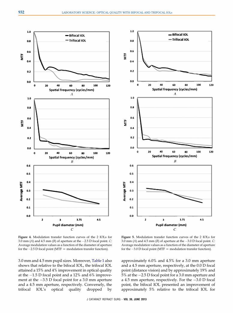
Figure 4. Modulation transfer function curves of the 2 IOLs for3.0 mm (A) and 4.5 mm (B) of aperture at the �2.5 D focal point. C:Averagemodulation values as a function of the diameter of aperturefor the �2.5 D focal point (MTFZmodulation transfer function).
Figure 5. Modulation transfer function curves of the 2 IOLs for3.0 mm (A) and 4.5 mm (B) of aperture at the �3.0 D focal point. C:Average modulation values as a function of the diameter of aperturefor the �3.0 D focal point (MTFZmodulation transfer function).
932 LABORATORY SCIENCE: OPTICAL QUALITY WITH BIFOCAL AND TRIFOCAL IOLS
3.0 mm and 4.5 mmpupil sizes. Moreover, Table 1 alsoshows that relative to the bifocal IOL, the trifocal IOLattained a 15% and 4% improvement in optical qualityat the �1.5 D focal point and a 12% and 6% improve-ment at the �3.5 D focal point for a 3.0 mm apertureand a 4.5 mm aperture, respectively. Conversely, thetrifocal IOL’s optical quality dropped by
J CATARACT REFRACT SURG
approximately 6.0% and 4.5% for a 3.0 mm apertureand a 4.5 mm aperture, respectively, at the 0.0 D focalpoint (distance vision) and by approximately 19% and5% at the�2.5 D focal point for a 3.0 mm aperture anda 4.5 mm aperture, respectively. For the �3.0 D focalpoint, the bifocal IOL presented an improvement ofapproximately 5% relative to the trifocal IOL for
- VOL 39, JUNE 2013

Figure 6. Modulation transfer function curves of the 2 IOLs for3.0 mm (A) and 4.5 mm (B) of aperture at the �3.5 D focal point. C:Averagemodulation values as a function of the diameter of aperturefor the �3.5 D focal point (MTFZmodulation transfer function).
Figure 7. Modulation transfer function curves of the 2 IOLs for3.0 mm (A) and 4.5 mm (B) of aperture at the �4.0 D focal point. C:Average modulation values as a function of the diameter of aperturefor the �4.0 D focal point (MTFZmodulation transfer function).
933LABORATORY SCIENCE: OPTICAL QUALITY WITH BIFOCAL AND TRIFOCAL IOLS
a 3.0 mm aperture, similar to the results for 4.5 mm. Inaddition, the 2 IOLs provided similar optical quality atthe �2.0 D and �4.0 D focal points. Hence, the mostsignificant results in Table 1 suggest that the bifocalIOL provides better optical quality than the trifocalIOL at the 0.0 D focal point (far vision) for smallerand larger pupils (dim and bright light conditions).
J CATARACT REFRACT SURG
For the near focal points, the bifocal IOL also obtainedbetter optical quality results at �2.5 D for both aper-tures and at �3.5 D for a 3.0 mm aperture. For thenear focal points, smaller pupils become more impor-tant due to the pupillary miosis provoked by theaccommodative reflex20–22 and by the bright lightconditions under which the near tasks are performed.
- VOL 39, JUNE 2013

Table 1. Modulation transfer function values at a discrete fre-quency of 50 cycles/mm of the 2 IOLs for the smaller and thelarger diameters of aperture and for 7 focal points (add powers).
AddPower (D)
3.0 mm Aperture 4.5 mm Aperture
BifocalIOL
TrifocalIOL
BifocalIOL
TrifocalIOL
0.0 0.440 0.379 0.475 0.4301.5 0.026 0.176 0.016 0.0592.0 0.027 0.043 0.008 0.0322.5 0.227 0.035 0.083 0.0323.0 0.258 0.205 0.112 0.1313.5 0.059 0.182 0.026 0.0874.0 0.004 0.035 0.013 0.034
Add Z addition; IOL Z intraocular lens
Figure 8. Through-focusMTF curves for both IOLs at 3.0mm (A) and4.5 mm (B) apertures. The curves were calculated for a spatial fre-quency of 50 cycles/mm (MTFZmodulation transfer function).
934 LABORATORY SCIENCE: OPTICAL QUALITY WITH BIFOCAL AND TRIFOCAL IOLS
On the other hand, the trifocal IOL showed better op-tical quality at the �1.5 D and �3.5 D focal points forboth apertures.
The objective evaluation of through-focus opticalquality gives valuable information about the behaviorof both IOLs under analysis. In the current study, thethrough-focus MTF curve was obtained by movingthe detector along the optical axis and subsequentlycomputing the value of theMTF at that focus for a spa-tial frequency of 50 cycles/mm (which correspondsapproximately to an optotype for 0.5 Snellen-equivalent visual acuity in white light) (Figure 8). Inagreement with the results of Gatinel et al.,10 trifocalIOL data showed 3 peaks corresponding to far, inter-mediate, and near vision while the bifocal IOL yieldedthe widely described4–9 V-pattern of 2 peaks corre-sponding to the vergences of near and distance vision.Figure 8, A, shows that for a 3.0 mm diameter, the tri-focal IOL outperformed the bifocal IOL at the �1.5 Dand �3.5 D focal points only, while having a similarperformance at�2.0 D and�4.0 D and yielding worseresults at the 0.0 D, �2.5 D, and �3.0 D focal points.Similarly, in Figure 8, B, for the 4.5 mm aperture, thetrifocal IOL obtained better values at the intermediatefocal points and at �3.5 D only.
The variation in the optical quality of the IOLs(given by the mean modulation value, sections C,Figures 1 to 7) with pupil size was also analyzed. Wefound that both designs have significant pupil depen-dence. The relationship between optical quality andpupil size varied with the viewing distance of the ob-ject (focal points). For instance, for the 0.0 D focal point(far vision), we concluded that the larger the pupil, thebetter the optical quality for both IOLs (Figure 1, C).However, for the intermediate and near focal points,the optical quality was worse with larger pupils andbetter with smaller ones. This behavior stems from
J CATARACT REFRACT SURG
the design of both IOLs, which is characterized bya gradual decrease in step height toward the periph-ery. These results are in good agreement with thosein previous studies that analyzed the Acrysof RestorIOL’s dependence of optical quality with pupil size15
and with those in Gatinel et al.’s study,10 which fo-cused on the Finevision IOL.
Comparing both IOLs at each focal point and foreach pupil size showed that at the 0.0 D focal point(distance vision), the bifocal IOL showed the highestmean modulation value for all pupil sizes, suggestingthat this IOL will provide a better distance visual per-formance than the trifocal IOL under dim and brightillumination conditions. Conversely, at the �1.5 D fo-cal point (which corresponds approximately to 80 cmat the spectacle plane), the mean modulation valueswere higher for the trifocal IOL than for the bifocalIOL. Logically, this improvement in performance canbe explained by the presence in the trifocal IOL ofthe additional intermediate focus, as seen in Figure 8.Regarding near vision (�2.5 D and �3.0 D, corre-sponding to 50 cm and 40 cm viewing distances at
- VOL 39, JUNE 2013

935LABORATORY SCIENCE: OPTICAL QUALITY WITH BIFOCAL AND TRIFOCAL IOLS
the spectacle plane), for the �2.5 D focal point, thebifocal IOL provided better optical quality than the tri-focal IOL, mainly with the smaller pupil. At the�3.0 Dfocal point, after analyzing the curves for the 2 IOLs,we observed that they intersected at a pupil-size valueof approximately 3.5 mm. For pupils larger than3.5 mm, the trifocal IOL provided a better mean mod-ulation value, while for pupil sizes below 3.5 mm, thebifocal IOL yielded better optical quality. At the�3.5 D focal point, the mean modulation values forall the apertures were higher for the trifocal IOL.Near activity is usually performed under photopicconditions, and bright light makes the pupils contract;at the same time, the pupil size decreases due to the ac-commodative reflex.20–22 Hence, taking into accountthe optical quality results and their relationship withhuman pupil dynamics, it could be suggested thatthe bifocal IOL provides better optical performanceat distance and at the �2.5 D and �3.0 D focal points,being comparable at �2.0 D and �4.0 D, while the tri-focal IOL is better at the�1.5 D and�3.5 D focal points(which approximately correspond to 80 cm and 35 cm,respectively, at the spectacle plane).
Moreover, it has been suggested that contrast sensi-tivity function has a direct relationship to MTF. There-fore, the optic quality of IOLs (by means of the MTF)should be expected to have an impact on the visualquality of patients.23 Therefore, in light of the resultsshown in Table 1 and considering that 3.0 mm and4.5 mm apertures could represent the average pupilsizes that patients over 60years of agehaveunderphot-opic conditions and mesopic conditions, respectively,one could suggest the better optical results obtainedwith the bifocal IOL could be related to better visualquality for the most important distances of vision.
It is also important that these results were obtainedin an ideal, well-centered IOL situation. It has beenshown that IOL tilt and decentration have an impor-tant impact on visual performance for a wide rangeof IOL designs.24–30 It was recently reported that theoptical quality yielded by the Acrysof RestorSNA6D1 IOL at the distance focus decreased whenthe IOL was tilted or decentered.31 Similarly, Gatinelet al.10 showed that 1.0 mm decentration of a Fine-vision IOL produced an attenuation of the diffractivetrifocal pattern and that as a result, the IOL becomesmore distance-vision dominant. Hence, according tothese previous findings, it would be interesting toperform additional studies to assess the impact ofdifferent degrees of tilt and decentration on the visualperformance of both IOLs at different focal points.These evaluations would provide valuable informa-tion to surgeons regarding visual and optical perfor-mance when these IOLs are misaligned. At the sametime, further studies should be developed to add
J CATARACT REFRACT SURG
different corneal profiles to assess the coupling effectof the IOLs with the cornea and thus obtain clearerclinical conclusions. The ideal conditions under whichthe IOLs studied are designed (see Materials andMethods section) are sometimes altered. For example,for distance-vision activities in strong daylight condi-tions, the pupil constricts due to the marked increasein illumination; thus, patients will probably use a sim-ilar or smaller area of the IOL than they do when theywork at near distance in indoor conditions. Therefore,different illumination conditions should be consideredwhen evaluating the clinical results of these IOLs.
In conclusion, the results in the present study sug-gest that the bifocal IOL under study could providea greater range of vision from distance to near thanthe trifocal IOL, with the exception of the intermediatefocal point, for which the trifocal IOL provides betteroptical quality.
-
WHAT WAS KNOWN
� Presbyopic patients have more intermediate visiondemands and seek a spectacle-free image. Aiming to im-prove intermediate vision, new trifocal designs have beendeveloped.
WHAT THIS PAPER ADDS
� The bifocal IOL provided better optical performance at the0.0 D (far vision), �2.5 D, and �3.0 D focal points, whilethe trifocal IOL was better at the �1.5 D focal point. It isexpected that bifocal IOLs could provide a greater range ofvision from distance to near than trifocal IOLs.
REFERENCES1. Luo BP, BrownGC, Luo SC, BrownMM. The quality of life asso-
ciated with presbyopia. Am J Ophthalmol 2008; 145:618–622
2. Maxwell WA, Lane SS, Zhou F. Performance of presbyopia-
correcting intraocular lenses in distance optical bench tests.
J Cataract Refract Surg 2009; 35:166–171
3. Santhiago MR, Wilson SE, Netto MV, Ghanen RC,
Monteiro MLR, Bechara SJ, Espana EM, Mello GR, Kara-
Junior N. Modulation transfer function and optical quality after
bilateral implantation of aC3.00 D versus aC4.00 D multifocal
intraocular lens. J Cataract Refract Surg 2012; 38:215–220
4. Petermeier K, Messias A, Gekeler F, Szurman P. Effect of
C3.00 diopter andC4.00 diopter additions in multifocal intraoc-
ular lenses on defocus profiles, patient satisfaction, and contrast
sensitivity. J Cataract Refract Surg 2011; 37:720–726
5. Maxwell WA, Cionni RJ, Lehmann RP, Modi SS. Functional out-
comes after bilateral implantation of apodized diffractive
aspheric acrylic intraocular lenses with a C3.0 or C4.0 diopter
addition power; randomized multicenter clinical study.
J Cataract Refract Surg 2009; 35:2054–2061
6. Alfonso JF, Fern�andez-Vega L, Puchades C, Mont�es-Mic�o R.
Intermediate visual function with different multifocal intraocular
lens models. J Cataract Refract Surg 2010; 36:733–739
VOL 39, JUNE 2013
936 LABORATORY SCIENCE: OPTICAL QUALITY WITH BIFOCAL AND TRIFOCAL IOLS
7. Madrid-Costa D, Cervi~no A, Ferrer-Blasco T, Garc�ıa-L�azaro S,
Mont�es-Mic�o R. Visual and optical performance with hybrid mul-
tifocal intraocular lenses. Clin Exp Optom 2010; 93:426–440
8. Pepose JS,WangD, AltmannGE. Comparison of through-focus
image sharpness across five presbyopia-correcting intraocular
lenses. Am J Ophthalmol 2012; 154:20–28
9. Alfonso JF, Fern�andez-Vega L, Bl�azquez JI, Mont�es-Mic�oR. Vi-sual function comparison of 2 aspheric multifocal intraocular
lenses. J Cataract Refract Surg 2012; 38:242–248
10. Gatinel D, Pagnoulle C, Houbrechts Y, Gobin L. Design and
qualification of a diffractive trifocal optical profile for intraocular
lenses. J Cataract Refract Surg 2011; 37:2060–2067
11. International Organization for Standardization. Ophthalmic
Implants – Intraocular Lenses – Part 2: Optical Properties and
Test Methods. Geneva, Switzerland, ISO, 1999 (ISO 11979–
2); technical corrigendum 1, 2003
12. International Organization for Standardization. Ophthalmic
Implants – Intraocular Lenses – Part 9. Multifocal intraocular
lenses. Geneva, Switzerland, ISO, 2006 (ISO 11979–9)
13. RawerR, StorkW,Spraul CW, Lingenfelder C. Imaging quality of
intraocular lenses. J Cataract Refract Surg 2005; 31:1618–1631
14. Kawamorita T, Uozato H. Modulation transfer function and pupil
size in multifocal and monofocal intraocular lenses in vitro.
J Cataract Refract Surg 2005; 31:2379–2385
15. Artigas JM, Menezo JL, Peris C, Felipe A, D�ıaz-Llopis M. Image
quality with multifocal intraocular lenses and the effect of pupil
size; comparison of refractive and hybrid refractive-diffractive
designs. J Cataract Refract Surg 2007; 33:2111–2117
16. AltmannGE, Nichamin LD, Lane SS, Pepose JS. Optical perfor-
mance of 3 intraocular lens designs in the presence of decentra-
tion. J Cataract Refract Surg 2005; 31:574–585
17. Lorente A, Pons AM, Malo J, Artigas JM. Standard criterion for
fluctuations of modulation transfer function in the human eye:
application to disposable contact lenses. Ophthalmic Physiol
Opt 1997; 17:267–272
18. Artigas JM, Peris C, Felipe A, Menezo JL, S�anchez-Cortina I,
L�opez-Gil N. Modulation transfer function: rigid versus foldable
phakic intraocular lenses. J Cataract Refract Surg 2009;
35:747–752
19. Marsack JD, Thibos LN, Applegate RA. Metrics of optical quality
derived from wave aberration predict visual performance. J Vis
2004; 4:322–328. Available at: http://www.journalofvision.org/
content/4/4/8.full.pdf. Accessed January 17, 2013
20. Loewenfeld IE. The Pupil; Anatomy, Physiology, and Clinical
Applications. Woburn, MA, Butterworth- Heinemann, 1993;
295–317
21. Myers GA, Stark L. Topology of the near response triad.
Ophthalmic Physiol Opt 1990; 10:175–181
22. Madrid-Costa D, Ruiz-Alcocer J, Radhakrishnan H, Ferrer-
Blasco T, Mont�es-Mic�o R. Changes in accommodative
J CATARACT REFRACT SURG
responses with multifocal contact lenses: a pilot study. Optom
Vis Sci 2011; 88:1309–1316
23. Peris-Mart�ınez C, Artigas JM, S�anchez-Cortina I, Felipe A,
D�ıez-Ajenjo A, Menezo JL. Influence of optic quality on contrast
sensitivity and visual acuity in eyes with a rigid or flexible phakic
intraocular lens. J Cataract Refract Surg 2009; 35:1911–1977
24. Madrid-CostaD,Ruiz-AlcocerJ,P�erez-VivesC,Ferrer-BlascoT,L�opez-Gil N, Mont�es-Mic�o R. Visual simulation through different
intraocular lenses using adaptive optics: effect of tilt and decen-
tration. J Cataract Refract Surg 2012; 38:947–958
25. Madrid-CostaD,P�erez-VivesC,Ruiz-AlcocerJ,Albarr�an-DiegoC,Mont�es-Mic�o R. Visual simulation through different intraocular
lenses in patients with previous myopic corneal ablation using
adaptive optics: effect of tilt and decentration. J Cataract Refract
Surg 2012; 38:774–786
26. Ruiz-Alcocer J, P�erez-Vives C, Madrid-Costa D, L�opez-Gil N,
Mont�es-Mic�o R. Effect of simulated IOL tilt and decentration
on spherical aberration after hyperopic LASIK for different intra-
ocular lenses. J Refract Surg 2012; 28:327–334
27. Mont�es-Mic�o R, Ferrer-Blasco T, Cervi~no A. Analysis of the
possible benefits of aspheric intraocular lenses: review of the
literature. J Cataract Refract Surg 2009; 35:172–181
28. PiehS, FialaW,Malz A, StorkW. In vitro Strehl ratioswith spher-
ical, aberration-free, average, and customized spherical
aberration-correcting intraocular lenses. Invest Ophthalmol Vis
Sci 2009; 50:1264–1270. Available at: http://www.iovs.org/
content/50/3/1264.full.pdf. Accessed January 17, 2013
29. Eppig T, Scholz K, L€offler A, Meßner A, Langenbucher A. Effect
of decentration and tilt on the image quality of aspheric intraoc-
ular lens designs in a model eye. J Cataract Refract Surg 2009;
35:1091–1100
30. McKelvie J, McArdle B, McGhee C. The Influence of tilt, decen-
tration, and pupil size on the higher-order aberration profile
of aspheric intraocular lenses. Ophthalmology 2011; 118:
1724–1931
31. Mont�es-Mic�o R, L�opez-Gil N, P�erez-Vives C, Bonaque S,
Ferrer-Blasco T. In vitro optical performance of nonrotational
symmetric and refractive–diffractive aspheric multifocal intraoc-
ular lenses: impact of tilt and decentration. J Cataract Refract
Surg 2012; 38:1657–1663
- VO
L 39, JUNE 2013First author:Robert Mont�es-Mic�o, PhD
Optometry Research Group,Optics Department, University ofValencia, Valencia, Spain