intussusception - cocukradyolojisi.org · diagnosis of intussusception describe advantages and...
TRANSCRIPT

INTUSSUSCEPTION
Oscar Navarro K.
The Hospital for Sick Children
University of Toronto

Objectives
Review role of ultrasonography in the diagnosis of intussusception
Describe advantages and disadvantages of the different enema reduction techniques
Discuss particular types of intussusception, including those due to pathologic lead points and those limited to the small bowel

What is intussusception?
Invagination of a segment of bowel into another
Broad definition that includes
conditions of no clinical significance
others that require prompt treatment due to the risk of bowel necrosis

What type of intussusception?
Limited to small bowel vs. colonic involvement
Asymptomatic vs. symptomatic
Due to lymphoid hyperplasia vs. pathologic lead point

Ileocolic and ileo-ileocolic intussusceptions
Most common of those requiring treatment
Usually symptomatic
Often due to lymphoid hyperplasia

Clinical diagnosis
Abdominal pain 83-85%
Vomiting 48-80%
Abdominal mass 22-65%
Blood in stool (occult or gross) 43-60%
Clinician often has to rely on imaging

Diagnosis
Sonography is cornerstone modality
Highly accurate
May identify pathologic lead points
Lack of radiation and non-invasiveness

Sonography: High accuracy
100% accuracy
Pracros et al, 1987 (Lyon, France)
Wang & Liu, 1988 (Shenyang, China)
Woo et al, 1992 (Daegu, South Korea)
Riebel et al, 1993 (Berlin, Germany)
Lim et al, 1994 (Seoul, South Korea)
Del Pozo et al, 1996 (Madrid, Spain)

Sonographic appearance
2-5 cm mass
Just deep to abdominal wall
Characteristic appearance
- “doughnut” and “target” signs
- “pseudokidney” and “sandwich” signs

del-Pozo et al. Radiographics 1999; 19:299-319

Sonography in intussusception False positives
Bowel wall thickening
Volvulus
Psoas muscle
Feces

Treatment of intussusception
Non-operative management: enema
High reduction rates
Less invasiveness and morbidity
No anesthesia
Short hospital stay
Decreased costs

Candidates for enema reduction
Relative contraindications:
dehydration - correction before
shock enema
Absolute contraindication:
peritonitis - surgery

Clinical findings and reducibility
Younger age
Rectal bleeding
Radiographic signs of obstruction
Longer duration of symptoms
Successful reduction can be achieved in the presence of any of these factors

Sonographic assessment of reducibility
Thick peripheral hypoechoic rim
Free intraperitoneal fluid
Trapped fluid within intussusception
Enlarged lymph nodes in intussusceptum
Pathologic lead point
Absence of blood flow

7m♀: bloody stools, lethargic

Sonographic assessment of reducibility
The preoperative diagnosis of bowel necrosis remains a challenge

Enema reduction techniques
Hydrostatic vs. Pneumatic
Fluoroscopy vs. Sonography
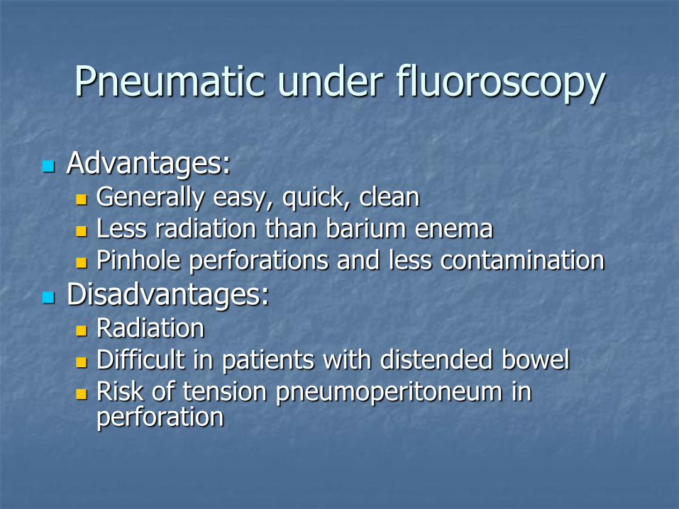
Pneumatic under fluoroscopy
Advantages: Generally easy, quick, clean Less radiation than barium enema Pinhole perforations and less contamination
Disadvantages: Radiation Difficult in patients with distended bowel Risk of tension pneumoperitoneum in
perforation


Hydrostatic under sonography
Advantages:
No radiation
Direct visualization of reduction, edematous ileo-cecal valve, residual intussusception and pathologic lead point
Disadvantages:
?Larger tears / more contamination in perforation
Longer procedure
Messier (excess fluids)

Complications of any technique: perforation
Present prior to enema due to necrosis
Secondary to enema even at low pressure
Rare event <1%
More common in young children <6-7mo with a longer duration of symptoms >36-48h
Perforations are smaller and easier to deal with when secondary to pneumatic reduction
Experimental peritoneal soiling: safety
air > saline > water-soluble contrast > barium


Delayed repeated reduction attempts
10-14% of irreducible intussusceptions undergo spontaneous reduction
51-66% manually reduced without bowel resection
Partial reduction may relieve congestion facilitating subsequent reduction attempt

Delayed repeated reduction attempts
Series Interval Cases Success rate (%)
Red rate without
Red rate with
Navarro, 2004
18m-12h 26 (12%) 50 84 90
Sandler, 1999
2h-19h 17 (n/a) 59 n/a n/a
González-Spínola, 1999
30m-24h n/a n/a 71 82
Gorenstein, 1998
45m-60m 19 (52%) 83 48 91
Saxton, 1994
30m-3h 21 (15%) 52 78 86

Delayed repeated reduction attempts
Safe and effective
Performed if initial attempt moves intussusceptum
Patient clinically stable
No optimal interval
Number of attempts weighed against risks of radiation

Recurrence of intussusception
Literature ~ 10% (SickKids 2004 19%)
2/3 have only 1 recurrence
Usually within first few days
High reducibility
Most no documented pathologic lead point 67-100% (SickKids 2004 75%)

Success of non-operative management
Not only enema reduction rate (highest possible)
Also number of manually reduced at surgery without bowel resection (lowest possible)

Intussusception due to pathologic lead points
Incidence 1.5-12% (SickKids 2004 8%)
Wide spectrum
Varied and nonspecific presentation
Diagnosis is relevant as management may be different

Pathologic lead points
Clinical clues:
Predisposing disease (Peutz-Jeghers, polyposis, HSP, celiac disease, CF, neutropenic colitis)
Age of patient
Lymphoma suspected in child >3y, long duration of symptoms and weight loss
Recurrences (SickKids 2004 25% vs. 5%)

Pathologic lead points
Focal:
Meckel diverticulum
Intestinal polyp
Duplication cyst
Lymphoma
Diffuse:
Henoch-Schönlein purpura
Cystic fibrosis
Celiac disease

Diagnosis of pathologic lead points
Sonography is modality of choice
Detection of 74% of focal PLP
Specific diagnosis of 32% of focal PLP
Diagnosis of all cases of duplication cyst
Meckel diverticula and polyps are more difficult to detect


Management of intussusceptions due to pathologic lead points
Successful reduction with AE in 60-64%
Enema reduction also recommended in PLP that will require surgery

Small bowel intussusception
Spectrum of presentations
Includes common, benign, transient event in asymptomatic or mildly symptomatic children
Persistent, large, symptomatic intussusception with a pathologic lead point

Transient small bowel intussusception
More frequently reported with increasing use of sonography and CT
In asymptomatic patients or mildly symptomatic children with or without underlying predisposing pathology (gastroenteritis, celiac disease, abdominal trauma, ALL-neutropenic colitis)

Transient small bowel intussusception
One or more
In left hemiabdomen or centroabdominal
Active peristalsis
Small diameter US: < 2.4cm (mean 1.8cm)
CT: < 3cm (mean 2.1cm)
Short segment US: < 2.7cm (mean 2.3cm)
CT: < 5cm (mean 2.2cm)


5y, ALL, neutropenia, abdominal pain
Initial CT Delayed images

5y MVA- Duodenal hematoma Pelvic fractures
11m SCID Candidiasis
4y ALL Neutropenia Abdominal pain
9y Fell from bike Liver trauma with portal vein injury
4y Heart tx Routine f/u
13y Celiac disease


Transient small bowel intussusception
Not all intussusceptions require reduction
Spontaneous reduction expected in children with characteristic appearance of transient small bowel intussusception

Symptomatic small bowel intussusception
Symptoms similar to ileocolic
Higher incidence of PLP
Most eventually require surgery

Symptomatic small bowel intussusception
Larger than transient SB intussusception diameter 2-4.3cm (mean 2.9 cm)
Variable length
PLP may be identified on sonography

Ileo-ileal intussusception - HSP
3y♀: abdominal pain x 5d


Summary
Sonography is modality of choice for diagnosis
Multiple management options
Decreasing radiation and complications
Patient’s comfort and safety
Continuous evaluation of our techniques
Open to accept new techniques