introduction to preimplantation genetic diagnosis -...
TRANSCRIPT

Section 1 Chapter Background
Introduction to preimplantation genetic diagnosis Joyce C. Harper
Key points
Preimplantation genetic diagnosis (PGD) was • fi rst applied in 1988 using a polymerase chain reaction (PCR) protocol to amplify a sequence on the Y chromosome for embryo sexing for patients carrying X-linked disease. Patients have to go through in vitro • fertilization (IVF) so that their embryos may be generated in vitro. Cells are removed from oocytes or embryos and used for the genetic diagnosis. Unaff ected embryos are transferred to the patient. Th e most common biopsy procedure is • cleavage-stage biopsy, but biopsy of polar bodies and trophectoderm cells is performed clinically. Th e indications for PGD are: monogenic • disorders, chromosome abnormalities, sexing, or specifi c diagnosis of X-linked disease. PGD technology has been used to try and • improve the pregnancy rate for infertile patients by screening for aneuploidies. Indications include advanced maternal age, repeated implantation failure, and repeated miscarriages (preimplantation genetic screening; PGS). Fluorescent • in situ hybridization (FISH) is the technique used to analyze chromosomes in the biopsied cells, and is the method of choice for embryo sexing. It is also used for chromosome abnormalities and aneuploidy screening. PCR is the technique used to detect • monogenic disorders but it has been hampered by problems with contamination and allele dropout. PGD has stimulated much ethical debate. • Many countries have legislation controlling
Preimplantation genetic diagnosis (PGD) was• fi rst applied in 1988 using a polymerase chain reaction (PCR) protocol to amplify a sequence on the Y chromosome for embryo sexing for patients carrying X-linked disease. Patients have to go through in vitro• fertilization (IVF) so that their embryos may be generated in vitro. Cells are removed fromoocytes or embryos and used for the geneticdiagnosis. Unaff ected embryos are transferredto the patient. Th e most common biopsy procedure is • cleavage-stage biopsy, but biopsy of polar bodies and trophectoderm cells is performedclinically. Th e indications for PGD are: monogenic • disorders, chromosome abnormalities, sexing,or specifi c diagnosis of X-linked disease. PGD technology has been used to try and• improve the pregnancy rate for infertile patients by screening for aneuploidies.Indications include advanced maternal age,repeated implantation failure, and repeatedmiscarriages (preimplantation geneticscreening; PGS). Fluorescent • in situ hybridization (FISH) is the technique used to analyze chromosomes in the biopsied cells, and is the method of choice for embryo sexing. It is also used forchromosome abnormalities and aneuploidy screening. PCR is the technique used to detect • monogenic disorders but it has beenhampered by problems with contaminationand allele dropout. PGD has stimulated much ethical debate.•Many countries have legislation controlling
PGD and in some countries cleavage-stage and blastocyst biopsy are illegal. Social sexing is illegal in Europe and other countries. Th e fi rst 20 years has shown major advances • in the fi eld of PGD. Th e next 20 years may include the use of arrays for examining all the chromosomes, multiple genes and gene expression. PGD may be used for all IVF patients to select the genetically “best” embryo. Th e European Society for Human • Reproduction and Embryology (ESHRE) PGD Consortium has collected nine years of data on PGD and PGS. Five working groups have been set up to look at PGS, accreditation, the database, guidelines, and misdiagnosis. Additionally a pediatric follow-up and external quality assessment for FISH and PCR have been developed.
Introduction Preimplantation genetic diagnosis (PGD) was devel-oped out of a need to provide an alternative to prenatal diagnosis for couples at risk of transmitting a genetic disease to their children. Th e options for such couples are: to remain childless; not to undergo genetic test-ing (reproductive roulette); or to go through prenatal diagnosis, PGD, gamete donation, or adoption. Th ese are all diffi cult reproductive options. Th e majority of couples will opt for prenatal diagnosis by chorionic vil-lus sampling (CVS) or amniocentesis (see Chapter 5 ). Th e procedures themselves take a few minutes, and for recessive disorders the couple have only a 25 per-cent chance of an aff ected pregnancy; with a dominant disorder this rises to 50 percent. But if the pregnancy is aff ected the couple have to decide if they wish to continue or consider termination. Neither is an easy option. Another advantage of prenatal diagnosis is
PGD and in some countries cleavage-stageand blastocyst biopsy are illegal. Social sexing is illegal in Europe and other countries. Th e fi rst 20 years has shown major advances•in the fi eld of PGD. Th e next 20 years may include the use of arrays for examining all the chromosomes, multiple genes and gene expression. PGD may be used for allIVF patients to select the genetically “best”embryo. Th e European Society for Human• Reproduction and Embryology (ESHRE) PGD Consortium has collected nine years of data on PGD and PGS. Five working groups have been set up to look at PGS, accreditation, the database, guidelines, and misdiagnosis. Additionally a pediatric follow-up and external quality assessment for FISH and PCR have been developed.
1
Cambridge University Press978-0-521-88471-6 - Preimplantation Genetic Diagnosis: Second EditionEdited by Joyce C. HarperExcerptMore information
www.cambridge.org© in this web service Cambridge University Press

Section 1: Background
2
Table 1.1 The three methods of embryo biopsy used in preimplantation genetic diagnosis (PGD)
Day performed
Types of cells removed
Indications Zona drilling
Cell removal
Limitations
Polar body First PB day 0 Second PB day 1 Or simultaneously on day 1
First and second polar bodies
PGS Monogenics carried by mother
Laser Mechanical Beveled pipette
Aspiration Only maternal chromosomes/genes
Cleavage-stage
Day 3 Blastomeres PGS Monogenics Sexing Chromosome abnormalities
Laser Mechanical Acid Tyrodes
Aspiration Displacement
Postzygotic mosaicism
Blastocyst Day 5 Trophectoderm PGS Monogenics Sexing Chromosome abnormalities
Laser Mechanical Acid Tyrodes
Herniation Postzygotic mosaicism Some embryos will arrest prior to biopsy Short time for diagnosis
Table 1.2 Methods used for preimiplantation genetic diagnosis (PGD)
Indications Cell preparation Protocol Limitations
FISH Sexing Chromosome abnormalities PGS
Spreading cells using methanol:acetic acid or Tween HCl
Fix Denature Hybridization Wash off unbound probe Visualize
Cumulus contamination Mosaicism Overlapping signals Failure of probes to bind
PCR Sexing Monogenic disorders
Tubing cells into lysis buff er
Lyse cell Cycles of denaturing, annealing, elongation, Detect products
Cumulus contamination Sperm contamination (use ICSI) Other contamination Amplifi cation failure Allele dropout
Metaphase CGH
Sexing Chromosome abnormalities PGS
Tubing cells into lysis buff er
Lyse cell, whole genome amplifi cation Co-hybridization with control sample on to metaphase spread Analysis of each chromosome using CGH software
Contamination Mosaicism Procedure takes several days and so currently embryos are frozen Requires many skills, PCR, and cytogenetics
FISH, fl uorescent in situ hybridization; PGS, preimplantation genetic selection; PCR, polymerase chain reaction; ICSI, intracytoplasmic sperm injection; CGH, comparative genomic hybridization.
that in most countries this will be paid for by the health service . If the couple decide to go through PGD they have to undergo IVF procedures to produce embryos in vitro even though they are oft en fertile. IVF is a highly invasive procedure with a relatively low chance
of success, and adding on PGD does not improve the chances of delivering a baby. Cells need to be removed from the embryo to allow single-cell genetic testing. Th ese may be removed from the oocyte/zygote (fi rst and second polar body biopsy), blastomeres may
Cambridge University Press978-0-521-88471-6 - Preimplantation Genetic Diagnosis: Second EditionEdited by Joyce C. HarperExcerptMore information
www.cambridge.org© in this web service Cambridge University Press

Chapter 1: Introduction to preimplantation genetic diagnosis
3
be taken from cleavage-stage embryos, or trophec-toderm cells taken from blastocysts ( Table 1.1 ) Th e polymerase chain reaction (PCR) is used for the diag-nosis of monogenic disorders, and fl uorescent in situ hybridization (FISH) is used for chromosome analy-sis ( Table 1.2 ).
PGD is a complicated procedure. As well as involv-ing the IVF team, it requires a diagnostic team who are experts in single-cell diagnosis. Besides the technical diffi culties, internationally PGD is a controversial pro-cedure as there are ethical and moral concerns about genetic testing of the early embryo .
In this book every aspect involved in PGD is con-sidered, from IVF, prenatal diagnosis, and genetic counseling to quality assurance and ethical considera-tions. Th is chapter off ers the reader a history of PGD, an outline of each chapter, and a report on the ESHRE PGD Consortium.
History of PGD
Animal studies and preclinical work Th e fi rst biopsies on embryos were performed by removing one cell from two-cell embryos by Seidel ( 1952 ) and Tarkowski and Wróblewska ( 1967 ), work-ing on rabbits and mice, respectively. Th e fi rst PGD was performed by Gardner and Edwards ( 1968 ), who biopsied a small portion of the trophectoderm from rabbit blastocysts, sexed the embryos by identifying sex chromatin (which identifi es females), and replaced them into recipient females. For a rabbit blastocyst to implant it needs to be expanded with an intact zona and so Richard Gardner made a very neat slit in the zona, sucked out a small amount of trophectoderm, pinched it off , and hoped that the remaining trophectoderm would block the hole in the zona. Th e off spring were found to be of the predicted sex (Edwards & Gardner, 1967 ; Gardner & Edwards, 1968 ). Th is technique was later tried on human blastocysts without success (Steptoe et al ., 1971 ) .
In 1985, at a Ciba Foundation meeting in London, scientists were discussing the possibility of diagnosing genetic disease in a human preimplantation embryo. It was generally agreed that there were no single-cell diag-nostic techniques available, and that the biopsied cell(s) would have to be cultured to obtain suffi cient cells for the diagnosis. Th e revolutionary PCR procedure had just been developed (Saiki et al ., 1985 ) but it was not envisaged that PCR could work on a single cell.
Subsequently, the idea of performing PGD was reviewed by a number of people. Penketh and McLaren ( 1987 ) wrote a review on “Prospects for prenatal diag-nosis during preimplantation human development” and Edwards and Hollands ( 1988 ) wrote a review on “New advances in human embryology; implications for preimplantation diagnosis of genetic disease” (Edwards & Hollands, 1988 ). Edwards and Hollands ( 1988 ) sug-gested that sexing sperm would be easier than sexing embryos but they said the advantage of typing embryos would be that the cells would be “fully representative of the embryonic genome.” Th ese authors suggested that noninvasive techniques in which the medium was examined would be the simplest; either secretion or uptake of substances from the culture medium would be possible. It is only now that noninvasive methods seem a possibility (Seli et al ., 2007 ; Vergouw et al ., 2008 ) . Edwards and Holland ( 1988 ) suggested that if invasive methods were used they would involve dis-solving the zona, disaggregating the embryo, sepa-rating the cells and culturing them for diagnosis, and putting the embryo back in an artifi cial zona for trans-fer. Th ey further suggested performing this technique on two-cell embryos.
Several diff erent approaches to embryo biopsy were being investigated in the late 1980s. In Australia, Leeanda Wilton was developing methods of remov-ing cells from mouse embryos (Wilton & Trounson, 1986; Kola & Wilton, 1991 ) ( Figure 1.1 ); André Van Steirteghem was exploring removing one cell from two-cell embryos (Nijs & Van Steirteghem, 1987 ); and Marilyn Monk and Alan Handyside were investigat-ing taking one or two cells from an eight-cell embryo for diagnosis of hypoxanthine phosphoribosyl-trans-ferase (HPRT) defi ciency (Monk et al ., 1987 ). Audrey Muggleton-Harris and Marilyn Monk demonstrated that PGD in a mouse model for Lesch–Nyhan disease could also be done by biopsy and analysis of a few tro-phectoderm cells extruded through the zona pellucida, a technique perfected by Audrey Muggleton-Harris in David Whittingham’s unit (Monk et al ., 1988 ). Trophectoderm biopsy was also tested by Dokras et al . ( 1990 ) and Summers et al . ( 1988 ) . Another approach to obtaining blastocysts was to perform uterine lavage where embryos would be fl ushed on day fi ve of devel-opment (Buster et al ., 1985 ). Th e diagnosis and transfer of blastocysts would avoid the low implantation rate of in vitro fertilized cleavage-stage embryos, which was only 15 percent at that time. Bruno Brambati suggested that uterine lavage would be an effi cient, practical, and
Cambridge University Press978-0-521-88471-6 - Preimplantation Genetic Diagnosis: Second EditionEdited by Joyce C. HarperExcerptMore information
www.cambridge.org© in this web service Cambridge University Press

Section 1: Background
4
safe method to obtain blastocysts for PGD (Brambati & Tului, 1990 ). However, the problem with using lavage was that it would be impossible to be sure that all of the blastocysts had been fl ushed, allowing the possibility that undiagnosed embryos could implant. Whatever method was used, it was predicted that the biopsy technique would almost certainly aff ect implantation (Edwards & Hollands, 1988 ) .
Th e challenge of the introduction of molecular biology for PGD was the move from working with millions of cells to the very few cells of the embryo. Edwards and Hollands ( 1988 ) suggested that the most reliable method for single-cell diagnosis would be “to use DNA probes for identifying the genotype of the human embryo” and they predicted that high levels of chromosome abnormalities (Plachot et al ., 1987 ) would “lead to complications in the interpre-tation of some diagnostic tests.” Monk, working in Anne McLaren’s MRC Mammalian Development Unit at University College London in the 1970s, had already developed an array of single-cell-sensitive molecular procedures for the study of gene expression and its regulation in early mouse development, most notably for the study of X chromosome inactivation in female embryonic development. In the late 1980s Alan Handyside collaborated with Marilyn Monk to carry out mouse embryo biopsies of single blast-omeres, single-cell diagnosis, and embryo transfers
to show that Monk’s single-cell molecular diagnoses were correct. Handyside had been working on mouse embryo biopsies at Cambridge University and joined Robert Winston at the Hammersmith Hospital . Th e fi rst single-cell diagnoses were performed on embryos from the fi rst genetically engineered mouse carrying a defect in the HPRT gene, the mouse model for Lesch–Nyhan syndrome in the human. Th e mouse was cre-ated by mutation of the HPRT gene in embryonic stem cells in culture, transferring some of these mutated cells to a host blastocyst, and returning that blastocyst to the uterus of a foster mother to produce a chimeric male off spring carrying the mutated gene in his sperm (Hooper et al ., 1987 ). Th us, some of his daughters were heterozygous for the HPRT mutation. Monk was able to use biopsied cells from embryos from this hetero-zygous female mouse to diagnose the mutant embryos (half the males) carrying the mutation on their sin-gle X chromosome . Th is was the fi rst demonstration that preimplantation diagnosis by biopsy and analy-sis of a single blastomere for a single gene defect was a feasible proposition (Monk et al ., 1987 ) and many key early papers followed (Monk et al ., 1988 ; Benson & Monk, 1988 ; Monk, 1988 ; Monk & Handyside, 1988 ; Holding & Monk, 1989 ; Monk & Holding, 1990 ; Monk, 1990a , 1990b , 1990c ; Monk, 1991 a, 1991b , 1991c ). Work with human embryos also began at this time; in collaboration with Braude and Johnson at Cambridge
Figure 1.1 Leeanda Wilton doing embryo biopsy in Melbourne, Australia in 1986.
Cambridge University Press978-0-521-88471-6 - Preimplantation Genetic Diagnosis: Second EditionEdited by Joyce C. HarperExcerptMore information
www.cambridge.org© in this web service Cambridge University Press

Chapter 1: Introduction to preimplantation genetic diagnosis
5
University, Monk assayed HPRT gene activity in single blastomeres biopsied from human preimplantation embryos (Braude et al ., 1989 ), although, in the human, the maternally inherited enzyme at the eight-cell stage would obscure the diagnosis of Lesch–Nyhan syn-drome by this method .
In many of the fi rst papers the procedure was called “preimplantation diagnosis” (PID), as an extension of prenatal diagnosis (PND). However, the name was changed to “preimplantation genetic diagnosis” (PGD) by people entering the fi eld later on to avoid confus-ing the acronym PID with that for pelvic infl ammatory disease.
Marilyn Monk and Cathy Holding set out to create further single-cell enzyme assays for common inherited genetic diseases as well as maintaining their interest in sin-gle-cell assays for X-linked genes to further their studies on the regulation of X chromosome inactivation in develop-ment ( Figure 1.2 ). One of these was adenosine deaminase (a defi ciency in this enzyme is the basis of severe combined immunodefi ciency disease (SCID)) (Benson & Monk, 1988 ). In Brussels, too, that same line of research led Karen Sermon, in André Van Steirteghem’s team, to evaluate the possibility of diagnosing Tay–Sachs disease through meas-uring the enzyme beta-N-acetylhexosaminidase activities in single blastomeres (Sermon et al ., 1991 ). Th ey could show that it would work in the mouse, but, unfortunately, not in the human. Later, the same group (Van Blerk et al ., 1991 ) showed the same for β-glucuronidase, the lysosomal enzyme defi cient in mucopolysaccharidosis type VII .
Holding and Monk, in collaboration with Cathy Abbott, were moving tubes from water bath to water
Figure 1.2 Marilyn Monk and Cathy Holding at Anne McLaren’s MRC Mammalian Development Unit in the Galton Laboratory, University College London, 1998/1999.
bath to try to develop the procedures of PCR and test-ing out the new PCR machine that was being devel-oped by Martin Evans and BioCam in Cambridge. Th ey wanted to look directly at the actual mutation in the DNA of a specifi c gene in a single cell. It was an immense struggle to fi nd the way to make PCR work at the single-cell level but their hard work and perse-verance led to eventual success using a mouse model for β thalassemia (Holding & Monk, 1989 ). Th ey used nested primers, fi rst amplifying the larger sequence and then, in a new reaction, amplifying an inner sequence with the inner primers. Th is vastly increased the spe-cifi city and sensitivity of the reaction, and they were able to analyze single cells and publish the fi rst nested PCR on a single cell detected by a simple agarose gel assay (Holding & Monk, 1989 ) as well as establishing PGD for β thalassemia in a mouse model system .
In 1990, Holding and Monk extended their single- cell PCR analyses to the human to develop single-cell detection of the sickle cell mutation in the betaglobin gene in human oocytes. In collaboration with Peter Braude, then at the Rosie Maternity Unit, Addenbrookes Hospital, in Cambridge, they were the fi rst to show that it was possible to diagnose gen-etic disease by analysis of the polar body of a human unfertilized egg, thus avoiding working on the human embryos themselves (Monk & Holding, 1990 ) .
Development of human embryo biopsy In the late 1980s many teams worldwide were attempt-ing clinical PGD, including the Hammersmith team in London, Jacques Cohen’s team in New York, and Yury Verlinksy’s team in Chicago. Th e fi rst two groups were attempting cleavage-stage biopsy and the Verlinsky team was working on polar body biopsy.
Th e Hammersmith Hospital team, led by Handyside and Winston, tried day two and day three human embryo biopsy. Alan Handyside, with the help of Kate Hardy, applied his mouse cleavage-stage biopsy techniques to day three human embryos using acid Tyrodes to drill a hole in the zona and aspirating one or two cells from eight-cell embryos, and allowed the embryos to grow on to day fi ve of development. Hardy used diff erential staining to count the number of trophectoderm and inner-cell mass cells of the control (32 embryos) and biopsied (45 embryos) to determine if the biopsy technique aff ected blastocyst develop-ment and measured the uptake of pyruvate and glucose (Hardy et al ., 1990 ). Since this study showed little eff ect on the ratio of the inner-cell mass and trophectoderm
Cambridge University Press978-0-521-88471-6 - Preimplantation Genetic Diagnosis: Second EditionEdited by Joyce C. HarperExcerptMore information
www.cambridge.org© in this web service Cambridge University Press

Section 1: Background
6
cells, or on metabolism, it gave the green light to human cleavage-stage biopsy. Today the same basic biopsy technique (of day three cleavage-stage biopsy) is used (Harper et al ., 2008a ). Th e zona is breached and single blastomeres are aspirated. Studies on day two biopsy did not show such favorable results as day three biopsy (Tarin et al ., 1992 ) .
The fi rst clinical cases Elena Kontogianni was studying for her PhD at the Hammersmith Hospital, on single-cell PCR for sexing, which she did by amplifying a repeated region of the Y chromosome ( Figure 1.3 ). It was this approach that was used for the world’s fi rst PGD cases (Handyside et al ., 1990 ). Female embryos were selectively transferred in fi ve couples at risk of X-linked disease, resulting in two twins and one singleton pregnancy. Because the Y chromosome region Kontogianni was amplifying con-tained many repeats, it gave fewer problems than trying to amplify a unique region. A band on the PCR gel indi-cated that the embryo was male and the absence of a band indicated that the embryo was female. However, failure to tube the cell, an anucleate blastomere, or failure of the PCR also resulted in absence of a band on the PCR gel. A total of 21 cycles were performed in two series and one misdiagnosis occurred. To reduce the risk of misdiagno-sis, Kontogianni went on to co-amplify sequences on the X and Y (Kontogianni et al ., 1991 ). At that time nothing was known about allele dropout, cumulus cell contami-nation, or amplifi cation failure from single cells.
During the 1980s, human IVF embryos were exclusively transferred on day two of development
as the culture medium used was incapable of reliably growing embryos past this stage. Since the biopsy was to be performed on day three, the fi rst diagnoses were all performed in one day, with transfer of the embryos late on day three. A comparison of day two and day three transfers indicated that this would not adversely aff ect pregnancy rates (Dawson et al ., 1995 ). Th e worry of embryos arresting was so high that some transfers took place in the early hours of day four so that the embryos were removed from culture as soon as possible. Th ere were many evenings at the Hammersmith when a trans-fer was performed at 1 a.m. on day four and researchers returned to the laboratory at 7 a.m. to start the next case. Winston helped deliver most of the fi rst PGD babies .
Development of FISH During the same period that single-cell enzyme activ-ity and gene mutation detection were being developed in the UK, others were analyzing whole chromosomes. Kola and Wilton ( 1991 ) biopsied single cells from embryos from mice that were carrying a Robertsonian translocation. Th ese single cells were karyotyped and normal embryos transferred. Analysis of the fetuses showed that the PGD was 100 percent accurate. Th is was the fi rst PGD of aneuploidy. In the late 1980s Wilton moved to London to work at the Institute of Zoology, and began to collaborate with Handyside at the Hammersmith Hospital.
Jones et al . ( 1987 ) were the fi rst to report the use of highly specifi c DNA probes to detect the human Y chromosome which could successfully be used on chromosomes from single cells of human embryos.
Figure 1.3 Elena Kontogianni, PhD viva. From left to right: Murdo Elder, Charles Rodeck, John West, Elena Kontogianni, Alan Handyside, and Robert Winston, Department of Obstetrics and Gynaecology, Hammersmith Hospital, 1993.
Cambridge University Press978-0-521-88471-6 - Preimplantation Genetic Diagnosis: Second EditionEdited by Joyce C. HarperExcerptMore information
www.cambridge.org© in this web service Cambridge University Press

Chapter 1: Introduction to preimplantation genetic diagnosis
7
Joy Delhanty was working with Richard Penketh at the Galton Laboratory, University College London and they thought about sexing embryos using radiolabeled probes, but detection of hybridization by autoradiogr-aphy took several days and was not reliable enough at the single-cell level. Th ey reported on the rapid sexing of human embryos by use of biotinylated probes in 1989 (Penketh et al ., 1989 ).
Th e Hammersmith team was aware that its PCR sexing protocol was fl awed, so Delhanty contacted them to say that she had taken on a PhD student (Darren Griffi n) to set up the new fl uorescence in situ hybridization (FISH) technology that she thought would be ideal for PGD ( Figures 1.4 , 1.5 , and 1.6 ). Griffi n started his PhD with Delhanty in 1988 and his fi rst job was to get FISH working, which involved learn-ing the radioactive and enzymatic in situ hybridization (ISH) approaches then adapting them to a fl uorescent approach (i.e. FISH). In those days there were no com-mercial FISH probes and everything had to be pre-pared in-house; this led to some stressful times when things stopped working. Th e fi rst set of experiments using single-color FISH with a Y probe were relatively successful; about 50 percent of the cells had a single signal as expected. Blastomeres for research were hard to come by and these single cells were initially spread by Penketh (Griffi n et al ., 1991 ). But, for PGD, both X and Y probes were required. Delhanty and Griffi n thought their salvation would come with the newly available Oncor X probe. Handyside spread the cells this time and some were from whole embryos as well as single cells. It was here they got the fi rst inkling of how
Figure 1.4 Members of the Galton Laboratory, University College London, in 1990. From left to right: Sioban SenGupta, Rajai Al Jehani, Joy Delhanty, Darren Griffi n, Kiran Gulati, and Sarah Leigh.
Figure 1.5 Robert Winston and Darren Griffi n in Prague, 1990.
chromosomally abnormal human embryos were going to be, with some cells having two, three, four, fi ve, or more X chromosomes (Griffi n et al ., 1991 ). Two things happened to make dual FISH work in human embryos. Aft er a trip to Leiden Griffi n learned the dual FISH technique and Leeanda Wilton joined the team, being very productive in spreading embryos. Wilton was working with the Hammersmith team trying to karyo-type human blastomeres but was struggling to obtain reliably spread chromosomes. Handyside suggested that Wilton retrieve the fi xed nuclei from the bin and allow Griffi n and Delhanty to have a go at “FISHing” them, and to everyone’s amazement the FISH worked fi rst time. Th e team was still aware of a tiny fl aw in the plan as, at that time, FISH took 24 hours to com-plete. On February 11, 1991 (his 24th birthday) Griffi n fi nally cracked the means by which FISH could be done
Cambridge University Press978-0-521-88471-6 - Preimplantation Genetic Diagnosis: Second EditionEdited by Joyce C. HarperExcerptMore information
www.cambridge.org© in this web service Cambridge University Press

Section 1: Background
8
(a) (b)
Figure 1.7 Robert Winston’s skiing trip, Murren, Switzerland: (a) 1993 , John Mansfi eld, Robert Winston, Pierre Ray, Joyce Harper, Vivienne Hall, Fiona Robinson, Kate Hardy, Debbie Taylor, Ben Winston, and Joe Conaghan; (b) 1994, Asangla Ao, Joyce Harper, Kate Hardy, Antony Lighton, Thanos Paraschos, Pierre Ray, Debbie Taylor, and Joe Conaghan.
Figure 1.6 Darren Griffi n in the Galton Laboratory, University College London, 1993.
in seven hours. Th ings then moved very quickly, with Wilton now spreading the cells, and the following week they were doing a case (Delhanty et al ., 1993 ; Griffi n et al ., 1993 ; Griffi n et al. , 1994 ). Th e problematic PCR sexing protocol was abandoned in favor of the FISH technique, which could clearly identify a male embryo, a female embryo, and an embryo with a single X chro-mosome but no Y (Turner syndrome). Many people were involved in these early cases: Handyside doing the biopsy; Wilton spreading the cells; and Griffi n and Delhanty the FISH. Th ese were the world’s fi rst PGD cases using FISH (Griffi n et al ., 1993 ; Griffi n et al ., 1994 ) .
IVF was not quite as organized as it is today. In one of the fi rst PGD cycles using FISH, the patient forgot to attend for her egg collection, which went ahead 12 hours later. Winston famously took 10 of his staff ski-ing every year ( Figure 1.7(a) and (b)) , and the skiing party was due to leave the day aft er the case. Th is meant an evening biopsy, spreading just before midnight, and FISH through the night. At 7 a.m. Griffi n faxed the results off to the Hammersmith Hospital, picked up his skis, and got on a plane to Switzerland with the rest of the team.
Th e fi rst clinical cases of PGD coincided, perhaps not accidentally, with the years of debate leading up to the passage of the Human Fertilisation and Embryology Bill through the UK Parliament in 1990. Th e hard work by Winston, Monk, Handyside, Wilton, and Delhanty was a positive infl uence on the Bill. Anne McLaren played a key role in public debate and media coverage, as well as liaising with politicians during the debate in
Cambridge University Press978-0-521-88471-6 - Preimplantation Genetic Diagnosis: Second EditionEdited by Joyce C. HarperExcerptMore information
www.cambridge.org© in this web service Cambridge University Press
Chapter 1: Introduction to preimplantation genetic diagnosis
9
Parliament. Th e passage of the Bill through Parliament, which was to permit embryo research under license in the UK, was greatly infl uenced by this early pioneering work demonstrating the clinical relevance of embryo research for PGD, which featured at this time as a sig-nifi cant medical breakthrough .
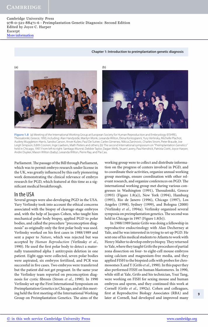
In the USA Several groups were also developing PGD in the USA. Yury Verlinsky took into account the ethical concerns associated with the biopsy of cleavage-stage embryos and, with the help of Jacques Cohen, who taught him mechanical polar body biopsy, applied PGD to polar bodies, and called the procedure “preconception diag-nosis” as originally only the fi rst polar body was used. Verlinsky worked on his fi rst cases in 1988/1989 and sent a paper to Nature , which was rejected but was accepted by Human Reproduction (Verlinsky et al ., 1990 ). He used the fi rst polar body to detect a mater-nally transmitted alpha 1 antitrypsin deletion in one patient. Eight eggs were collected, seven polar bodies were aspirated, six embryos fertilized, and PCR was successful in fi ve cases. Two embryos were transferred but the patient did not get pregnant . In the same year the Verlinksy team reported on preconception diag-nosis for cystic fi brosis (Strom et al ., 1990 ). In 1990 Verlinsky set up the First International Symposium on Preimplantation Genetics in Chicago, and at this meet-ing held the fi rst meeting of the International Working Group on Preimplantation Genetics. Th e aims of the
(a) (b)
Figure 1.8 (a) Meeting of the International Working Group at European Society for Human Reproduction and Embryology (ESHRE), Thessaloniki, Greece, 1993, including: Alan Handyside, Marilyn Monk, Leeanda Wilton, Elena Kontogianni, Yury Verlinsky, Michelle Plachot, Audrey Muggleton-Harris, Sandra Carson, Anver Kuliev, Paul De Sutter, Carles Gimenez, Nikica Zaninovic, Charles Strom, Peter Braude, Joe Leigh Simpson, Edith Coonen, Inge Liaebers, Math Pieters and others; (b) The second international symposium on “Preimplantation Genetics” held in Chicago, 1997. From left to right: Santiago Munné, Debbie Taylor, Dagan Wells, Stuart Lavery, Paul Kendrick, Patrizia Ciotti, Joyce Harper, Andre Duyker, Mason Wilton (baby), Leeanda Wilton, Pierre Ray, and Pia Cau.
working group were to collect and distribute informa-tion on the progress of centers involved in PGD, and to coordinate their activities, organize annual working group meetings, ensure coordination with other rel-evant research, and organize conferences on PGD. Th e international working group met during various con-gresses in Washington (1991), Th essaloniki, Greece (1993) ( Figure 1.8(a) ), New York (1994), Hamburg (1995), Rio de Janero (1996), Chicago (1997), Los Angeles (1998), Sydney (1999), and Bologna (2000) (Verlinsky et al ., 1994a ). Verlinsky organized several symposia on preimplantation genetics. Th e second was held in Chicago in 1997 ( Figure 1.8(b) ) .
In 1988/1989 Jamie Grifo was doing a fellowship in reproductive endocrinology with Alan Decherney at Yale, and he was interested in trying to set up PGD. He sent one of his medical students to Atlanta to work with Henry Malter to develop embryo biopsy. Th ey returned to Yale, where they taught Grifo the procedure of partial zona dissection on four- to eight-cell mouse embryos using calcium and magnesium-free media, and they applied FISH to the biopsied cells with probes for chro-mosomes X and Y (Grifo et al ., 1990 ). In this paper they also performed FISH on human blastomeres. In 1990, while still at Yale, Grifo and his technician, Ysui Tang, were working on FISH for sexing mouse and human embryos and sperm, and they continued this work at Cornell (Grifo et al ., 1992a ). Cohen and colleagues, fi rst at Reproductive Biology Associates (RBA) and later at Cornell, had developed and improved many
Cambridge University Press978-0-521-88471-6 - Preimplantation Genetic Diagnosis: Second EditionEdited by Joyce C. HarperExcerptMore information
www.cambridge.org© in this web service Cambridge University Press

Section 1: Background
10
micromanipulation techniques and Grifo joined the Cornell team (Cohen, Malter, Talanski, Rosenwaks, and Berkley) ( Figure 1.9 ). Th e Cornell team performed its fi rst PGD cases by sexing single-cell blastomeres using co-amplifi cation of DNA on the X and Y chromosome (Grifo et al ., 1992b ). Santiago Munné had studied male infertility and cytogenetics of mouse embryos with Anna Estop and Josep Egozcue, a pioneer in the study of cytogenetics of gametes and embryos. He joined the Cornell team in 1991, bringing fi xation skills with him, and developed the FISH technique. In 1992, in collabo-ration with Ulli Weier, he was the fi rst to apply FISH with directly labeled probes (Munné et al ., 1993a ) .
Development of PCR for monogenic disorders Several groups were now working on using PCR for the detection of specifi c gene mutations for PGD (Li et al ., 1988 ; Holding & Monk, 1989 ; Monk & Holding, 1990 ; Bradbury et al ., 1990 ; Coutelle et al ., 1989 ; Gomez et al ., 1990 ; Navidi & Arnheim, 1991 ; Sermon et al ., 1991 ; Sermon et al ., 1992 ). Mark Hughes came to the Hammersmith from the USA to develop single-cell PCR for cystic fi brosis ( Figure 10(a) , (b) , and (c) ). Along with Pierre Ray, who was studying for his PhD, Hughes developed nested PCR to amplify the ΔF508 region followed by heteroduplex formation for rapid detection of the deletion (Lesko et al ., 1991 ; Handyside et al ., 1992 ; Liu et al ., 1992 ). It is amazing that the cystic fi brosis gene was only described in 1989 (Riordan et al ., 1989 ), and by 1992 the fi rst diagnosis of cystic fi bro-sis in a single cell was possible. Th e fi rst report was on just three couples, all carrying the ΔF508 mutation, of which one woman became pregnant (Handyside et al ., 1992 ). At the same time, the Brussels team developed
Figure 1.9 Cohen and Munnés team, 1994 including Jacque Cohen, Mina Alicani, Santiago Munné, and others.
its own protocol for cystic fi brosis (Liu et al ., 1992 ; Liu et al ., 1993 ) and later was the fi rst team to perform PGD for Duchenne muscular dystrophy (Liu et al ., 1995 ) .
At the Genetics & IVF Institute (GIVF) in Virginia, USA, Gary Harton was in the process of developing PGD in 1992, and he performed the Institute’s fi rst clinical case in 1993 (Levinson et al ., 1992 ). Work focused on monogenic disease diagno-sis, including tests for cystic fi brosis, Huntington’s disease (non-disclosing), Fragile X, and the fi rst birth of an unaff ected child following PGD for spinal muscular atrophy (SMA) (Fallon et al ., 1999 ), as well as the fi rst clinical PGD test for an autosomal domi-nant disease, Marfan syndrome (Harton et al ., 1996 ). GIVF also pioneered the separation of X and Y sperm using MicroSort ® (Levinson et al ., 1992 ; Schulman & Karabinus, 2005 ).
Marilyn Monk’s team developed mouse PGD for Lesch–Nyhan syndrome, SCID, thalassemia, and sickle cell disease, X-linked Duchenne muscular dys-trophy, Fragile X, myotonic dystrophy, and Kennedy disease (Daniels et al ., 1995 ; Monk et al ., 1995 ). Monk published the fi rst quality control experiments to ver-ify sensitivity, effi ciency, and accuracy to lay down the standards for this sensitive work and to convince the fi eld that single-cell PCR was indeed possible (Monk et al ., 1993). Monk’s group was already aware of the problem of carryover contamination (millions of cop-ies of product were being produced). Cathy Holding separated the sites of loading samples into the PCR tubes (which were carried out in the Galton Laboratory car garage) and the PCR procedure in the laboratory. Later, Monk and colleagues began developing single-cell technology for the triplet repeat diseases – Fragile X and myotonic dystrophy (Daniels et al ., 1995 ; Daniels et al ., 1996 ) and imprinted genes (Daniels et al ., 1995 ; Daniels et al ., 1996 ; Daniels et al ., 1997 ; Huntriss et al ., 1998 ; Salpekar et al ., 2001 ). Monk’s team also developed a method they called “cell recycling,” in which a single cell could be analyzed by PCR for a specifi c gene defect (Duchenne muscular dystrophy) as well as the same single cells being studied for sex by in situ hybridiza-tion (Th ornhill et al ., 1994 ; Th ornhill & Monk, 1996 ) ( Figure 1.11 ).
Th e Cornell group published one of the fi rst papers on whole-genome amplifi cation using primer exten-sion preamplifi cation (PEP) (Xu et al ., 1993 ). Th e group developed three PEP protocols on single blast-omeres from arrested embryos. Th ree aliquots of each PEP product were used as templates for exon 10 of the cystic fi brosis gene, or the human X chromosome .
Cambridge University Press978-0-521-88471-6 - Preimplantation Genetic Diagnosis: Second EditionEdited by Joyce C. HarperExcerptMore information
www.cambridge.org© in this web service Cambridge University Press