introduction to medical image processing - 國立臺灣大學b95041/biomedical/12_21_mip2010.pdf ·...
TRANSCRIPT

1
Introduction to Medical Image Processing
Δ Essential environments of a medical imaging system
● Image processing may be a post-imaging or pre-analysis operator.
● Functions of Image processing and Image analysis may overlap each other.
Imaging
System
System
Image
Processing
Images
Feature
Images
Energy
Image Analysis Subject

2
Δ Goals of medical image analysis techniques:
● Quantification: Measuring the features on medical images, eg.,
helpng radiologist obtain measurements from medical images (e.g., area or volume).
* To make the features measurable, it is necessary to extract objects from images by
segmentation.
● Computer Aided Diagnosis (CAD): given measurements and features make a
diagnosis. Help radiologists on their diagnosis procedure for accuracy and efficiency.
● Evaluation and validation techniques.

3
Δ General image analysis (regardless of its application) encompasses:
● incorporation of prior knowledge
● classification of features
● matching of model to sub-images
● description of shape
● many other problems and approaches of AI...
Δ About Digital Image Processing
● What is an Image?
– Formal definition: A digital image is a multi-dimensional signal that is sampled in
space and/or time and quantized in amplitude.
– Looser definition: An image is a “picture."
The brightness values in the picture may represent
distance, reflectivity, density, temperature, etc.
– The image may be 2-D (planar), 3-D (volumetric), or N-D.

4
– Image elements: An image may be composed of:
• 2-D: pixels = picture elements
• 3-D: voxels = volume elements
• 4-D and higher: hypervoxels = hypervolume elements
● What do images represent?
For medical images, pixels may represent parameters such as:
– X-ray attenuation (density)
– Water (proton) density
– Acoustic impedance (distribution)
– Optical reflectivity (or impedance)
– Electrical activity , etc. . . .
● How are images processed?
– Manual analysis: processed by human
– Semi-automatic analysis: human and computer work together to process image
– Automatic analysis: computer processes image, human reviews the results

5
● Purposes of image processing:
– Preprocess image to reduce noise and blur (filtering)
– Identify structures within the image (segmentation)
– Extract “useful” information from the image (quantification)
– Prepare the image for visualization (enhancement, reconstruction)
* Exact processing steps depend on the application.
● What we do to images:
– Enhancement:
– Noise reduction
– Deblur
– Improve contrast
– Identify structures in image (segmentation):
– Identify homogeneous regions in an image (label image pixels = segmentation)
– Measure:
6
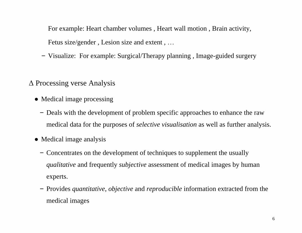
For example: Heart chamber volumes , Heart wall motion , Brain activity,
Fetus size/gender , Lesion size and extent , …
– Visualize: For example: Surgical/Therapy planning , Image-guided surgery
Δ Processing verse Analysis
● Medical image processing
– Deals with the development of problem specific approaches to enhance the raw
medical data for the purposes of selective visualisation as well as further analysis.
● Medical image analysis
– Concentrates on the development of techniques to supplement the usually
qualitative and frequently subjective assessment of medical images by human
experts.
– Provides quantitative, objective and reproducible information extracted from the
medical images

7
Δ So many subjects:
57 chapters in Handbook of Medical Image Processing and Analysis ,
by Isaac Bankman (Ed.), Academic Press, 2009.
I Enhancement; Fundamental Enhancement Techniques; Adaptive Image Filtering; Enhancement by Multiscale
Nonlinear Operators; Medical Image Enhancement with Hybrid Filters; II Segmentation; Overview and
Fundamentals of Medical Image Segmentation; Image Segmentation by Fuzzy Clustering: Methods and Issues;
Segmentation with Neural Networks; Deformable Models; Shape Information in Deformable Models; Gradient Vector
Flow Deformable Models; Fully Automated Hybrid Segmentation of the Brain; Unsupervised Tissue Classification;
Partial Volume Segmentation with Voxel Histograms; Higher Order Statistics for Tissue Segmentation; III Quantification; Two-dimensional Shape and Texture Quantification; Texture Analysis in Three Dimensions for
Tissue Characterization; Computational Neuroanatomy Using Shape Transformations; Tumor Growth Modeling in
Oncological Image Analysis; Arterial Tree Morphometry; Image-Based Computational Biomechanics of the
Musculoskeletal System; Three-Dimensional Bone Angle Quantification; Database Selection and Feature Extraction
for Neural Networks; Quantitative Image Analysis for Estimation of Breast Cancer Risk; Classification of Breast
Lesions in Mammograms; Quantitative Analysis of Cardiac Function; Image Processing and Analysis in Tagged
Cardiac MRI; Analysis of Cell Nuclear Features in Fluorescence Microscopy Images; Image Interpolation and
Resampling; IV Registration; Physical Basis of Spatial Distortions in Magnetic Resonance Images; Physical and
Biological Bases of Spatial Distortions in PET Images; Biological Underpinnings of Anatomic Consistency and
Variability in the Human Brain; Spatial Transformation Models; Validation of Registration Accuracy; Landmark-
based Registration Using Features Identified through DifferentialGeometry; Image Registration Using Chamfer
Matching; Within-Modality Registration Using Intensity-Based Cost Functions; Across-Modality Registration Using
Intensity-Based Cost Functions; Talairach Space as a Tool for Intersubject Standardization in the Brain; Warping
Strategies for Intersubject Registration; Optimizing the Resampling of Registered Images; Clinical Applications of
Image Registration; Registration for Image-Guided Surgery; Image Registration and the Construction of
Multidimensional Brain Atlases; V Visualization; Visualization Pathways in Biomedicine; Three-Dimensional
Visualization in Medicine and Biology; Volume Visualization in Medicine; Fast Isosurface Extraction Methods for
Large Image Data Sets; Computer Processing Methods for Virtual Endoscopy; VI Compression, Storage, and Communication; Fundamentals and Standards of Compression and Communication; Medical Image Archive and
Retrieval; Image Standardization in PACS; Imaging and Communication in Medical and Public Health Informatics;
Dynamic Mammogram Retrieval from Web-Based Image Libraries; Quality Evaluation for Compressed Medical
Images: Fundamentals; Quality Evaluation for Compressed Medical Images: Diagnostic Accuracy; Quality Evaluation
for Compressed Medical Images: Statistical Issues; Three-Dimensional Image Compression with Wavelet Transforms.

8
Δ Why Image Enhancement?
● Can’t distinguish between tissues
The nature of the physiological system under investigation and the procedures used
in imaging may diminish the contrast and the visibility of details.
● Data is too noisy for computer algorithm to perform well
Medical images are often deteriorated by noise due to various sources of
interference and other phenomena that affect the measurement processes in
imaging and data acquisition systems.
● Imaging artifacts interfere with visualization or computer processing
Δ How to Enhance Image? By operations to
● Increase contrast
● Remove noise
● Emphasize edges: Edge boost, Unsharp masking.
● Modify shapes

9
* Image enhancement techniques range from linear to nonlinear,
from fixed to adaptive, and from pixel-based to multiscale methods, …
Δ Contrast Enhancement by Histogram Equalization

10
Δ Enhancement by adaptive wavelet shrinkage denoising
(only subimages with diagnostic information are reconstructed).

11
Δ Enhancement by adaptive filtering: noise or speckle reduction.
● Adaptive image filtering needs some a priori information about the image.
( This is an image modeling problem)
● Geometric or statistical information are the primary information used.

12
Δ Medical Image Segmentation
● Segmentation, separation of structures of interest from the background and from
each other, is an essential analysis function for which numerous algorithms have
been developed in the field of image processing.
● The principal goal of the segmentation process is to partition an image into regions
that are homogeneous with respect to one or more characteristics or features.
● Segmentation is an important tool in medical image processing, and it has been
useful in many applications.
The applications include detection of the coronary border in angiograms,
multiple sclerosis lesion quantification, surgery simulations, surgical planning,
measurement of tumor volume and its response to therapy, functional mapping,
automated classification of blood cells, study of brain development,
detection of microcalcifications on mammograms, image registration,
atlas-matching, heart image extraction from cardiac cineangiograms, detection of
tumors, etc.

13
● In medical imaging, segmentation is important for feature extraction, image
measurements, and image display.
In some applications it may be useful to classify image pixels into anatomical
regions, such as bones, muscles, and blood vessels, while in others into pathological
regions, such as cancer, tissue deformities, and multiple sclerosis lesions.
=> Segmentation can be thought as the preprocessor for further analysis.
● A wide variety of segmentation techniques have been proposed.
However, there is no standard segmentation technique that can produce satisfactory
results for all imaging applications.

14
● Segmentation techniques can be divided into classes in different ways.
e.g., based on the classification scheme:
– Manual, semiautomatic, and automatic
– Pixel-based (local methods) and region-based (global methods).
– Low-level segmentation (thresholding, region growing, etc.), and
Model-based segmentation (multispectral or feature map techniques, Marcov
random field, deformable models, etc.).
* Model-based techniques are suitable for segmentation of images that have
artifacts, noise, and weak boundaries between structures.
* Deformable models: Snake model and Level Sets
– Classical (thresholding, edge-based, and region-based techniques),
Statistical, Fuzzy, and Neural network techniques.

15
Δ Histogram analysis
● Histogram analysis is usually required before doing segmentation,
it is a pixel-based technique.
● An example
0 10 20 30 40 50 60 70 80 90 0
200
400
600
800
1000
The information from histogram
Number of modes
Mode to mode contrast
Spread of each mode
(seperability)

16
x < 30 30 < x <50 x > 50
x < 30 contains mostly the bone
30 < x < 50 contains mostly the fluid region
x > 50 contains mostly the tissue region,
All of them are noisy and partially correct
=> Simple thresholding can not provide satisfactory segmentation

17
Δ The general question is:
● How are the pixels grouped?
=> Contour detection and linking or Region growing approaches
● What is criterion of the homogeneity (or connectivity) for growing?
* Pixel connectivity can be deterministic or probabilistic.
Δ Edge detection
● Edges detected using Sobel’s and Canny’s detectors
● As the result of pixel-thresholding, detected edges are noisy and erroneous.
● Pre-processing for noise suppresion or contour modeling are required.

18
Δ Image Modeling by Random Process (Field)
● Images are in general treated as pieces of information, which are essentially random
processes. Random process theory provides rich ways to handle the information
carried in images
● When images are represented as random process, an image ( , )x i j is treated as a
realization of the random process ( , )X i j . Thus, an ( )N N image is considered as
the outcomes of 2N random number generators.
● The 2D index ( , )i j is referred as site ( , )s i j , and collection of all sites { }S s is
named as a site system or lattice system in the “field of statistical mechanical
system”.
…
… x S sx

19
Δ Definition of Neighborhood System
● Definitions
s - site or lattice point, s S S - set of lattice points
sX - the value of X at s s - the neighboring points of s
● A neighborhood system s must be symmetric
alsor s s r s s
● Example of 8 point neighborhood
Neighbors of ( , )2 2x
{( , ),( , ),( , ),( , ),( , ),( , ),( , ),( , )}s 1 1 1 2 1 3 2 1 2 3 3 1 3 2 3 3
( , )s 2 2

20
Δ A simple Markov Random Field (MRF) model
● Definition of MRF: A random field X on the lattice S with neighborhood system s
is said to be a Markov random field, if for all s S , ( | ) ( | )s i s s sp x x p x x
● A very simple MRF model: the Ising model , it is described by the conditional
probability model
''
''
exp( )
exp
s ss ss s
ss s
x xp x x
1 x
, with { , }sX 0 1
● For small , the model represents fine texture images
For large , the model represents coarse texture images

21
Δ Bayesian image restoration (or segmentation)
MAP Restoration
= Maximizing ( | ) ( | ) ( )p x y p y x p x
p(x) p(y|x)
MRF Models (either learned or known)
Restoration
( | )p x y
Optimal
estimate
x* =
Imaging y = x =

22
From Bayes theorem:
•Likelihood PriorPosterior
Normalization Factor
( | ) ( )( | )
( )
P Observation True Image P True ImageP True Image Observation
P Observation
Both the prior and the likelihood allow the introduction of expert knowledge:
● The prior can express assumptions about what the “true scene” should be,
e.g., piecewise smooth, which is essentially a Markovian assumption.
● The likelihood can incorporate knowledge about the degradation and distortions
introduced in the imaging process. It gives the probability about what the
observations should be based on the “true scene”, when degraded by additive or
multiplicative noise, blurring, etc.
Prior: ( )P True Image = Probabilitic Model of image
Likelihood: ( | )P Observation True Image = Probabilitic Model of Imaging Process

23
Δ Application-texture segmentation
This image can be segmented as the followings
by using different value of
This image can be segmented to be any one of the followings
by using different value of

24
= 0.1 = 0.2
= 0.5 = 1.0
= 2.0 = 4.0
* A three level MRF model is used.