introduction to freesurfer · to caffeinate or not to caffeinate? please don’t spill coffee (or...
TRANSCRIPT


Post Your Questions!
http://surfer.nmr.mgh.harvard.edu/cgi-bin/fsurfer/questions.cgi

To Caffeinate or not to
Caffeinate?
Please don’t spill coffee (or anything else on the
laptops), or if you feel you must, please be prepared to
fund a replacement!
(we will have coffee this afternoon at the break)

Why FreeSurfer?
1. Anatomical analysis is not like functional analysis –
it is completely stereotyped.
2. Registration to a template (e.g. MNI/Talairach)
doesn’t account for individual anatomy.
3. Even if you don’t care about the anatomy, anatomical
models allow functional analysis not otherwise
possible.

ICBM Atlas
Why not just register to an
ROI Atlas?
12 DOF
(Affine)

Subject 1Subject 2 aligned with Subject 1
(Subject 1’s Surface)
Problems with Affine (12 DOF)
Registration (you will get sick of this slide)

Surface and Volume Analysis
Cortical Reconstruction
and Automatic LabelingInflation and Functional
Mapping
Surface Flattening
Surface-based Intersubject
Alignment and Statistics
Automatic Subcortical
Gray Matter Labeling
Automatic Gyral White
Matter Labeling

Talk Outline
1. Cortical (surface-based) Analysis.
2. Volume Analysis.

Talk Outline
1. Cortical (surface-based) Analysis.
2. Volume Analysis.

What Can One Do With A Surface
Model?
left primary visual cortex right visual hemifield
desired shape of activity pattern required shape of stimulus
goal: use model to imposed desired activity pattern on V1
Collaboration with Jon Polimeni and Larry Wald.
w=k log(z+a)

Tangential Resolution Measured with
Surface-based Analysis
Collaboration with Jon Polimeni and Larry Wald.

Tangential Resolution Measured with
Surface-based Analysis
Collaboration with Jon Polimeni and Larry Wald.

NeuroMarketing!
Thanks to Larry Wald for this slide.
Aim 1 of our NCRR Center Grant, spelling:
“MGH Center for Functional Neuroimaging Technologies; an NCRR Center for Research Resources.”
(just kidding)

Surfaces: White and Pial
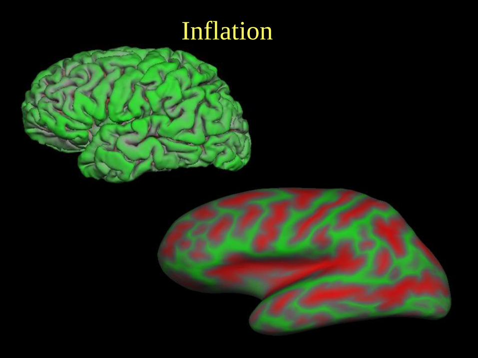
Inflation

Inflated surface with cutssuperior temporal
Metrically optimal flat map
calcarine
central
sylvian
anterior
posterior
Surface Flattening – Whole
Hemisphere

Cortical Thickness
white/gray surface
pial surface
lh.thickness, rh.thickness
• Distance between white and pial surfaces
• One value per vertex

A Surface-Based Coordinate System

Comparing Coordinate Systems
and Brodmann Areas
Cumulative histogram
(red=surface, blue=nonlinear
Talairach)
Ratio of surface accuracy to
volume accuracy

Automatic Surface Segmentation
Precentral GyrusPostcentral Gyrus
Superior Temporal GyrusBased on individual’s folding pattern

Inter-Subject AveragingS
ubje
ct 1
Subje
ct 2
NativeSpherical Spherical
Surface-to-
Surface
Surface-to-
Surface
GLM
Demographics
mri_glmfit cf. Talairach

Borrowed from (Halgren et al., 1999)
Visualization

Rosas et al., 2002
Kuperberg et al., 2003
Gold et al., 2005
Rauch et al., 2004Salat et al., 2004
Fischl et al., 2000
Sailer et al., 2003

Talk Outline
1. Cortical (surface-based) Analysis.
2. Volume Analysis.

Volume Analysis: Automatic
Individualized Segmentation
Surface-based coordinate system/registration
appropriate for cortex but not for thalamus, ventricular
system, basal ganglia, etc…
Anatomy is extremely variable – measuring the
variance and accounting for it is critical (more in the
individual subject talk)!

Volumetric Segmentation (aseg)
Caudate
Pallidum
Putamen
Amygdala
Hippocampus
Lateral Ventricle
Thalamus
White Matter
Cortex
Not Shown:
Nucleus Accumbens
Cerebellum

Volume Differences Predictive of AD
Data courtesy of Drs Marilyn Albert and Ron Killiany

Combined Segmentation
aparc+aseg
aseg
aparc

Gyral White Matter Segmentation
Nearest Cortical Label
to point in White Matter
wmparc
+ +
aparc
aparc+aseg

Summary• Why Surface-based Analysis?
– Function has surface-based organization
– Visualization: Inflation/Flattening
– Cortical Morphometric Measures
– Inter-subject registration
• Automatically generated ROI tuned to each subject individually
Use FreeSurfer Be Happy

MGH
Allison Stevens
Nick Schmansky
Andre van der Kouwe
Doug Greve
David Salat
Evelina Busa
Lilla Zollei
Koen Van Leemput
Sita Kakunoori
Ruopeng Wang
Rudolph Pienaar
Krish Subramaniam
Diana Rosas
Acknowledgements
UC San DiegoAnders Dale
MIT
Polina Golland
B. T. Thomas Yeo
Mert Sabuncu
Florent Segonne
Peng Yu
Ramesh Sridharan
MGH
Jean Augustinack
Martin Reuter
Anastasia Yendiki
Jon Polimeni
Kristen Huber
UCLMarty Sereno
NINDS
MGH (past)
Brian T Quinn
Xiao Han
Niranjini Rajendran
Jenni Pacheco
Sylvester Czanner
Gheorghe Postelnicu
Sean Marrett

Why Is a Model of the
Cortical Surface Useful?
Local functional organization of cortex is largely 2-dimensional! Eg, functional mapping of primary visual areas:
From (Sereno et al, 1995, Science). Also, smooth along surface

Flat Map of Monkey Visual Areas
D.J. Felleman and D.C. Van Essen, CC, 1991