intrinsic fluorescene of cowpea-chlorotic-mottle-virus protein
TRANSCRIPT

Eur. J. Biochem. 95, 21-29 (1979)
Intrinsic Fluorescence of Cowpea-Chlorotic-Mottle-Virus Protein Jacob KRUSE, Benedictus J. M. VERDUIN, and Antonie J. W. G. VISSER
Departments of Biochemistry and Virology, Agricultural University, Wageningen
(Received September 4 / December 1, 1978)
Changes in the environment of the aromatic amino acid residues in the protein subunits of cowpea chlorotic mottle virus were studied with fluorescence techniques. The fluorescence properties of the protein in nucleoprotein particles, empty protein shells and dimers of the coat protein subunit were determined.
According to fluorescence spectra (excitation at 295 nm, emission maxima at 342 - 346 nm depending on pH and ionic strength) the tryptophan residues are located in a polar environment. With excitation at 235 nm a strong shoulder at about 300 nm in the total emission spectrum can be observed. This emission must be attributed to the tyrosine residues in the protein and can also be observed as a fast component in the fluorescence decay curve. The lifetimes were compared with those of the other bromoviruses which are different in amino acid composition. From excitation spectra of cowpea chlorotic mottle virus it can be concluded that energy is transferred from tyrosine to tryptophan residues.
Increase in the quantum yield of the tryptophan emission of the nucleoprotein particles upon raising the ionic strength and pH are attributed to a diminishing interaction between the protein and the RNA. The fluorescence properties of the protein hardly change when the empty protein shells are dissociated into protein dimers.
Quenching studies with potassium iodide and acrylamide indicate that the tryptophan residues in the protein are attainable more easily at low ionic strength than at high ionic strength.
Cowpea chlorotic mottle virus (CCMV) is a multi- component spherical plant virus, which belongs, to- gether with brome mosaic virus (BMV) and broad bean mottle virus (BBMV), to the group of bromo- viruses [l, 21. Their purified preparations consist of almost equal proportions of three different nucleo- protein particles. Two of them contain one RNA strand of M , about 1.1 x lo6 and the third has two smaller RNA components with molecular weights of about 0.8 x 10⁶ and 0.3 x lo6. The protein coat of these particles is similar and consists of 180 identical polypeptides with a molecular weight of 19400, arranged on the surface of an icosahedron.
The nucleoprotein particles, which are stable at pH 5.0, increase in hydrodynamic volume when the pH is raised to 7.5. The resulting decrease in sedimen- tation coefficient from 88 S to 78 S has been explained, by radial expansion of protein and nucleic acid due to electrostatic repulsion [2]. Increasing the ionic strength to 1.0 M at pH 7.5 dissociates the swollen
Abbreviations. CCMV, cowpea chlorotic mottle virus; BBMV, broad bean mottle virus; BMV, brome mosaic virus; fwhm, full width at half maximum.
virus into coat protein dimers [3] and RNA [1,2]. When the RNA is removed, the proteins dimers can reversibly associate into empty spherical protein shells called pseudo-top component. The arrangement of the polypeptides in these shells is similar to that in the nucleoprotein coat [4].
In each CCMV protein subunit three tryptophan, five tyrosines and four phenylalanines are present. For BMV and BBMV proteins the number of aromatic amino acid residues are, respectively, two and zero tryptophans, five and four tyrosines and five and seven phenylalanines [ 5 ] . By observing the fluorescence properties of the aromatic amino residues, informa- tion can be obtained about the direct environment and the localization of these residues in the protein particles [6,7].
This paper reports on fluorescence of CCMV protein. The fluorescence properties studied were peak location and band width of emission spectra, excitation spectra, lifetimes and degree of polarization. In particular, the changes in these parameters induced upon passing through the different states of aggrega- tion of the virus have been measured and correlated with structural features of the virus particles.

22 Fluorescence of Cowpea-Chlorotic-Mottle-Virus Protein
MATERIALS AND METHODS
Virus
CCMV was isolated from 7-day-infected cowpea leaves as described before [8]. The coat protein was prepared according to the CaC12 method and pseudo- top component was obtained by dialysing against 1 M NaCl, pH 5.0 [9]. BMV and BBMV were grown in barley and horse bean respectively. Virus and coat protein were isolated by the same procedures as de- scribed for CCMV. The nucleoprotein and protein preparations were dialyzed against 0.02 M cacodylic acid buffer of the desired pH and ionic strength. All measurements were done in this buffer. Preparations of each bromovirus contained the three different nucleoprotein particles in the same ratio as the iso- lates from the plants. Both protein and nucleoprotein were used in a concentration of about 0.1 mg/ml.
Absorption Spectra
spectrophotometer. Absorption spectra were recorded with a Cary 14
Fluorescence Spectra
Fluorescence spectra were measured with a Hi- tachi-Perkin Elmer MPF 2A spectrofluorimeter equip- ped with a thermostated cell holder and with a home-built apparatus. It was composed of a 450-W xenon lamp and an M4QIII Zeiss monochromator for excitation and a Jarrell-Ash 0.25-m Ebert mono- chromator (model 82.410) with a cooled EM1 9659 QB S20 photomultiplier for detection. The light beam was chopped at 124 Hz. The photomultiplier signal was amplified by a PAR 128A lock-in amplifier, supplied with a reference signal from the light chopper. The cell holder could be thermostated between - 180 "C and room temperature, using either liquid nitrogen or circulating methanol as coolant. The fluorescence spectra were recorded at 4 "C.
The sensitivity of the detection system was cali- brated with a standard tungsten lamp [lo]. The quan- tum yield was determined by comparing an integrated and corrected spectrum with that of a tryptophan solution of known concentration (Q = 0.14) [ l l ] .
Fluorescence lifetimes were determined with a phase fluorometer [12,13]. Fluorescence decay curves were obtained with a single-photon counting apparatus consisting of a gated flash lamp unit (Applied Photo- physics Ltd), a 56 DUVP/03 Philips photomultiplier, operated at 2400 V, NIM modules of the Ortec 9200 system and a Laben multichannel analyzer. A flash in deuterium (1 atm) passing through a Jarrell Ash 0.25-m monochromator (model 82.410) was used for excitation. The emission from the thermostated sample cuvette was observed through a set of filters consisting of a Schott WG 305, a Chance Pilkington OX7 and a
I 0 K) 20 30
Time (ns)
Fig. 1. Example of the analysis ofafluorescence decay curve measured by means of singlephoton counting. (1) Excitation profile, calculated by deconvoluting the decay curve of the quantum counter with a known lifetime. (2) The dots represent experimental data, the drawn line is the calculated decay curve. (3) The deviation function DV(i) = [ F c ( i ) - F ( i ) ] / m ) where F(i) is the content of the ith channel and F,(i) is the calculated value of this content. The scale is linear
NiS04/CoS04 liquid filter with a maximum trans- mission at 317 nm and a full width at half maximum (fwhm) of 42 nm [14] or through a Balzer K 36 band filter (A,,, = 366 nm, fwhm = 47 nm). The filters used were chosen in such a way that a glycogen solution with a turbidity comparable to that of the virus solutions resulted in a negligeable number of counts. Because of its wavelength dependence the response function of the apparatus was determined with a reference compound by a method described elsewhere [15]. This reference compound was a degassed solu- tion of p-diphenylbenzene (BDH) in cyclohexane (Merck, Uvasole). The life-time of this compound was determined as 0.95 ns with the phase fluorimeter operating at 60 MHz. The lifetime was homogeneous with respect to measurements of phase shift and modu- lation [12]. The decay experiments were performed at 9 "C. Analysis of the data was performed using a non-linear regression algorithm for the deconvolution of the fluorescence decay [16] described by:
(1) t
Fc(t) = Jg(T) . I ( t -T)dT where g(T) is the instrumental response function and
Z ( t ) is the decay law assumed to be: ,i aJe- r~ i~ , with n = 1 (mono-exponential), n = 2 (bi-exponential) or n = 3 (tri-exponential). After adjustment of the parameters aj and ~j the fit was checked by calculating the convolution product of I(t) and g( t ) [Eqn (l)] and by visual comparison of the experimental F(t) and the calculated curves Fc(t) (Fig. 1). The criterion of best fit was attained after a search for the minimum value of x2 :
in which the summation is made over N channels.
J = 1
x - - I = I 3 wi [F(i) - Fc(i)]2 (2)

J. Kriise, B. J. M. Verduin, and A. J. W. G. Visser
D Tyr TrP
23
0.3
m E 0.2
SI D L
n 4 0.1
0
Wavelength (nrn) Wavelength (nrn)
C I 2 0 0
150
100
53
0
Fig. 2. Absorption andfluorescence spectra Of'CCMVprotein and nucleoprotein. (A) Absorption spectra of CCMV protein at pH 1.5 + 1 M NaCl (----), and CCMV nucleoprotein at pH 5 (-). (B) Corrected fluorescence spectra of CCMV nucleoprotein excited at 235 nm: (1) pH 5.0, (2) pH 5.0 + 1.0 M NaCl, (3) pH 7.5, (4) pH 7.5 + 1.0 M NaCI. (C) As for B with excitation at 295 nm. (D) Characteristic excitation spectrum of CCMV nucleoprotein. This spectrum is not corrected for lamp intensity at different wavelengths. The detection wavelength is at 350 nm
(3)
wherefis a number related to the number of lifetime components in which deconvolution takes place : 2, 4 or 6 for a mono, di, or tri-exponential fit respec- tively. The factor (N-f) denotes the degrees of freedom.
Also plotted is a diviation function of the residuals defined as :
for each channel i.
(4)
Fluorescence Polarization
Fluorescence polarization values were determined on an apparatus built according to the idea of Weber and Bablouzian [17]. Excitation was at 292 nm (fwhm = 10 nm) and the emission was detected with a com- bination of the Schott filters WG 320 + UGI (A,,, = 365 nm, fwhm = 52 nm).
RESULTS
Absorption Spectra
The virus exhibited an absorption maximum at 260 nm, mainly originating from the RNA. The con-
tribution of the protein appeared as a shoulder in the absorption spectrum of the nucleoprotein. The spectrum of the protein showed a maximum at 277 nm and a shoulder at 290 nm originating from the tyro- sine and the tryptophan residues respectively (Fig. 2A). The tryptophan shoulder was shifted as compared to the shoulder at 288 nm in the absorption spectrum of a solution of free tryptophan. The differences between free tryptophan and tryptophan in the protein might reflect a less polar environment of the tryptophans in the protein [6,7].
Fluorescence Spectra
The fluorescence was observed with a band width of 3.3 nm. Excitation was at 285 and 295 nm. Pseudo- top component and the protein dimers exhibited similar fluorescence spectra that were independent of their aggregation states as influenced by pH and ionic strength. On the other hand, a change in pH and ionic strength had a remarkable effect on the protein fluorescence of the nucleoprotein particles (Fig. 2 B). At pH 5.0 and low ionic strength, where the virus is stable, the 'emission spectrum showed a maximum at 342 nm when excited in the tryptophan absorption band at 295 nm. No RNA fluorescence could be de- tected. An increase of the ionic strength at pH 5.0

24 Fluorescence of Cowpea-Chlorotic-Mottle-Virus Protein
Table 1. Emission parameters of CCMVprotein fluorescence tein (Fig.2D) a band at 235 nm is also present when Excitation was at 295 nm (fwhm = 2.2 nm). Measurement were at detection is at 350 nm. This is probably due to energy 4 " c unless indicated otherwise. The accuracy of the emission wave- transfer from tyrosine to tryptophan residues within length maximum (Amax) was 2 1 nm and of the quantum yield (Q) about 5 % of the value. fwhm is the width of the emission curve the protein* at half intensity of the maximum Excitation at 235 nm of the virus solutions (fwhm
of excitation is 4 nm) resulted in a dependence of the Component Solution conditions Q fwhm Amax tryptophan emission intensity at 350 nm on pH and
ionic strength, comparable to the observations upon excitation at 295 nm. When exciting the nucleoprotein
Nucleoprotein pH 5.0 0.075 60 342 at 235 nm, a strong shoulder is observed at 300 nm PH 5.0 + 1 M NaCl 0.13 59 346 in the emission spectrum. This shoulder can be attrib- pH 7.5 '.16 59 345 uted to the presence of tyrosine residues, since free pH 7.5 + 1 MNaCl 0.19 59 346 tyrosine exhibits an emission maximum in this region
Protein O.I9 60 346 whereas a free tryptophan solution hardly shows any contribution at this wavelength. The intensity at 300 nm does not depend on the different conformations of the virus (Fig. 2B and C).
nm ~~
Protein and nucleoprotein at room temperature pH 7.5 + I M NaCi 0.13 346
to 1 M NaCl resulted in a higher quantum yield and a shift of the emission maximum to 346 nm. Under these conditions no changes in hydrodynamic prop- erties have been observed (Verduin, unpublished results). At low ionic strength ( I < 0.1 M) and pH 7.5, where the virus is swollen and assumed to be stabilized mainly by electrostatic protein - nucleic-acid inter- actions, a further increase in the quantum yield was observed. The quantum yield was slightly higher than at 1 M NaCl pH 5.0 and the maximum shifts to 345 nm. Increasing the ionic strength at pH 7.5, thereby causing dissociation of the virus into protein dimers and RNA, enhanced the quantum yield even more. Characteristics of the fluorescence spectra of CCMV protein are collected in Table 1.
The presence of 0.01 M MgC12 in a solution of low ionic strength at pH 7.5, that partly prevents the swell- ing of the nucleoprotein particles, had too small an effect on the fluorescence intensity to be expressed in a value of the quantum yield. In all cases the full width at half maximum (fwhm) of the tryptophan emission band was 60 f 1 nm. The fluorescence quantum yield of the undissociated nucleoprotein was always lower than that of the RNA-free protein solutions (cf. Table 1). The fact that the fluorescence spectra of the RNA-free protein showed no observ- able dependence on the pH and ionic strength was in sharp contrast to the sensitivity towards NaCl con- centration of the nucleoprotein emission. This sug- gests that an RNA-protein interaction might be re- sponsible for the changes observed within the virus.
In excitation spectra of a diluted tyrosine solution (detection at 300 nm) a peak at 235 nm can be located. This maximum is absent in excitation spectra of either tryptophan solutions or diluted tryptophan and tyro- sine solutions mixed in a ratio, identical to that found in the coat protein, when detection is at 350 nm. How- ever, in an excitation spectrum of CCMV nucleopro-
Excitation Spectra and Energy Transfer
Uncorrected fluorescence excitation spectra with a detection wavelength at 350 nm (fwhm = 4 nm) showed three peaks: at 235 nm, mainly from the tyro- sine residues, at 285 nm, and at 292 nm (Fig. 2 D). The shape of the excitation spectrum did not change when the detection wavelength was varied between 350 and 400 nm. The peak at 285 nm is partly due to the tyrosine residues. This was confirmed by measur- ing the excitation spectrum with 300 nm as detection wavelength, in which case only bands with maxima at 235 and 285 nm could be observed. Studies on solutions of tryptophan, tyrosine and a mixture of these amino acids also indicated that this conclusion was justified (results not shown). The detection of the band at 235 nm was possible because of the low ab- sorbance of the cacodylic acid solution at this wave- length. With the diluted protein and nucleoprotein solutions (approximately 0.1 mgiml), where no cor- rections for screening effects due to high absorbance were necessary, no changes in the relative intensities of the peaks at 285 and 292 nm in the excitation spectra were observed, when pH and ionic strength were varied. This demonstrated that the energy transfer from tyrosines to tryptophans was not very dependent on the structure of the virus particles. The amount of energy transfer could be estimated from absorption and from lamp-intensity-corrected excitation spectra of the protein [18 --201. Upon comparing these spectra with the absorption spectra of aqueous solutions of tryptophan and of a mixture of tryptophan and tyro- sine (3: 5), it was shown that the efficiency of energy transfer from tyrosine to tryptophan was about 30 %.
For a donor-acceptor pair the efficiency E is de- fined as :
1191 (5 ) 1
E =

J. Kriise, B. J. M. Verduin, and A. J. W. G. Visser 25
Table 2. Fluorescence lifetimes of tryptophun and tyrosine residues in bromovirus proteins The fluorescence lifetimes were measured by the single photon counting technique. Ai is the amplitude of the exponential function e - r / T , ,
multiplied with the fluorescence lifetime ~ i . Deconvolution with three time constants gave the best fit. (z) is the averaged lifetime (,Z,Aizi)/ (ZAi). xz is a function of the deviation between the measured and the calculated decay curve (see Methods). In all experiments the temperature was 9 "C. The aggregation state is characterized by pH and molarity of NaCl in the solution
Virus Wavelength of Component Aggregation state A1 rl A2 rZ A3 53 (t} xz excitation detection PH [NaCl]
nm
CCMV 235 317 nucleoprotein
235 366
295 366
protein
nucleoprotein
protein
nucleoprotein
protein
BMV 235 317 nucleoprotein
5 5 7.5 7.5 5 7.5
5 5 7.5 7.5 5 7.5
5 5 7.5 7.5 5 7.5
M
0 1 .o 0 1 .o 1 .o 1 .o 0 1 .o 0 1 .o 1 .o 1 .o 0 1 .o 0 1 .o 1 .o 1 .o
0.12 0.12 0.12 0.03 0.07 0.04
0.07 0.03 0.05 0.01 0.04 0.02
0.15 0.06 0.07 0.04 0.07 0.04
ns
0.39 0.42 0.40 0.1 1 0.26 0.53
0.57 0.26 0.89 0.12 0.92 0.15
0.41 0.37 0.63 0.31 0.51 0.28
5 0 0.07 0.21 5 1 .o 0.15 0.21 7.5 1 .o 0.23 0.38
BBMV 235 317 nucleoprotein 5 0 005 0 10 5 1 0 004 0.15 7 5 1 0 0 0 4 0 14
0.48 0.39 0.37 0.38 0.37 0.41
0.16 0.12 0.15 0.07 0.08 0.08
0.18 0.14 0.14 0.09 0.10 0.09
0.49 0.54 0.55
0.11 0.15 0.12
~~
__
ns
2.1 2.3 2.4 2.4 2.1 2.4
2.3 2.1 3.5 1.9 2.7 2.6
2.7 1.9 2.9 2.3 2.8 3.1
1.08 1.52 1.87
1.68 1 .so 1.58
0.40 0.49 0.51 0.59 0.57 0.55
0.77 0.85 0.80 0.92 0.88 0.90
0.67 0.80 0.79 0.87 0.83 0 87
0.44 0.31 0.22
0.84 0.81 0.84
_ _
~
ns
6.3 6.9 7.1 7.0 6.8 1.2
7.2 7.2 7.1 7.3 1.3 7.5
7.4 7.3 7.6 7.5 7.5 7.7
3.0 3.8 4.0
3.8 3.8 3.9
-
-
~
3.6 4.3 4.6 5.1 4.7 5.0
6.0 6.4 6.7 6.9 6.1 7.0
5.5 6.1 6.5 6.7 6.5 7.0
1.9 2.0 2.0
3.4 3.4 3.5
~
1.1 1.2 0.85 1.3 1.1 1.3
1.3 1.3 1.1 1.1 1.6 1.4
1.3 1.4 1.3 I .2 1.5 1.1
1.4 1.1 1.1
1.4 1.2 1.2
where Y = distance between donor and acceptor and RO = the Forster critical distance where E is 50%. For the donor-acceptor pair Tyr-Trp, a Ro of 1.31 - 1.5 nin is assumed [19,20].
Since there are five tyrosine and three tryptophan residues per protein subunit, multiple pathways of energy transfer can exist, making it difficult to draw conclusions about relative distances and orientations from the value of the efficiency. The observed increase in the fluorescence quantum yield of the virus particles, when ionic strength or pH were raised, was not ac- companied by a change in the shape of the excitation spectra monitoring tryptophan emission at 350 nm. The efficiency of energy transfer from tyrosine to tryptophan residues thus appeared to be independent of this increase in quantum yield. This must be ex- plained by assuming that the originally non-fluorescent tryptophan residues are equally effective as an accep- tor of the tyrosine excitation energy.
Fluorescence Lifetimes
CCMV protein and nucleoprotein solutions were excited at 235 and 295 nm. The time-resolved emission was observed through filters having transmission maxima at 317 and 366nm. A typical example of such a fluorescence decay experiment is shown in Fig. 1. At 317 nm both tyrosine and the blue side of tryptophan emissions could be observed, while at 366 nm only tryptophan emission is transmitted. The results are collected in Table 2. It should be noted that in all solutions studied the decay function could not be assumed to be mono-exponential. On the other hand the decay profiles measured with a standard compound like N-acetyl-tryptophanamide in buffer pH 7.0 [21] could be fitted with a single lifetime. For comparison, the results obtained with other bromo- viruses are also included. In order to emphasize the relative importance of the different lifetime com-
26 Fluorescence of Cowpea-Chlorotic-Mottle-Virus Protein
ponents, the amplitudes of the exponential functions are multiplied with their lifetimes. This product is directly comparable to the contributions of the dif- ferent chromophores to the time-independent emis- sion intensity at a particular wavelength region. It is to be noted that the relatively large differences in
3.5 I-----
0.5 ; 0 0.1 0.2 0.3 0.4 0.5 0.6 0.7
[KI] ( W 3.5
3.0
2.5
1.5
1.0
0.5; I I
01 0.2 0.3 0.4 0.5 0.6 0.7 [ K I l (M)
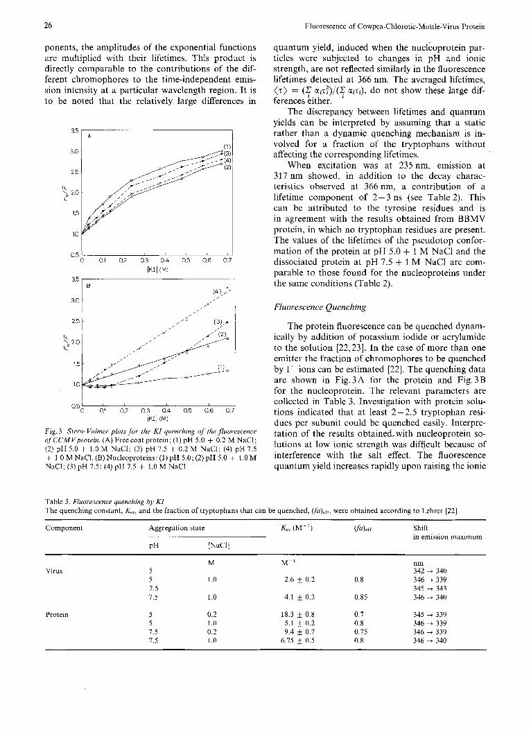
Fig. 3. Stern- Volmer plots fo r the KI quenching of the fluorescence of CCMVprotein. (A) Freecoat protein; (1) pH 5.0 + 0.2 M NaCl; (2) pH 5.0 + 1.0 M NaCl; (3) pH 7.5 + 0.2 M NaC1; (4) pH 7.5 + 1 .O M NaCl. (B) Nucleoproteins: (1) pH 5.0; (2) pH 5.0 + 1.0 M NaCl; (3) pH 7.5; (4) pH 7.5 + 1.0 M NaCl
quantum yield, induced when the nucleoprotein par- ticles were subjected to changes in pH and ionic strength, are not reflected similarly in the fluorescence lifetimes detected at 366 nm. The averaged lifetimes, (7) = (& uizT) / (c uizi), do not show these large dif- ferences kither.
The discrepancy between lifetimes and quantum yields can be interpreted by assuming that a static rather than a dynamic quenching mechanism is in- volved for a fraction of the tryptophans without affecting the corresponding lifetimes.
When excitation was at 235 nm, emission at 317 nm showed, in addition to the decay charac- teristics observed at 366 nm, a contribution of a lifetime component of 2-3 ns (see Table 2). This can be attributed to the tyrosine residues and is in agreement with the results obtained from BBMV protein, in which no tryptophan residues are present. The values of the lifetimes of the pseudotop confor- mation of the protein at pH 5.0 + 1 M NaCl and the dissociated protein at pH 7.5 + 1 M NaCl are com- parable to those found for the nucleoproteins under the same conditions (Table 2).
Fluorescence Quenching
The protein fluorescence can be quenched dynam- ically by addition of potassium iodide or acrylamide to the solution [22,23]. In the case of more than one emitter the fraction of chromophores to be quenched by I- ions can be estimated [22]. The quenching data are shown in Fig.3A for the protein and Fig.3B for the nucleoprotein. The relevant parameters are collected in Table 3 . Investigation with protein solu- tions indicated that at least 2 - 2.5 tryptophan resi- dues per subunit could be quenched easily. Interpre- tation of the results obtained,with nucleoprotein so- lutions at low ionic strength was difficult because of interference with the salt effect. The fluorescence quantum yield increases rapidly upon raising the ionic
Table 3. Fluorescence quenching by KI The quenching constant, K,,, and the fraction of tryptophans that can be quenched, vu)eff, were obtained according to Lehrer [22]
Component ~
Aggregation state Ksv 0'-') Vu),rr Shift
PH [NaCl] in emission maximum
Virus
Protein
5 5 7.5 7.5
5 5 7.5 7.5
M
1 .o
1 .o
0.2 1 .o 0.2 1 .o
M-' nm 342 + 340
2.6 k 0.2 0.8 346 + 339 345 + 343
4.1 k 0.2 0.85 346 + 340
18.3 0.8 0.7 345 -+ 339 5.1 & 0.2 0.8 346 + 339 9.4 k 0.7 0.75 346 + 339
6.75 f 0.5 0.8 346 + 340

J. Kriise, B. J. M. Verduin, and A. J. W. G. Visser: 21
strength and then decreases with addition of I- ions. Acrylaniide does not show this effect. The proteins at high ionic strength were quenched less easily than at lower ionic strength (Fig.3A). This indicates that at high salt concentrations the chromophores are more shielded than at low ionic strength. In all cases the emission maximum is shifted to lower wavelengths after addition of quencher, indicating the presence of more buried tryptophan residues.
Polurization
The degree of polarization of the tryptophan emis- sion in the intact nucleoprotein particles has a value of 0.22 when excited at 292nm. This value slightly decreases to 0.19 when the particles dissociate. The decrease can be explained in terms of decrease of the rotational correlation time of the protein. This is apparent from the Perrin equation [24] :
Since the volume of the particles decreases upon dissociation and the averaged lifetime remains con- stant, the last term in the equation, RTz/qV,,, where Vm is the molar volume, thus increases, assuming all other quantities to be invariant. From the observed depolarization, we can roughly estimate the dimen- sion of the protein dimers, when the polarization of the non-dissociated particles is adopted as the limiting polarization Po. Assuming the dimers to be spherical particles, we obtain a diameter of about 5.5 nm. When we estimate the dimension of the dimers using neutron scattering data from Jacrot et al. [25], we obtain a comparable magnitude. The degree of polarization of the pseudo-top component has the same value as the nucleoprotein solutions and also decreases upon dissociation.
DISCUSSION
The changes in the fluorescence quantum yield observed when ionic strength or pH are raised, can be explained in terms of weakening of interaction between the protein subunits and the RNA strand. A direct interaction between RNA and some trypto- phan residues might play a role or, alternatively, the influence of RNA exerted on the protein subunits causes changes in the internal structure of the protein. The phenomena observed at low ionic strength ( I < 0.1 M) in the nucleoprotein particles cannot be compared with the effects in empty protein shells at low ionic strength, because in the latter case the protein precipitates. The interpretation of the changes in the fluorescence quantum yield is supported by the following arguments.
First, the changes occur within the nucleoprotein ,particles and not within the protein in the pseudo-top component or dimer. (The emission from nucleopro- tein particles increases upon raising ionic strength from 0.2 M to 1.0 M, but the emission from the RNA- free particles remains constant under these conditions.)
Second, an increase of salt concentration, known to disrupt ionic linkages, causes an enhancement of fluorescence quantum yield even at pH 5.0.
Third, weakening and disappearance of interaction between protein and RNA was also observed in BMV by Pfeiffer and Hirth [26,27]. By increasing the ionic strength at pH 5.5 they were able to remove RNA from the nucleoprotein particles. Also, from our own observations with CCMV, we know that it is possible to make nucleic-acid-free protein particles by increasing the ionic strength of the solutions at pH 6.5 (Verduin, unpublished results). Probably no complete dissociation is needed in this process.
Last, Chauvin et al. [28] observed a decrease in the radial extension of BMV RNA when virus at pH 5.5 was moved from 0.2 M KCl to 1.5 M KCI. No change in the radial position of the coat protein was noticed upon this increase of ionic strength.
The influence of RNA on the fluorescence of CCMV protein was not observed with BMV [29]. From comparison of the latter observations on BMV [29] with our own experiments on BMV and CCMV, it is obvious that the tryptophan residues in BMV protein have completely different fluorescence char- acteristics (lifetimes, quantum yields, emission max- ima) to those in CCMV protein. These differences cannot be explained exclusively by the difference in the number of tryptophan residues in CCMV ( 3 ) and BMV (2). In contrast with the situation in CCMV, the environment of the tryptophan residues in BMV probably does not change when ionic strength and pH are raised.
The shift in emission maximum that accompanies the increase in quantum yield must be attributed to the fact that the tryptophans that become more fluo- rescent upon weakening of the interaction between RNA and the protein, are now located in a slightly more polar environment (the outer regions of the protein subunits).
The increase in fluorescence quantum yield of CCMV protein, mentioned before, is not manifested in a proportional change in the lifetimes of the tryp- tophan fluorescence. This can be explained by assum- ing that a static quenching process dominates, which means that the quenching group or residue is in inter- action with the quenched tryptophan during a time interval that is at least equal or longer than the fluo- rescence lifetime. This is in contrast to a dynamic quenching process as is caused by addition of KI. Here the quenching is caused by a collision between an I- ion and a chromophore that exists in an excited

28 Fluorescence of Cowpea-Chlorotic-Mottle-Virus Protein
state. In this process a decrease in lifetime roughly proportional to the change in quantum yield is ob- served.
Since the fluorescence decay curves of the tryp- tophan fluorescence (excitation at 295 nm, detection at 366 nm) cannot be fitted with one single time con- stant, we must conclude that a heterogeneity exists in the tryptophan residues of the protein. Excitation at 235 nm and detection at 317 nm shows, in addition to a lifetime component of about 2 ns (mainly due to the tyrosine emission), a component of about 7.0 ns (due to tryptophan emission). The contribution of this last component increases when the quantum yield of the tryptophan emission increases. Comparing the results of the lifetime measurements on CCMV with those of BMV (our averaged lifetimes are in good agreement with the results of Herzog et al. [29]) one sees that the fluorescence in BMV is dominated by the tyrosines and tryptophans with short lifetimes. According to Burstein et al. [7], the short tryptophan lifetimes indicate that these residues are shielded from the solution. This interpretation might explain the lack of interaction between the tryptophans and the RNA in BMV.
The heterogeneity in the tryptophan residues of CCMV is also observed in the KI quenching experi- ment. The different tryptophan emissions cannot be quenched equally well and the quenching causes a shift of the emission maximum to a lower wavelength This suggests that the tryptophan residues with an emission maximum at relatively short wavelength (335 -- 340 nm) are shielded more effectively from the solvent than the ‘red-shifted’ tryptophans (340 - 350 nm). The classification that Burstein gave of the tryptophan residues in different environments can be applied to CCMV in a satisfactory way. From the observed lifetimes, quantum yields and widths of the emission band (Table 2) we may conclude that the tryptophan residues are in a more or less polar en- vironment [7].
Polarization of the tryptophan fluorescence in CCMV and of free tryptophan in a rigid solution [6] shows that the tryptophan residues are rigidly attached to the protein frame work. Also the intact virus par- ticles and pseudo-top component must have a rather rigid structure. This can be concluded from the values of the fluorescence polarization and from the calcu- lated values of the rotational correlation times.
The observed energy transfer from tryptophan to tyrosine seems independent of the conformation state of the protein and nucleoprotein particles. The insen- sitivity can be partly due to averaging of the energy transfer from each of the five tyrosines to each of the three tryptophans. On the other hand, large changes in the tertiary structure of the protein should be reflected in a pronounced change of efficiency of energy transfer from tyrosine to tryptophan.
From this study we can learn that the major effects are caused by a change on the outside of the protein particles. No evidence has been found that the internal structure of the protein particles is changed. The internal probes used here have the ad- vantage over external probes that they do not disturb the structure of the particles.
Excitation at 235 nm appears to be a fast and accu- rate method in characterising the different conforma- tions of CCMV and its isolated protein.
In conclusion, the fluorescence parameters both of products of reassembly experiments and of native virus may provide useful information in the investiga- tion of the assembly mechanism. Since the effects mentioned here are absent in BBMV and BMV, attempts will also be made to use external fluorescent probes as a tool in further characterization of the association-dissociation mechanism of the bromo- viruses.
We thank Drs A. van Kammen, J. Lyklema, C. Veeger and J. P. H. van der Want for regular discussions and Dr P. Pfeiffer for sending us a manuscript prior to publication. We are indebted to Mrs K . M. Kriise-Wolters for preparing virus and protein, Mr B. J. Sachteleben for drawing the figures and Mrs J. C. Toppen- berg-Fang for typing the manuscript. This research was in part sup- ported by the Netherlands Foundation for Chemical Research (SON) with financial aid from the Netherlands Organization for the Advancement of Pure Research (ZWO).
REFERENCES
1. 2. 3.
4.
5.
6.
7.
8. 9.
10. 11. 12.
13.
14.
15.
16.
17.
18.
Bancroft, J. B. (1970) Adv. Virus Res. 16, 99-134. Lane, L. C. (1974) Adv. Virus Res. 19, 151-220. Adolph, K . W. & Butler, P. J. G. (1977) J . Mof . Biol. 109,
Finch, J . T. & Bancroft, J. B. (1968) Nature (Lond.) 220,
Bancroft, J. B., Rees, M. W. & Short, M. N. (1971) Virology,
Konev, S. V. (1967) Fluorescence and Phosphorescence of Pro-
Burstein, E. A,, Vedenkind, N. S. & Ivkova, M. N. (1973) Pho-
Verduin, B. J . M. (1978) J . Gen. Virof. 39, 131 - 147. Verduin, B. J. M. (1974) FEBS Lett. 45, 50 - 54. Parker, C. A. & Rees, W. T. (1960) Analyst, 85, 587-600. Eisinger, J . (1969) Photochem. Photobiol. 9, 247 -258. Spencer, R. D. & Weber, G. (1969) Ann. N . Y. Acad. Sci. 158,
Schiirer, K., Ploegaert, P. G. F. & Wennekes, P. G. M. (1976) J . Sci. Instrum. 9, 8121 -8124.
Passner, A,, McCall, S. L. & Leventhal, M. (1976) Rev. Sci. Instrum. 47, 1221 - 1222.
Wahl, Ph., Auchet, J . C. & Donzel, B. (1974) Rev. Sci. In- strum. 46, 28 - 32.
Grinvald, A. & Steinberg, I. Z. (1974) Anal. Biochem. 59, 583- 598.
Visser, A. J . W. G., Grande, H. J., Miiller, F. & Veeger, C. (1974) Eur. J . Biochem. 45, 99-107.
Forster, Th. (1965) in Modern Quantum Chemistry (Sinanoglu, O., ed.) vol. 3, pp. 93- 137, Academic Press, New York.
345 - 357.
835-816.
45, 707.
teins and Nucleic Acids, Plenum Press, New York.
tochem. Photobiol. 18,263 -279.
361 - 376.

J. Kriise, B. J. M. Verduin, and A. J. W. G. Visser 29
19. Eisinger, J., Feuer, B. & Lamola, A. A. (1969) Biochemistry,
20. Steinberg, I. Z. (1971) Annu. Rev. Biochern. 40, 83-114. 21. Grinvald, A. (1976) Anal. Bioclzern. 75, 260-280. 22. Lehrer, S. S. (1971) Biochemistry, 10, 3254-3263. 23. Eftink, M. R. & Ghiron, C. A. (1976) Biochemistry, I S , 672-
24. Perrin, F. (1929) Ann. Phys. (Paris) 12, 169-275.
25. Jacrot, G., Pfeiffer, P. & Witz, J . (1976) Phil. Trans. R. Soc.
26. Pfeiffer, P. & Hirth, L. (1974) Virology, 58, 362-368. 27. Herzog, M., PfeiKer, P. & Hirth, L. (1976) Virology, 69, 394-
28. Chauvain, C., Pfeiffer, P., Witz, J. & Jacrot, B. (1978) Virology,
29. Herzog, M., Gerard, D., Hirth, L. & Laustriat, G. (1977) Bio-
8, 3908 - 3915. Lond. B276, 108-112.
407.
680. in the press.
chim. Biophys. Acta, 493, 167- 117.
J. Kriise, B. J . M. Verduin, and A. J . W. G. Visser, Laboratoria voor Biochemie en voor Virologie, Landbouwhogeschool, De Dreijen 11, NL-6703 BC Wageningen, The Netherlands