intracranial stenting for cerebrovascular pathologypabrainspine.com/media/articles/intracranial...
TRANSCRIPT

VOLUME 23 • NUMBER 25
December 15, 2001
Only within the past few years have technologicaladvances made it possible to produce stents capable of nego-
tiating the tortuosity of the intracranial circulation. Recently,the development of smaller, more pliable stents and deliv-ery devices has greatly broadened the application of intracra-nial stenting for various clinicopathologic disease processes.In this review, we discuss the utility of stent implantation forthe following cerebrovascular diseases: intracranial athero-sclerosis and stenosis; arterial dissection; fusiform and wide-necked aneurysms; venous occlusive disease; and acute vesselthrombosis. We also describe some of the basic technicalaspects of intracranial stent insertion, along with the peripro-cedural medical management of patients.
Historical BackgroundAlthough intracranial applications for stent technology
are merely a few years old, these intravascular devices havebeen in existence for more than three decades. In a reviewof neurovascular stenting by Horowitz and Purdy, pio-neering work by several groups is described. Dotter andJudkins first proposed the concept of intravascular stent-ing in 1964. Shortly thereafter, animal research reported byDotter in 1969 demonstrated long-term patency in the dogpopliteal artery following intravascular placement of a coil-spring tube graft. More than a decade later, a second reportby Dotter described the use of nitinol stents in the caninevasculature. Throughout the early 1980s, several investi-gators tested stents in various animal models. In 1987,Rousseau et al. described the first case of stent placementwithin a human coronary artery.
Intracranial Stenting for Cerebrovascular PathologyElad I. Levy, M.D., Alan S. Boulos, M.D., Bernard R. Bendok, M.D.,
Michael B. Horowitz, M.D., Stanley H. Kim, M.D., Adnan I. Qureshi, M.D.,
Lee R. Guterman, Ph.D., M.D., and L. Nelson Hopkins, M.D.
Learning Objectives: After reading this article, the participant should be able to:1. Describe the use of stent-assisted angioplasty for intracranial atherosclerotic disease.2. Recall the present status of stent-assisted managment of intracranial aneurysms.3. Describe the role of stenting and its technical aspects in the treatment of arteriovenous fistulae and acute stroke.
A BIWEEKLY PUBLICATION FOR CLINICAL NEUROSURGICALCONTINUING MEDICAL EDUCATION
Category: Vascular
Key Words: Cerebrovascular, Atherosclerosis, Aneurysm, Stroke, Arte-riovenous fistula
Dr. Levy is Resident, Department of Neurosurgery, University of Pitts-burgh Medical Center–Presbyterian Hospital, 200 Lothrop Street, Pitts-burgh, PA 15213; Dr. Boulos is Assistant Instructor of ClinicalNeurosurgery; Dr. Bendok is Assistant Professor of Neurosurgery; Dr.Kim is Assistant Professor of Neurosurgery; Dr. Qureshi is AssistantProfessor of Neurosurgery and Professor of Neurology; Dr. Gutermanis Assistant Professor of Neurosurgery; and Dr. Hopkins is Director,Department of Neurosurgery and Toshiba Stroke Research Center,School of Medicine and Biomedical Sciences, University at Buffalo,State University of New York, Department of Neurosurgery, 3 GatesCircle, Buffalo NY 14209-1194; and Dr. Horowitz is Associate Profes-sor of Neurosurgery and Radiology, Departments of NeurologicalSurgery and Radiology, University of Pittsburgh Medical Center–Pres-byterian University Hospital, 200 Lothrop Street, Pittsburgh PA15213.
Drs. Levy, Boulos, Bendok, Horowitz, and Kim have disclosed thatthey have no significant relationships with or financial interests in anycommercial organizations pertaining to this educational activity.
Dr. Qureshi has disclosed that he receives grant/research supportfrom Cordis, Cor Therapeutics, Centocor, C.R. Bard, and Pfizer. Dr.Guterman has disclosed that he receives grant/research support fromToshiba Medical Systems and Centocor; he is a consultant for Guidant,Possis Medical, Sapient Capital, and Pequot Capital; and he is a Med-ical Advisor for AxiaMed, Embolic Protection, Kyphon, and MicroTher-apeutics. Dr. Hopkins has disclosed that he receives grant/researchsupport from and is a consultant for Boston Scientific, Cordis, EPI,EndoTex, Guidant, Medtronic, and Micrus; he is a major stock share-holder of EPI, and he has other financial/material interests in EPI,EndoTex, Guidant, Medtronic, and Micrus.
The authors have disclosed that the use of stents and angioplasty bal-loons has not been approved by the U.S. Food and Drug Adminis-tration for cerebrovascular pathology as discussed in this article. Pleaseconsult product labeling for approved uses of these products.

2
The origins of the word “stent” remain unclear. Horowitzand Purdy have previously referenced a stent as the prosthe-sis developed by British dentist Charles Thomas Stent in themid-1800s. They also mention that “stent” may be an alterationof the word “stint.” More recently, Palmaz defined stents asexpandable coil or mesh tube prostheses that can be introducedinto the body through a catheter. As technological advancescontinue to supply clinicians with transcatheter intra-arterialdevices, novel applications may provide alternate therapiesfor various clinicopathologic entities.
Atherosclerotic Disease: Stent-Assisted AngioplastyBefore the availability of intracranial stents, symptomatic,
medically refractory atherosclerosis of the intracranial vasculature was treated primarily with balloon angioplasty.Although balloon angioplasty has demonstrated efficacyin the coronary vasculature, cerebral vessels have less adven-titia and are surrounded by cerebrospinal fluid and, there-fore, may be at greater risk for dissection and abrupt closure.Subsequent to the initial report by Sundt et al. in 1980, sev-eral advances in both technique and devices have increasedthe efficacy of intracranial balloon angioplasty. Nearly adecade would pass before Ahuja et al. and other cliniciansreported multiple cases with more widespread applicationof intracranial angioplasty.
In a recent report by Connors and Wojak, a cohort of 50patients received angioplasty in which the balloons wereundersized and were inflated slowly and deliberately. Eventhough 16% of this group had residual stenosis of morethan 50% of the luminal diameter, none of them had strokesor suffered acute occlusion. These authors admit that theslow inflation technique and undersizing may yield sub-optimal angiographic results but feel that it greatlydecreased the likelihood of intimal damage and acute throm-bosis. Although postangioplasty results may appear sub-optimal in many cases (because of recoil and residualstenosis), Derdeyn et al. have described restoration of nor-mal cerebral blood flow and oxygen extraction despite resid-ual stenosis of 40% in the setting of supraclinoid internalcarotid artery stenosis that caused misery perfusion (inad-
equate perfusion relative to metabolic demands of theparenchyma ipsilateral to the stenosis). Similar improve-ments in cerebral blood flow and oxygen extraction havebeen reported after basilar artery angioplasty.
As suggested by Mori et al., the morphology of the lesionoften determines the suitability of balloon angioplasty.Lesions that are tortuous, angulated, or longer than 10 mm(type C) have a restenosis rate of 100% at 1 year and a strokerisk of 87% at 1 year, with only a 33% likelihood for imme-diate success. Lesions less than 5 mm long with a concen-tric configuration (type A) are well suited for angioplasty,with a 92% immediate success rate and no restenosis at 1year. Type B lesions are 5 to 10 mm in length and eccentric,and may or may not be best suited for angioplasty, as evi-denced by a 33% rate of recurrent stenosis at 1 year.
To date, several small retrospective series have examinedthe angiographic and clinical outcomes in patients withintracranial atherosclerotic disease treated by use of intracra-nial stents. In a series reported by Gomez et al., 12 basilarartery stenoses were treated with stents. There were no per-manent significant complications such as vessel occlusion,stent thrombosis, or vessel rupture. The mean residual steno-sis was 10% (before stenting, the mean stenosis was 71%).Although the results of this series suggest that intracranialstenting is feasible with low morbidity, other clinicians havereported significant mortality and morbidity rates follow-ing stenting of the intracranial posterior circulation. In aseries of 11 patients treated with stent-assisted vertebrobasilarangioplasty, Levy et al. reported three periprocedural deathsand one delayed death from procedure-related brain steminfarction. Among the survivors, follow-up angiographyrevealed in-stent hyperplasia in one patient, a new stenosisproximal to the stented lesion in a second patient, and in-stent aneurysm formation in a third patient.
Some atherosclerotic lesions in the intracranial circula-tion may be less amenable to balloon angioplasty alonebecause of the bony confinements of the cerebral vascula-ture. The petrous portion of the carotid artery is especiallyrefractory to balloon dilatation. In a report by Fessler et al.,angioplasty of a flow-limiting stenotic lesion of the petrous
The continuing education activity in Contemporary Neurosurgery is intended for neurosurgeons, neurologists, neu-roradiologists, and neuropathologists.
Contemporary Neurosurgery
(ISSN 0163-2108) is published bi-weekly by Lippincott Williams & Wilkins, Inc., 16522 Hunters Green Parkway,Hagerstown, MD 21740-2116. Phone (800) 787-8981 or (410) 528-8572. 24-Hour Fax (410) 528-4105 or [email protected]. Visit our website at www.lww.com.
Copyright 2002 Lippincott Williams & Wilkins, Inc. All rights reserved. Priority Postage paid at Hagerstown, MD, andat additional mailing offices. POSTMASTER: Send address changes to Contemporary Neurosurgery, SubscriptionDept., Lippincott Williams & Wilkins, 16522 Hunters Green Parkway, Hagerstown, MD 21740-2116.
Publisher: Daniel E. Schwartz • Senior Managing Editor: Michael Levin-EpsteinMarketing Manager: Michele Swain • Customer Service Manager: Audrey Dyson
Subscription rates: Personal: $462 US and Foreign. Institutional: $462 US and Foreign. In-training: $91 US and Foreign.GST Registration Number: 895524239. Send bulk pricing requests to Publisher. Single copies: $18. COPYING: Con-tents of Contemporary Neurosurgery are protected by copyright. Reproduction, photocopying, and storage or trans-mission by magnetic or electronic means are strictly prohibited.Violation of copyright will result in legal action, includingcivil and/or criminal penalties. Permission to reproduce in any way must be secured in writing from: PermissionsDept., Lippincott Williams & Wilkins, 530 Walnut Street, Philadelphia, PA 19106-3261, Fax (215) 521-8466.
Contemporary Neurosurgery is independent and not affiliated with any organization, vendor, or company.Opinions expresseddo not necessarily reflect the views of the Publisher, Editor, or Editorial Board. A mention of products or services does notconstitute endorsement. All comments are for general guidance only; professional counsel should be sought for specific situations. Indexed by Bio-Sciences Information Services. For information on CME accreditation, see back page.
EDITOR: Ali F. Krisht, M.D.*University of Arkansas for Medical SciencesPRODUCTION ASSISTANT: Ronalda WilliamsEDITORIAL BOARD:Ossama Al-Mefty, M.D.Evandro De Oliveira, M.D.Curtis A. Dickman, M.D.Rudolph Fahlbusch, M.D.John Fox, M.D.Allan Friedman, M.D.Douglas Kondziolka, M.D.Mark Linskey, M.D.Tom Origitano, M.D.Glenn Pait, M.D.Kalmon Post, M.D.Chandra Sen, M.D.Robert Solomon, M.D.Martin Weiss, M.D.Gazi Yasargil, M.D.
* Dr. Krisht has disclosed that he has nosignificant relationships with or financialinterests in any commercial organizationspertaining to this educational activity.

carotid artery resulted in minimal improvement in the diam-eter of the vessel lumen. This suboptimal result most likelywas due to the limited capability for vessel expansion inthe carotid canal. In such cases, stenting may improvepostangioplasty vessel lumen diameter. Stenting for ather-osclerotic stenosis also may be especially useful for stenoticlesions that recur after angioplasty or following minimallyresponsive luminal dilatation to angioplasty alone. Fol-lowing angioplasty, restenosis results from intimalinjury–induced fibrosis. Stenting of these lesions has a lowerrisk of embolic shower as compared to stenotic lesions result-ing from atheromatous plaques. In addition, these fibrouslesions tend to have better post-stent angiographic results.
Technical performance of intracranial stenting involvesthe following key stent parameters: sizing of the stent tothe vessel lumen; “trackability” (the ease with which thetortuosity of the intracranial vessels can be negotiated);thickness; porosity; flexibility; balloon overhang (length ofstent relative to balloon); adherence; rate of balloon infla-tion; and outward radial force exerted by the stent. Severalpatient-dependent anatomical considerations often dictatewhich stent will be used. These factors include vessel tor-tuosity and flexibility, lumen diameter, distortability of thevasculature, and the inherent curvature of the vasculature(which is extremely low for vessels surrounded by a bonycanal). Aortic arches with bovine configurations or a severelyangulated vessel off of the aortic arch present even morecomplexity. All of these anatomic factors must be appreci-ated when choosing the optimal stent design for the pro-cedure. Stent selection should be based on these parametersas they relate to the individual patient’s anatomy.
For atherosclerotic lesions of the intracranial circulation,it may not be necessary to achieve a vessel diameter equiv-
alent to that of the parent vessel. On the other hand, a poortechnical result may increase the possibility for subacutestent thrombosis or restenosis (Fig. 1). These lesions developslowly, often allowing the brain to obtain supply from thecollateral circulation as a compensatory mechanism for aug-menting flow to hypoperfused regions. Thus, even mod-erate improvements in luminal diameter may providesufficient restoration of flow. As is observed in conjunctionwith carotid endarterectomy procedures, recanalization ofa clinically significant, high-grade intracranial stenosis maycause reperfusion hemorrhage. Oversizing stents in anattempt to achieve minimal residual stenosis also increasesthe risk of vessel rupture and dissection. In addition, stentsthat are deployed rapidly and with high outward radialforce increase the risk of dissection. Finally, stents differ intrackability. It is important that the stent chosen for stent-assisted angioplasty be able to navigate through turns with-out significant resistance to increase the likelihood ofplacement and reduce the risk of vessel injury.
Stent-Assisted Aneurysm ManagementIn the future, endovascular techniques eventually will
replace surgery for intracranial aneurysms. Current tech-niques result in a significant rate of regrowth because ofcoil compaction and incomplete obliteration. Hemodynamicstudies suggest that endosaccular occlusion with currentcoil technology most likely will not cure aneurysms with-out complete elimination of neck remnants, especially atthe inflow zone. Stents represent a method of neck remod-eling with significant diversion of flow from the aneurysminflow zone. Stenting of fusiform aneurysms, described byHigashida et al. and by our group, may obviate the needfor these technically challenging and risky procedures.
3
Figure 1. A, Digital subtraction angiogram of a right vertebral arteryinjection demonstrates bilateral vertebrobasilar junction high-gradestenosis with reflux of contrast down the left vertebral artery. B,Angiogram following stent placement in the distal right vertebralartery demonstrates excellent resolution of the stenotic segment.Reflux of contrast into the left vertebral artery is no longer seen.
A
B

Initially, both Guglielmi detachable coils (Boston Scien-tific/Target, Fremont, CA) and a stent were used for theendovascular management of these types of cases. Currently,however, we and other investigators are advocating the use oflow-porosity stents (i.e., without coils) as the primary treat-ment for vertebrobasilar fusiform aneurysms. Using in vitroside-wall aneurysm modeling, Lieber et al. demonstrated thataneurysm flow from the parent vessel lumen and into theaneurysm could be controlled with low-porosity stents. It seemslogical that stents with sufficiently low porosity should be ableto recreate normal intraluminal laminar flow within the par-ent vessel with concomitant aneurysm thrombosis. Using laser-based imaging technology, investigators have demonstratedreduced flow vortices into aneurysms, with resultant stagna-tion and eventual thrombosis following stent placement.
In addition to fusiform vertebrobasilar aneurysms, intimaldissections (which may develop into pseudoaneurysms) canbe treated effectively with stents. As detailed by Lylyk et al.,dissecting aneurysms can be treated with stent-assisted coil-ing. In another report, Mericle et al. described stenting of apseudoaneurysm arising from a dissection of the horizontalpetrous internal carotid artery. Stenting of this lesion pre-
vented coil herniation into the parent vessel and obviated theneed for tight coil packing into a friable pseudoaneurysmdome. Malek et al. reported on an iatrogenic dissection of thebasilar artery following angioplasty and stenting of a stenoticsegment. This dissection was managed by deploying a tan-dem stent that covered the intimal flap, thereby restoring flowthrough the basilar artery. It has been our experience that low-porosity MagicWall stents (Boston Scientific, Natick, MA)may be sufficient to treat these fusiform and pseudoaneurysmsand dissections without the additional need for coils.
Before the development of remodeling techniques andintracranial stents, wide-necked aneurysms posed great chal-lenges to endovascular surgeons. Coil herniation into parentvessels is more likely to occur in aneurysms with fundus-to-neck ratios of less than 2. In addition, tight packing of theseaneurysms is more difficult, with follow-up angiographydemonstrating coil compaction over time. Stent-assisted coil-ing of these aneurysms provides a buttress to protect the par-ent vessel from coil herniation, diverts flow from the aneurysminflow zone, and allows for tighter coil packing (Fig. 2). Casereports from Sekhon et al., Lavine et al., and others havedemonstrated optimal angiographic results following stent-assisted coiling of wide-necked aneurysms.
In a series of 10 patients reported on by Lanzino et al.,aneurysms were treated with either stent-assisted coiling or
4
Figure 2. A, Anteroposterior (left) and lateral (right) angiographic views demonstrates a large basilar trunk aneurysm. B, Unsubtractedangiogram demonstrates the stent acting as a buttress against the coil mass. C, Angiogram of the basilar artery following successfulstent-assisted coiling of the aneurysm with preservation of the anterior inferior cerebellar arteries.
A
CB

5
stenting alone. No permanent complications occurred, andgood angiographic results (>90% occlusion) were achievedin patients treated initially with stent-assisted coiling. In awide-necked aneurysm model created by Imbesi and Kerber, the placement of a stent across the aneurysm neckwas sufficient to disturb the slipstreams entering theaneurysm. Additional coil deposition caused even furtherdisruptions of the slipstreams into the aneurysm. Such hemo-dynamic alteration may result in thrombosis of the aneurysm.
New liquid embolic agents show promise in the stent-assisted management of wide-necked aneurysms. Theadvantage of these agents over coils is that they can assumethe configuration of the aneurysm sac without leaving resid-ual dead space. Ideal coil packing of an aneurysm resultsin only 30% to 40% occlusion of the fundus volume.Although higher rates of complete endosaccular occlusionmight be achieved with the use of liquid embolics, parentvessel protection often is required to prevent extrusion ofthe agent into the vessel. Parent vessel protection can beachieved by means of balloon occlusion or stent placement.Animal studies by Murayama et al. and by our group havedemonstrated excellent results with a combination of liq-uid embolic agent and stent placement (with and withoutconcomitant deposition of coils) for the treatment of wide-necked aneurysms. Although some fear that jailing of ves-sels by stent struts may lead to small branch vessel occlusion,the strut diameter that is currently used intracranially isinsufficient to occlude these vessels. As demonstrated byWakhloo et al. in animal and in vitro studies, the pressuredifferential between parent arteries and their side branchesmaintains vessel patency across stent struts that do not covermore than 50% of the branch vessel orifice. Risks incurredby treatment of aneurysms with liquid embolics includeleakage of the agent into the parent vessel, resulting inischemia and possible vessel thrombosis.
Arteriovenous Fistulae and Venous OcclusionsDural sinus thrombosis resulting in intracranial hyper-
tension may cause symptoms ranging from headaches toblindness and even brain death. In many cases, dural sinusthromboses can be treated by using pharmacologicalthrombolysis, mechanical clot evacuation, or anticoagu-lation therapy. Complex, symptomatic arteriovenous fis-tulae may require a combination of these interventions.
Murphy et al. describe a case of a type IV transversesinus arteriovenous fistula with a transverse sinus throm-bosis in which the patient was treated with mechanicalthrombolysis followed by the insertion of multiple stents.Stents were placed from the transverse sinus to the inter-nal jugular vein. This procedure re-established normaldrainage and simultaneously obliterated the dural arteri-ovenous fistulae. Malek et al. describe the case of a 13-year-old-boy with aphasia and right hemiparesis secondaryto multiple dural arteriovenous fistulae and posterior sagit-tal, bilateral transverse, and occipital sinus occlusions.After failure of medical therapy, angioplasty of the lefttransverse sinus and occipital sinus was performed. Overthe next few days, the occlusions recurred. Repeat angio-
plasty followed by stenting of the occipital sinus was per-formed. At 3 months’ follow-up, the occipital sinus waspatent and was the significant conduit for drainage ofvenous outflow. The patient was asymptomatic at the 12-month follow-up evaluation.
Stenting of occluded venous sinuses should not be con-sidered first-line therapy but, rather, as one option for failedmedical therapy or failed thrombolysis (medical and mechan-ical). The choice of a low-porosity stent in this setting seemspreferable, because the stent may function both to maintainvessel patency and to seal arteriovenous fistulae sites.
Stent Placement for Acute Stroke Despite many novel treatment devices and pharma-
cotherapies, acute stroke remains a leading cause of mor-bidity and mortality in the United States. In 1996, there werean estimated 600,000 to 730,000 new strokes. Although intra-arterial thrombolytic agents have enabled clinicians to recanal-ize vessels, maintaining lumen patency—especially in theface of underlying focal stenosis—can be more difficult.
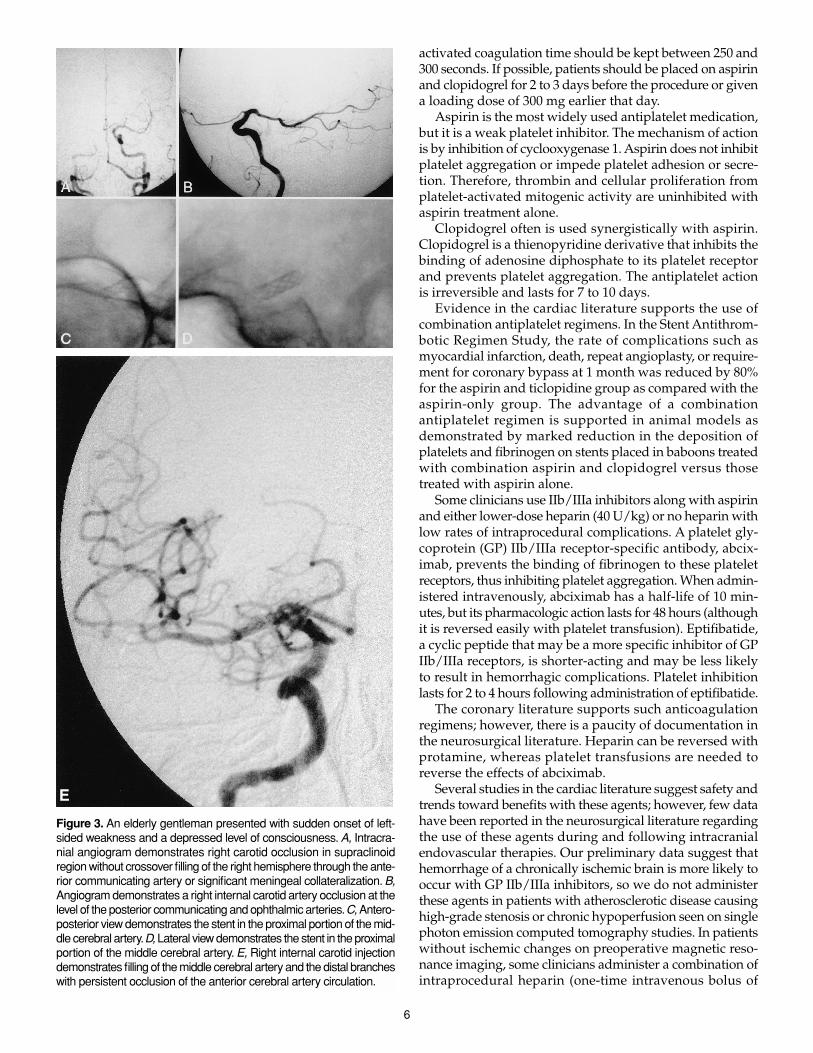
Following failed thrombolysis with intra-arterially admin-istered therapeutic agents such as reteplase or abciximab,balloon angioplasty or microsnares sometimes are used tomorselize clot. Should the clot be effectively lysed, the pres-ence of residual stenosis or dissection can present a dilemma.The stenosis can be managed by subsequent stent insertion.In this setting, our group occasionally has used stents intracra-nially to treat significant residual stenosis. The rationalebehind such an intervention is that residual narrowing mayinduce sluggish flow through diseased portions of the ves-sel, resulting in recurrent acute thrombosis. Recently, we haveinserted stents in vessels that continue to occlude acutelydespite maximal pharmacotherapeutic thrombolysis. In thissetting, stent insertion has been used as a “last resort” fol-lowing hours of failed mechanical and pharmacologic throm-bolysis. Perhaps stents should be considered sooner forocclusions that seem resistant to conventional intra-arterialthrombolysis. In our experience, excellent angiographic resultswere achieved, but clinical outcome was poor due to pro-longed ischemic time from large vessel occlusion (Fig. 3).
Periprocedural Medical ManagementCatheters, wires, balloons, and stents all have the poten-
tial to cause intimal injury and subsequent thrombosis, embo-lus, and vessel occlusion. In addition, all devices arethrombogenic. When blood encounters foreign substances,a monolayer of platelets and fibrin becomes adherent, depend-ing on the surface charge, chemical properties, and surfaceirregularities of the foreign body. Therefore, proper antico-agulation and antiplatelet premedication is essential beforethese devices are introduced within the intracranial circula-tion. Additionally, clinicians must be careful when using anddosing a variety of anticoagulants in settings of acute strokeor critical stenosis, as intracerebral hemorrhage can occur.
For most intracranial stent procedures, an intra-arterialor intravenous bolus of 70 U/kg of heparin is administeredfollowing catheterization of the common carotid artery. Allsaline flush bags are primed with heparin (1 U/mL). The
activated coagulation time should be kept between 250 and300 seconds. If possible, patients should be placed on aspirinand clopidogrel for 2 to 3 days before the procedure or givena loading dose of 300 mg earlier that day.
Aspirin is the most widely used antiplatelet medication,but it is a weak platelet inhibitor. The mechanism of actionis by inhibition of cyclooxygenase 1. Aspirin does not inhibitplatelet aggregation or impede platelet adhesion or secre-tion. Therefore, thrombin and cellular proliferation fromplatelet-activated mitogenic activity are uninhibited withaspirin treatment alone.
Clopidogrel often is used synergistically with aspirin.Clopidogrel is a thienopyridine derivative that inhibits thebinding of adenosine diphosphate to its platelet receptorand prevents platelet aggregation. The antiplatelet actionis irreversible and lasts for 7 to 10 days.
Evidence in the cardiac literature supports the use ofcombination antiplatelet regimens. In the Stent Antithrom-botic Regimen Study, the rate of complications such asmyocardial infarction, death, repeat angioplasty, or require-ment for coronary bypass at 1 month was reduced by 80%for the aspirin and ticlopidine group as compared with theaspirin-only group. The advantage of a combinationantiplatelet regimen is supported in animal models asdemonstrated by marked reduction in the deposition ofplatelets and fibrinogen on stents placed in baboons treatedwith combination aspirin and clopidogrel versus thosetreated with aspirin alone.
Some clinicians use IIb/IIIa inhibitors along with aspirinand either lower-dose heparin (40 U/kg) or no heparin withlow rates of intraprocedural complications. A platelet gly-coprotein (GP) IIb/IIIa receptor-specific antibody, abcix-imab, prevents the binding of fibrinogen to these plateletreceptors, thus inhibiting platelet aggregation. When admin-istered intravenously, abciximab has a half-life of 10 min-utes, but its pharmacologic action lasts for 48 hours (althoughit is reversed easily with platelet transfusion). Eptifibatide,a cyclic peptide that may be a more specific inhibitor of GPIIb/IIIa receptors, is shorter-acting and may be less likelyto result in hemorrhagic complications. Platelet inhibitionlasts for 2 to 4 hours following administration of eptifibatide.
The coronary literature supports such anticoagulationregimens; however, there is a paucity of documentation inthe neurosurgical literature. Heparin can be reversed withprotamine, whereas platelet transfusions are needed toreverse the effects of abciximab.
Several studies in the cardiac literature suggest safety andtrends toward benefits with these agents; however, few datahave been reported in the neurosurgical literature regardingthe use of these agents during and following intracranialendovascular therapies. Our preliminary data suggest thathemorrhage of a chronically ischemic brain is more likely tooccur with GP IIb/IIIa inhibitors, so we do not administerthese agents in patients with atherosclerotic disease causinghigh-grade stenosis or chronic hypoperfusion seen on singlephoton emission computed tomography studies. In patientswithout ischemic changes on preoperative magnetic reso-nance imaging, some clinicians administer a combination ofintraprocedural heparin (one-time intravenous bolus of
6
E
Figure 3. An elderly gentleman presented with sudden onset of left-sided weakness and a depressed level of consciousness. A, Intracra-nial angiogram demonstrates right carotid occlusion in supraclinoidregion without crossover filling of the right hemisphere through the ante-rior communicating artery or significant meningeal collateralization. B,Angiogram demonstrates a right internal carotid artery occlusion at thelevel of the posterior communicating and ophthalmic arteries.C, Antero-posterior view demonstrates the stent in the proximal portion of the mid-dle cerebral artery.D, Lateral view demonstrates the stent in the proximalportion of the middle cerebral artery. E, Right internal carotid injectiondemonstrates filling of the middle cerebral artery and the distal brancheswith persistent occlusion of the anterior cerebral artery circulation.

7
70 U/kg for a target activated coagulation time of 200 sec-onds) and a 0.25 mg/kg intravenous bolus of abciximab over10 to 60 minutes before the procedure. Following the proce-dure, abciximab is infused intravenously for 12 hours at a rateof 10 µg/min. In place of abciximab, eptifibatide may beadministered with a loading dose of 135 (g/kg followed bya 20- to 24-hour infusion of 0.5 (g /kg. Immediately after stent-ing, patients who might be considered for short-term ther-apy with GP IIb/IIIa inhibitors must undergo a computedtomography scan to exclude postprocedural intracranial hem-orrhage, which would preclude the use of these agents.
Following routine stent placement, heparin therapy isdiscontinued but not actively reversed. Patients typicallyare maintained on clopidogrel (75 mg daily) for 1 monthand on aspirin indefinitely. Modifications of anticoagula-tion regimens depend on the indication for stenting (foracute stroke, some interventionists would not discontinueheparin immediately following the procedure) and anycomplication that may have resulted.
Technique for Stent PlacementThe technique used for stent placement varies slightly
from institution to institution. The following is a generaloutline of the steps our group follows to place stents in theintracranial circulation for stenting for most indications.
The procedure is performed in a suite with biplane dig-ital subtraction angiography and fluoroscopic imaging capa-bilities. Following femoral artery puncture, a 6-French sheathis inserted. A 5-French catheter is advanced over a 0.035-inch hydrophilic wire into the aortic arch, and the artery ofinterest is catheterized. Road-mapping technique is used.The sheath and catheter are removed with the wire left inplace, and a 6-French guide catheter is then placed into thevessel. Routine transfemoral angiography is performed,and a 3-French catheter is advanced coaxially over a 0.014-inch microwire into the intracranial vessel. The wire isremoved, and a stiffer 300-cm 0.014-inch exchange wire isthen placed through the microcatheter. This system is thenmanipulated across the lesion. The microcatheter is with-drawn, and a balloon-mounted, over-the-wire stent is nav-igated across the area of interest where it is deployed at 6to 8 atmospheres of pressure. Angiograms are obtained fol-lowing balloon deflation.
Future DirectionsNovel modifications of stent surfaces using biomaterial
science and surface-coating techniques have opened a newarea in stent research. Investigators have demonstratedsome promising results in both human and animal studieswith the use of heparin-coated stents. Although rates ofsubacute thrombosis with heparin-coated stents are lowerin some series, consistently significant reductions in resteno-sis rates have not been demonstrated. Stents that haveshown promise in the cardiac literature are those coatedwith phosphorylcholine, paclitaxel (Taxol), nitric oxide, andrapamycin (sirolimus).
Of the many drug-coated stents under clinical investi-gation, rapamycin-coated stents seem exceptionally promis-ing. Zohlnhˆfer et al. have shown that gene-expression
patterns of human neointima demonstrate the upregula-tion of FKBP12 at the mRNA and protein level of humanneointima. FKBP12 is involved in controlling transform-ing growth factor (TGF) beta receptor I signaling.Rapamycin, which acts at the FKBP12 receptor site, hasbeen shown to reduce neointima formation in animal mod-els. It may be that arrest of neointimal formation and resul-tant in-stent restenosis are due to the rapamycin effects ofblocking protein synthesis and concomitant induction ofcell-cycle arrest. Recent human trials suggest thatrapamycin-coated stents reduce restenosis rates. In one ofthe first human trials of sirolimus-coated stents, reportedby Sousa et al., none of the patients treated with the coatedstents demonstrated restenosis at the 4- to 6-month follow-up evaluation. The results from the Randomized Studywith the Sirolimus-coated Stent (RAVEL) European studyof 220 patients were recently disclosed. RAVEL was a ran-domized, multicenter, double-blinded study of sirolimusballoon-expandable stents in patients with single lesionsin the coronary vasculature. According to the results of thisstudy, no restenosis was found at 6 months in patientstreated with the coated stents, and neointimal volume was2% versus 37% in those treated with non-coated stents.Although these data come from findings following stent-ing of cardiac vessels, it may be that the same response willbe demonstrated in the intracranial vasculature.
A second drug-coated stent that has recently showed clin-ical promise for inhibiting in-stent restenosis is the pacli-taxel-coated stent. Paclitaxel inhibits restenosis is by alteringthe stability of mictrotubules. This alteration leads to theinhibition of cell replication and intracellular signaling. Inanimal models, reductions in neointimal proliferation atblood concentrations 100 times lower than antineoplasticlevels have been shown. In a recent pilot clinical trial, noneof the 21 patients demonstrated restenosis of stented coro-nary lesions. The Asian Paclitaxel-Eluting Stent Clinical Trial(ASPECT) trial compared the safety and efficacy of high-dose and low-dose paclitaxel-coated stents versus uncoatedstents in patients with single lesions in coronary arteries. At6 months, restenosis rates had dropped from 27% in the con-trol group to 4% in the high-dose paclitaxel group. Thesefindings are attributed to reductions in volume of neointi-mal hyperplasia. It will be interesting to see how therapamycin- and paclitaxel-coated stents will be applied inthe future for stenting of intracranial stenotic lesions.
Much interest exists in radioactive stents; however, theresults have been variable and seem to be dependent on theanimal model, time to sacrifice, and the radioactivity of thestents. It is hoped that development and further research ofcoated stents will provide clinicians with prosthetic devicesthat locally inhibit platelet-fibrin deposition, mitigate clot for-mation and restenosis, and promote local endothelialization.
CaveatsIntracranial stenting is a novel technique, still in its early
developmental stages. Although intracranial stents are beingused with increasing frequency, it is important to remem-ber that there are no long-term data regarding rates ofpatency, restenosis, or vessel injury. Additionally, the effects

of stent-induced intimal hyperplasia are not known in thecerebral vasculature, and the treatment for in-stent steno-sis may be problematic.
Stenting of intracranial vascular pathology may provideclinicians with therapeutic interventions where none existedpreviously. The subset of patients who are not surgical can-didates, for reasons such as severe cardiopulmonary prob-lems, now has alternative nonsurgical options for intracranialrevascularization for severe stenoses or aneurysm occlusion.Clearly, long-term, prospective data are needed to betterunderstand and define the efficacy of intracranial stentingfor diverse cerebrovascular disease processes.
AcknowledgmentsWe thank Paul H. Dressel for preparation of the illus-
trations and the staff at Kaleida Gates Hospital Library forassistance obtaining the reference articles.
Readings
Ahuja A, Guterman LR, Hopkins LN: Angioplasty for basilar artery ather-osclerosis. J Neurosurg 77:941, 1992
Ausman JI, Shrontz CE, Pearce JE, et al: Vertebrobasilar insufficiency. ArchNeurol 42:803, 1985
Bhatt DL, Kapadia SR, Yadav JS, Topol EJ: Update on clinical trials of antiplatelettherapy for cerebrovascular diseases. Cerebrovasc Dis 10:34, 2000
Buergler JM, Tio FO, Schulz DG, et al: Use of nitric-oxide-eluting polymer-coated coronary stents for prevention of restenosis in pigs. Coron ArteryDis 11:351, 2000
Callahan AS III, Berger BL: Basilar artery endoprosthesis placement: rescuetherapy for recurrent thrombosis. J Neuroimaging 10:47, 2000
Carter AJ, Scott D, Bailey L, et al: Dose-response effects of 32P radioactive stentsin an atherosclerotic porcine coronary model. Circulation 100:1548, 1999
Connors JJ 3rd, Wojak JC: Percutaneous transluminal angioplasty for intracra-nial atherosclerotic lesions: evolution of technique and short-term results.J Neurosurg 91:415, 1999
Derdeyn CP, Cross DT 3rd, Moran CJ, Dacey RG Jr: Reversal of focal misery per-fusion after intracranial angioplasty: case report. Neurosurgery 48:436, 2001
Dotter CT: Transluminally-placed coilspring endarterial tube grafts: Long-term patency in canine popliteal artery. Invest Radiol 4:327, 1969
Dotter CT, Buschman PAC, McKinney MK, Rosch J: Transluminal expand-able nitinol coil stent grafting: preliminary report. Radiology 147:259, 1983
Dyet JF: Endovascular stents in the arterial system: current status. Clin Radiol52:83, 1997
Fessler RD, Guterman LR, Qureshi A, Hopkins LN: State of neuroendovas-cular surgery: endoluminal reconstruction of basilar artery fusiformaneurysms. Transcatheter Cardiovascular Therapeutics: 11th AnnualSymposium, Washington, DC, 1999.
Fessler RD, Ringer AJ, Qureshi AI, et al: Intracranial stent placement to trapan extruded coil during endovascular aneurysm treatment: technicalnote. Neurosurgery 46:248, 2000
Fontaine AB, Borsa JJ, Dos Passos S, et al: Evaluation of local abciximab deliv-ery from the surface of a polymer-coated covered stent: in vivo caninestudies. J Vasc Interv Radiol 12:487, 2001
Galli M, Bartorelli A, Bedogni F, et al: Italian BiodivYsio open registry (Bio-divYsio PC-coated stent): study of clinical outcomes of the implant of aPC-coated coronary stent. J Invasive Cardiol 12:452, 2000
Gomez CR, Misra VK, Campbell MS, Soto RD: Elective stenting of sympto-matic middle cerebral artery stenosis. Am J Neuroradiol 21:971, 2000
Harker LA, Bruno JJ: Ticlopidine’s mechanism of action on platelets, in HassWK, Easton JD (eds): Ticlopidine, Platelets and Vascular Diseases. New York:Springer-Verlag, 1993, pp 41–59
Heldman AW, Cheng L, Jenkins GM, et al: Paclitaxel stent coating inhibitsneointimal hyperplasia at 4 weeks in a porcine model of coronary resteno-sis. Circulation 103:2289, 2001
Higashida RT, Smith W, Gress D, et al: Intravascular stent and endovascu-lar coil placement for a ruptured fusiform aneurysm of the basilar artery:case report and review of the literature. J Neurosurg 87:944, 1997
Hopkins LN, Martin NA, Hadley MN, et al: Vertebrobasilar insufficiency. JNeurosurg 66:662, 1987
Horowitz MB, Levy EI, Koebbe C, Jungreis CC: Transluminal stent-assistedcoil embolization of a vertebral confluence aneurysm: case report. SurgNeurol 55:291, 2001
Horowitz MB, Pride GL, Graybeal DF, Purdy PD: Percutaneous transluminalangioplasty and stenting of midbasilar stenoses. Neurosurgery 45:925, 1999
Horowitz MB, Purdy PD: The use of stents in the management of neu-rovascular disease: a review of historical and present status. Neurosurgery46:1335, 2000
Imbesi SG, Kerber CW: Analysis of slipstream flow in a wide-necked basi-lar artery aneurysm: evaluation of potential treatment regimens. AJNR:Am J Neuroradiol 22:721, 2001
Kutryk MJB, Serruys PW: Stenting, in Topol EJ (ed): Comprehensive Cardio-vascular Medicine. Philadelphia: Lippincott-Raven, 1998, pp 2307–2338
Lanzino G, Fessler RD, Miletich RS, et al: Angioplasty and stenting of basi-lar artery stenosis. Neurosurgery 45:404, 1999
Lanzino G, Wakhloo AK, Fessler RD, et al: Efficacy and current limitationsof intravascular stents for intracranial internal carotid, vertebral, and basi-lar artery aneurysms. J Neurosurg 91:538, 1999
Lavine SD, Larsen DW, Giannotta SL, Teitelbaum GP: Parent vessel Guglielmidetachable coil herniation during wide-necked aneurysm embolization:treatment with intracranial stent placement—two technical case reports.Neurosurgery 46:1013, 2000
Levy E, Horowitz MB, Jungreis C, et al: Transluminal stent assisted angio-plasty of the intracranial vertebrobasilar system for medically refractoryposterior circulation ischemia: early results. Neurosurgery 48:1215, 2001
Levy EI, Firlik AD, Wisniewski S, et al: Factors affecting survival rates foracute vertebrobasilar artery occlusions treated with intra-arterial throm-bolytic therapy: a meta-analytical approach. Neurosurgery 45:539; 1999
Lieber BB, Stancampiano AP, Wakhloo AK: Alteration of hemodynamics inaneurysm models by stenting: influence of stent porosity. Ann BiomedEng 25:460, 1997
Lopes DK, Ringer AJ, Boulos A, et al: Patency of arterial branches crossedby intracranial stents: fate of the “jailed” artery (abstract). J Neurosurg94:169A, 2001
Lylyk P, Ceratto R, Hurvitz D, Basso A: Treatment of a vertebral dissecting aneurysmwith stents and coils: technical case report. Neurosurgery 43:385, 1998
Malek AM, Higashida RT, Balousek PA, et al: Endovascular recanalizationwith balloon angioplasty and stenting of an occluded occipital sinus fortreatment of intracranial venous hypertension: technical case report. Neu-rosurgery 44:896, 1999
Malek AM, Higashida RT, Halbach VV, et al: Tandem intracranial stentdeployment for treatment of an iatrogenic, flow-limiting, basilar arterydissection. Neurosurgery 45:919, 1999
Mericle RA, Lanzino G, Wakhloo AK, et al: Stenting and secondary coilingof intracranial internal carotid artery aneurysm: technical case report.Neurosurgery 43:1229, 1998
Mori T, Kazita K, Chokyu K, et al: Short-term arteriographic and clinical outcomeafter cerebral angioplasty and stenting for intracranial vertebrobasilar andcarotid atherosclerotic occlusive disease. AJNR Am J Neuroradiol 21:249, 2000
Mori T, Kazita K, Mori K: Cerebral angioplasty and stenting for intracranialvertebral atherosclerotic stenosis. AJNR Am J Neuroradiol 20:787, 1999
Murayama Y, Vinuela F, Tateshima S, et al: Endovascular treatment of exper-imental aneurysms by use of a combination of liquid embolic agents andprotective devices. AJNR Am J Neuroradiol 21:1726, 2000
Murphy KJ, Gailloud P, Venbrux A, et al: Endovascular treatment of a grade IVtransverse sinus dural arteriovenous fistula by sinus recanalization, angio-plasty, and stent placement: technical case report. Neurosurgery 46:497, 2000
Palmaz JC: Intravascular stenting: From basic research to clinical applica-tion. Cardiovasc Intervent Radiol 15:279, 1992
Phatouros CC, Higashida RT, Malek AM, et al: Endovascular stenting of anacutely thrombosed basilar artery. Neurosurgery 44:667, 1999
Piotin M, Mandai S, Sugiu K, et al: Endovascular treatment of cerebralaneurysms: an in vitro study with detachable platinum coils and tricel-lulose acetate polymer. AJR Am J Roentgenol 176:235, 2001
Qureshi AI, Luft AR, Sharma M, et al: Prevention and treatment of throm-boembolic and ischemic complications associated with endovascular
8

procedures: Part I—Pathophysiological and pharmacological features.Neurosurgery 46:1344, 2000
Qureshi AI, Luft AR, Sharma M, et al: Prevention and treatment of thromboem-bolic and ischemic complications associated with endovascular procedures:Part II—Clinical aspects and recommendations. Neurosurgery46:1360, 2000
Rousseau H, Puel J, Joffre F, et al: Self-expanding endovascular prosthesis:an experimental study. Radiology 164:709, 1987
Sekhon LH, Morgan MK, Sorby W, Grinnell V: Combined endovascular stentimplantation and endosaccular coil placement for the treatment of a wide-necked vertebral artery aneurysm. Neurosurgery 43:380, 1998
Shin EK, Son JW, Sohn MS, et al: Efficacy of heparin-coated stent in early set-ting of acute myocardial infarction. Catheter Cardiovasc Interv 52:306, 2001
Sousa JE, Costa MA, Abizaid A, et al: Lack of neointimal proliferation afterimplantation of sirolimus-coated stents in human coronary arteries: aquantitative coronary angiography and three-dimensional intravascularultrasound study. Circulation103:192, 2001
Spetzler RF, Hadley MN, Martin NA, et al: Vertebrobasilar insufficiency. JNeurosurg 66:648, 1987
Sundt TM Jr, Smith HC, Campbell JK, et al: Transluminal angioplasty forbasilar artery stenosis. Mayo Clin Proc 55:673, 1980
Vrolix MC, Legrand VM, Reiber JH, et al: Heparin-coated Wiktor stents inhuman coronary arteries (MENTOR trial): MENTOR Trial Investigators.Am J Cardiol 86:385, 2000
Wakhloo AK, Tio FO, Lieber BB, et al: Self-expanding nitinol stents in caninevertebral arteries: hemodynamics and tissue response. AJNR Am J Neu-roradiol 16:1043, 1995
Zidar JP: Rationale for low-molecular weight heparin in coronary stenting.Am Heart J 134:S81, 1997 (suppl)
Zohlnhöfer D, Klein CA, Richter T, et al: Gene expression profilingof human stent-induced neointima by cDNA array analysis ofmicroscopic specimens retrieved by helix cutter atherectomy: detec-tion of FK506-binding protein 12 upregulation. Circulation 103:1396, 2001
9

10
To earn CME credit, you must read the CME article and complete the quiz on the enclosed form, answering at least 70% of the quizquestions correctly. Select the best answer and place a check mark or an X mark in the corresponding space on the enclosed answerform. Do not fill in the answer box completely. Please indicate any name and address changes directly on the answer form. If your nameand address do not appear on the answer form, please print that information in the blank space at the top left of the page.Make a photocopyof the completed answer form for your own files and mail the original answer form in the enclosed postage-paid business reply envelopeby December 14, 2002. For more information, call (800) 787-8981.
Please do not use the Scantron cards and business reply envelopes that you may previously have received for this or anyfuture issue of Contemporary Neurosurgery.
Lippincott Williams & Wilkins (LWW) is accredited by the Accreditation Council for Continuing Medical Education to provide continuingmedical education for physicians.
LWW designates this educational activity for a maximum of 1.5 hours in category 1 credit toward the AMA’s Physician’s RecognitionAward. Each physician should claim only those hours of credit that he/she actually spent in the educational activity.
1. Stenotic lesions that are less than 5 mm long with a concen-tric configuration are best suited for angioplasty.
True or False?
2. During a stenting procedure for intracranial occlusive disease,a moderate improvement in luminal diameter is usually suffi-cient to adequately improve cerebral blood flow.
True or False?
3. Oversizing stents to achieve minimal residual stenosis hasno risk of reperfusion hemorrhage.
True or False?
4. Stent-omitted coiling of intracranial aneurysm has the poten-tial advantage of protecting parent vessels from coil hernia-tion, especially when used with wide-neck aneurysms.
True or False?
5. Stent placement for acute stroke has no role when throm-bolytic therapy is used.
True or False?
6. There is no evidence that stenting could help in the treatmentof venous sinus thrombosis.
True or False?
7. IIb/IIIa inhibitors seem to predispose to hemorrhage after reper-fusion of cerebral regions in patients with chronic hypoperfusion.
True or False?
8. There is no evidence from the cardiac literature that combi-nation antiplatelet regimens reduce the rate of periprocere-bral complications and the need for repeat angioplasty.
True or False?
9. Rapamycin (sirolimus) has been used in Rapamycin-coatedstents to inhibit restenosis by altering the stability of micro-tubules and thus inhibiting cell replication.
True or False?
10. The use of paclitaxel-coated stents in coronary stenting tri-als decreased the 6-month restenosis rates from 27% in thecontrol group to 4% in the paclitaxel group.
True or False?