intracranial avms: comparison of volumes generated from orthogonal measurements and integrated 3d...
TRANSCRIPT

Intracranial AVMs: Comparison of volumes generated from
orthogonal measurements and integrated 3D analysis
Faiz I Syed MD MS1, Lubomir Hadjiiski PhD1, Aditya S Pandey MD2, Heang-
Ping Chan PhD1, Ashok Srinivasan MD1
Poster #: EP-62
1 Department of Radiology, University of Michigan2 Department of Neurosurgery, University of MichiganContact: Faiz I. Syed, [email protected]

Disclosures: None

Purpose
• Intracranial AVM volume estimation is important for– guiding therapeutic options– monitoring treated lesions
• Volume estimation based on orthogonal measurements is commonly used

Purpose
• The purpose of this study was to compare the intracranial AVM volumes calculated using two different methods:
– calculating volume of an ellipsoid from three orthogonal measurements of the AVM
– 3D integrated volumetric analysis of the AVM.

Methods
• After IRB clearance, imaging studies from 11 patients with known brain AVMs were reviewed.
• Studies included CT of the head with contrast, CTA of the head and MRI of the head with contrast.
• All studies were analyzed by one board certified neuroradiologist (Reader 1) and a second year neuroradiology fellow (Reader 2) on custom software developed at the University of Michigan.

Methods
• For each patient, the AVM volume was calculated by two methods:
– Ellipsoid formula ABC/2 – A, B, C: maximal orthogonal
measurements of AVM nidus
– 3D integrated volume – calculated by tracing the boundaries of
the AVM nidus on each axial slice of a cross-sectional study

Methods
Figure1. Measuring orthogonal projections of the AVM nidus for calculating ellipsoid volume

Methods
Figure 2. Tracing AVM nidus boundaries to calculate 3D integrated volume

• Average volume of AVMs in our study determined by Ellipsoid and 3D Integrated volume calculations
• The volumes were calculated based on estimation by two readers
Reader 1 Reader 2
Ellipsoid volume (mL)
4.2 Range: 0.4-16.6
7.6Range: 0.4-29.6
3D Integrated volume (mL)
6.2Range: 0.6-23.8
6.9Range: 0.6-21.7
Table 1. Average intracranial AVM volumes by Ellipsoid and 3D Integrated volume
calculations based on estimation by two readers
Results
3D Integrated Volume [cm3]
0 5 10 15 20 25 30
Ell
ipso
id V
olu
me
[cm
3 ]
0
5
10
15
20
25
30
Results
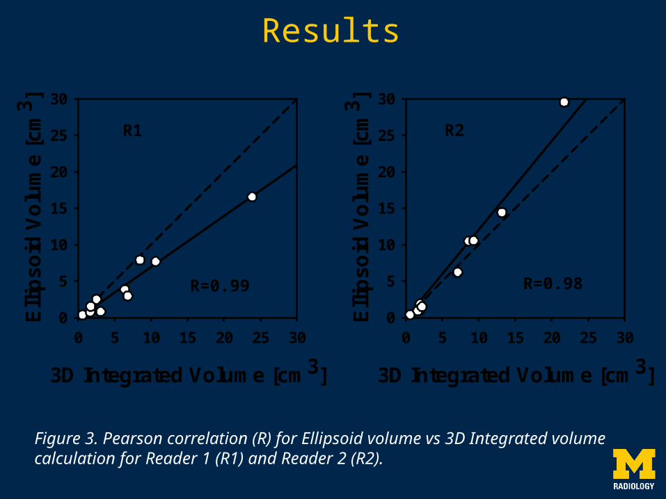
Figure 3. Pearson correlation (R) for Ellipsoid volume vs 3D Integrated volume calculation for Reader 1 (R1) and Reader 2 (R2).
R1
3D Integrated Volume [cm3]
0 5 10 15 20 25 30
Ell
ipso
id V
olu
me
[cm
3 ]
0
5
10
15
20
25
30
R2
R=0.99 R=0.98

Results
Figure 4. Pearson correlation (R) for Reader 1 (R1) vs Reader 2 (R2) for the Ellipsoid and 3D Integrated volume calculations.
R1 Integrated Volume [cm3]
0 5 10 15 20 25 30R2
Inte
gra
ted
Vo
lum
e [c
m3 ]
0
5
10
15
20
25
30
R1 Ellipsoid Volume [cm3]
0 5 10 15 20 25 30R2
Ell
ipso
id V
olu
me
[cm
3 ]
0
5
10
15
20
25
30
R=0.88 R=0.87

Conclusion
• There was a high inter-reader correlation for both the Ellipsoid and 3D Integrated volumes.
• There was a high correlation between the Ellipsoid and 3D Integrated volumes.
• Orthogonal volume measurement of intracranial AVMs based on the ellipsoid formula may be sufficient.

Limitations
• Our study had a small sample size of 11 patients
• Many of the studies analyzed had low spatial resolution

References
• Foroni R, Gerosa M, Pasqualin A, et al. Shape recovery and volume calculation from biplane angiography in the stereotactic radiosurgical treatment of arteriovenous malformations. Int J Radiat Oncol Biol Phys. 1996 Jun 1;35(3):565-77
• Forkert ND, Illies T, Goebell E, Fiehler J, Säring D, Handels H. Computer-aided nidus segmentation and angiographic characterization of arteriovenous malformations. Int J Comput Assist Radiol Surg. 2013 Sep;8(5):775-86. doi: 10.1007/s11548-013-0823-9. Epub 2013 Mar 7