intracellular metabolism of tenofovir alafenamide...
TRANSCRIPT

Mackenzie L. Cottrell, Katy L. Garrett, Cindi W. Emerson,
Amanda Schauer, Craig Sykes, Angela D.M. Kashuba
University of North CarolinaUNC Eshelman School of Pharmacy
Intracellular Metabolism of Tenofovir
Alafenamide in Cervical and Vaginal
Epithelial Cells

TAF Exhibits Favorable Pharmacology
Adapted from Gupta S. IAS 2015 Oral Abstract #TUAB0103.
HIV TARGET CELL
AMIDATE
ESTER
DIANION
GI TRACT
Tenofovir
alafenamide
(TAF)
Tenofovir
disoproxil
fumarate
(TDF)
Tenofovir
(TFV) Parent
Nucleotide
PLASMA
TAF25 mg
TDF 300 mg
TFV
TFV
TFV
TFV HIVTFVdpT1/2 = 0.4 min
†
T1/2 = 90 min†

TAF Achieves Higher TFVdp in PBMCs
Ruane P. CROI 2012 Oral Abstract #103.
TAF
8mg
TAF
25mg
TAF
40mg
TDF
300mg

TFVdp is Lower in FGT Tissues for TAF
Garrett K. CROI 2016 Oral Abstract #102LB.
Time (hr)
0 12 24 36 48 60 72
TF
V o
r T
FV
dp
(pM
)
103
104
105
●TFV
▼TFVdp
300mg TDF single dose

TFVdp is Lower in FGT Tissues for TAF
Garrett K. CROI 2016 Oral Abstract #102LB.
300mg TDF
Time (hr)
0 12 24 36 48 60 72
TF
V o
r T
FV
dp
(pM
)
103
104
105
300mg TDF single dose 25mg TAF single dose
●TFV
▼TFVdp
TFV 2-fold
TFVdp 1.3-fold

Mucosal Tissue Microanatomy
Adapted from Margolis L. 2006 Nature Reviews Microbiology.
Langerhans Cells
Dendritic Cells
Macrophages
T-Lymphocytes
Epithelial Cells

Mucosal Tissue Microanatomy
Adapted from Margolis L. 2006 Nature Reviews Microbiology.
Langerhans Cells
Dendritic Cells
Macrophages
T-Lymphocytes
Epithelial Cells
Whole blood Ovary Vagina Uterus
Cathepsin A Gene Expression
http://www.gtexportal.org/home/gene/CTSA
Lo
g10
RP
KM

Hypothesis: TFVdp will be
lower in epithelial cells dosed
with TAF vs TFV

In Vitro Approach
0.5 μΜ
PBMC
0.5 μM
Ect1
0.5 μM
VK2
10 μM
PBMC
10 μM
Ect1
10 μM
VK2
0.5 μΜ
PBMC
0.5 μM
Ect1
0.5 μM
VK2
10 μM
PBMC
10 μM
Ect1
10 μM
VK2
TAF TFV
3
hr
12
hr
24
hr
48
hr
72
hr
Cells: Freshly isolated PBMCs (single healthy volunteer*),
Ect1/E6E7 ectocervical and VK2/E6E7 vaginal cell lines (ATCC®)
Treat with:
Harvest at:
Count on: Muse™ Cell Analyzer
Lyse in: 70:30 methanol:water
*UNC Center for AIDS Research Sample Collection Protocol IRB 08-0047
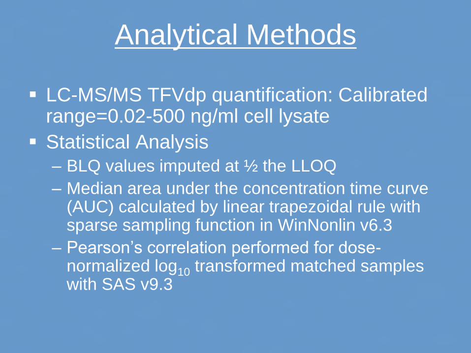
Analytical Methods
LC-MS/MS TFVdp quantification: Calibrated range=0.02-500 ng/ml cell lysate
Statistical Analysis
‒ BLQ values imputed at ½ the LLOQ
‒ Median area under the concentration time curve (AUC) calculated by linear trapezoidal rule with sparse sampling function in WinNonlin v6.3
‒ Pearson’s correlation performed for dose-normalized log10 transformed matched samples with SAS v9.3

TFVdp Median (Min, Max) Concentration
vs Time: TAF (solid)
Time (hour)
0 12 24 36 48 60 72
TF
Vdp(f
mol/m
illio
n c
ells
)
100
101
102
103
104
105
106
PBMC
Time (hour)
0 12 24 36 48 60 72
TF
Vdp (
fmol/m
illio
n c
ells
)
100
101
102
103
104
105
106
PBMC
10μM Dosing 0.5μM Dosing

TFVdp Median (Min, Max) Concentration
vs Time: TAF (solid)
Time (hour)
0 12 24 36 48 60 72
TF
Vdp(f
mol/m
illio
n c
ells
)
100
101
102
103
104
105
106
Ect1
PBMCVK2
0.5μM Dosing
Time (hour)
0 12 24 36 48 60 72
TF
Vdp (
fmol/m
illio
n c
ells
)
100
101
102
103
104
105
106
Ect1
PBMCVK2
10μM Dosing

TFVdp Median (Min, Max) Concentration
vs Time: TAF (solid) & TFV (dashed)
TAF AUC0-72hr 192-1305 fold Higher compared to TFV
Time (hour)
0 12 24 36 48 60 72
TF
Vdp(f
mol/m
illio
n c
ells
)
100
101
102
103
104
105
106
Ect1
PBMCVK2
Time (hour)
0 12 24 36 48 60 72
TF
Vdp (
fmol/m
illio
n c
ells
)
100
101
102
103
104
105
106
Ect1
PBMCVK2
0.5μM Dosing 10μM Dosing

TFVdp AUC0-72hr
Cell Type DoseμM
TAFMean AUC (SE)
pmol*hr/million cells
TFVMean AUC (SE)
pmol*hr/million cells
PBMC0.5 2122 (209) 1.63 (0.173)
10 12646 (702) 33.5 (5.61)
Ect10.5 10037 (989) 26.7 (1.78)
10 210382 (18701) 469 (28.1)
VK20.5 3587 (826) 18.6 (1.60)
10 60604 (35752) 256 (11.1)
TAF: PBMC 1.7-16.6 fold Lower vs Epithelial
TFV: PBMC 7.6-16.4 fold Lower vs Epithelial

TFVdp Rate of Accumulation
0.5μM TAF 0.5μM TFV
Epithelial cells: 6
Times Faster
Epithelial cells: 17-30
Times Faster
Time (hour)
0 3 6 9 12
TF
Vd
p (
fmo
l/1
06
ce
lls)
0
20000
40000
60000
80000
100000
120000
140000
160000
PBMC
VK2
Ect1Rate=11204, r
2=0.97
Rate=10608, r2=0.94
Rate=1772, r2=0.75
Time (hour)
0 3 6 9 12
TF
Vd
p (
fmo
l/1
06 c
ells
)
0
100
200
300
400
500
600
PBMC
VK2
Ect1
Rate=23.61, r2=0.86
Rate=41.60, r2=0.92
Rate=1.383, r2=0.61

TFVdp Correlates Between Cell Types
Pearson’s r Ect1 VK2 PBMC
Ect1 1 0.71† 0.64*
VK2 0.90† 1 0.45*
PBMC 0.58* 0.42* 1
*p<0.05; †p<0.001
PBMC Log10
TFVdp (fmol/106 cells)
3.5 4.0 4.5 5.0 5.5
Ect1
Log
10 T
FV
dp (
fmo
l/1
06
ce
lls)
3.5
4.0
4.5
5.0
5.5
6.0
PBMC Log10
TFVdp (fmol/106 cells)
0.5 1.0 1.5 2.0 2.5 3.0 3.5
Ect1
Log
10 T
FV
dp (
fmol/10
6 c
ells
)
0.5
1.0
1.5
2.0
2.5
3.0
3.5
TFVTAF

Limitations
Static exposure over 72 hours
Immortalized cells exhibit altered
phenotypes − Ect1 TFVdp ~1.7-fold higher compared to
primary ectocervical cells1
1Shen Z. 2014 PlosOne.

Summary and Conclusions
TFVdp was HIGHER with matched TAF vs TFV dosing
− >100-fold AUC0-72hr in PBMCs, Ect1 and VK2 cells
TFVdp was HIGHER in epithelial cells vs PBMCs
− VK2 ≥ 1.7 fold AUC0-72hr
− Ect1 ≥ 4.7 fold AUC0-72hr
TFVdp was significantly correlated among cell types
− Ect1 vs PBMC TFVdp r2=0.58, p<0.05
− VK2 vs PBMCs TFVdp r2=0.42, p<0.05
Presence of epithelial cells within tissue homogenates does
not explain previous observations of low TFVdp
concentrations in tissue biopsies from women dosed with TAF.
This work was supported in part by the Centers for AIDS Research (Grant P30 AI50410)
and the John A. and Deborah S. McNeill Jr. Distinguished Professorship