intra-arterial embolization of lumbar artery pseudoaneurysm following percutaneous nephrolithotomy
TRANSCRIPT
INTRODUCTIONRenovascular injury is a well-recognized complication of percu-
taneous renal procedures such as renal biopsies, percutaneous
nephrostomy (PCN) and percutaneous nephrolithotomy (PCNL).
Needle injury of a branch of the renal artery may result in the
formation of a pseudoaneurysm or an arteriovenous fistula
(AVF). The high-pressure leak from a lacerated artery is trans-
mitted through the tract into a lower resistance system of a vein or
a connective tissue space, such as is found around the pelvis or
calyces. A CT examination immediately following renal biopsies
has revealed haematomas in 90% of cases;1 whereas renal
angiography performed immediately after renal biopsies has
shown haematomas in 23%, arteriovenous fistulas in 8%, and
renal infarcts in 6% of patients.2 Most of these lesions close
spontaneously, but a few remain open and slowly increase in
size, leading to life-threatening haematuria that requires thera-
peutic intervention.
Iatrogenic lumbar arterial injuries related to percutaneous
renal procedures are very rare and may go undetected because
of a problem in the localization of the accompanying haemor-
rhage.3–5 A case of pseudoaneurysm of a lumbar artery
following vertebral biopsy has also been reported.6 We describe
here a case of lumbar artery pseudoaneurysm following PCNL,
which required intra-arterial embolization of the lumbar artery
using a metallic coil and gel foam particles.
CASE REPORTA 45-year-old man presented with significant haematuria of 10
days duration following left PCNL. Before the procedure his
blood pressure was 140/90 mmHg, and his bleeding and
coagulation parameters were normal; that is, prothrombin time
(PT) was 13.6 s (control 12.8 s), partial thromboplastin time
(PTT) was 30 s (control 27 s) and platelet count was 500 �
109/L. Six hours after an uneventful procedure, the patient
complained of pain in the lumbar region and started having
haematuria. Ultrasound examination revealed a left hydro-
nephrosis with residual calculi and hyperechoic medium level
echoes, which suggested clots in the collecting system. The
haematocrit dropped from 39 to 25%. Even after an infusion of
six units of blood and four packs of platelets, the patient
continued to have haematuria; hence, the patient was referred
for an angiographic evaluation.
A selective left renal angiogram obtained via a right
transfemoral artery route showed a main renal artery supplying
the upper and midpolar regions of the left kidney along with
incidental reflux of contrast into the first left lumbar artery. A
small pseudoaneurysm was seen overlapping the midpolar
region of the kidney (Fig. 1) and, on fluoroscopy, was seen not
to move with the kidney in different phases of respiration.
Selective injection of the left first lumbar artery revealed a small
pseudoaneurysm filling from it. No radiculomedullary artery
Case Report
Intra-arterial embolization of lumbar arterypseudoaneurysm following percutaneousnephrolithotomyR Jain, S Kumar, RV Phadke, SS Baijal and RB GujralDepartment of Radiodiagnosis, Sanjay Gandhi Post-Graduate Institute of Medical Sciences, Lucknow, Uttar Pradesh, India
SUMMARY
The management of a patient with haematuria following percutaneous nephrolithotomy is described. The patientunderwent renal angiography to assess the cause of bleeding. A pseudoaneurysm arising from first left lumbar arterywas incidentally discovered, which was then successfully embolized using an indigenously fabricated metallic coil andgel foam particles in the same sitting.
Key words: Lumbar artery embolization; percutaneous nephrolithotomy; pseudoaneurysm.
R Jain MD; S Kumar MD; RV Phadke MD; SS Baijal MD; RB Gujral MD.
Correspondence: Dr Sunil Kumar, Additional Professor, Department of Radiodiagnosis, Sanjay Gandhi Post-Graduate Institute of Medical Sciences,
Rae Bareily Road, Lucknow, UP 226014, India. Email: [email protected]
Submitted 15 October 1999; resubmitted 22 February 2000; accepted 5 April 2000.
Australasian Radiology (2001) 45, 383–386

(artery of Adamkiewicz) was seen originating from the first left
lumbar artery. An accessory renal artery arising from the aorta
at L3–4 level was seen supplying the lower polar region of the
left kidney and no intrarenal vascular lesion could be detected.
This pseudoaneurysm was embolized first with an injection
of gel foam particles using a 5 F multipurpose catheter. A small,
indigenously fabricated metallic coil, measuring 2 mm in dia-
meter and 12 mm in length, was then placed in the lumbar
artery followed by a gel foam pledget to occlude the artery
completely. Post-embolization check aortogram revealed no
flow into the left first lumbar artery and no filling of the
pseudoaneurysm (Fig. 2). The patient had pain in the lumbar
region, which settled with analgesics, but had no neurological
deficit. The heamaturia decreased after embolization, but after
three days the patient again underwent angiographic evaluation
as the haematuria persisted. Repeat examination did not reveal
any vascular lesion and the haematuria settled completely after
four days.
DISCUSSIONThe first four lumbar arteries are usually paired arteries and
arise from the posterior aspect of the aorta. The fifth lumbar
artery is usually small in size and may originate from the median
sacral artery. Immediately after arising from the aorta, the
lumbar arteries curve around the bodies of the lumbar
vertebrae and give rise to small retroperitoneal branches to the
psoas muscle and to the spinal artery before dividing into
anterior and posterior branches.
The anterior branch gives branches to the quadratus
lumborum and sacrospinalis muscle as it runs between them,
and then branches to the muscles and skin of the flank. The
posterior branch supplies branches to the sacrospinalis muscle
and skin of the back. These muscular branches running dorsal
to the kidney are susceptible to injury during percutaneous
renal procedures as well as during percutaneous vertebral
biopsy. Accumulation of blood in the posterior pararenal space
should suggest lumbar artery injury because of its close
proximity to the lumbar arteries.
The reported incidence of renal pseudoaneurysm is 0.6–
1.0% following PCNL,7,8 and 2.0–3.4% following percutaneous
needle biopsy of a renal allograft.7–9 Deaths secondary to
bleeding have occurred in 0.07–0.17% of cases.10 However,
there are only three reported cases of lumbar artery injury
following renal biopsies and none following PCN or PCNL. In the
384 R JAIN ET AL.
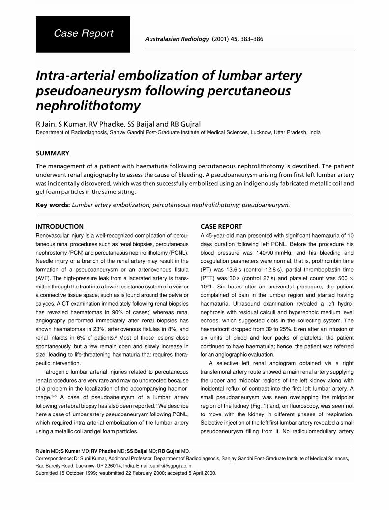
Fig. 1. (a) Selective left renal arteriogram shows a small pseudoaneurysm (arrow) overlapping the midpolar region along with reflux of contrast into
the first left lumbar artery. (b) Nephrographic phase shows filling of the first left lumbar artery and the pseudoaneurysm.

385LUMBAR ARTERY PSEUDOAENURYSM
first reported case, the exact site of haemorrhage was difficult to
localize even at surgical exploration.3 In the second case report,
there were two sites of haemorrhage: (i) from a branch of the left
renal artery, which was successfully controlled by embolization;
and (ii) from the left fourth lumbar artery, which was identified
two days after initial embolization. Even after successful
embolization of the lumbar artery bleeding with gel foam
pledgets, the patient developed fatal complications from
prolonged hypotension.5 In the third case, initial abdominal
aortograms did not reveal any vascular lesion, while a repeat
oblique aortogram demonstrated contrast extravasation from
the first left lumbar artery, which was embolized using two
straight microcoils.4 In the present case, a lumbar artery
pseudoaneurysm was detected incidently because of reflux of
contrast material into the lumbar artery while obtaining a renal
arteriogram to look for some renal vasculature abnormality. All
these cases demonstrate that lumbar arterial injury following
percutaneous renal procedures is difficult to detect. Hence,
clinical suspicion of lumbar arterial injury should be high, and
angiograpy considered in appropriate cases.
Intra-arterial embolization for traumatic arterial bleeding may
be considered if the injured vessel is expendable or if the vessel is
surgically inaccessible or difficult. Lumbar arterial injury meets
both of these characteristics in most situations. Operative control
of lumbar arterial bleeding is often difficult because the site
of origin may not be readily isolated. Although embolization
appears to be a relatively safe and successful method of obtain-
ing haemostasis, it is not without complications. Spinal cord and
peripheral nerve infarction are the major complications. The
lumbar artery should be scrutinized carefully for major spinal
branches, such as the great anterior radicular artery (artery of
Adamkiewicz). It may arise anywhere from sixth intercostal to the
second lumbar artery and has the characteristic angiographic
configuration of a hair pin.However, it may be difficult to depict the
great radicular artery angiographically, even on selective lumbar
injections. Digital subtraction techniques, however, can greatly
enhance its delineation. Spinal cord injury can occur even during
diagnostic angiography. Thus, forceful injection of too much
contrast medium, particularly through a catheter wedged in the
lumbar artery giving rise to a radiculomedullary artery, should be
avoided. Placing the tip of the catheter distal to the spinal
branches of the lumbar artery before embolization may decrease
the risk of spinal cord or nerve root infarction. Embolic particle
size also should be large enough to preclude embolization of the
small radicular branches that supply the cord and the roots.Small
vessel occlusives such as cyanoacrylate or alcohol should not
be used because they defeat the purpose of vascular occlusion
in traumatic bleeding; that is, to facilitate haemostasis without
unnecessarily infarcting otherwise healthy tissue.11 Acute para-
spinal muscle infarction following lumbar artery embolization
has also been reported.12 Metallic wire coils of 12 mm in
length and 2 mm in diameter are compatible with the diameter
of the typical lumbar artery and can be used, as was done
successfully in the present case, along with gel foam particles
and pledgets.
In summary, lumbar artery injury following percutaneous
renal procedures is a rare occurrence and should be con-
sidered while doing angiography, especially if no intrarenal
vascular injury is seen. Once detected it can be embolized in the
same sitting after identifying and avoiding the radiculomedullary
feeders, if any.
REFERENCES1. Ralls PW, Barakos JA, Kaptein EM et al. Renal biopsy related
hemorrhage: Frequency and comparison of CT and sonography.
J Comput Assist Tomogr 1987; 11: 1031–4.
2. Jorstad S, Borander U, Berg KJ et al. Evaluation of complications
due to percutaneous renal biopsy: A clinical and angiographic
study. Am J Kidney Dis 1984; 4: 162–5.
3. Jamison MH, Coward RA. Severe haemorrhage from a lumbar
artery as a complication of percutaneous renal biopsy.Postgr Med J
1985; 61: 69–70.
4. Kim KT, Kim BS, Park YH et al. Embolic control of lumbar artery
hemorrhage complicating percutaneous renal biopsy with a 3-F
coaxial catheter system: Case report. Cardiovasc Intervent Radiol
1991; 14: 175–8.
Fig. 2. Post-embolization aortogram shows non-filling of the first left
lumbar artery and the pseudoaneurysm with metallic coil (arrow) at
L1 level.

386 R JAIN ET AL.
5. Wall B, Keller FS, Spalding DM et al. Massive hemorrhage from a
lumbar artery following percutaneous renal biopsy. Am J Kidney
Dis 1986; 7: 250–3.
6. Stevens KJ, Gregson RH, Kerslake RW. False aneurysm of a
lumbar artery following vertebral biopsy.Eur Spine J 1997;6: 205–7.
7. Phadke RV, Sawlani V, Rastogi H et al. latrogenic renal vascular
injuries and their radiological management. Clin Rad 1997; 52:
119–23.
8. Murthy TS, Coleman CC, Hunter DW et al. Percutaneous
uroradiologic techniques. In: Castaneda-Zuniga WR, Murthy
Tadavarthy S (eds). Interventional Radiology. Williams & Wilkins,
Baltimore, 1988; 423–621.
9. Bennett AR, Wiener SN. Intrarenal arteriovenous fistula and
aneurysm. AJR 1965; 95: 372–82.
10. Kark RM. Renal biopsy. JAMA 1968; 205: 220–6.
11. Salvatore JAS, Lauren OF, Thomas FP et al. Lumbar arterial
injury: Radiologic diagnosis and management. Radiology 1987;
165: 709–14.
12. Doppman JL, Di Chiro G. Paraspinal muscle infarction: A painful
complication of lumbar artery embolization associated with
pathognomonic radiographic and laboratory findings. Radiology
1986; 161: 319–21.