intestinal parasites. intestinal protozoa rhaizopode: entamoeba histolytica, entamoeba coli...
TRANSCRIPT

Intestinal Parasites

Intestinal protozoa
• Rhaizopode:
Entamoeba histolytica,
Entamoeba coli Flagellate:
Giardia lamblia.

Entamoeba histolytica
• Pathogenic (amoebic dysentery).
• Central endosome (karyosome).
• Regular distribution of peripheral chromatin.
• Trophozoite stage : amoeboid shape, metabolically active
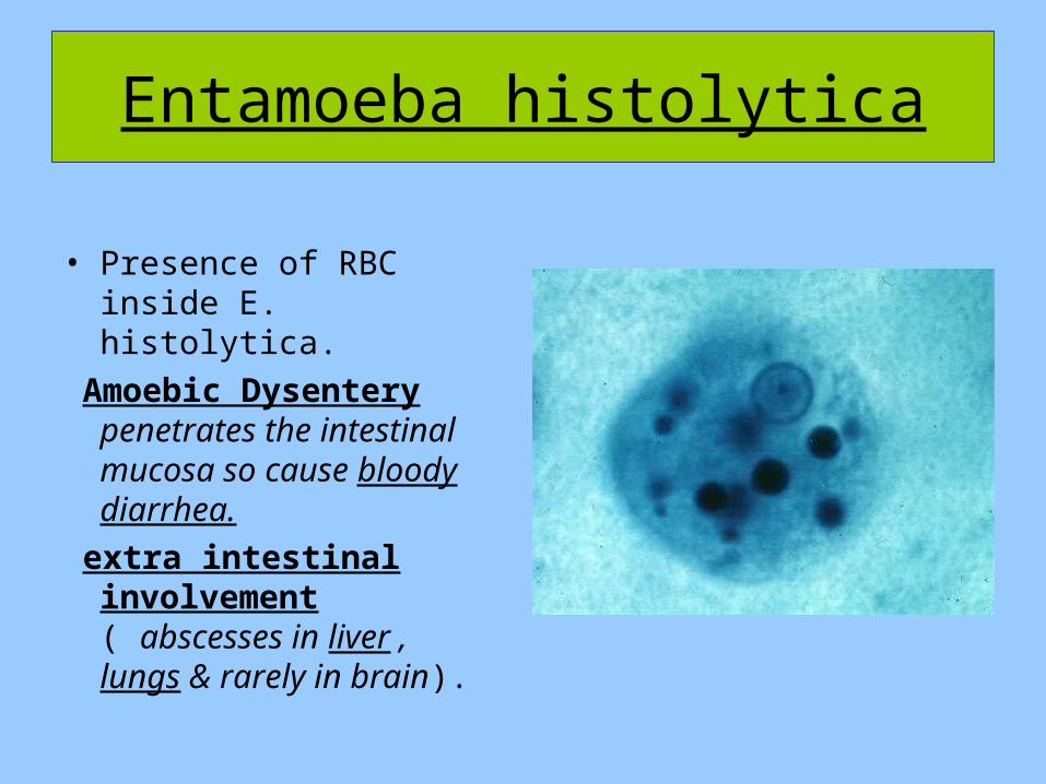
Entamoeba histolytica
• Presence of RBC inside E. histolytica.
Amoebic Dysentery penetrates the intestinal mucosa so cause bloody diarrhea.
extra intestinal involvement ( abscesses in liver , lungs & rarely in brain).

Entamoeba histolyticacyst
cyst stage: round shape , metabolically inactive, infective
stage .

Entamoeba histolyticaLife cycle

Entamoeba coli
• Non pathogenic.• Eccentric
karyosomes.• Irregular peripheral
chromatin.• Cyst contain 8
nucleus.

Giardia lamblia
• Trophozoite pear shape, tennis rickets.
• 2 nuclei without peripheral chromatin.
• Pathogenic Giardiasis.

Giardia lamblia
• Trophozoite contain 4 pairs of flagella.
• 2 sucking disc in the ventral part.

Giardia lamblia cyst
Cyst stage: Oval shape ( in
amoeba it was round)
(1-4) nuclei. Mature cysts have 4 nuclei. Either 2 anterior & 2 posterior OR the 4 on the same side.

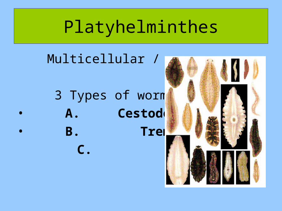
Multicellular / worms
3 Types of worms :• A. Cestodes• B. Trematodes C. Nematodes
Platyhelminthes

Cestodes :
• Taenia saginata
(Beef tapworm)• Taenia solium
(Pork tapworm)• Diphylobothrium
latum.
(fish worm)

Taenia saginata• Cestode worm is
composed of 3 parts: • 1- scolex : responsible
for the attachment to intestinal mucosa by suckers.
• 2- Neck: responsible for strobilization / production of body segments.
• 3- Body : compose of segments (proglottids)
•

It has 3 types of proglottids (segments) :
1-Immature: near the neck,
2- Mature : developing of sex organs occur.
3- Gravid : the segment is mature & uterus is filled with eggs.

Taenia solium
• 4 sucker with
rostellum and hooks

Taenia eggs
• # Eggs have embryo called :
hexacanth embryo


Diphyllobothrium latum
• Intermediate host is fish infected with larva
Scolex of D.latum:• Has no suckers /
instead it has 2 grooves called bothria ( di-phyllo-bothria )

D. latum gravid proglottides
• Gravid segment of D.latum
• Can appear in stool(like taenia)
• Width is more than the length
• Uterus has rossete shape ( no longitudinal branches like tinea)
Central genital pore (lateral in taenia)

D.latum Eggs:
• Like trematode eggs
• Oval shape• Transparent,
hasn’t thick wall• Operculated:
( has a cap , the cap open so embryo will be released from egg.


Comparison of Cestodes
Taenia saginata
Taenia solium
Diphyllobothrium latum
Scolex
Sucking Disks Only
Sucking Disks Plus a Rostellum with Hooklets
Sucking Grooves (Bothria)
Proglottid
Longer than it is Wide
Longer than it is Wide
Wider than it is Long
Uterus
Lateral Branching > 13 Branches (About 16)
Lateral Branching< 13 Branches (About 8)
Rosette in Shape
Genital Pore
Located on the Lateral Margin
Located on the Lateral Margin
Located Centrally

Trematodes
• 1. Clonorchis sinensis:
• Distinctive membrane
• Shouldered operculum
• Has posterior projection

Heterophyes heterophyes
• Same characteristics of C.sinesis eggs but NOT shouldered operculum

Fasciolopsis buski found in intestine
3.5 cm in length• Fasciola hepatica • found in the liver or
bilary duct. 3 cm in length.

Schistosoma mansoni
• Separate sexes• Lateral
spine

Nematodes

Enterobius vermicularis.
• *intestinal worm, small size, cause anal itching at night (mostly in children ).
• *Transparent egg: Presence of larva stage inside egg
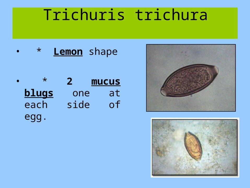
Trichuris trichura
• * Lemon shape
• * 2 mucus blugs one at each side of egg.
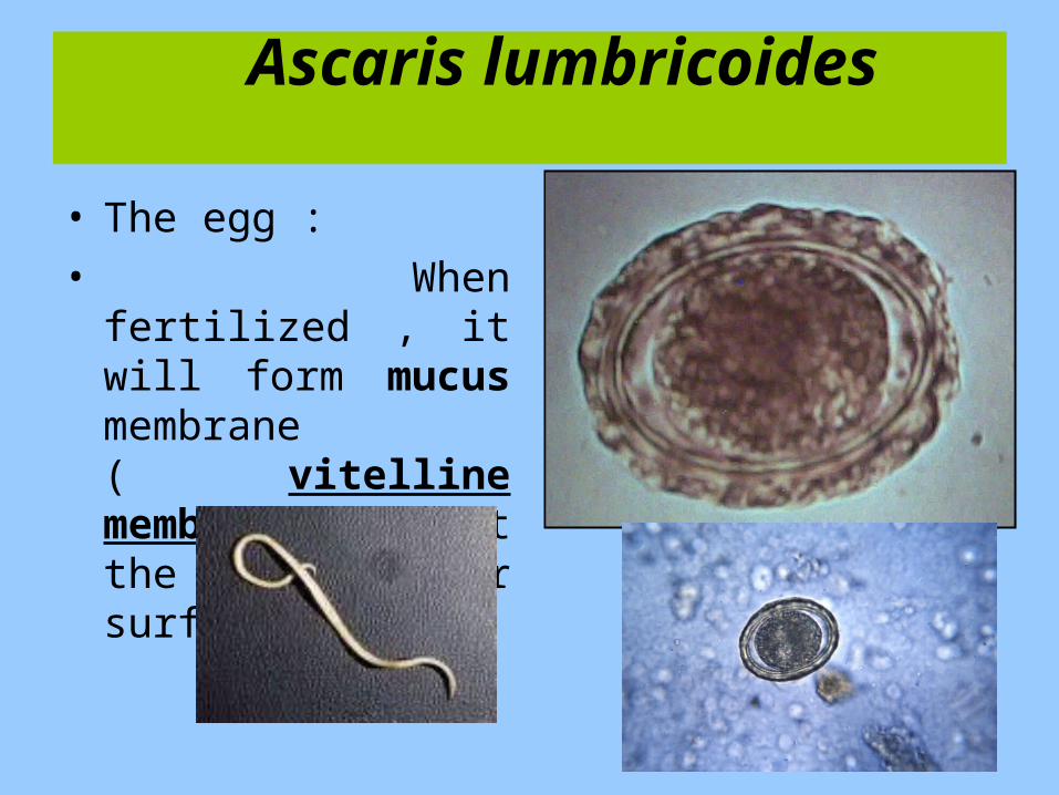
Ascaris lumbricoides
• The egg :• When fertilized ,
it will form mucus membrane ( vitelline membrane ) at the outer surface.

Hookworms
( Ancylostoma duodenale, Necator americana)
Hook worm egg (found in stool):
It has cell stage embryo (form multiple cells

Strongyloides stercoralis
•
• the larva stage NO EGG
• Male : smaller than female, has pointed curved end
• Female : larger,straight tail

THANK YOU