intervaginal space injection of a liquid metal can prevent
TRANSCRIPT

This journal is©The Royal Society of Chemistry and the Chinese Chemical Society 2020 Mater. Chem. Front., 2020, 4, 1397--1403 | 1397
Cite this:Mater. Chem. Front.,
2020, 4, 1397
Intervaginal space injection of a liquid metal canprevent breast cancer invasion and better-sustainconcomitant resistance
Yupeng Cao,ab Xiajun Hu,c Qiang Zhang,a Wenda Hua,a Nan Hu,abd Yifeng Nie,ab
Xue Xu,e Yonggang Xu,f Chongqing Yang,g Xiaohan Zhou,ab Wentao Liu*a andDong Han *ab
Chest wall (CW) invasion by recurrent or locally advanced breast cancer (BC) requires further treatment
with more potential complications. The negative margin of a tumor has been proposed as a critical
factor in BC surgery; however, the ideal margin remains controversial, and data suggest that this
parameter cannot further reduce CW invasion. Herein, a liquid metal (LM) was introduced as an
interstitial stream (IS) to separate the BC and CW to prevent tumor invasion into the CW. Successful
continuous separation and the prevention of CW invasion were observed in 4T1 tumor-bearing mice by
magnetic resonance imaging (MRI) and anatomical observation, respectively. Furthermore, the inhibition
of primary tumor growth and the prevention of accelerated secondary tumor growth suggest that this
method potently retains concomitant resistance (CR). Subsequently, the enhanced maturation of
dendritic cells (DCs) from the LM group suggests that this method may activate T cell-dependent
immunity to retain CR through DC maturation. Additionally, LM was shown to have limited toxicity. In
this study, LM was introduced as an IS into the interstitial space, which potentially links superficial and
deep sites in the body, and showed excellent ability to prevent CW invasion, thus also providing a
potential way to efficiently deliver drugs.
Introduction
Breast cancer (BC) is one of the most common cancers infemales worldwide.1,2 Depending on the stage of BC at diagnosis,numerous therapeutic options are currently available, includingsurgery, chemotherapy, radiation therapy and immunotherapy.3
For early-stage BC, breast conserving therapy (BCT) is commonlyperformed as a surgical treatment,4,5 but this treatment strategycan fail, leading to local recurrence.6 Recurrences in the chest wall
(CW) require further treatment,2 as does locally advanced BCinvolving the CW.3 However, these further treatments are asso-ciated with more complications. For instance, pulmonary andinfectious complications following CW resection and reconstruc-tion have been reported.7 Therefore, avoiding CW invasion iscritical for obtaining a favorable prognosis.
The negative margin width for the surgical resection of BCis related to the risk of recurrence.8 To reduce recurrenceinvolving the CW, many studies have focused on methods forthe intraoperative determination of tumor margins, includingmagnetic resonance imaging (MRI), computed tomographyand invisible near-infrared fluorescent light imaging.9–13
In addition, treatment of locally advanced BC that has notinvaded the CW requires recognizing the negative tumormargins.3 However, the optical negative margin width is stillcontroversial regarding minimizing the recurrence risk afterthe surgical resection of BC.8,14–16 More effective methods areneeded to reduce CW invasion. In recent years, the biologicalutilisation of the intervaginal space in medical applications hasbeen emerging.17–20 In particular, a previous study on the flowbehavior of LMs in the connected fascial space (FS) showed thatthe IS in the FS system is a potential pathway to deliver drugstargeting certain regions of the body.19
a CAS Center for Excellence in Nanoscience, National Center for Nanoscience and
Technology, Beijing 100190, China. E-mail: [email protected],
[email protected] School of Future Technology, University of Chinese Academy of Sciences,
Beijing 100049, Chinac Department of Cardiology, Union Hospital, Tongji Medical College,
Huazhong University of Science and Technology, Wuhan 430022, Chinad Department of Traditional Chinese Medicine, Chengde Medical University,
Chengde 066000, Chinae Department of Gerontology, The Second Hosipital of Shandong University, Jinan,
Chinaf Department of Hematology, Xiyuan Hospital, China Academy of Chinese Medical
Sciences, Beijing 100091, Chinag Department of Pathology, Beijing Hospital, Beijing 100730, China
Received 12th December 2019,Accepted 17th January 2020
DOI: 10.1039/c9qm00753a
rsc.li/frontiers-materials
MATERIALS CHEMISTRYFRONTIERS
RESEARCH ARTICLE
Ope
n A
cces
s A
rtic
le. P
ublis
hed
on 3
0 M
arch
202
0. D
ownl
oade
d on
1/1
2/20
22 9
:57:
36 P
M.
Thi
s ar
ticle
is li
cens
ed u
nder
a C
reat
ive
Com
mon
s A
ttrib
utio
n-N
onC
omm
erci
al 3
.0 U
npor
ted
Lic
ence
.
View Article OnlineView Journal | View Issue

1398 | Mater. Chem. Front., 2020, 4, 1397--1403 This journal is©The Royal Society of Chemistry and the Chinese Chemical Society 2020
In this study, we introduced an LM, a gallium-based alloy, asan obstruction to reduce the CW invasion of BC in breasttumor-bearing mice. Intervaginal space injection (ISI) of a LMinto the wrist was performed in tumor-bearing mice. MRIwas used to observe the obstructive effect of the LM. Grossdissection was performed to detect the ability of this method toblock CW invasion. In addition, the potential immunologicaleffect of this method was observed by flow cytometry (FCM).Histological sections of various viscera were examined toconfirm the safety of LM in this study.
ExperimentalISI in tumor-bearing mice
A gallium-based alloy composed of 75% gallium and 25%indium melted at 15 1C was provided by the Technical Instituteof Physics and Chemistry at the Chinese Academy of Sciences.Female Balb/c mice (with a body weight of approximately 20 g)were purchased from Beijing Vital River Experimental AnimalCorporation (Beijing, China). The 4T1 BC cell line used in thisstudy was purchased from the Type Culture Collection (ChineseAcademy of Sciences, Shanghai, China).
All the animal experiments were approved by the Committeeat the Institute of the Chinese Academy of Medical Science. 4T1cells were cultured in Dulbecco’s modified Eagle’s mediumsupplemented with 10% fetal bovine serum (Gibco), 100 U mL�1
penicillin, and 1 mg mL�1 streptomycin at 37 1C in an incubatorwith 95% air and 5% CO2. 4T1 cells (1 � 106) suspended in PBSwere subcutaneously injected into the left flank of each femaleBALB/c mouse. On the seventh day, 4T1 cells (2 � 105) suspendedin PBS were injected into the right flank of each female BALB/cmouse. The tumor volume was calculated according to thefollowing formula: width2 � length � 0.5. The tumor volume ata specific time point divided by the tumor volume on day 8 isreported. On the eighth day, each mouse in the LM groupunderwent ISI with LM (0.1 mL), and each mouse in the PBSgroup underwent ISI with PBS (0.1 mL). The control group was notsubjected to ISI.
In vivo MRI
The mice were observed by MRI (Bruker, 7T) before and afterISI on day 8. In addition, MRI was performed on day 12.T2-weighted MRI was used with the following parameters:TR = 2000 ms; TE = 30 ms; FOV = 28 � 28 mm2; and imagesize = 256 � 256.
Ex vivo analysis of DCs in the spleen
To study DC maturation, the spleens were harvested from themice and stained with anti-CD11c-PE (Biolegend, Clone: N418,Catalog: 117307), anti-CD86-APC (Biolegend, Clone: GL-1, Cat-alog: 105011), and anti-CD80-Alexa Fluors 488 (Biolegend,Clone: 16-10A1, Catalog: 104715) antibodies according to themanufacturer’s protocols. A Beckman Coulter Navios flowcytometer system (USA) was used. The data were analyzed bythe flow cytometer workstation.
Histopathological analysis
This analysis aimed to examine the potential toxicity of LMin mice. Heart, liver, spleen, lung and kidney tissues wereharvested from the mice and fixed overnight in 4% neutral-buffered paraformaldehyde. The histological slides were stainedwith hematoxylin and eosin (HE), followed by dehydrationthrough a graded series of ethanol solutions from 75% to100%, and examined in a blinded manner by a well-trainedpathologist. Histological observations and photomicrographywere performed using a digital slide scanner (NanoZoomer-XRC12000, Japan). Damage to the above visceral tissues wasexamined in an average of five fields of view (�200).
Results and discussion
To directly assess the separation effect of LM, we utilizedT2-weighted MRI to observe the positions of the tumor andCW in mice. Because the transverse relaxation (T2) value oftumors is high in this paper, the signal of the tumors was highin the T2-weighted MRI. Because the T2 value of PBS is higherthan that of tumors, the signal of PBS is higher than that of thetumor in the T2-weighted MRI.21 Before ISI, the tumor exhibitedhigh signal intensity. Hydrogen in mice was used for visualiza-tion by MRI as LM does not contain hydrogen. Hence, we canobserve LM in mice through its low signal intensity, as shownin Fig. 1b. In addition, a portion of the injected LM rapidlyentered the region between the tumor and CW within tenminutes after ISI. Similarly, phosphate-buffered saline (PBS)was distributed in the region between the tumor and CW. Theresults suggest that fluids, including PBS and LM, can flowthrough the FS to the region near a tumor.
LM outperformed PBS in mice in this study. Specifically, themobilities of these two agents were different. LM distributedmore in the region between the tumor and the CW, whilePBS distributed in the region between the tumor and arm.Furthermore, LM remained in the mice longer than PBS.As shown in Fig. 1b, LM remained at four days after injection,but PBS could not be observed at this time point. PBS is a water-based solution and hence can be absorbed by mice. It isdifficult for LM to diffuse throughout the body due to its highsurface tension.19 Therefore, LM has the potential to remain ina region in mice for a long period and resist tumor invasion.Subsequently, we compared the separation effect between theLM and blank control groups. As shown in Fig. 2, LM remainedbetween the CW and the tumor. However, the tumors in theother two groups grew adjacent to CW tissues. As evidenced bytumors explanted during the growth period, LM and PBS didnot flow to the region near the tumor on the opposite flank.This result suggests that LM is limited to a certain region intumor-bearing mice.
Dissection was performed to examine the ability of LM toseparate the tumor and the CW. At the molecular level, theinvasion of the tumor involves the tumor cells secretingenzymes to degrade the extracellular matrix adjacent to thetumor. At the tissue level, invasion refers to the direct extension
Research Article Materials Chemistry Frontiers
Ope
n A
cces
s A
rtic
le. P
ublis
hed
on 3
0 M
arch
202
0. D
ownl
oade
d on
1/1
2/20
22 9
:57:
36 P
M.
Thi
s ar
ticle
is li
cens
ed u
nder
a C
reat
ive
Com
mon
s A
ttrib
utio
n-N
onC
omm
erci
al 3
.0 U
npor
ted
Lic
ence
.View Article Online

This journal is©The Royal Society of Chemistry and the Chinese Chemical Society 2020 Mater. Chem. Front., 2020, 4, 1397--1403 | 1399
and penetration by cancer cells into neighboring tissues. Thebarrier between the tumor and the chest wall muscle wasbreached.22 As shown in Fig. 3, CW invasion occurred in micein the control group. The muscles of the CW adjacent to thebreast tumor were seriously damaged in the control group, andthe tumor margin was ambiguous. Compared to the control group,the LM group exhibited better separation of the tumor margin.
After dissection, LM was found to have remained in the regionbetween the tumor and the CW, and the CW muscles were notdamaged. Importantly, LM was enwrapped by a film of fascia,a type of interstitial tissue. After ISI, LM flowed from theinjection site and continuously distributed in the regionbetween the tumor and the CW through the FS. Direct tumorinvasion disrupting the fascia film has been reported.23
In addition, tumor invasion occurring on the adjacent normaltissues due to the acidic microenvironment has beenreported.24 Because of the acidic environment, the gallium-based liquid metal might be degraded to form gallium(III)ions, which can be used as an anticancer agent.25 In thisstudy, LM was shown to play a role in separating the tumorand the CW and hence resisting the CW invasion by thetumor. Due to the direct physical separation created by LM,the tumor cells cannot invade fascia films and other tissues.Moreover, as LM localized adjacent to the tumor, the tumormargin was distinct, which facilitates tumor recognition. Inaddition, the fascia enwrapping the CW remained smoothbecause it was not invaded. Hence, the fascia enwrapping thebreast tumor was easily dissected, which will benefit BCT.
Fig. 1 Study course and MRI of the LM and PBS groups. The green dashedline represents PBS, the blue full lines indicate the tumor, the red dottedlines represent LM, and days 8-0 and 8-1 indicate images taken before andafter ISI, respectively. (a) The study course, including the first and secondtumor inoculations, ISI with LM and MRI. (b) Observation of mice in the LMand PBS groups before and after ISI with LM and on the following 4th day.The low signal intensity representing LM was distributed in the regionbetween the tumor and the CW, whereas PBS was distributed near thetumor after ISI, but disappeared on the following 4th day.
Fig. 2 MRI observations of the LM and blank control groups. LM remainingin the targeted region is indicated by red dotted lines and separates the CWand the tumor, which is indicated by blue full lines.
Fig. 3 Anatomical observation of mice in the LM and control groups uponsurgical dissection. The black arrows represent invasion regions, the bluetriangles represent LM, the blue arrows represent tumors and the reddashed lines represent the region of interest (ROI); mosaics were used tocover the heads and abdomens of the mice. LM separated the tumor andthe CW; however, CW invasion occurred in mice of the control group.
Fig. 4 Tumor growth analysis. (a) The primary tumor volumes in the threegroups were plotted. The primary tumor volumes on days 11 to 14 in theLM group were significantly lower than those in the other groups. (b) Thesecondary tumor weights in the three groups were not significantlydifferent. p values were determined with the t-test (*p o 0.05).
Materials Chemistry Frontiers Research Article
Ope
n A
cces
s A
rtic
le. P
ublis
hed
on 3
0 M
arch
202
0. D
ownl
oade
d on
1/1
2/20
22 9
:57:
36 P
M.
Thi
s ar
ticle
is li
cens
ed u
nder
a C
reat
ive
Com
mon
s A
ttrib
utio
n-N
onC
omm
erci
al 3
.0 U
npor
ted
Lic
ence
.View Article Online
1400 | Mater. Chem. Front., 2020, 4, 1397--1403 This journal is©The Royal Society of Chemistry and the Chinese Chemical Society 2020
Relapse after surgery results in worse patient outcomes.However, in experimental and clinical settings, primary tumordissection has been shown to contribute to the acceleratedgrowth of spontaneous metastases.26–28 This phenomenon,the inhibition of metastases by the primary tumor, is calledconcomitant resistance (CR) and is relieved by dissection of theprimary tumor.26,29 Hence, the reduction in CR of metastaticfoci after treatment should be taken into consideration.In this experiment, secondary tumor weight and primary tumorgrowth were compared in the three groups. As shown in Fig. 4a,
the volume of the primary tumor was significantly lower in theLM group than in the PBS and control groups from day 11 today 14 after ISI. In addition, the secondary tumors were notheavier in the LM group than in the other two groups, as shownin Fig. 4b. Therefore, LM inhibited primary tumor growth andconcurrently retained potent CR.
T cell-dependent immunity plays a pivotal role in CR.30
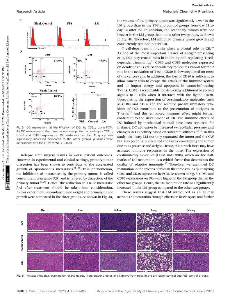
As one of the most important classes of antigen-presentingcells, DCs play crucial roles in initiating and regulating T cell-dependent immunity.31 CD80 and CD86 molecules expressedon dendritic cells are co-stimulatory molecules known for theirrole in the activation of T-cell. CD80 is downregulated on mostof the cancer cells. In addition, the loss of CD80 is sufficient toallow cancer cells to escape the attack of the immune systemand to impart anergy and apoptosis in tumor-infiltratingT cells. CD86 is responsible for delivering additional or secondsignals to T cells when it interacts with the ligand CD28.Upregulating the expression of co-stimulatory molecules suchas CD80 and CD86 and the secreted pro-inflammatory cyto-kines of DCs contribute to the presentation of antigens toT cells.32 And this enhanced immune effect might furthercontribute to the sustainment of CR. The immune effects ofDC induced by mechanical stimuli have been reported; forinstance, DC activation by increased extracellular pressure andchanges in DC activity based on substrate stiffness.33–35 In thisstudy, the heavy LM not only separated the tumor and the CWbut also potentially stretched the fascia enwrapping the tumordue to its presence and weight. Hence, this stretch force may haveactivated immune responses in the mice. The expression ofco-stimulatory molecules (CD80 and CD86), which are the hall-marks of DC maturation, is a critical factor that determines thequality of adaptive immunity.36 Therefore, we examined DCmaturation in the spleens of mice in the three groups by analyzingCD80 and CD86 expression by FCM. As shown in Fig. 5, CD80 andCD86 expressions on DCs were higher in the LM group than in theother two groups. Hence, the DC maturation rate was significantlyincreased in the LM group compared to the other two groups.
These results suggest that LM introduced as an IS mayactivate DC maturation through effects on fascia space and further
Fig. 5 DC maturation. (a) Identification of DCs by CD11c using FCM.(b) DC maturation in the three groups was plotted according to CD11c,CD80 and CD86 expressions. DC maturation in the LM group wassignificantly increased compared to the other groups. p values weredetermined with the t-test (***p o 0.001).
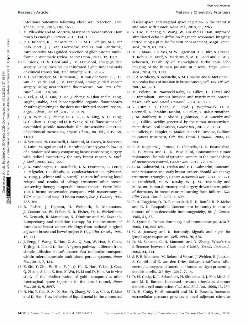
Fig. 6 Histopathological examination of the hearts, livers, spleens, lungs and kidneys from mice in the LM, blank control and PBS control groups.
Research Article Materials Chemistry Frontiers
Ope
n A
cces
s A
rtic
le. P
ublis
hed
on 3
0 M
arch
202
0. D
ownl
oade
d on
1/1
2/20
22 9
:57:
36 P
M.
Thi
s ar
ticle
is li
cens
ed u
nder
a C
reat
ive
Com
mon
s A
ttrib
utio
n-N
onC
omm
erci
al 3
.0 U
npor
ted
Lic
ence
.View Article Online

This journal is©The Royal Society of Chemistry and the Chinese Chemical Society 2020 Mater. Chem. Front., 2020, 4, 1397--1403 | 1401
prevent the accelerated growth of metastases after inhibitingprimary tumor growth. The combination of enhanced DC matura-tion and checkpoint blockade therapy significantly inhibited tumorgrowth.37 Hence, this method has the potential to prevent thegrowth of metastases caused by a reduction in CR and enhancethe immune response in combination with checkpoint blockadetherapy.
LM is inorganic matter with reportedly low toxicity.38
However, the dose of LM used in this study was quite highfor mice. We performed a histological examination to evaluatethe toxicity of LM. As shown in Fig. 6, vacuolar degeneration,cytoplasm swelling and degeneration and inflammation occurredin the heart, liver and lung tissues of mice in all three groups. Thespleen and kidney histological results revealed no abnormalities.Although some negative effects occurred, the histological resultsdid not suggest that LM impaired the viscera of mice.
To avoid the risk of toxicity from LM, alternative materialsthat satisfy the aforementioned features and show appropriatebiosafety and biocompatibility may be a better choice to facili-tate the clinical application of this method. As suggested by aprevious study on ISs19 and this work, the materials injected inthe FS must have three features: mobility, the ability to diffuseinto tissues, and the ability to resist degradation. For instance,collagen and hyaluronic acid used in the clinic have thepotential to replace the liquid metal. Additionally, loadinganticancer drug onto the substitute may be a more efficientand effective treatment.39
Conclusions
In summary, we have described a method of introducing LMas an IS injection into the FS to efficiently link superficial todeep sites and to successfully separate the CW and the BC intumor-bearing mice. LM flowed through the FS under theinjection site to the region between the primary tumor andthe CW. During the experimental period, LM did not flow to theregion between the secondary tumor and the CW. LM remainedfor a long period and continuously resisted CW invasion by theprimary tumor. Based on the primary tumor growth andsecondary tumor weight results, the impact of this method onCR was discussed. Subsequently, the significant increase in DCmaturation in the spleens from mice in the LM group suggeststhat sustained CR may stem from T cell-dependent immunityregulated by DCs. In addition, the combination of this methodand checkpoint blockade as a potential therapy was proposedfor the treatment of BC, not only as a preventative strategy forCW invasion. Additionally, the advantages and disadvantagesof LM were discussed. Additional potential replacement mate-rials were also discussed. This method utilized the interstitialspace, which is between two adjacent interstitial tissues, as apathway for targeted delivery to a particular region. The agentscan be injected at a superficial site and flow through theinterstitial space to deep within the body, exhibiting highlyefficient passage from superficial sites to specific deep regions.More investigations on the IS, i.e., the link between superficial
sites and deep sites, will be of great value and facilitate thedevelopment of therapeutics that will contribute to humanhealth.
Author contributions
Dong Han and Wentao Liu designed research, analyzed data,and wrote the paper. Yupeng Cao analyzed data and wrote thepaper. Xiajun Hu, Qiang Zhang, Wenda Hua, Nan Hu, YifengNie and Xue Xu performed the anatomy experiment. YupengCao performed the magnetic resonance imaging experiment.Yonggang Xu and Yupeng Cao performed the flow cytometerexperiment. Congqing Yang analyzed the histopathologicaldata. Xiaohan Zhou performed the mouse tumor model.
Conflicts of interest
There are no conflicts to declare.
Acknowledgements
We are sincerely thankful to Technical Institute of Physics andChemistry at the Chinese Academy of Sciences for providing theliquid metal. This work was supported by the Source InnovationResearch Program of Frontier Sciences, CAS, Grant No. ZDBS-LY-SLH036.
Notes and references
1 B. W. Stewart and C. P. Wild, World Cancer Report 2014,International Agency for Research on Cancer, 2014.
2 E. Wakeam, S. A. Acuna and S. Keshavjee, Chest wallresection for recurrent breast cancer in the modern era asystematic review and meta-analysis, Ann. Surg., 2018,267, 646.
3 B. Sepesi, Management of breast cancer invading chest wall,Thorac. Surg. Clin., 2017, 27, 158.
4 M. Morrow, R. A. Schmidt and C. Bucci, Breast conservationfor mammographically occult carcinoma, Ann. Surg., 1998,227, 502.
5 M. R. Grootendorst, M. Cariati, S. E. Pinder, A. Kothari,M. Douek, T. Kovacs, H. Hamed, A. Pawa, F. Nimmo, J. Owen,V. Ramalingam, S. Sethi, S. Mistry, K. Vyas, D. S. Tuch,A. Britten, M. Van Hemelrijck, G. J. Cook, C. Sibley-Allen,S. Allen and A. Purushotham, Intraoperative assessmentof tumor resection margins in breast-conserving surgeryusing F-18-FDG cerenkov luminescence imaging: A first-in-human feasibility study, J. Nucl. Med., 2017, 58, 891.
6 M. Clemons, S. Danson, T. Hamilton and P. Goss,Locoregionally recurrent breast cancer: incidence, risk factorsand survival, Cancer Treat. Rev., 2001, 27, 67.
7 J. D. Spicer, J. B. Shewale, M. B. Antonoff, A. M. Correa,W. B. Hofstetter, D. C. Rice, A. A. Vaporciyan, R. J. Mehran,G. L. Walsh, J. A. Roth and B. Sepesi, The influence ofreconstructive technique on perioperative pulmonary and
Materials Chemistry Frontiers Research Article
Ope
n A
cces
s A
rtic
le. P
ublis
hed
on 3
0 M
arch
202
0. D
ownl
oade
d on
1/1
2/20
22 9
:57:
36 P
M.
Thi
s ar
ticle
is li
cens
ed u
nder
a C
reat
ive
Com
mon
s A
ttrib
utio
n-N
onC
omm
erci
al 3
.0 U
npor
ted
Lic
ence
.View Article Online
1402 | Mater. Chem. Front., 2020, 4, 1397--1403 This journal is©The Royal Society of Chemistry and the Chinese Chemical Society 2020
infectious outcomes following chest wall resection, Ann.Thorac. Surg., 2016, 102, 1653.
8 M. Pilewskie and M. Morrow, Margins in breast cancer: Howmuch is enough?, Cancer, 2018, 124, 1335.
9 P. L. Kubben, K. J. ter Meulen, O. E. M. G. Schijns, M. P. terLaak-Poort, J. J. van Overbeeke and H. van Santbrink,Intraoperative MRI-guided resection of glioblastoma multi-forme: a systematic review, Lancet Oncol., 2011, 12, 1062.
10 S. Gioux, H. S. Choi and J. V. Frangioni, Image-guidedsurgery using invisible near-infrared light: fundamentalsof clinical translation, Mol. Imaging, 2010, 9, 237.
11 A. L. Vahrmeijer, M. Hutteman, J. R. van der Vorst, C. J. H.van de Velde and J. V. Frangioni, Image-guided cancersurgery using near-infrared fluorescence, Nat. Rev. Clin.Oncol., 2013, 10, 507.
12 Z. Lei, X. Li, X. Luo, H. He, J. Zheng, X. Qian and Y. Yang,Bright, stable, and biocompatible organic fluorophoresabsorbing/emitting in the deep near-infrared spectral region,Angew. Chem., Int. Ed., 2017, 56, 2979.
13 Q. X. Wen, Y. J. Zhang, C. Y. Li, S. S. Ling, X. H. Yang,G. C. Chen, Y. Yang and Q. B. Wang, NIR-II fluorescent self-assembled peptide nanochain for ultrasensitive detectionof peritoneal metastasis, Angew. Chem., Int. Ed., 2019, 58,11001.
14 U. Veronesi, N. Cascinelli, L. Mariani, M. Greco, R. Saccozzi,A. Luini, M. Aguilar and E. Marubini, Twenty-year follow-upof a randomized study comparing breast-conserving surgerywith radical mastectomy for early breast cancer, N. Engl.J. Med., 2002, 347, 1227.
15 J. A. Vandongen, H. Bartelink, I. S. Fentiman, T. Lerut,F. Mignolet, G. Olthuis, E. Vanderschueren, R. Sylvester,D. Tong, J. Winter and K. Vanzijl, Factors influencing localrelapse and results of salvage treatment after breast-conserving therapy in operable breast-cancer – Eortc Trial-10801, breast conservation compared with mastectomy inTNM stage-I and stage-II breast-cancer, Eur. J. Cancer, 1992,28A, 801.
16 B. Fisher, J. Dignam, N. Wolmark, E. Mamounas,J. Costantino, W. Poller, E. R. Fisher, D. L. Wickerham,M. Deutsch, R. Margolese, N. Dimitrov and M. Kavanah,Lumpectomy and radiation therapy for the treatment ofintraductal breast cancer: Findings from national surgicaladjuvant breast and bowel project B-17, J. Clin. Oncol., 1998,16, 441.
17 J. Feng, F. Wang, X. Han, Z. Ao, Q. Sun, W. Hua, P. Chen,T. Jing, H. Li and D. Han, A ‘‘green pathway’’ different fromsimple diffusion in soft matter: fast molecular transportwithin micro/nanoscale multiphase porous systems, NanoRes., 2014, 7, 434.
18 X. Shi, Y. Zhu, W. Hua, Y. Ji, Q. Ha, X. Han, Y. Liu, J. Gao,Q. Zhang, S. Liu, K. Ren, X. Wu, H. Li and D. Han, An in vivostudy of the biodistribution of gold nanoparticles afterintervaginal space injection in the tarsal tunnel, NanoRes., 2016, 9, 2097.
19 N. Hu, Y. Cao, Z. Ao, X. Han, Q. Zhang, W. Liu, S. Liu, F. Liaoand D. Han, Flow behavior of liquid metal in the connected
fascial space: Intervaginal space injection in the rat wristand mice with tumor, Nano Res., 2018, 11, 2265.
20 Y. Cao, Y. Zhang, Y. Wang, W. Liu and D. Han, Improvedstimulated echo in diffusion magnetic resonance imaging:introducing a pi pulse for SNR enhancement, Magn. Reson.Med., 2019, 81, 2905.
21 M. C. Maas, E. K. Vos, M. W. Lagemaat, A. K. Bitz, S. Orzada,T. Kobus, O. Kraff, S. Maderwald, M. E. Ladd and T. W. J.Scheenen, Feasibility of T-2-weighted turbo spin echoimaging of the human prostate at 7 tesla, Magn. Reson.Med., 2014, 71, 1711.
22 E. A. McSherry, S. Donatello, A. M. Hopkins and S. McDonnell,Molecular basis of invasion in breast cancer, Cell. Mol. Life Sci.,2007, 64, 3201.
23 M. Polette, B. Nawrocki-Raby, C. Gilles, C. Clavel andP. Birembaut, Tumour invasion and matrix metalloprotei-nases, Crit. Rev. Oncol. Hematol., 2004, 49, 179.
24 V. Estrella, T. Chen, M. Lloyd, J. Wojtkowiak, H. H.Cornnell, A. Ibrahim-Hashim, K. Bailey, Y. Balagurunathan,J. M. Rothberg, B. F. Sloane, J. Johnson, R. A. Gatenby andR. J. Gillies, Acidity generated by the tumor microenviron-ment drives local invasion, Cancer Res., 2013, 73, 1524.
25 P. Collery, B. Keppler, C. Madoulet and B. Desoize, Galliumin cancer treatment, Crit. Rev. Oncol. Hematol., 2002, 42,283.
26 R. A. Ruggiero, J. Bruzzo, P. Chiarella, O. D. Bustuoabad,R. P. Meiss and C. D. Pasqualini, Concomitant tumorresistance: The role of tyrosine isomers in the mechanismsof metastases control, Cancer Res., 2012, 72, 1043.
27 C. M. Galmarini, O. Tredan and F. C. Galmarini, Concomi-tant resistance and early-breast cancer: should we changetreatment strategies?, Cancer Metastasis Rev., 2014, 33, 271.
28 R. Demicheli, M. W. Retsky, W. J. M. Hrushesky andM. Baum, Tumor dormancy and surgery-driven interruptionof dormancy in breast cancer: learning from failures, Nat.Clin. Pract. Oncol., 2007, 4, 699.
29 R. A. Ruggiero, O. D. Bustuoabad, R. D. Bonfil, R. P. Meissand C. D. Pasqualini, Concomitant immunity in murinetumors of non-detectable immunogenicity, Br. J. Cancer,1985, 51, 37.
30 B. Quesnel, Tumor dormancy and immunoescape, APMIS,2008, 116, 685–694.
31 C. A. Janeway and K. Bottomly, Signals and signs forlymphocyte-responses, Cell, 1994, 76, 275.
32 D. M. Sansom, C. N. Manzotti and Y. Zheng, What’s thedifference between CD80 and CD86?, Trends Immunol.,2003, 24, 314.
33 S. F. B. Mennens, M. Bolomini-Vittori, J. Weiden, B. Joosten,A. Cambi and K. van den Dries, Substrate stiffness influ-ences phenotype and function of human antigen-presentingdendritic cells, Sci. Rep., 2017, 7, 14.
34 D. H. Craig, K. L. Schaubert, H. Shiratsuchi, J. Kan-Mitchelland M. D. Basson, Increased pressure stimulates aberrantdendritic cell maturation, Cell. Mol. Biol. Lett., 2008, 13, 260.
35 D. H. Craig, H. Shiratsuchi and M. D. Basson, Increasedextracellular pressure provides a novel adjuvant stimulus
Research Article Materials Chemistry Frontiers
Ope
n A
cces
s A
rtic
le. P
ublis
hed
on 3
0 M
arch
202
0. D
ownl
oade
d on
1/1
2/20
22 9
:57:
36 P
M.
Thi
s ar
ticle
is li
cens
ed u
nder
a C
reat
ive
Com
mon
s A
ttrib
utio
n-N
onC
omm
erci
al 3
.0 U
npor
ted
Lic
ence
.View Article Online

This journal is©The Royal Society of Chemistry and the Chinese Chemical Society 2020 Mater. Chem. Front., 2020, 4, 1397--1403 | 1403
for enhancement of conventional dendritic cell maturationstrategies, Biochem. Biophys. Res. Commun., 2009, 387, 174.
36 M. I. Bonetti, L. Pieri, L. Domenici, S. Urbani, G. Romano,A. Aldinucci, C. Ballerini, M. Monici, R. Saccardi, V. Basile,A. Bosi and P. Romagnoli, Dendritic cells with lymphocyte-stimulating activity differentiate from human CD133 positiveprecursors, Blood, 2011, 117, 3983.
37 C. Wang, L. G. Xu, C. Liang, J. Xiang, R. Peng and Z. Liu,Immunological responses triggered by photothermal therapy
with carbon nanotubes in combination with anti-CTLA-4therapy to inhibit cancer metastasis, Adv. Mater., 2014, 26,8154.
38 S. A. Chechetka, Y. Yu, X. Zhen, M. Pramanik, K. Y. Pu andE. Miyako, Light-driven liquid metal nanotransformers forbiomedical theranostics, Nat. Commun., 2017, 8, 19.
39 G. Lemperle, V. Morhenn and U. Charrier, Human histologyand persistence of various injectable filler substances for softtissue augmentation, Aesthetic Plast. Surg., 2003, 27, 354.
Materials Chemistry Frontiers Research Article
Ope
n A
cces
s A
rtic
le. P
ublis
hed
on 3
0 M
arch
202
0. D
ownl
oade
d on
1/1
2/20
22 9
:57:
36 P
M.
Thi
s ar
ticle
is li
cens
ed u
nder
a C
reat
ive
Com
mon
s A
ttrib
utio
n-N
onC
omm
erci
al 3
.0 U
npor
ted
Lic
ence
.View Article Online