interpretation and use of frax in clinical practice - esceo interprretation... · interpretation...
TRANSCRIPT
POSITION PAPER
Interpretation and use of FRAX in clinical practice
J. A. Kanis & D. Hans & C. Cooper & S. Baim &
J. P. Bilezikian & N. Binkley & J. A. Cauley &
J. E. Compston & B. Dawson-Hughes &
G. El-Hajj Fuleihan & H. Johansson & W. D. Leslie &
E. M. Lewiecki & M. Luckey & A. Oden &
S. E. Papapoulos & C. Poiana & R. Rizzoli & D. A. Wahl &E. V. McCloskey & Task Force of the FRAX Initiative
Received: 18 May 2011 /Accepted: 7 June 2011 /Published online: 21 July 2011# International Osteoporosis Foundation and National Osteoporosis Foundation 2011
AbstractSummary The introduction of the WHO FRAX® algo-rithms has facilitated the assessment of fracture risk onthe basis of fracture probability. Its use in fracture riskprediction has strengths, but also limitations of which the
clinician should be aware and are the focus of thisreviewIntroduction The International Osteoporosis Foundation(IOF) and the International Society for Clinical Densitometry(ISCD) appointed a joint Task Force to develop resource
J. A. Kanis (*)WHO Collaborating Centre for Metabolic Bone Diseases,University of Sheffield Medical School,Beech Hill Road,Sheffield S10 2RX, UKe-mail: [email protected]
D. HansCenter of Bone Diseases, Bone and Joint Department,Lausanne University Hospital,Lausanne, Switzerland
C. CooperMRC Lifecourse Epidemiology Unit, University of Southampton,Southampton, UK
C. CooperNIHR Musculoskeletal Biomedical Research Unit,Institute of Musculoskeletal Sciences, University of Oxford,Oxford, UK
S. BaimDivision of Endocrinology, University of Miami,Miller School of Medicine,Coral Gables, FL, USA
J. P. BilezikianColumbia University College of Physicians and Surgeons,New York, NY, USA
N. BinkleyOsteoporosis Clinical Research Program,University of Wisconsin,Madison, WI, USA
J. A. CauleyDepartment of Epidemiology, Graduate School of Public Health,University of Pittsburgh,Pittsburgh, CA, USA
J. E. CompstonDepartment of Medicine, Addenbrooke’s Hospital,Cambridge University,Cambridge, UK
B. Dawson-HughesJean Mayer United States Department of Agriculture HumanNutrition Research Center on Aging, Tufts University,Boston, MA, USA
G. El-Hajj FuleihanWHO Collaborating Center for Metabolic Bone Disorders,American University of Beirut,Beirut, Lebanon
H. Johansson :A. OdenCentre for Bone Research at the Sahlgrenska Academy,Institute of Medicine, University of Gothenburg,Gothenburg, Sweden
W. D. LeslieUniversity of Manitoba,Winnipeg, MB, Canada
E. M. LewieckiNew Mexico Clinical Research & Osteoporosis Center,University of New Mexico School of Medicine,Albuquerque, NM, USA
Osteoporos Int (2011) 22:2395–2411DOI 10.1007/s00198-011-1713-z

documents in order to make recommendations on how toimprove FRAX and better inform clinicians who use FRAX.The Task Force met in November 2010 for 3 days to discussthese topics which form the focus of this review.Methods This study reviews the resource documents andjoint position statements of ISCD and IOF.Results Details on the clinical risk factors currently used inFRAX are provided, and the reasons for the exclusion ofothers are provided. Recommendations are made for thedevelopment of surrogate models where country-specificFRAX models are not available.Conclusions The wish list of clinicians for the modulationof FRAX is large, but in many instances, these wishescannot presently be fulfilled; however, an explanation andunderstanding of the reasons may be helpful in translatingthe information provided by FRAX into clinical practice.
Keywords Bone mineral density. Clinical risk factors .
Fracture probability. Risk assessment
Preamble
In October 2009, the International Osteoporosis Foundation(IOF) and the International Society for Clinical Densitometry(ISCD) agreed to appoint a joint task force to review thestrengths and limitations of FRAX under the flag of ‘theFRAX initiative’. Several Task Forces were formed to developresource documents based on literature reviews to answerspecific queries on the use of clinical risk factors, theapplication of bone mineral measurements to FRAX and the
epidemiological basis for its international development. InNovember 2010, the IOF and the ISCD held a joint meetingthat invited the Task Force members, an independent panel ofexperts and an audience to debate the findings of the TaskForce. The objective was to make recommendations toimprove FRAX and better inform clinicians who use this tool.An outcome of the conference was a position developmentpaper, endorsed by both organisations and published in theJournal of Clinical Densitometry [1] and the clinical reviewin this paper. Papers to support the position statements and aselection of the resource documents are to be published in theJournal of Clinical Densitometry and Archives of Osteoporo-sis, respectively. The Task Force membership and ExpertPanel are listed at the end of this manuscript.
Introduction
FRAX® is a computer-based algorithm (http://www.shef.ac.uk/FRAX) developed by the World Health OrganizationCollaborating Centre for Metabolic Bone Diseases and firstreleased in 2008. The algorithm, intended for primary care,calculates fracture probability from easily obtained clinicalrisk factors (CRFs) in men and women [2–4]. The output ofFRAX is the 10-year probability of a major osteoporoticfracture (hip, clinical spine, humerus or wrist fracture) andthe 10-year probability of hip fracture.
Probability is calculated from age, sex, body mass index(BMI) and dichotomized risk factors comprising prior fragilityfracture, parental history of hip fracture, current tobaccosmoking, ever use of long-term oral glucocorticoid use,rheumatoid arthritis, other causes of secondary osteoporosisand high alcohol consumption (Fig. 1). Femoral neck bonemineral density (BMD) can be optionally input to enhancefracture risk prediction [5]. Fracture probability is computedtaking both the risk of fracture and the risk of death intoaccount. This is important because some of the risk factorsaffect the risk of death as well as the fracture risk. Examplesinclude increasing age, sex, low BMI, low BMD, use ofglucocorticoids and smoking. Other risk algorithms calculatethe probability of a clinical event without taking into accountthe possibility of death from other causes [6, 7]. In addition,the FRAX® models are calibrated for different countriesusing country-specific fracture and mortality rates.
The relationships between risk factors and fracture proba-bility have been constructed using information derived fromthe primary data of nine population-based cohorts fromaround the world, including centres from North America,Europe, Asia and Australia. Clinical risk factors for fracturewere identified that provided independent information onfracture risk based on a series of meta-analyses. The FRAXalgorithm has been validated in 11 independent cohorts with asimilar geographic distribution with in excess of 1 million
M. LuckeySt. Barnabas Osteoporosis & Metabolic Bone Disease Center,Livingston, NJ, USA
S. E. PapapoulosDepartment of Endocrinology and Metabolic Diseases,Leiden University Medical Center,Leiden, the Netherlands
C. PoianaDepartment of Endocrinology,Carol Davila University of Medicine and Pharmacy,Bucharest, Romania
R. RizzoliDivision of Bone Diseases, Department of Rehabilitation andGeriatrics, Geneva University Hospitals and Faculty of Medicine,Geneva, Switzerland
D. A. WahlInternational Osteoporosis Foundation,Nyon, Switzerland
E. V. McCloskeyAcademic Unit of Bone Metabolism, University of Sheffield,Sheffield, UK
2396 Osteoporos Int (2011) 22:2395–2411

patient-years [5]. The use of primary data for the modelconstruct permits the determination of the predictive importancein a multivariable context of each of the risk factors, as well asinteractions between risk factors, and thereby optimises theaccuracy by which fracture probability can be computed [8, 9].
Fracture probability varies markedly in different regions ofthe world [10]. Thus, the FRAX® models need to becalibrated to those countries where the epidemiology offracture and death is known. Models are currently available for31 countries across the world: Argentina, Australia, Austria,Belgium, Canada, China, Denmark, Taiwan, Colombia,Finland, France, Germany, Hong Kong, Hungary, Italy, Japan,Jordan, Lebanon, Malta, Mexico, the Netherlands, NewZealand, Philippines, Singapore, South Korea, Spain, Sweden,Switzerland, Turkey, the UK and the USA. The model isavailable in 13 languages: Arabic, English, traditional andsimplified Chinese, Danish, Finnish, French, German,Japanese, Polish, Russian, Spanish, Swedish and Turkish.
The obvious application of FRAX is in the assessment ofindividuals to identify those who would be candidates forBMD screening or pharmacological intervention. Otheruses of FRAX, for guideline development, drug registrationand health economic applications, are reviewed elsewhere[11, 12]. It has been widely used for the assessment ofpatients since the launch of the website in 2008 andcurrently receives about 200,000 hits per working day.Following regulatory review by the US Food and DrugAdministration, FRAX was incorporated into DXA scan-ners to provide FRAX probabilities at the time of DXAscanning. For those without internet access, handheld
calculators and an application for the iPhone® and iPad®have been developed by the IOF (http://itunes.apple.com/us/app/frax/id370146412?mt=8). The FRAX pad allowspatients to input risk variables prior to medical consultationand is available from the IOF (www.iofbonehealth.org) inseveral languages.
FRAX is now, or is in the process of being, incorporated inmany clinical guidelines [13–27]. Despite the wide accep-tance of the tool, FRAX should not be considered as a goldstandard in patient assessment, but rather as a referenceplatform. The same argument applies to BMD testing. Thus,the fracture risk estimates derived from FRAX (or BMDalone) should not be uncritically used in the management ofpatients without an appreciation of its limitations as well asits strengths (Table 1). In some instances, limitations (e.g. toexperts in bone disease) are perceived as strengths to others(e.g. primary care physicians). Several of these limitations(perceived and real) were considered by the IOF–ISCD TaskForces and are discussed below.
Clinical risk factors currently used in FRAX
Risk factors included in FRAX were chosen carefully to limitthe number and complexity, for ease of input, and to includeonly well-recognised, independent contributors to fracturerisk. In addition, it was important that the factors usedidentified a risk that was amenable to an intervention [3].
The FRAX tool has been appreciated for its simplicityfor use in primary care but criticised for the same reasonbecause it does not take account of exposure response. Forexample, the risk of fracture increases with exposure toglucocorticoids, but FRAX only accommodates a yes/noresponse to the relevant question. Other well-researchedexamples of ‘dose–response’ include the number of priorfractures and the consumption of alcohol. If FRAX is to bemade more accurate by the inclusion of different degrees ofexposure, then information is required not only on the riskof fracture associated with these exposures but also on theirdependence on the other risk variables in FRAX and theirindependent effect on the death hazard. This demands thecollection of new population cohorts that include suchinformation as well as the other FRAX variables insufficient numbers and with wide geographical representa-tion. In the meanwhile, the available research informationcan inform the clinician how to temper clinical judgementon the existing output of the FRAX models.
Prior fragility fracture
The FRAX tool inputs a history of a prior fragility fractureasking for a yes/no response. There is, however, goodevidence that the risk of fracture depends on the number of
Fig. 1 Screen page for the input of FRAX variables (UK model,version 3.2. http://www.shef.ac.uk/FRAX)
Osteoporos Int (2011) 22:2395–2411 2397
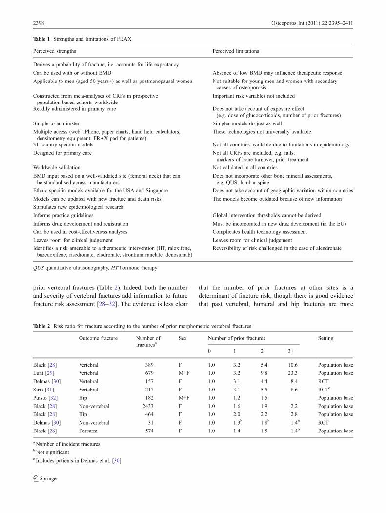
prior vertebral fractures (Table 2). Indeed, both the numberand severity of vertebral fractures add information to futurefracture risk assessment [28–32]. The evidence is less clear
that the number of prior fractures at other sites is adeterminant of fracture risk, though there is good evidencethat past vertebral, humeral and hip fractures are more
Table 1 Strengths and limitations of FRAX
Perceived strengths Perceived limitations
Derives a probability of fracture, i.e. accounts for life expectancy
Can be used with or without BMD Absence of low BMD may influence therapeutic response
Applicable to men (aged 50 years+) as well as postmenopausal women Not suitable for young men and women with secondarycauses of osteoporosis
Constructed from meta-analyses of CRFs in prospectivepopulation-based cohorts worldwide
Important risk variables not included
Readily administered in primary care Does not take account of exposure effect(e.g. dose of glucocorticoids, number of prior fractures)
Simple to administer Simpler models do just as well
Multiple access (web, iPhone, paper charts, hand held calculators,densitometry equipment, FRAX pad for patients)
These technologies not universally available
31 country-specific models Not all countries available due to limitations in epidemiology
Designed for primary care Not all CRFs are included, e.g. falls,markers of bone turnover, prior treatment
Worldwide validation Not validated in all countries
BMD input based on a well-validated site (femoral neck) that canbe standardised across manufacturers
Does not incorporate other bone mineral assessments,e.g. QUS, lumbar spine
Ethnic-specific models available for the USA and Singapore Does not take account of geographic variation within countries
Models can be updated with new fracture and death risks The models become outdated because of new information
Stimulates new epidemiological research
Informs practice guidelines Global intervention thresholds cannot be derived
Informs drug development and registration Must be incorporated in new drug development (in the EU)
Can be used in cost-effectiveness analyses Complicates health technology assessment
Leaves room for clinical judgement Leaves room for clinical judgement
Identifies a risk amenable to a therapeutic intervention (HT, raloxifene,bazedoxifene, risedronate, clodronate, strontium ranelate, denosumab)
Reversibility of risk challenged in the case of alendronate
QUS quantitative ultrasonography, HT hormone therapy
Table 2 Risk ratio for fracture according to the number of prior morphometric vertebral fractures
Outcome fracture Number offracturesa
Sex Number of prior fractures Setting
0 1 2 3+
Black [28] Vertebral 389 F 1.0 3.2 5.4 10.6 Population base
Lunt [29] Vertebral 679 M+F 1.0 3.2 9.8 23.3 Population base
Delmas [30] Vertebral 157 F 1.0 3.1 4.4 8.4 RCT
Siris [31] Vertebral 217 F 1.0 3.1 5.5 8.6 RCTc
Puisto [32] Hip 182 M+F 1.0 1.2 1.5 Population base
Black [28] Non-vertebral 2433 F 1.0 1.6 1.9 2.2 Population base
Black [28] Hip 464 F 1.0 2.0 2.2 2.8 Population base
Delmas [30] Non-vertebral 31 F 1.0 1.3b 1.8b 1.4b RCT
Black [28] Forearm 574 F 1.0 1.4 1.5 1.4b Population base
a Number of incident fracturesb Not significantc Includes patients in Delmas et al. [30]
2398 Osteoporos Int (2011) 22:2395–2411

predictive of future fractures than are fractures at other sites[33].
For the reasons noted above, there are insufficient dataavailable for the adjustment of the current FRAX models toinclude information on the number, site and severity of pastfractures. With regard to the number of prior fractures, itshould also be recognised that the cohorts used to buildFRAX included individuals with a prior fracture irrespectiveof their number (which was inconsistently documented).Thus, the probability assessment is based on the riskassociated with a single, two and more than two previousfractures. Since the ratios of men and women with thesecharacteristics are not known, this further complicates anyadjustment of FRAX. In the absence of quantitative informa-tion, the clinician should recognise that fracture probabilitiesshould be upward revised in patients with a history of multipleprior fractures (i.e. more than average) and greater weightaccorded to a prior vertebral, hip or humeral fracture than tofractures at other sites.
Long-term use of oral glucocorticoids
Ever use of systemic glucocorticoids is a dichotomous riskfactor (yes/no) in FRAX and does not therefore takeaccount of the dose of glucocorticoids. Neither does it takeaccount of the duration of its use, except that exposures ofless than 3 months should not be taken into account [34].For longer-term use, FRAX assumes an average risk,providing hazard ratios for an average dose and durationof exposure to glucocorticoids [35]. The most extensiveassessment of dose–response effects of glucocorticoids onfracture risk is found in the studies of van Staa et al [36–38]which examined the general practitioner records of the UKusing the General Practice Research Database (GPRD). Asmight be expected, higher than medium daily doses of oralglucocorticoids (2.5–7.5 mg prednisolone or equivalent)were associated with higher risks of fracture and vice versa(Table 3).
Kanis et al. have recently explored the possible impact ofdifferent doses of glucocorticoids on fracture probabilityusing the UK FRAX model and the fracture risks reportedin GPRD [39]. Relatively simple arithmetic procedureswere formulated which can be applied to conventionalFRAX estimates of probabilities of hip fracture and a major
osteoporotic fracture to modulate the probability assessmentwith knowledge of dose of glucocorticoids (Table 4).
As expected, probabilities were dose dependent withhigher probabilities associated with the high dose and viceversa. At medium doses (2.5–7.5 mg daily or equivalent),the unadjusted FRAX value can be used. For low-doseexposure (<2.5 mg daily of prednisolone or equivalent), theprobability of a major fracture was decreased by about 20%depending on age. For high doses (>7.5 mg daily),probabilities can be upward revised by about 15%.Conversion factors were also determined for the adjustmentof hip fracture probability. It is important to note that theadjustments to FRAX in Table 4 were derived from theUK version of FRAX and dose–responses (also from theUK) in an independent study without the possibility ofexamining the impact of different doses on all other FRAXrisk factors. This and a large number of other assumptionswere made so that caution should be exercised on the useof the adjustment factors until they are independentlyvalidated.
This caveat aside, it is important to note that theincrement in fracture probability was less than would bepredicted only from the hazard ratio of fracture. Forexample, the hazard ratio for hip fracture between themedium and high dose (hazard ratio=1.28) would predict a28% higher hip fracture probability with the high dose,whereas the calculated increment was 18% (rounded to15% in Table 4). Conversely, fracture probabilities werereduced with the low dose compared to the medium dosebut by less than that expected from the hazard ratio forfracture. The reason is that glucocorticoid exposureincreases the risk of death as well as for fracture and bothcompete, therefore in the calculation of fracture probability.
FRAX does not take account of the duration of exposureto glucocorticoids. There is evidence that higher cumulativedoses impart a higher fracture risk [36, 37]. Intermittent useof glucocorticoids will also increase fracture risk withhigher doses and more frequent use leading to greater risk[34]. Because of the variability in dose and schedule ofdosing, quantification of this risk is not possible. Low dosesof inhaled glucocorticoids, most commonly given forchronic obstructive pulmonary disease (COPD) or asthma,are not associated with an increase in fracture risk [34]. Bycontrast, a meta-analysis suggests that inhaled doses of
Fracture outcome Low dose (n=50,649) Medium dose (n=104,833) High dose (n=87,949)
Non-vertebral 1.17 (1.10–1.25) 1.36 (1.28–1.43) 1.64 (1.54–1.76)
Forearm 1.10 (0.96–1.25) 1.04 (0.93–1.17) 1.19 (1.02–1.39)
Hip 0.99 (0.82–1.20) 1.77 (1.55–2.02) 2.27 (1.94–2.66)
Vertebral 1.55 (1.20–2.01) 2.59 (2.16–3.10) 5.18 (4.25–6.31)
Table 3 Adjusted relative risk(95% CI) of fracture comparedto controls according to doseof oral glucocorticoids [36](with permission)
Osteoporos Int (2011) 22:2395–2411 2399

beclomethasone above 800 μg/day are associated with asmall increase in fracture risk [40]. The relative risks forany fracture and hip fractures were 1.30 (95% confidenceinterval (CI)=1.07–1.58) and 1.32 (95% CI=0.90–1.92),respectively. It should be noted that COPD itself is a riskfactor for fracture, so that the association may not be causal[41]
Consumption of alcohol
The input to FRAX asks for a positive entry if the patienttakes three or more units of alcohol daily. A unit of alcoholvaries slightly in different countries from 8 to 10 g ofalcohol. This is equivalent to a standard glass of beer(285 ml), a single measure of spirits (30 ml), a medium-sized glass of wine (120 ml) or one measure of an aperitif(60 ml). Intake of alcohol appears to have a dose-dependenteffect, i.e. the higher the exposure, the greater the risk. Thisis not taken into account and the computations in FRAXassume average exposure in those above or below the limit.Clinical judgement should be used for low or high
exposures, and some guidance is given in Table 5, whichis drawn from the meta-analysis used for the population ofthe FRAX models [42]. Risk ratios were similar in men andwomen, though the prevalence of high intakes of alcohol ishigher in men than in women.
A similar conclusion was reached in another independentmeta-analysis published by Berg and colleagues [43]. Amore recent meta-analysis was undertaken for the FRAXinitiative [44], which showed no significant effect ofalcohol exposure below an intake of 6 U/day. Unfortunately,the analysis was not based on individual data and wasdominated by a large study of 2.3 million women in whomgeneral practitioner records were used of unknown butdubious validity [6].
Parental history of fracture
FRAX enquires for a history of hip fracture in the patient’smother or father which is entered as a yes/no response. Itmight be expected that a family history of fracture at othersites would be a risk factor and there is some evidence tosupport this view [33, 45–47]. The effect, however, is smalland in the FRAX cohorts, for example, a family history ofany osteoporotic fracture amongst first degree relatives wasassociated with a small but significant increase in fracture riskin women (relative risk (RR)=1.21; 95% CI=1.09–1.35). Theassociation was stronger when hip fracture was the outcomemeasured (RR=1.40; 95% CI=1.09–1.80).
It has been suggested that a family history in any firstdegree relative would be a better variable to use inFRAX than a parental history. This would increase theprevalence of the risk factor. A sibling history of anyosteoporotic fracture was associated with a small increasein fracture risk in women (RR=1.13; 95% CI=0.94–1.36) butin neither case was the association significant [47]. FRAXelected to use a parental history of hip fracture because of thestrong and consistent association and to exclude a siblinghistory, since the probability of a sibling varies markedlyworldwide.
Dose Prednisoloneequivalent (mg/day)
Age (years) All ages
40 50 60 70 80 90
Hip fracture
Low <2.5 −40 −40 −40 −40 −30 −30 −35Mediuma 2.5–7.5
High ≥7.5 +25 +25 +25 +20 +10 +10 +20
Major osteoporotic fracture
Low <2.5 −20 −20 −15 −20 −20 −20 −20Mediuma 2.5–7.5
High ≥7.5 +20 +20 +15 +15 +10 +10 +15
Table 4 Percentage adjustmentof 10-year probabilities of a hipfracture or a major osteoporoticfracture by age according todose of glucocorticoids [39](with kind permission fromSpringer Science+BusinessMedia B.V.)
a No adjustment
Table 5 Hazard ratio and 95% CI according to intake of alcohol inmen and women combined with and without BMD [42] (with kindpermission from Springer Science+Business Media B.V.)
Category (units/day) Without BMD With BMD
RR 95% CI RR 95% CI
Osteoporotic fracture
3 or more 1.38 1.16–1.65 1.36 1.13–1.63
4 or more 1.55 1.26–1.92 1.53 1.23–1.91
5 or more 1.70 1.30–2.22 1.64 1.24–2.17
Hip fracture
3 or more 1.68 1.19–2.36 1.70 1.20–2.42
4 or more 1.92 1.28–2.88 2.05 1.35–3.11
5 or more 2.26 1.35–3.79 2.39 1.39–4.09
95% CI 95% confidence intervals
2400 Osteoporos Int (2011) 22:2395–2411

On this basis, it is difficult to give firm advice on thefurther interpretation of FRAX probability estimates outsidethe limits of the FRAX questionnaire that is supported byan evidence base. In the future, it will be important toassess the independent contribution of genetic risk markersto fracture probability in large epidemiologic cohorts.
Current tobacco smoking
Tobacco smoking is entered into FRAX as yes or nodepending on whether the patient currently smokes tobacco.The fracture risks are derived from meta-analyses of theWHO cohorts [48] and are consistent with other largesurveys [49, 50]. There is some evidence for dose–responseeffects on fracture risk [51]. First, the risk is higher in menthan in women, possibly related to the higher exposure inmen [48]. Second, the risk of hip fracture rises progres-sively with age [48, 49], possibly related to the duration ofexposure. Third, the risk is lower in ex-smokers comparedwith current smokers [48, 51]. These observations areconsistent with a dose–response relationship but do notprovide information that helps in the interpretation ofFRAX probabilities. In addition, smoking increases themortality risk which may offset in part or in whole theincrease in fracture risk.
Rheumatoid arthritis
Although input for rheumatoid arthritis in the FRAXalgorithm is a dichotomous variable, intuitively, one wouldexpect that more severe or active disease would be associatedwith more severe osteoporosis. Associations have beenreported between functional disability and clinical fracturerisk in patients with rheumatoid arthritis [52], butevidence is limited that more severe or active disease isassociated with a greater risk for fracture. There is alsolittle evidence that interventions for rheumatoid arthritis(other than glucocorticoids) adversely affect fracture risk.Indeed, anti-TNF therapies may have beneficial effects onbone mineral density [53] though the impact on fracture riskis unknown.
The risk associated with rheumatoid arthritis may beunderestimated by FRAX due to reporting bias. The preva-lence of rheumatoid arthritis in the FRAX cohorts isapproximately 4% [35], whereas the true prevalence in thegeneral population may be closer to 1–2%. Thus, theapparent risk may be diluted somewhat by patients reportingosteoarthritis as rheumatoid arthritis. Osteoarthritis is, ifanything, protective. The underestimate may be partly offsetby improved therapy of rheumatoid arthritis, though thisremains to be quantified. Nonetheless, reliance should not beplaced on a patient’s report of ‘arthritis’ unless there isclinical or laboratory evidence to support the diagnosis.
Risk factors not considered in FRAX
Many clinicians have a wish list of risk factors not consideredin FRAX. These include enlargement of the number ofsecondary causes of osteoporosis, the inclusion of falls risk,markers of bone turnover and bone mineral measurements atother sites (e.g. lumbar spine) and with technologies otherthan DXA (e.g. quantitative ultrasonography, QUS).
Other causes of secondary osteoporosis
Many clinicians have suggested secondary causes of osteo-porosis that should be considered in FRAX. For example, ithas been noted that there are more than 80 causes of secondaryosteoporosis given in the US Surgeon General’s report onosteoporosis [54] of which a minority are described in FRAX[55]. Of the many secondary causes of osteoporosis, those inTable 6 have been consistently documented to be associatedwith a significant increase in fracture risk. With the exceptionof chronic obstructive pulmonary disease [56, 57], theseremain as listed in the Technical Report on FRAX [3]. Ofthese, their effect on fracture risk can usually be explained bythe effect of the disease on decreasing bone density.Rheumatoid arthritis is the only secondary cause ofosteoporosis that is considered independent of BMD in theFRAX algorithm [35]. Whereas this may hold true for someother secondary causes of osteoporosis, the evidence base isweak.
Falls
A criticism of the FRAX model by some users has been thelack of consideration of falls or falls risk in predictingfractures. A remit of the FRAX Clinical Task Force was toreview the evidence and consider if falls should be incorpo-rated into the FRAX model or alternatively provide guidanceto clinicians on how a history of falls should be used inconjunction with FRAX in clinical decision making.
The Task Force strongly recommended that falls should beincorporated into FRAX [58]. Whereas this view is a soundacademic conclusion from the literature on falls risk, theincorporation into FRAX is problematic for several reasons.First, existing data are not of adequate quality to incorporatequantitative adjustment to FRAX at the present time.Information on falls was available in a minority of cohortsused to derive or validate FRAX. In addition, the constructof questions on falls was very heterogeneous, and perhapsfor this reason, meta-analysis showed no significant increasein fracture risk (E McCloskey, JA Kanis, H Johansson,unpublished data provided to the Task Force, 2010).
Second, falls risk is inherently taken into account in thealgorithm, though not as an input variable. Thus, thefracture probability given for any combination of risk
Osteoporos Int (2011) 22:2395–2411 2401

factors assumes that the falls risk is that observed (but notdocumented) in the cohorts used to construct FRAX. In thisregard, it is of interest that a small study in a hospitalsetting showed that FRAX identified fallers who fracturewith a higher predictive value than the STRATIFY (St.Thomas risk assessment tool in falling elderly) [59] instru-ment designed to predict falls [60]. In order to incorporatethe falls risk, the fracture risk must first be quantified in theFRAX cohorts for individuals who do not fall frequently.These data do not exist in sufficient cohorts. Third, theinterrelationship of falls risk with the other FRAX variableshas been inadequately explored on an international basis.Fourth, the relationship between the risk variable andmortality needs to be accounted for, but there are no dataavailable.
These technical problems aside, FRAX is intended toidentify a risk that is amenable to a therapeutic intervention.In a single study, a post hoc analysis of a community-basedintervention study with clodronate in elderly women [61]showed that fracture reduction was similar in women withor without recent multiple falls or in those with impairedability in rising from a chair [62]. This finding suggests thatfalls or falls risk may identify a risk amenable tointervention. In contrast, in the phase III trial of risedronate,where hip fracture was the primary end point, hip fracture
risk was not significantly decreased in patients over the ageof 80 years, the majority of whom were purportedlyselected on the basis of falls risk [63]. Thus, more dataare required before falls can be safely incorporated intoassessment algorithms.
Unfortunately, falls risk is not routinely incorporated intophase III studies of osteoporosis treatment. In contrast, therisk factors used in the current version of FRAX have allundergone a thorough investigation in the majority of phaseIII studies to determine that they identify a risk amenable totherapeutic intervention—‘reversible risk’ or more accu-rately—reversibility of risk [11, 12].
It has been argued that falls can be prevented and thatthis provides a further reason for the inclusion of falls riskin FRAX [58]. However, the relevant question is whetherintervention for falls decreases the risk of fracture. Whereassome studies report that falls may be prevented by multi-dimensional interventions, the evidence that these reducethe risk of fracture is plausible but not proven in meta-analysis [64–66], with the possible exception of exerciseinterventions. There is also evidence that vitamin D maydecrease the risk of fracture by preventing falls [67], butthis is uncertain [64]. The case for incorporating falls intorisk algorithms would be strengthened by knowledge thatthe treatment of falls risk can reduce fracture risk. In the
Secondary cause Example
Glucocorticoids Any dose, by mouth for three months or more
High doses of inhaled glucocorticoids
Cushing’s disease
Rheumatoid arthritis
Chronic liver disease Alcoholism
Untreated hypogonadism in men and women Bilateral oophorectomy or orchidectomy
Anorexia nervosa
Chemotherapy for breast cancer
Tamoxifen in premenopausal women
Aromatase inhibitors
GnRH inhibitors for prostate cancer
Hypopituitarism
Prolonged immobility Spinal cord injury
Parkinson’s disease
Stroke
Muscular dystrophy
Ankylosing spondylitis
Organ transplantation
Type I diabetes
Thyroid disorders Untreated hyperthyroidism
Over-treated hypothyroidism
Gastrointestinal disease Crohn’s disease
Ulcerative colitis
Chronic obstructive pulmonary disease
Table 6 Secondary causes ofosteoporosis associated with anincrease in fracture risk (adaptedfrom [3]; with permission fromthe WHO Collaborating Centrefor Metabolic Bone Diseases,University of Sheffield MedicalSchool)
2402 Osteoporos Int (2011) 22:2395–2411

absence of this information, risk assessment algorithms thatincorporate falls fail in their primary intent [6, 7].
Thus, it is not possible to provide good clinical advice toclinicians on how a history of falls should be used inconjunction with FRAX in their clinical decision making,despite recommendations to the contrary [58]. The onlysound advice is that individuals who fall more frequentlythan average are likely to have a higher fracture probabilitythan that provided by FRAX. The obverse is also true inthat individuals who fall less frequently than average arelikely to have a lower fracture probability than thatprovided by FRAX.
Biochemical markers of bone turnover
Bone turnover markers (BTMs) reflect the metabolic activityof bone. They are traditionally categorised as markers of boneformation or bone resorption. Oestrogen deficiency, associatedwith menopause, results in a generalised increase in boneremodelling and an imbalance between bone formation andresorption that is maintained for several decades after themenopause and is associated with accelerated bone loss andfracture risk [68]. Thus, it is logical to consider that high boneturnover might predict fracture and a large number of studiessupport this view [69–72]. The evidence is less convincing inmen. Some, but not all, studies have shown that in womenwith a low BMD, the presence of increased BTMs has aneffect on fracture risk that is independent of BMD. This hasled to calls for the incorporation of these markers into FRAX.
There are a number of limitations to the incorporation ofmarkers into risk prediction models. They include theirbiological variability and, in some cases, the multiplemethodologies used for the same analyte (e.g. the assays forosteocalcin). It is not surprising, therefore, that associationsbetween markers and fracture outcomes have been heteroge-neous [68]. Moreover, there are few studies that haveexamined the interactions between the BTMs and otherFRAX variables and none that takes an internationalperspective.
For these reasons, it is not yet possible to provideinformation that helps in the interpretation of FRAXprobabilities. In the future, it is expected that the adoptionof reference analytes and analytical standards will allowstudies to be pooled more easily and determine their role infracture risk assessment [68].
Assessment of BMD at the lumbar spine and elsewhere
Lumbar spine BMD is frequently measured by DXA andindeed is incorporated into several clinical guidelinesincluding those of ISCD [13, 23, 73, 74]. It is the sitefavoured for monitoring treatment, and there is thus muchinterest in the incorporation into FRAX of measurements at
the lumbar spine. The same is true for peripheral measure-ments (and QUS) where there are no facilities for centralDXA.
The femoral neck is the only skeletal region of interestcurrently validated for use with FRAX [3]. The T-scorederived from measurement of BMD at the femoral neck byDXA is also the WHO international reference standard forthe diagnosis of osteoporosis [2, 75]. It has the advantagethat for any given age and BMD, the fracture risk isapproximately the same in men and women [76]. Becauseof this, the T-score used in FRAX is derived from a singlereference standard (the NHANES III database for femaleCaucasians aged 20–29 years) [77].
The principal reason for the use of femoral neck BMD inFRAX was the wide availability of measurements at thissite. For example, in the meta-analysis of the source cohortsused to construct FRAX, femoral neck BMD was availablein about 40,000 individuals whereas information wasavailable in approximately half this number for BMD atthe lumbar spine [76] and half again for BMD at peripheralsites. Moreover, femoral neck BMD is associated with ahigher gradient of risk (increase in fracture risk/unitdecrease in BMD) for hip fracture than BMD measure-ments at the lumbar spine [78]. The same probably holdstrue for the prediction of major fractures when appropriateadjustment is made to the units of BMD [79]. Notwith-standing, measurements of BMD at sites other than thefemoral neck provide significant information on fracturerisk [76, 78, 79].
In the absence of specific models that incorporate BMDat the lumbar spine, the question arises whether lumbarspine BMD or the T-score might be used in FRAX whereinformation is lacking on femoral neck BMD. The shortanswer is no. A major impediment is that the age-relateddecrease in T-score differs at different skeletal sites [80] andthat the gradient of risk (the increase in fracture risk/unitdecrease in BMD) for specific fracture outcomes is knownto differ by site [78]. Additional difficulties include the age-related degenerative changes at the lumbar spine, theuncertain interactions of spine measurements with the otherFRAX variables and the lack of an international referencestandard for lumbar spine BMD [81].
Although the measurement of two skeletal sites does notimprove the general performance characteristics (sensitivity/specificity) of the BMD test in a given population [82–84],there are situations where there is a large discordance in theT-score at different skeletal sites in individuals for whom theuse of this information will enhance the accuracy for thecharacterisation of risk, particularly if they lie close to anintervention threshold. An example is provided from theCanadian guidelines that recommend treatment in men andwomen with 10-year probabilities for a major fracture thatexceed 20% [23]. A 70-year-old woman with a maternal
Osteoporos Int (2011) 22:2395–2411 2403

history of hip fracture, a BMI of 22 kg/m2 and a T-score of−2.2 SD at the femoral neck has a fracture probability of19% when calculated with FRAX (Canadian model, version3.1). With a T-score of −3.5 SD at the lumbar spine, it isexpected that her true risk would be higher and likely lieabove the treatment threshold of 20%. The impact of spine/femoral neck T-score discordance has recently been exploredin a large BMD-referral population from Manitoba, Canada.Fracture outcomes were available over a 10-year time frame.There was an approximately 10% change in fracture risk foreach unit of T-score discordance [85].
On this basis, the authors propose that the clinician may‘Increase/decrease FRAX estimate for a major fracture byone-tenth for each rounded T-score difference between thelumbar spine and femoral neck’. An example is provided inthe case above in which the T-score for femoral neck BMDwas −2.2 SD with a FRAX-calculated fracture probabilityof 19%. In this case, the T-score discordance was 1.3 SD(3.5–2.2). If the figure is rounded off (to 1.0 SD), theestimated probability with the inclusion of lumbar BMD isupward revised by 10% (19+1.9) to 21%.
The rule is intended to provide some guidance forphysicians, particularly those that report on the output ofDXA to primary care. Approximately 16% of individuals willhave a T-score at the lumbar spine that is lower by 1 SD ormore than that at the femoral neck. Conversely, approximately16% of individuals will have a T-score at the lumbar spine thatis higher by 1 SD than that at the femoral neck. There are,however, several caveats to consider. Firstly, this approachrequires external validation in independent cohorts and withother DXA technologies. Secondly, its application should benot only to revise probabilities upwards. The obverse may alsoapply and probabilities revised downwards in patients withhigher T-scores at the lumbar spine than at the femoralneck. Thirdly, the adjustment is only relevant for those closeto an intervention threshold.
The incorporation of quantitative ultrasonography
QUS is an attractive method for assessing fracture riskbecause it is portable, inexpensive, without ionizingradiation and available in areas of the world where DXAis not readily accessible or affordable. Validated techniquesfor QUS at the heel have been shown to predict hip fractureand other fractures as well as central DXA [81, 86]. As inthe case of lumbar spine BMD, and for the same reasons,ultrasound measurements should not be entered in FRAXmodels in place of femoral neck BMD.
One study has shown that the consideration of clinicalrisk factors can enhance fracture risk prediction with QUS[87]. Age, BMI, history of fracture, results of the chair test,a history of a fall over the last 12 months, current cigarette
smoking and diabetes mellitus were independent covariates.The value of this approach requires further developmentbefore clinical application but illustrates a proof of principlethat the addition of clinical risk factors may improve theperformance characteristics of QUS alone.
Other limitations of FRAX
The Task Force examined several other limitations ofFRAX (see Table 1), some of which are reviewed below.
Concurrent treatment
FRAX is intended to identify patients for treatment. Thus,FRAX is unnecessary in patients for whom treatment isclearly indicated, e.g. an elderly patient with multiple fragilityfractures [81]. In those receiving treatment for osteoporosis,FRAX is likely to overestimate fracture probability sincetreatment effects are not accommodated in the model. Theempirical data suggest that FRAX remains a good predictivetool in women currently or previously on treatments forosteoporosis [88], possibly related to the contribution oftreatment-induced changes in BMD.
Within country variation in fracture rates
In addition to large variations in fracture rates around theworld, fracture rates may vary within countries. In additionto ethnic-specific differences [3, 89, 90], up to two-folddifferences in hip fracture incidence have been reportedusing common methodology with the higher rates in urbancommunities in Argentina [91], Turkey [92], Sweden [93],Norway [94–96] Switzerland [97], Croatia [98] and in theUSA [99, 100]. Where possible, FRAX models are builtusing national rather than regional data, the formergenerally being of higher quality and based on largepopulation-based sample sizes. It is not feasible to buildregional models and probably not desirable, but cliniciansshould be aware of this variation.
An additional feature of multi-ethnic populations is thatfracture risk and mortality may vary widely across ethnicgroups. Where sufficient information is available, ethnic-specific FRAX models have been built (for the USA andfor Singapore). In most other societies, there are insufficientdata on fracture rates and mortality in ethnic minorities,though differences in fracture risk may be less than thatsuggested from the ethnicity of origin. For example, blacksin the US have lower fracture probabilities than Caucasians,but the probability of fracture in US blacks is much higherthan in African blacks [90] in part due to the higher fracturerates and lower mortality risks in those from the USA. A
2404 Osteoporos Int (2011) 22:2395–2411

similar ‘acclimatisation’ is seen in the Japanese populationof Hawaii [101].
In view of the variations in hip fracture risks betweenurban and rural communities, it is relevant to questionwhether ethnic-specific models are of real utility where thedifferences in fracture rates are modest. In addition, fracturerates may vary within ethnic groups. A case in point is theUS Hispanic population which is very diverse in origin andethnic background with both ‘black’ and ‘white’ Hispanics.As a result, the risk of fracture may differ substantially forHispanics in different areas of the country [102]. Regionssuch as New York with more black Hispanics from PuertoRico have lower hip fracture risk than Florida which hasmany Cuban Americans. Hispanics in the Southwest havehip fracture risks that are in between. At the end of the day,it may be more profitable to recognise these differences totemper interpretation of FRAX results rather than to strivefor a spurious sense of accuracy.
Secular trends
In addition to the large geographic variation reported in theincidence of hip fracture throughout the world, the age- andsex-specific incidence of fracture is changing. This has beenwell characterised for hip fracture, but also noted at othersites of fracture [103, 104]. Estimates of incidence trendshave varied widely and variously reported an increase,plateau and decrease, in age-adjusted incidence rates for hipfracture among both men and women. Studies in westernpopulations, whether in North America, Europe or Oceania,have generally reported increases in hip fracture incidencethrough the second half of the last century, but those studiescontinuing to follow trends over the last two decades havefound that rates stabilise, with age-adjusted decreases beingobserved in certain centres. In contrast, the mortality hazardhas continued to decrease in most regions of the world.
These shifts in fracture and death hazards have implica-tions for FRAX. In the USA, the FRAX model was revisedin 2009 to take account of changing death and hip fracturerates and improvements in the descriptive epidemiology ofother outcome fractures. Recent data from the USA suggestthat, while hip fracture rates are declining among Cauca-sians, there has been an increase in the age- and sex-specific hip fracture risk in Hispanic Americans fromCalifornia [105], possibly related to social admixture. Ifconfirmed, then the Hispanic model may need revision orthe Caucasian model used instead [90]. In Turkey, a countrywith low fracture probabilities, the FRAX model is basedon hip fracture rates acquired more than 20 years ago.Studies in progress, stimulated by the availability of FRAXfor Turkey, will address whether hip fracture incidence haschanged in the interim. Thus, FRAX models may need to
be updated from time to time to take account of changingepidemiology.
Probability of clinical spine, humeral and forearm fracturefractures
A minority of countries that have a FRAX model also haverobust information on the risk of the other major fractures(clinical spine, forearm and humerus). Where available,these are incorporated in the models (e.g. UK, USA,Switzerland, Sweden, Japan, Mexico). In the absence ofinformation, FRAX models are based on the assumptionthat the age- and sex- specific pattern of these fractures issimilar to that observed in Sweden.
Despite a large number of studies that have examined theincidence of fractures by age and sex, there are problems indefining the pattern of fractures in different countries [106].For example, there are differences in the population studied.Some studies have been from random samples of thegeneral population, from self-selected populations, fromaccident departments, radiology departments, fracture clinicsor inpatient records. Clinical vertebral fractures are variouslydefined, even within a single study [107]. These differentsampling frames give rise to large differences in the patternof fractures reported. Moreover, several surveys do not studyor report all fracture types relevant to the outcomes ofFRAX, have small samples, an age range not relevant toosteoporosis or do not include men. A further problem isthat the incidence and therefore the apparent pattern offracture may change with time-discordant samples, sothat historical data may not be relevant. The most completeinformation comes from Sweden, UK, Canada, Australia andthe USA.
The available information suggests that the pattern offractures is similar in the Western world and Australia,despite differences in incidence [106]. In the USA, Swedenand the UK the incidence of forearm, proximal humeral andhip fracture varies. For example, in women aged 80–84 years, the rates of these fractures are 3,206, 5,157 and2,558/100.000 in the USA, Sweden and UK, respectively[108–110], but the pattern of these fractures with age isremarkably similar (Fig. 2). The relationship between theincidences of hip, clinical vertebral and forearm fracture isalso similar between Sweden and Australia [111]. Withinthe USA, the pattern appears to be similar amongst blacksand whites. For example, amongst white women aged 65–79 years, the ratio of frequency of hip, distal forearm andproximal humerus is 43%, 38% and 19%, respectively. Forblack women, the ratio is 45%, 36% and 18% [112].
This commonality of pattern is supported by registerstudies, which indicate that in those regions where hipfracture rates are high, so too is the risk of forearm fracture
Osteoporos Int (2011) 22:2395–2411 2405

and vertebral fractures (requiring hospital admission)(Fig. 3) [113, 114].
Since the pattern of osteoporotic fractures appears to bebroadly similar in the Western world, this suggests that theimputed rates clinical (but not morphometric) vertebral,forearm and humeral fractures used in FRAX are unlikelyto be grossly over- or underestimated. The pattern offractures elsewhere is, however, less secure [90].
Surrogate models
Fracture probability varies markedly in different regions ofthe world [10] (Fig. 4). Thus, the FRAX models need to becalibrated to those countries where the epidemiology of
fracture and death is known. At present, FRAX models areavailable for 31 countries. Other models are beingdeveloped, where sufficient data are available.
Thus, in the absence of a FRAX model for a particularcountry, a surrogate country should be chosen, preferablybased on the likelihood that it is representative of the indexcountry. Where limited data of uncertain quality areavailable, it may be appropriate to choose a surrogate thatbest approximates the fracture risk of the index country.Mortality data are available for nearly all countries, so thatthe Task Force recommend that the FRAX model shouldincorporate the death hazard of the index country [90]. Acase in point at present is Poland which has adopted the UKas its surrogate country. The entire UK FRAX model wasused whereas it may have been more appropriate to
0
50
100
150
0 500 1000 1500Hip (rate/100,000)
0 100 200 300 400 500 0
200
400
600
800
Distal forearm (rate/100,000)Spine (rate/100,000)
R2 = 0.58 R2 = 0.80
Fig. 3 Discharge rates for ver-tebral fractures and hip fracturesin Europe (left) and dischargerates for forearm fractures andhip fractures in Westerncountries (right). Data from[113] and [114]
0
20
40
60
80
100
0
20
40
60
80
100
Age (years)
%
Hip Distal forearm Proximal humerus
Men
Women
United States of AmericaSweden United Kingdom
55- 65- 85+ 50- 60- 70- 80- 75-
%
55- 65- 85+ 50- 60- 70- 80- 75- 55- 65- 85+ 50- 60- 70- 80- 75-
Fig. 2 Pattern of common oste-oporotic fractures expressed as aproportion (%) of the total in theUS, Sweden and the UK [106](with kind permission fromSpringer Science+BusinessMedia B.V.)
2406 Osteoporos Int (2011) 22:2395–2411

accommodate the death hazard for Poland if the recom-mendation was followed. New data on fracture rates inPoland will, however, obviate the need for adjustment.
In the absence of any country-specific data, it has beensuggested that a regional model might be constructed, butthe heterogeneity of fracture risks in different regions of theworld makes this approach less secure (see Fig. 4). On theother hand, accuracy errors have little impact on the rankorder with which the FRAX tool categorises risk in a givenpopulation [115], but they do change the absolute numbergenerated and thus have implications where treatmentguidelines are based on cost-effectiveness or the economicburden of disease.
For these reasons, the FRAX tool should not beconsidered by physicians as a gold standard, but rather asa platform technology on which to build as new validatedrisk indicators become available. Notwithstanding, thepresent model provides an aid to enhance patient assess-ment by the integration of clinical risk factors alone and/orin combination with BMD.
Conclusions
FRAX represents a significant advance in the assessment ofboth women and men at risk for osteoporosis-relatedfracture and allows the tailoring of pharmacologicalinterventions to high-risk subjects. While FRAX does notdefine intervention thresholds, which depend on country-specific considerations, it provides a platform to assessfracture probability which is needed to make rationaltreatment decisions by clinicians and public health agen-cies. The tool is, however, far from perfect, but better than
BMD alone. The widespread use and interest in FRAX andits adoption into management guidelines has fuelled interestas to how models can be improved, extended to othercountries and, in particular, how the limitations of FRAXshould temper clinical judgement.
The wish list of clinicians for the modulation of FRAXis large, and in many instances, these wishes cannotpresently be fulfilled, but an explanation and understandingof the reasons may be helpful in translating the informationprovided by FRAX into clinical practice.
Acknowledgements This paper was reviewed and endorsed by theCommittee of Scientific Advisors of the IOF. The FRAX Initiativecomprised the organisers, task force liaisons, the members of the threeTask Forces and the expert panel.
Organisers Didier Hans (Co-chair), Cyrus Cooper (co-chair),Sanford Baim, Bess Dawson-Hughes, John A Kanis, William DLeslie, Marjory Lucky, Rene Rizzoli, Catalina Poiana, John PBilezikian (moderator) and Socrates E Papapoulos (moderator).
FRAX Clinical Eugene McCloskey (Chair); Neil Binkley (Co-chair); Jonathan Adachi and Sanford Baim (programme committeeliaison); Dennis Black, Robert Blank, Susan Broy and Bess Dawson-Hughes (programme committee liaison) and Sergio Ortolani, HansPeter Dimai, Michael Kleerekoper, Marc-Antoine Krieg, BenteLangdahl, Andrew Laster, Edward Leib, Tahir Masud, Mike McClung,Rene Rizzoli, Kenneth Saag and Ethel Siris.
FRAX BMD E Michael Lewiecki (Chair); Juliet Compston (Co-chair); Jonathan Adachi Judith Adams, Robert Adler, Doug BauerGlen Blake, Patricia Clark, Adolfo Diez-Perez, Didier Hans and JohnA Kanis (programme committee liaison); Marc-Antoine Krieg, DidierHans and William Leslie (programme committee liaison) and AlirezaMoayyeri, Robert Josse, David Kendler, Aliya Khan, Brian Lentle,Roman Lorenc, Basel Masri and Paul Miller.
Female aged 65 years, prior fragility fracture, T-score -2.5 SD
10 year probability (%)
0
2
4
6
8
10
Denm
ark
Sw
eden
Taiw
an
Austria
Malta
Belgium
S'pore C
hinese
Sw
itzerland
S'pore Indian
S K
orea
Italy
UK
Argentina
US
Caucasian
Hungary
Hong K
ong
Germ
any
Finland
Canada
Mexico
Netherlands
France
New
Zealand
Japan
Jordan
Spain
Australia
Colom
bia
Lebanon
US
Black
China
Philippines
Turkey
Fig. 4 Ten-year probability ofhip fracture in women aged65 years with a prior fractureand a T-score of −2.5 SD at thefemoral neck in the differentcountries with FRAX models(FRAX version 3.2)
Osteoporos Int (2011) 22:2395–2411 2407

FRAX International Jane Cauley (Chair); Ghada El-Hajj Fuleihan(Co-chair); Asma Arabi, Andrew Calderon, Zhao Chen, Siok BeeChinoh, Jeffry Curtis, Michelle Danielson, Saeko Fujiwara, DavidHanley, Heikki Kroger, Annie Kung, Olga Lesnyak, Anne Looker andMarjorie Luckey (programme committee liaison); Dan Mellstrom, JeriNieves, Wojciech Pluskiewicz, Rola El Rassi and René Rizzoli(programme committee liaison) and Sergio Ragi-Eis, Anna MarieSchott-Pethelaz and Stuart Silverman.
Expert panel Socrates E Papapoulos (moderator), John P Bilezikian(moderator) and Jonathan Adachi, Robert D Blank, Roland Chapurlat,Wu (Paulo) Chih-Hsing, Edward Czerwinski, Aldolfo Diez-Perez,Hans Peter Dimai, Ghada El-Hajj Fuleihan, Saeko Fujiwara, RuxandraM Ionescu, John A Kanis, Mike McClung, Paul Miller, Sergio Ragi-Eis,Jan Stepan, Kenneth Saag, John T Schousboe, Wei Yu and CristianoZerbini.
Support staff Peter D. Brown (ISCD), Patrice McKenney (IOF) andHelena Johansson, Judit Nagy, Anders Oden and Denys AWahl.
Conflicts of interest S Baim, JP Bilezikian, JE Compston, G El-Hajj Fuleihan, A Oden, B Dawson-Hughes, EM Lewiecki, DAWahl,H Johansson and D Hans have no competing interests to declare withregard to this manuscript. JA Kanis and E McCloskey: Consultingfees, paid advisory boards, lecture fees, and/or grant support from themajority of companies concerned with skeletal metabolism. WDLeslie: Speaker fees and unrestricted research grants from Amgen andMerck Frosst; unrestricted research grants from Sanofi-Aventis,Warner Chilcott (formerly Procter & Gamble), Novartis, Amgen,Genzyme; advisory boards for Genzyme, Novartis, and Amgen. JCauley: Consultant to Novartis for the HORIZON steering committeeand receives research funding for the HORIZON trial. C Cooper:Consulting fees and paid advisory boards for Alliance for Better BoneHealth, Glaxo Smith Kline, Roche, Merck Sharp and Dohme, Lilly,Amgen, Wyeth, Novartis, Servier, and Nycomed. R Rizzoli: Paidadvisory boards and lecture fee for Merck Sharp and Dohme, Eli Lilly,Amgen, Wyeth, Novartis, Servier, Nycomed, and Danone. N Binkley:Consulting fees and research support from Amgen, Lilly, Merck andTarsa. M Luckey: Consulting fees and lecture fees from Merck Sharpand Dohme, Eli Lilly, Amgen, Novartis and Tarsa Pharmaceuticals. CPoiana: Lecture fees and paid advisory boards for Amgen, Merck Sharpand Dohme, Glaxo Smith Kline and Novartis. S Papapoulos: Consultingand/or lecture fees from Alliance for Better Bone Health, Amgen, EliLilly, Glaxo Smith Kline, Merck & Co, Novartis and Wyeth.
References
1. Hans D, Kanis JA, Baim S et al, on behalf of the FRAX PositionConference Members (2011) Joint Official Positions of theInternational Society for Clinical Densitometry and InternationalOsteoporosis Foundation on FRAX®. J Clin Densitom (in press)
2. World Health Organization (2007) Assessment of osteoporosis atthe primary health care level. WHO, Geneva. www.who.int/chp/topics/rheumatic/en/index.html
3. Kanis JA, on behalf of the World Health Organization ScientificGroup (2008) Assessment of osteoporosis at the primary health-care level. Technical report. WHO Collaborating Centre, Uni-versity of Sheffield, UK. Available at http://www.shef.ac.uk/FRAX/index.htm
4. Kanis JA, Johnell O, Oden A, Johansson H, McCloskey EV(2008) FRAX™ and the assessment of fracture probability inmen and women from the UK. Osteoporos Int 19:385–397
5. Kanis JA, Oden A, Johnell O et al (2007) The use of clinical riskfactors enhances the performance of BMD in the prediction ofhip and osteoporotic fractures in men and women. OsteoporosInt 18:1033–1046
6. Hippisley-Cox J, Copuland C (2009) Predicting risk of osteopo-rotic fracture in men and women in England and Wales:prospective derivation and validation of QFractureScores. BMJ339:b4229
7. Nguyen ND, Frost SA, Center JR, Eisman JA, Nguyen TV (2008)Development of prognostic nomograms for individualizing 5-yearand 10-year fracture risks. Osteoporos Int 19:1431–1444
8. De Laet C, Oden A, Johansson H, Johnell O, Jonsson B, KanisJA (2005) The impact of the use of multiple risk indicators forfracture on case-finding strategies: a mathematical approach.Osteoporos Int 16:313–318
9. Kanis JA, Johnell O, Oden A, De Laet C, Jonsson B, Dawson A(2002) Ten-year risk of osteoporotic fracture and the effect ofrisk factors on screening strategies. Bone 30:251–258
10. Kanis JA, Johnell O, De Laet C, Jonsson B, Oden A, Oglesby A(2002) International variations in hip fracture probabilities: impli-cations for risk assessment. J Bone Miner Res 17:1237–1244
11. Kanis JA, Oden A, Johansson H, Borgström F, Ström O,McCloskey E (2009) FRAX® and its applications to clinicalpractice. Bone 44:734–743
12. Kanis JA, McCloskey E, Jönsson B, Cooper A, Ström O,Borgström F (2010) An evaluation of the NICE guidance for theprevention of osteoporotic fragility fractures in postmenopausalwomen. Arch Osteoporos 5:19–48
13. National Osteoporosis Foundation (2008) Clinician’s guide toprevention and treatment of osteoporosis. National OsteoporosisFoundation, Washington, DC. www.nof.org
14. Compston J, Cooper A, Cooper C, on behalf of the NationalOsteoporosis Guideline Group (NOGG) et al (2009) Guidelinesfor the diagnosis and management of osteoporosis in postmen-opausal women and men from the age of 50 years in the UK.Maturitas 62:105–108
15. Kanis JA, McCloskey EV, Johansson H, Strom O, Borgstrom F,Oden A, National Osteoporosis Guideline Group (2008) Casefinding for the management of osteoporosis with FRAX®—assessment and intervention thresholds for the UK. OsteoporosInt 19:1395–1408, Erratum 2009 Osteoporos Int 20, 499–502
16. Neuprez A, Johansson H, Kanis JA et al (2009) Rationalisationdu remboursement des médicaments de l’ostéoporose: de lamesure isolée de la densité osseuse à l’intégration des facteurscliniques de risque fracturaire. Validation de l’algorithmeFRAX®. La Revue Médicale de Liège 64:612–619
17. Fujiwara S, Nakamura T, Orimo H, Hosoi T, Gorai I, Oden A etal (2008) Development and application of a Japanese model ofthe WHO fracture risk assessment tool (FRAX™). OsteoporosInt 19:429–448
18. Lippuner K, Johansson H, Kanis JA, Rizzoli R (2010) FRAX®assessment of osteoporotic fracture probability in Switzerland.Osteoporos Int 21:381–390
19. Association Suisse contre l‘Ostéoporose (2011) Ostéoporose:recommandations 2010. ASCO, Lausanne
20. Czerwinski E, Kanis JA, Trybulec B, Johansson H, Borowy P,Osieleniec J (2009) The incidence and risk of hip fracture inPoland. Osteoporos Int 20:1363–1368
21. Kanis JA, Burlet N, Cooper C, on behalf of the EuropeanSociety for Clinical and Economic Aspects of Osteoporosis andOsteoarthritis (ESCEO) et al (2008) European guidance for thediagnosis and management of osteoporosis in postmenopausalwomen. Osteoporos Int 19:399–428
2408 Osteoporos Int (2011) 22:2395–2411

22. Grossman JM, Gordon R, Ranganath VK et al (2010) AmericanCollege of Rheumatology 2010 recommendations for theprevention and treatment of glucocorticoid-induced osteoporosis.Arthritis Care Res (Hoboken) 62:1515–1526
23. Papaioannou A, Morin S, Cheung AM et al (2010) 2010 clinicalpractice guidelines for the diagnosis and management ofosteoporosis in Canada: summary. CMAJ 182:1864–1873
24. Socialstyrelsen (2010) Nationella riktlinjer för rörelseorganenssjukdomar 2010—stöd för styrning och ledning. Preliminär version.Artikelnr 2010-11-15. Publicerad www.socialstyrelsen.se
25. Dawson-Hughes B, National Osteoporosis Foundation GuideCommittee (2008) A revised clinician’s guide to the preventionand treatment of osteoporosis. J Clin Endocrinol Metab93:2463–2465
26. U.S. Preventive Services Task Force (2011) Screening forosteoporosis: U.S. Preventive Services Task Force Recommen-dation Statement. Ann Intern Med 154:356–364
27. Johansson H, Kanis JA, McCloskey EV, Odén A, Devogelaer J-P,Kaufman J-M, Neuprez A, Hiliigsmann M, Bruyere O, ReginsterJY (2011) A FRAX® model for the assessment of fractureprobability in Belgium. Osteoporos Int 22:453–461
28. Black DM, Arden NK, Palermo L, Pearson J, Cummings SR(1999) Prevalent vertebral deformities predict hip fractures andnew vertebral deformities but not wrist fractures. Study ofOsteoporotic Fractures Research Group. J Bone Miner Res14:821–828
29. Lunt M, O'Neill TW, Felsenberg D et al (2003) Characteristics ofa prevalent vertebral deformity predict subsequent vertebralfracture: results from the European Prospective OsteoporosisStudy (EPOS). Bone 33:505–513
30. Delmas PD, Genant HK, Crans GG et al (2003) Severity ofprevalent vertebral fractures and the risk of subsequent vertebraland nonvertebral fractures: results from the MORE trial. Bone33:522–532
31. Siris ES, Genant HK, Laster AJ, Chen P, Misurski DA, Krege JH(2007) Enhanced prediction of fracture risk combining vertebralfracture status and BMD. Osteoporos Int 18:761–770
32. Puisto V, Heliovaara M, Impivaara O et al (2010) Severity ofvertebral fracture and risk of hip fracture: a nested case–controlstudy. Osteoporos Int 22:63–68
33. Blank R, on behalf of the FRAX Position Conference Memberset al (2011) Official positions for FRAX clinical regarding priorfractures. J Clin Densitom (in press)
34. Leib ES, Saag KG, Adachi JD, on behalf of the FRAX PositionConference Members et al (2011) The impact of the use ofglucocorticoids on the estimate by FRAX of the 10 year risk offracture. J Clin Densitom (in press)
35. Kanis JA, Johansson H, Oden A et al (2004) A meta-analysis ofprior corticosteroid use and fracture risk. J Bone Miner Res19:893–899
36. van Staa TP, Leufkens HG, Abenhaim L, Zhang B, Cooper C(2000) Use of oral corticosteroids and risk of fractures. J BoneMiner Res 15:993–1000
37. van Staa TP, Leufkens HGM, Abenhaim L, Zhang B, Cooper C(2000) Fracture and oral corticosteroids: relationship to daily andcumulative dose. Rheumatol 39:1383–1389
38. van Staa TP, Abenhaim L, Cooper C, Zhang B, Leufkens HG(2001) Public health impact of adverse bone effects of oralcorticosteroids. Br J Clin Pharmacol 51:601–607
39. Kanis JA, Johansson H, Oden A, McCloskey EV (2010)Guidance for the adjustment of FRAX according to the dose ofglucocorticoids. Osteoporos Int 22:809–816
40. Etminan M, Sadatsafavi M, Ganjizadeh Zavareh S, Takkouche B,FitzGerald JM (2008) Inhaled corticosteroids and the risk offractures in older adults: a systematic review and meta-analysis.Drug Saf 31:409–414
41. van Staa TP, Leufkens HG, Cooper C (2001) Use of inhaledcorticosteroids and risk of fractures. J Bone Miner Res 16:581–588
42. Kanis JA, Johansson H, Johnell O et al (2005) Alcohol intake asa risk factor for fracture. Osteoporos Int 16:737–742
43. Berg KM, Kunins HV, Jackson JL, Nahvi S, Chaudhry A, HarrisKA Jr, Malik R, Arnsten JH (2008) Association between alcoholconsumption and both osteoporotic fracture and bone density.Am J Med 121:406–418
44. Vestergaard P, Langdahl B (2010) Alcohol. ISCD/IOF FRAXClinical Task Force, Report prepared for the FRAX initiative
45. Cummings SR, Nevitt MC, Browner WS et al (1995) Riskfactors for hip fracture in white women. Study of OsteoporoticFractures Research Group. N Engl J Med 332:767–773
46. Fox KM, Cummings SR, Powell-Threets K, Stone K (1998) Familyhistory and risk of osteoporotic fracture. Study of OsteoporoticFractures Research Group. Osteoporos Int 8:557–562
47. Kanis JA, Johansson H, Oden A et al (2004) A family history offracture and fracture risk: a meta-analysis. Bone 35:1029–1037
48. Kanis JA, Johnell O, Oden A et al (2005) Smoking and fracturerisk: a meta-analysis. Osteoporos Int 16:155–162
49. Law MR, Hackshaw AK (1997) A meta-analysis of cigarettesmoking, bone mineral density and risk of hip fracture;recognition of a major effect. Br Med J 315:841–846
50. Vestergaard P, Mosekilde L (2003) Fracture risk associated withsmoking: a meta-analysis. J Intern Med 254:572–583
51. Dimai HP, Chandran M, on behalf of the FRAX PositionConference Members (2011) Official positions for FRAX clinicalregarding smoking. J Clin Densitom (in press)
52. Broy B, Tanner B, Krieg M-A, on behalf of the FRAX® PositionDevelopment Conference Members (2011) Official positions forFRAX clinical regarding rheumatoid arthritis. J Clin Densitom(in press)
53. Confavreux CB, Chapurlat RD (2011) Systemic bone effects ofbiologic therapies in rheumatoid arthritis and ankylosing spon-dylitis. Osteoporos Int 22:1023–1036
54. Office of the Surgeon General (US) (2004) Bone health andosteoporosis: a report of the surgeon general. Office of theSurgeon General (US), Rockville
55. Lakatos P, Balogh A, Czerwinski E et al (2010) New consid-erations on the management of osteoporosis in Central andEastern Europe (CEE): summary of the “3rd Summit onOsteoporosis—CEE”. Arch Osteoporos. doi:10.1007/s11657-010-0048-2
56. Dam TT, Harrison S, Fink HA, Ramsdell J, Barrett-Connor E,Osteoporotic Fractures in Men (MrOS) Research Group (2010)Bone mineral density and fractures in older men with chronicobstructive pulmonary disease or asthma. Osteoporos Int21:1341–1349
57. Kjensli A, Falch JA, Ryg M et al (2009) High prevalence ofvertebral deformities in COPD patients: relationship to diseaseseverity. Eur Respir J 33:1018–1024
58. Masud T, Binkley N, Boonen S, Hannan MT, on behalf of theFRAX Position Conference members (2011) Can falls and frailtybe used in FRAX? J Clin Densitom (in press)
59. Oliver D, Britton M, Seed P, Martin FC, Hopper AH (1997)Development and evaluation of evidence based risk assessmenttool (STRATIFY) to predict which elderly inpatients will fall:case-control and cohort studies. BMJ 315:1049–1053
60. Toyabe S (2010) World Health Organization fracture riskassessment tool in the assessment of fractures after falls inhospital. BMC Health Serv Res 10:106, PubMed PMID:20423520; PubMed Central PMCID: PMC2868843
61. McCloskey EV, Beneton M, Charlesworth D et al (2007)Clodronate reduces the incidence of fractures in communitydwelling elderly women unselected for osteoporosis: results of a
Osteoporos Int (2011) 22:2395–2411 2409

double-blind, placebo-controlled randomized study. J BoneMiner Res 22:135–141
62. Kayan K, Johansson H, Oden A et al (2009) Can fall risk beincorporated into fracture risk assessment algorithms: a pilot studyof responsiveness to clodronate. Osteoporos Int 20:2055–2061
63. McClung MR, Geusens P, Miller PD et al (2001) Effect ofrisedronate on the risk of hip fracture in elderly women. HipIntervention Program Study Group. N Engl J Med 344:333–340
64. Gillespie LD, Robertson MC, Gillespie WJ et al (2009)Interventions for preventing falls in older people living in thecommunity. Cochrane Database Syst Rev 2009, Issue 2. Art.No.:D007146. doi:10.1002/14651858.CD007146.pub2
65. Cameron ID, Murray GR, Gillespie LD, Robertson MC, HillKD, Cumming RG, Kerse N (2010) Interventions for preventingfalls in older people in nursing care facilities and hospitals.Cochrane Database Syst Rev 1:CD005465
66. Chang JT, Morton SC, Rubenstein L, Mojica W, Maglione M,Suttorp M, Roth E, Shekelle P (2004) Interventions for theprevention of falls in older adults: systematic review and meta-analysis of randomised clinical trials. BMJ 328:680
67. Bischoff-Ferrari HA, Dawson-Hughes B, Staehelin HB et al(2009) Fall prevention with supplemental and active forms ofvitamin D: a meta-analysis of randomised controlled trials. BMJ339:b3692. doi:10.1136/bmj.b3692
68. Vasikaran S, Eastell R, Bruyère O et al (2011) Markers of boneturnover for the prediction of fracture risk and monitoring ofosteoporosis treatment: a need for international reference stand-ards. Osteoporos Int 22:391–420
69. Vasikaran SD (2008) Utility of biochemical markers of boneturnover and bone mineral density in management of osteopo-rosis. Crit Rev Clin Lab Sci 45:221–258
70. Bergmann P, Body JJ, Boonen S et al (2009) Evidence-basedguidelines for the use of biochemical markers of bone turnoverin the selection and monitoring of bisphosphonate treatment inosteoporosis: a consensus document of the Belgian Bone Club.Int J Clin Pract 63:19–26
71. Brown JP, Albert C, Nassar BA et al (2009) Bone turnovermarkers in the management of postmenopausal osteoporosis.Clin Biochem 42:929–942
72. Szulc P, Delmas P (2008) Biochemical markers of bone turnover:potential use in the investigation and management of postmen-opausal osteoporosis. Osteoporos Int 19:1683–1704
73. Orimo H, Nakamura T, Fukunaga M (2006) Japanese guidelinesfor the prevention and treatment of osteoporosis, TranslatedAbridged Edition. Life Science, Tokyo
74. Baim S, Binkley N, Bilezikian JP et al (2008) Official Positionsof the International Society for Clinical Densitometry andexecutive summary of the 2007 ISCD Position DevelopmentConference. J Clin Densitom 11:75–91
75. Kanis JA, McCloskey EV, Johansson H, Oden A, Melton LJ III,Khaltaev N (2008) A reference standard for the description ofosteoporosis. Bone 42:467–475
76. Johnell O, Kanis JA, Oden A et al (2005) Predictive value ofBMD for hip and other fractures. J Bone Miner Res 20:1185–1194
77. Looker AC, Wahner HW, Dunn WL et al (1998) Updated data onproximal femur bone mineral levels of US adults. Osteoporos Int8:468–489
78. Marshall D, Johnell O, Wedel H (1996) Meta-analysis of howwell measures of bone mineral density predict occurrence ofosteoporotic fractures. BMJ 312:1254–1259
79. Stone KL, Seeley DG, Lui LYet al (2003) BMD at multiple sitesand risk of fracture of multiple types: long-term results from thestudy of osteoporotic fractures. J Bone Miner Res 18:1947–1954
80. Faulkner KG, von Stetten E, Miller P (1999) Discordance inpatient classification using T-scores. J Clin Densitom 2:343–350
81. Lewiecki EM, Compston JE, Miller PD, on behalf of the FRAXPosition Conference members et al (2011) J Clin Densitom (in press)
82. Kanis JA, Johnell O, Oden A et al (2006) The use of multiplesites for the diagnosis of osteoporosis. Osteoporos Int 17:527–534
83. Blake GM, Patel R, Knapp KM, Fogelman I (2003) Does thecombination of two BMD measurements improve fracturediscrimination? J Bone Miner Res 18:1955–1963
84. Leslie WD, Tsang JF, Caetano PA, Lix LM (2007) Number ofosteoporotic sites and fracture risk assessment: a cohort studyfrom the Manitoba Bone Density Program. J Bone Miner Res22:476–483
85. Leslie WD, Lix LM, Johansson H, Oden A, McCloskey E, Kanis JA(2011) Spine-hip discordance and fracture risk assessment: aphysician-friendly FRAX enhancement. Osteoporos Int 22:839–847
86. Adams J, Moayyeri A, Adler R, Lentle B, Blake G (2010) CanQUS of the calcaneus be used to assess fracture risk with FRAX?ISCD/IOF FRAX BMD Task Force, Middletown
87. Hans D, Durosier C, Kanis JA, Johansson H, Schott-PethelazAM, Krieg MA (2008) Assessment of the 10-year probability ofosteoporotic hip fracture combining clinical risk factors and heelbone ultrasound: the EPISEM prospective cohort of 12,958elderly women. J Bone Miner Res 23:1045–1051
88. Leslie WD, Lix LM, Johansson H, Oden A, McCloskey E, KanisJA (2010) Does osteoporosis therapy invalidate FRAX for fractureprediction? J Bone Miner Res 25(Suppl 1):S60, Abstract 1199
89. Levine S, Makin M, Menczel J, Robin G, Naor E, Steinberg R(1970) Incidence of fractures of the proximal end of the femur inJerusalem: a study of ethnic factors. J Bone Joint Surg Am52:1193–1202
90. Cauley JA, El-Hajj Fuleihan G, Luckey M, on behalf of theFRAX Position Conference Members et al (2011) Officialpositions for FRAX clinical regarding international differences.J Cin Densitom (in press)
91. Wittich A, Bagur A, Mautalen C et al (2010) Epidemiology of hipfracture in Tucuman, Argentina. Osteoporos Int 21:1803–1807
92. Elffors I, Allander E, Kanis JA et al (1994) The variableincidence of hip fracture in southern Europe: the MEDOS Study.Osteoporos Int 4:253–263
93. Jonsson B, Gardsell P, Johnell O, Redlund-Johnell I, Sernbo I(1992) Differences in fracture pattern between an urban and arural population: a comparative population-based study insouthern Sweden. Osteoporos Int 2:269–273
94. Finsen V, Benum P (1987) Changing incidence of hip fractures inrural and urban areas of central Norway. Clin Orthop Relat Res218:104–110
95. Bulajic-Kopjar M, Wiik J, Nordhagen R (1998) Regionaldifferences in the incidence of femoral neck fractures in Norway.Tidsskr Nor Laegeforen 118:30–33
96. Kaastad TS, Meyer HE, Falch JA (2008) Incidence of hipfracture in Oslo, Norway: differences within the city. Bone22:175–178
97. Chevalley T, Herrmann FR, Delmi M et al (2002) Evaluation ofthe age-adjusted incidence of hip fractures between urban andrural areas: the difference is not related to the prevalence ofinstitutions for the elderly. Osteoporos Int 13:113–118
98. Matković V, Kostial K, Simonović I, Buzina R, Brodarec A,Nordin BE (1979) Bone status and fracture rates in two regionsof Yugoslavia. Am J Clin Nutr 32:540–549
99. Madhok R, Melton LJ 3rd, Atkinson EJ, O'Fallon WM,Lewallen DG (1993) Urban vs rural increase in hip fractureincidence. Age and sex of 901 cases 1980–89 in OlmstedCounty, U.S.A. Acta Orthop Scand 64:543–548
100. Jacobsen SJ, Goldberg J, Miles TP, Brody JA, Stiers W, RimmAA (1990) Regional variation in the incidence of hip fracture.US white women aged 65 years and older. JAMA 264:500–502
2410 Osteoporos Int (2011) 22:2395–2411
101. Ross PD, Norimatsu H, Davis JWet al (1991) A comparison ofhip fracture incidence among native Japanese, JapaneseAmericans, and American Caucasians. Am J Epidemiol133:801–809
102. Lauderdale DS, Jacobsen SJ, Furner SE, Levy PS, Brody JA,Goldberg J (1998) Hip fracture incidence among elderlyHispanics. Am J Public Health 88:1245–1247
103. Cooper C, Cole ZA, Holroyd CR, Earl SC, Harvey NC,Dennison EM, and the IOF CSA Working Group on FractureEpidemiology (2011) Secular trends in the incidence of hip andother osteoporotic fractures. Osteoporos Int 22:1277–1288
104. Gullberg B, Johnell O, Kanis JA (1997) World-wide projectionsfor hip fractures. Osteoporos Int 7:407–413
105. Zingmond DS, Melton LJ 3rd, Silverman SL (2004) Increasinghip fracture incidence in California Hispanics, 1983 to 2000.Osteoporos Int 15:603–610
106. Kanis JA, Oden A, Johnell O, Jonsson B, de Laet C, Dawson A(2001) The burden of osteoporotic fractures: a method for settingintervention thresholds. Osteoporos Int 12:417–427
107. Bow CH, Cheung E, Cheung CL et al (2011). Ethnic differenceof clinical vertebral fracture risk. Osteoporos Int (in press)
108. Melton LJ, Crowson CS, O’Fallon WM (1999) Fractureincidence in Olmsted County, Minnesota: comparison of urban
and with rural rates and changes in urban rates over time.Osteoporos Int 9:29–37
109. Singer BR, McLauchlan CJ, Robinson CM, Christie J (1998)Epidemiology of fracture in 15.000 adults. The influence of ageand gender. J Bone Joint Surg 80B:234–238
110. Lippuner K, von Overbeck J, Perrelet R, Bossard H, Jaeger P(1997) Incidence and direct medical costs of hospitalizations dueto osteoporotic fractures in Sweden. Osteoporos Int 7:414–425
111. Sanders KM, Pasco JA, Ugoni AM et al (1998) The exclusion ofhigh trauma fractures may underestimate the prevalence of bonefragility fractures in the community: the Geelong OsteoporosisStudy. J Bone Miner Res 13:1337–1342
112. Baron JA, Barrett J, Malenka D et al (1994) Racial differences infracture risk. Epidemiol 5:42–47
113. Melton LJ (1995) Epidemiology of fractures. In: Riggs BL,Melton LJ (eds) Osteoporosis: etiology, diagnosis and manage-ment, 2nd edn. Lippincott-Raven, Philadelphia, pp 225–227
114. Johnell O, Gullberg B, Kanis JA (1997) The hospital burden ofvertebral fracture. A study of national register sources. Osteo-poros Int 7:138–144
115. Kanis JA, Johansson H, Oden A, Dawson-Hughes B, Melton LJ3rd, McCloskey EV (2010) The effects of a FRAX revision forthe USA. Osteoporos Int 21:35–40
Osteoporos Int (2011) 22:2395–2411 2411