international symposium brain immune …aab.sci.am/pix/abstracts.pdf · yerevan 2009 international...
TRANSCRIPT
YEREVAN 2009
INTERNATIONAL SYMPOSIUM
BRAIN IMMUNE SYSTEM: NEUROCHEMICAL AND NEUROENDOCRINE ASPECTS
thdedicated to the 80 Anniversary of Academician Armen GALOYAN

International Symposium
Immune System of the Brain:
Neurochemical and Neuroendocrine Aspects
Dedicated to
the 80th anniversary of Academician Armen A. Galoyan
October 6 - 8, 2009
Yerevan
Republic of Armenia
Abstracts

Editor-in-Chief Guevork Kevorkian
Editorial Board
Varduhi Knaryan
Karine Gevorgyan
Artashes Gevorgyan
Institution
H. Buniatian Institute of Biochemistry of the
National Academy of Sciences of the Republic of Armenia
Armenian Association of Biochemists
5/1 Paruir Sevak Str., 0014 Yerevan, Republic of Armenia
Tel: + 374-10- 28 18 40; 28 18 01 (office)
Fax: + 374-10- 29 73 43
E-mail: [email protected]
Web site: http://aab.sci.am
Contents
Organizers .................................................................................................................................................... 2
Organizing Committee ............................................................................................................................... 3
Preface .......................................................................................................................................................... 3
Speakers and Participants .......................................................................................................................... 6
Oral Presentations - Quick Reference ....................................................................................................12
Oral Presentations - Abstracts .................................................................................................................14
Young Scientists‘ Session - Quick Reference .......................................................................................24
Young Scientist‘s Session - Abstracts....................................................................................................26
Poster Presentations - Quick Reference .................................................................................................45
Poster Presentations - Abstracts ..............................................................................................................49
2
Organizers
The National Academy of Sciences of the Republic of Armenia (NAS RA)
Ministry of Education and Science,
Committee on Science of the Republic of Armenia
H. Buniatian Institute of Biochemistry of the NAS RA
Armenian Association of Biochemists, Constituent member of FEBS
Academy of Medical Sciences of Armenia
Sponsors
The National Academy of Sciences of the Republic of Armenia
Committee on Science of the Republic of Armenia
The International Society for Neurochemistry (ISN) supports
International Young Scientists‘ Session
Springer Publisher, New York
Likvor Pharmaceutical Company, Armenia
Contributors
N.K. Koltsov Institute of Developmental Biology, Russian Academy of Sciences
A.N. Bach Institute of Biochemistry, Russian Academy of Sciences
Russian Neurochemical Society
Pavlov Phyisyological Society, Russia
Publications
The original articles and full text papers of the reports presented at the Symposium will be published in the
Special issue of the Neurochemical Research, dedicated to Academician A.A. Galoyan. Editor in Chief: Abel
Lajtha, USA

3
Organizing Committee
INTERNATIONAL SYMPOSIUM
BRAIN IMMUNE SYSTEM: NEUROCHEMICAL AND NEUROENDOCRINE ASPECTS
dedicated to the 80th Anniversary of Academician Armen GALOYAN
Chairman
MARTIROSYAN R.M., Academician, Professor, President of the National Academy of Sciences of RA. Yerevan, the Republic of Armenia
Co-Chairmen
HAKOBYAN V.P., Academician, Professor, Academician-Secretary of the Natural Sciences Division, NAS RA. Yerevan, RA
KEVORKIAN G.A., Professor, Dr. Biol. Sci., Director of H.Buniatian Institute of Biochemistry, NAS RA. Yerevan, RA
MKRTCHYAN L.N., Professor, Dr. Biol. Sci., President of Medical Academy of Armenia. Yerevan, RA
Secretaries
GEVORGYAN K.G., Sci. secretary on international cooperation of H.Buniatian Institute of Biochemistry, NAS RA. Yerevan, RA
KNARYAN V.H., Cand. Biol. Sci. (Ph.D), Secretary of the Armenian Association of Biochemists, H.Buniatian Institute of Biochemistry, NAS
RA. Yerevan, RA
Honorary Committee
Local Committee
Abrahamyan R. Prof., Corr. member of NAS RA M.Heratsi
State Medical University Abrahamyan S. Dr. Biol. Sci., H.Buniatian Inst. of Biochemistry
Ashotyan A. Dr. Sci., RA Minister of Education and
Science, RA Aghadjanov M. Prof., M.Heratsi State Medical University
Besedovsky H. Prof.,Germany Alexanyan S. Dr. Biol. Sci., Gyumri State Pedagogical Institute
Grigorev A. Corr. member of RAS, Vice-President of RAS,
RF Aprikyan G. Dr. Biol. Sci., H.Buniatian Inst. of
Biochemistry
Grishin E. Academician, President of Russian
Neurochemical Society, RF Aprikyan V. Dr. Biol. Sci., H.Buniatian Inst. of Biochemistry
Hakobyan H. RA Minister of Diaspora Affairs Arakelyan L. Cand. Biol. Sci., Sci. secretary of H.Buniatian
Inst. of Biochemistry
Hamprecht B. Professor, Ph.D., Germany Aroutiounian R.
Corr.member of NAS RA., Yerevan State
University
Haroutunian S. Prof., Chairman of the State Committee on
Science at RA Ministry of Education and
Science
Arutjunyan A.V. Prof. , Saint Petersburg, RF
Ivanov V.
Academician, Director of Shemyakin-
Ovchinnikov Institute of Bioorganic Chemistry
of RAS, RF
Barkhudaryan N.
Dr. Biol. Sci., H.Buniatian Inst. of
Biochemistry
Kushkian H. Prof., Dr. Med. Sci., RA Minister of Health Chailian S.
Dr. Biol. Sci., H.Buniatian Inst. of
Biochemistry
Kvamme E. Ph.D., Norway Davtyan M. Academician, Yerevan State University
Lajtha A. Prof., Foreign member of NAS RA, USA Davtyan T.
Dr. Biol. Sci., H.Buniatian Inst. of Biochemistry,
and ―Hayk‖ Medical Center
Ostrovsky M. Academician, President of Pavlov
Physiological Society, RF
Hayrapetyan H. Cand. Biol. Sci., Sci. secretary of the Council for
defending thesis
Ozernyuk N. Prof., Director of Koltsov Institute of
Developmental Biol.ogy of RAS, RF
Kamalyan R. Dr. Biol. Sci., H.Buniatian Inst. of
Biochemistry
Popov V. Prof., Director of Bach Institute of
Biochemistry of RAS, RF
Karageuzyan K.
Academician, Sci.Technol. Center of Organic
and Pharm. Chemistry, NAS RA
Simonian S. Prof., Foreign member of NAS RA, USA Karapetyan N.
Prof., Moscow, RF
Tarverdyan A. Prof., Corr. member of NAS RA Rector of the
Armenian State Agrarian University, RA Kazaryan P. Dr. Biol. Sci., Prof., Haematological
Center of Health Ministry of RA
Trchounian A.
Corr.member of NAS RA., Head of Highest
Qualif. Committee of RA, RA Mardanyan S.
Dr. Biol. Sci., H.Buniatian Inst. of
Biochemistry
Ugryumov M.
Academician, RAS Presidium Councilor on
Intern. Connect., RF
Petrosyan A.
Cand. Biol. Sci., H.Buniatian Inst. of
Biochemistry
Weller D. Ph.D., the Pacific Northwest National
Laboratory, USA Simonyan A.
Dr. Biol. Sci., H.Buniatian Inst. of
Biochemistry
Simonyan M.
Dr. Biol. Sci., H.Buniatian Inst. of
Biochemistry
Srapionyan R.
Dr. Biol. Sci., H.Buniatian Inst. of
Biochemistry
Vahradyan H. Dr. Biol. Sci., Prof. M.Heratsi State Medical
University
Technical Support Committee (H.Buniatian Institute of Biochemistry)
Durgaryan A., Gevorgyan A., Margaryan A., Rafaelyan N., Seyferyan T.

4
Preface
Crosstalk between the CNS and the immune system was recently substantiated through the disclosure of the
molecular and cellular basis of their interaction. It was found, that mediators of the nervous system, in
addition to neurotransmitter function, influence immune cell function. In addition, mediators of the immune
system can bind to receptors in the nervous system, the function of which they regulate. Under physiological
conditions these interactions serve to maintain homeostasis in the organism.
There is growing evidence, that ‗misunderstandings‘ between these two major powerful systems enhance the
potential risk of neuroinflammation and neurodegeneration. Such observations significantly extended the
understanding of the nature of inflammatory neurodegenerative diseases, including multiple sclerosis,
Alzheimer‘s dementia, Parkinson‘s disease, etc. This is important for additional pathological CNS conditions
(stroke, cerebral ischemia, hypoxia, hematoma, etc.), in which inflammatory factors play an essential role in
the neurodegenerative processes. Therefore, neurochemical, morphological and neurophysiological
investigations along with immunological studies provide particular information on the mechanisms
underlying CNS diseases, information essential and applicable for the development of therapeutic strategies.
The Symposium is dedicated to the 80th anniversary of Academician, Prof. Armen Galoyan. During the past
20 years Prof. A.A. Galoyan was among the leading investigators in Neuroendocrine Immunology, signalling
molecules of the immune system of the brain. He initiated these studies in Armenian and Russian research
institutions, simultaneously leading the Departments of Neurohormone Biochemistry at the H.Buniatian
Institute of Biochemistry of the National Academy of Sciences of Armenia in Yerevan and the A.N. Bach
Institute of Biochemistry of the Russian Academy of Sciences in Moscow. Several overseas laboratories
have also participated in these studies via international collaborative grant programs. For the years of
meticulous investigations Prof. A.A.Galoyan and co-workers have succeeded in discovering and chemically
identifying a number of hypothalamic neuroactive peptides, the synthetic analogues of which are now
available. The immunomodulatory and neuroprotective effects of these compounds were demonstrated, and
the findings were published in peer-reviewed journals and book chapters (for reviews see Galoyan A.A. 2004
Brain Neurosecretory Cytokines: Immune Response and Neuronal Survival. Kluwer Academic / Plenum
Publishers, New York, p. 188; Lajtha A., Galoyan A., Besedovsky H., 2008 Handbook of Neurochemistry
and Molecular Neurobiology: Neuroimmunology, 3rd Edition. Springer, New York).
During the Symposium various aspects of brain neurochemistry, the neuroimmune organization of the brain
and pertinent regulatory mechanisms will be presented and discussed in depth by leading scholars from
Armenia and internationally distinguished scientists from Europe and overseas. Key presentations are
devoted to neuroendocrine regulation issues, including the roles of cytokines and signalling molecules, and
advanced methodological approaches in these studies. Emphasis will be put on the brain-borne hypothalamic
neuropeptides (proline-rich peptides) that were found to serve as neuromodulators in numerous biochemical
reactions and pathways not only in the CNS but also in other tissues and organs. These series of
presentations covering the latest findings will illustrate the challenges and research opportunities that exist in
this exciting field. The most recent data on antitumorigenic effect of neuropeptides will be presented publicly
for the first time.
Presentations devoted to the neurobiochemical mechanisms of the nervous system organization and
pathology, effects of hypothalamic neuropeptides on biochemical, morphological and behavioral
characteristics in Alzheimer‘s dementia, Parkinson‘ disease, eye disorders, and others constitute a special
section of the Symposium.

5
This Symposium would constitute a continuation of the previous international symposia held in Armenia that
were devoted to topics pertinent to both Neurochemistry and Neuroimmunology: in Yerevan-Dilijan (1997),
in Tsakhkadzor (2001) and in Yerevan-Gavar (2008).
An International Young Scientist‘s Session, which is supported by International Society for Neurochemistry,
is also organized, to give the podium to postgraduate students, young investigators and postdoctoral fellows,
coming from Armenia, Germany, France, and Russia. We hope that this event would be an execellent
opportunity for young generation of scientists for open interaction and shairing thoughts.
We will also offer an attractive social and cultural program, which will give you an oppoprtunity to get
acquainted with history and culture of ancient Armenia.
We foresee an exciting International Symposium and looking forward to see you in Yerevan.
Prof. Guevork Kevorkian
Director, H. Buniatian Institute of Biochemistry
Vice-President, Armenian Association of Biochemists

6
Speakers and Participants
Michail Aghajanov
M.D., Ph.D., Dr. Sci., Professor of Biochemistry. He was the head of the Department of Biochemistry
through 1986-2008, and Vice-Rector on science in 1989-1991 at the Yerevan State Medical University,
Armenia. Along with his main activity, i.e. delivering lectures on Biochemistry for medical students, Prof.
Aghajanov is the Academician-secretary and an active member of the Academy of Medical Sciences of
Armenia, the scientific leader of the Student‘s Scientific Committee, and the President of the Ethical
Committee of the YSMU. He is the founder and the President of the Alzheimer‘s disease Armenian
Association. His scientific research interests are focused on neurochemistry, particularly on investigation of
the mechanisms of neurodegeneration and the ways of its possible prevention.
Giorgi Alexidze
Full Professor of Saint Andrews Georgian University in Tbilisi, Correspondent member of the International
Academy of Medico-Social Sciences (IAMSS), the President of Intercontinental Medico-Biological
Scientific-Research Centre ―ALEXIS‖. The scientific field of activity is Biomedicine. He is author of more
than 100 scientific articles and 11 inventions. He is author of an anticancerogenic preparation GA-40 and
other new remedies.
Nugzar Aleksidze
Professor, member of the Georgian National Academy of Sciences, Tbilisi, Academician of the Natural
Sciences, ―Egrisi‖, Ecology, and Bio-Medicine. He is author of more than 300 scientific articles, 6
monographs, 8 textbooks, and 18 inventions. Scientific field of activity includes molecular and cellular
mechanisms of brain integrative activity, membrane-metabolic relationship of neuron and neuroglia and of
nerve tissue lectins.
Rouben M. Aroutiounian
Corresponding member of the National Academy of Sciences of RA, member of the Academy of Medical
Sciences of Armenia, Doctor of Biological Sciences, Professor, Head of the Department of Genetics and
Cytology of the Faculty of Biology in the Yerevan State University. He is the author of more than 170
scientific publications. Besides, he has two monographs and two textbooks on ecological genetics and
genetic toxicology.
Naren L. Banik
Professor, Director of Research, Department of Neurology, Medical University of South Carolina,
Charleston, USA. He also serves as a Professor at the Departments of Microbiology and Immunology,
External Faculty, of MUSC. Prof. L.Banik is an Associate Scientific Director of the State Spinal Cord
Injury Research Fund. He is the author of numerous manuscripts (over165 published) and abstracts (279),
chapters and review articles (46) published in peer-reviewed journals. He is an Editor of the Handbook of
Neurochemistry and Molecular Biology: Central Nervous System Injuries and Disorders in Brain and Spinal
Cord, Springer Publisher, NY, 2010.
Nina Barkhudaryan
Ph.D., Dr. Sci., Head of the Neuropeptides Biochemistry Laboratory at H. Buniatian Institute of
Biochemistry, NAS RA. She is an expert in the field of neurochemistry, particularly interactions between
the nervous and the immune systems. In past several years her research referred to the determination of
molecular mechanisms of homeostatic action of hemorphins on HPA axis activity in pathophysiology of
severe diseases (stress, diabetes, cancer). The investigations of the role of hemorphins in the regulation of
transcriptional activity via modulation of Ca2+/calmodulin/calcineurin signaling pathway are in progress.
Hugo O. Besedovsky
Emeritus Professor of the Medical Faculty, Philips-University, and Institute of Physiology and
Pathophysiology in Marburg, Germany. Author of numerous books and papers published in peer-reviewed
journals. Prof. H.Besedovsky also served as a member of Editorial boards of numerous periodicals in the
field of immunology, neuroimmune biology, and neuroendocrine immunology. Handbook of

7
Neurochemistry and Molecular Neurobiology: Neuroimmunology: Ed. A. Galoyan and H.O. Besedovsky,
Springer, Science+Bussines, Media, 2008. is one of those. Prof. H.Besedovsky is a Normann Cousins
Laureate and Herbert Spector Awardee.
Adriana del Rey
Biochemist, Ph.D., Professor of Physiology, Director of the Department of Immunophysiology, Institute of
Normal and Pathological Physiology, Medical Faculty, University Marburg, Germany. She is a receiver of
many Research Grants from German National Research Council. She combines research with public work,
such as organization of discussion groups and educational courses, membership in Steering Committees of
specialized societies, including German Society of Endocrinology, German Society of Immunology,
Psychoneuroimmunology Research Society, German-Brain-Immune-Network, International Society of
NeuroImmunoModulation. Prof. delRey is a member of the editorial board of the journal "Brain, Behavior
and Immunity", and Co-Editor of the journal "Neuroimmunomodulation".
In co-authorship Hugo Besedovsky and Adriana delRey published 160 manuscripts and articles in the field
of immune-neuro-endocrine interactions, clinical, experimental, and cell immunology, immunopharmacology, all in peer reviewed journals and book chapters.
Anna Boyajyan
Having involved in science directly from the university in 1976, defending her Ph.D. thesis in 1986, and
receiving Doctor of Science degree in 1997, Anna Boyajyan currently is the Director of the Institute of
Molecular Biology, NAS RA and the Head of the Department of Biotechnology of the International
Scientific and Educational Centre, NAS RA. She is internationally acknowledged scientist, Professor in
molecular and cellular biology, member of many national and international scientific societies. Anna
Boyajyan has more than 200 scientific publications, supervised 20 Ph.D. students, 30 undergraduates, and
received prestigious international and national awards for contribution in science and technology. Her
current research activities are focused on the investigation of the molecular pathomechanisms of generation
and development of immune system dysfunction leading to aberrant apoptosis, autoimmunity, inflammation
and auto-inflammation, alterations in neuro-immune-endocrine interactions and blood brain barrier
permeability.
Gayane Buniatian
She started her scientific carrier during her studies in the State Yerevan Medical Institute by training in field
of Vascular Pharmacology. Receiving in 1971 M.D. Diploma, she switched to Neurochemistry and worked
as a scientific researcher in the Institute of Biochemistry of the Armenian Academy of Sciences. In 1977 she
received Ph.D. Diploma (Candidate of Biological Sciences) from the Highest Attestation Commission of the
Ministry of Education of the USSR. In 1991 Dr. Buniatian was invited as a researcher to Germany and
conducted research work in different famous scientific centres, such as Max-Planck-Institute (MPI) for
Biochemistry and MPI for Psychiatry, Martinsried (Münich), Institute of Physiological Chemistry of the
University of Tübingen, MPI for Developmental Biology, Tübingen; MPI for Cell Biology, Heidelberg,
Institute of Biochemistry of Medical Faculty of the University of Leipzig. She is author of more than 70
scientific publications comprising articles and abstracts. Based on the results obtained, she put forward a
new concept about common protective functions of neural and non-neural perivascular cells. She is a
member of the German Societies for Biochemistry, German Association of Liver Molecular Biology,
Nephrology, Cell Biology, and Connective Tissue. She is a reviewer of several International scientific
Journals.
Ruben Chailakhyan
M.D., Ph.D., Dr. Sci., Professor, Head of the Laboratory of Stromal Regulation of Immunity in
N.F.Gamaley Scientific Research Institute of Experimental Microbiology of the Moscow State University.
He is also the Head of the Inter-regional Association on Cellular Technologies and Regenerative Medicine,
Moscow branch. His major scientific achievement was that at the end of 1960s for the first time using the
method of selective cloning, disclosed unique category of stromal cells, i.e. clonogenic stromal. Later, it was
confirmed that the revealed population of stromal cells-precursors served as stem cells of bone marrow
stroma. Based on the revealed osteogenetic and proliferative properties of these cells, Prof.Chailakhian and
co-workers elaborated new biotechnological method for regeneration of bones and hyaline articular cartilage
(1984-1985). This method was applied in the Institute of Traumatology and Orthopedics in Yerevan, and
also in many clinics in Moscow.
8
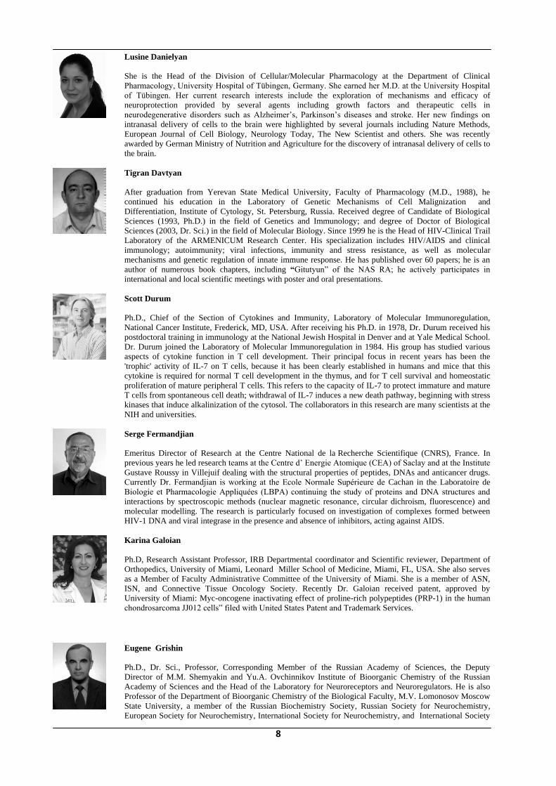
Lusine Danielyan
She is the Head of the Division of Cellular/Molecular Pharmacology at the Department of Clinical
Pharmacology, University Hospital of Tübingen, Germany. She earned her M.D. at the University Hospital
of Tübingen. Her current research interests include the exploration of mechanisms and efficacy of
neuroprotection provided by several agents including growth factors and therapeutic cells in
neurodegenerative disorders such as Alzheimer‘s, Parkinson‘s diseases and stroke. Her new findings on
intranasal delivery of cells to the brain were highlighted by several journals including Nature Methods,
European Journal of Cell Biology, Neurology Today, The New Scientist and others. She was recently
awarded by German Ministry of Nutrition and Agriculture for the discovery of intranasal delivery of cells to
the brain.
Tigran Davtyan
After graduation from Yerevan State Medical University, Faculty of Pharmacology (M.D., 1988), he
continued his education in the Laboratory of Genetic Mechanisms of Cell Malignization and
Differentiation, Institute of Cytology, St. Petersburg, Russia. Received degree of Candidate of Biological
Sciences (1993, Ph.D.) in the field of Genetics and Immunology; and degree of Doctor of Biological
Sciences (2003, Dr. Sci.) in the field of Molecular Biology. Since 1999 he is the Head of HIV-Clinical Trail
Laboratory of the ARMENICUM Research Center. His specialization includes HIV/AIDS and clinical
immunology; autoimmunity; viral infections, immunity and stress resistance, as well as molecular
mechanisms and genetic regulation of innate immune response. He has published over 60 papers; he is an
author of numerous book chapters, including “Gitutyun‖ of the NAS RA; he actively participates in
international and local scientific meetings with poster and oral presentations.
Scott Durum
Ph.D., Chief of the Section of Cytokines and Immunity, Laboratory of Molecular Immunoregulation,
National Cancer Institute, Frederick, MD, USA. After receiving his Ph.D. in 1978, Dr. Durum received his
postdoctoral training in immunology at the National Jewish Hospital in Denver and at Yale Medical School.
Dr. Durum joined the Laboratory of Molecular Immunoregulation in 1984. His group has studied various
aspects of cytokine function in T cell development. Their principal focus in recent years has been the
'trophic' activity of IL-7 on T cells, because it has been clearly established in humans and mice that this
cytokine is required for normal T cell development in the thymus, and for T cell survival and homeostatic
proliferation of mature peripheral T cells. This refers to the capacity of IL-7 to protect immature and mature
T cells from spontaneous cell death; withdrawal of IL-7 induces a new death pathway, beginning with stress
kinases that induce alkalinization of the cytosol. The collaborators in this research are many scientists at the
NIH and universities.
Serge Fermandjian
Emeritus Director of Research at the Centre National de la Recherche Scientifique (CNRS), France. In
previous years he led research teams at the Centre d‘ Energie Atomique (CEA) of Saclay and at the Institute
Gustave Roussy in Villejuif dealing with the structural properties of peptides, DNAs and anticancer drugs.
Currently Dr. Fermandjian is working at the Ecole Normale Supérieure de Cachan in the Laboratoire de
Biologie et Pharmacologie Appliquées (LBPA) continuing the study of proteins and DNA structures and
interactions by spectroscopic methods (nuclear magnetic resonance, circular dichroism, fluorescence) and
molecular modelling. The research is particularly focused on investigation of complexes formed between
HIV-1 DNA and viral integrase in the presence and absence of inhibitors, acting against AIDS.
Karina Galoian
Ph.D, Research Assistant Professor, IRB Departmental coordinator and Scientific reviewer, Department of
Orthopedics, University of Miami, Leonard Miller School of Medicine, Miami, FL, USA. She also serves
as a Member of Faculty Administrative Committee of the University of Miami. She is a member of ASN,
ISN, and Connective Tissue Oncology Society. Recently Dr. Galoian received patent, approved by
University of Miami: Myc-oncogene inactivating effect of proline-rich polypeptides (PRP-1) in the human
chondrosarcoma JJ012 cells‖ filed with United States Patent and Trademark Services.
Eugene Grishin
Ph.D., Dr. Sci., Professor, Corresponding Member of the Russian Academy of Sciences, the Deputy
Director of M.M. Shemyakin and Yu.A. Ovchinnikov Institute of Bioorganic Chemistry of the Russian
Academy of Sciences and the Head of the Laboratory for Neuroreceptors and Neuroregulators. He is also
Professor of the Department of Bioorganic Chemistry of the Biological Faculty, M.V. Lomonosov Moscow
State University, a member of the Russian Biochemistry Society, Russian Society for Neurochemistry,
European Society for Neurochemistry, International Society for Neurochemistry, and International Society

9
of Toxinology. His research interests and activities include Protein Chemistry (protein modification and
crystallization), Toxinology (isolation of polypeptide toxins from snake, scorpion, spider and ant venoms;
amino acid sequences and spatial structure of polypeptide toxins; cloning of cDNA encoding protein toxins;
neurotoxins as tools for study of receptor components), Molecular Neurobiology, and Neuroreceptors
(interaction of natural neurotoxins with sodium, potassium and calcium channels). Prof. Grishin has
published about 320 papers and abstracts.
Bernd Hamprecht
Ph.D., Professor, Interfaculty Institute for Biochemistry, University of Tübingen, Tübingen, Germany
Araksya Izmiryan
Graduated form French Lycée, Gumry, Armenia in 1996. She received B.Sc. (2000) and M.Sc. (2002) in
Biochemistry in Faculty of Biology of the Yerevan State University, Armenia. Thesis in Molecular Biology
at University Paris 7 was supervised by Prof. Denise Paulin. Since 2008 she is in postdoctoral position,
Inserm U781, France.
David Jessop
Dr. Jessop was awarded his Ph.D. from the University of London in 1988 for endocrine studies performed at
St. Bartholomew‘s Hospital. Since then he works at Westminster Hospital, London, and the University of
Bristol in the UK. Major areas of his research are brain responses to stress and the consequences of stress to
the immune system and disease. Dr. Jessop has also made major contributions to research into
neuropeptides synthesised within immune tissues. He was the first to identify the novel opioid peptides
endomorphins in the mammalian immune system and demonstrate their anti-inflammatory activities. His
current interests are: (i) the synthesis of endomorphins in immune cells and the therapeutic application of
these compounds, and their long-acting analogues, to the treatment of arthritis; (ii) influence of environment
and stress on the onset and severity of chronic inflammation; (iii) defects in the HPA axis in autistic
children. Dr. Jessop was awarded an honorary Professorship in Neuroimmunology by the Hong Kong
Polytechnic University in 2004.
Guevork Kevorkian
Ph.D., Dr. Sci., Professor, Director of the H. Buniatian Institute of Biochemistry, NAS RA, Head of the
Department of Pathological Biochemistry at the H. Buniatian Institute of Biochemistry. Prof. Kevorkian is
Vice-President of the Armenian Association of Biochemists and Vice-President of the Armenian Society of
Clinical Radiology and Molecular Imaging. He has published more than 290 abstracts and articles, and
supervised 11 Ph.D. theses. The field of his research interests includes Ca ions translocation and Ca2+-
binding properties, synthesis and degradation of membrane proteins at different experimental pathological
states, such as Crush syndrome, Bipolar disorders, as well as testing of different natural and synthetic
peptides for treatment.
Varduhi Knaryan
Ph.D., senior scientific staff at the Department of Neurohormones Biochemistry of H.Buniatian Institute of
Biochemistry, NAS RA, and a member of the Scientific Council of the Institute. Her current research
interests concern with the investigation of cellular and molecular mechanisms of neuronal degeneration at
experimental Parkinsonism and involvement of spinal cord in the progression of disease. Studies are
particularly focused on the role of proteolytic enzymes (calpain and caspase-3) in neurodegenerative
processes caused by Parkinsonian toxins. During 2005-2006 she was a Fulbright Scholar in USA,
conducting research work in the Department of Neurosciences/Neurology, Medical University of South
Carolina. She is a member of the Armenian Association of Biochemists, affiliated to FEBS, Armenian Brain
Research Association, affiliated to IBRO, and Society for Neuroscience. Dr. Knaryan is a Secretary of the
Armenian Association of Biochemists, and serves as an organizer of scientific meetings, conferences and
workshops. Recently, she has been elected by FEBS Council, as a member of the Working Group on
Assistance to Central and Eastern Europe (WOGCEE).
10
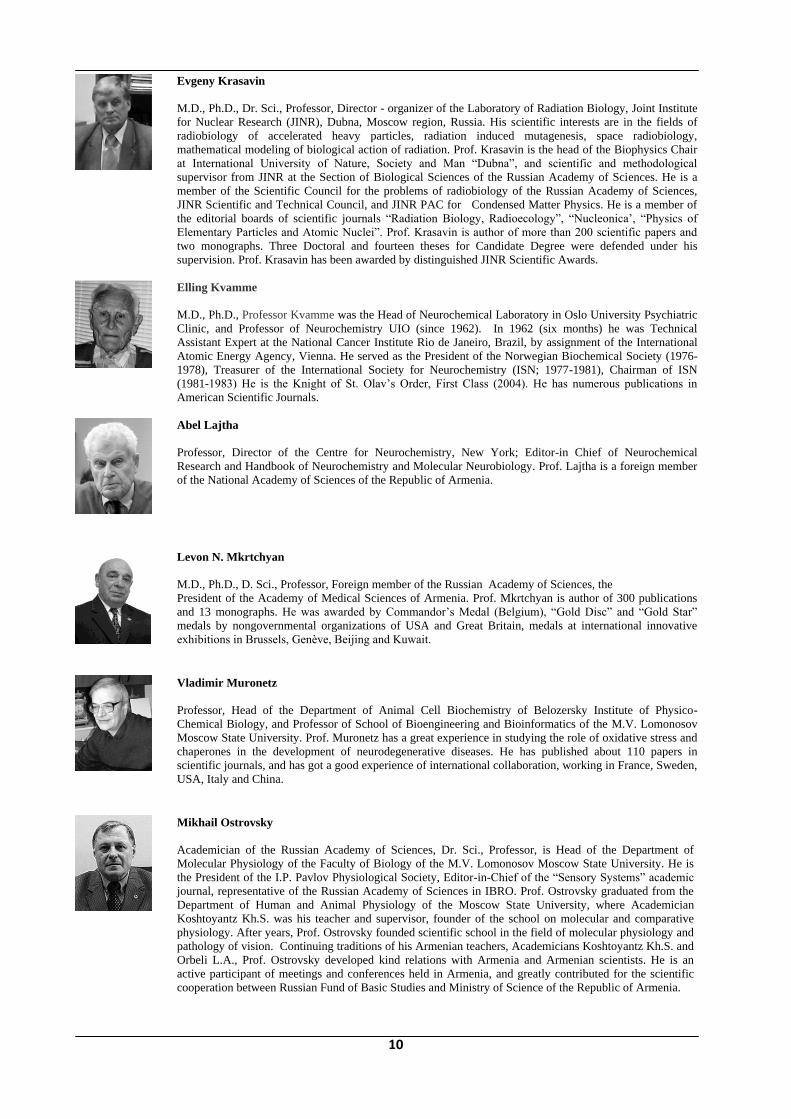
Evgeny Krasavin
M.D., Ph.D., Dr. Sci., Professor, Director - organizer of the Laboratory of Radiation Biology, Joint Institute
for Nuclear Research (JINR), Dubna, Moscow region, Russia. His scientific interests are in the fields of
radiobiology of accelerated heavy particles, radiation induced mutagenesis, space radiobiology,
mathematical modeling of biological action of radiation. Prof. Krasavin is the head of the Biophysics Chair
at International University of Nature, Society and Man ―Dubna‖, and scientific and methodological
supervisor from JINR at the Section of Biological Sciences of the Russian Academy of Sciences. He is a
member of the Scientific Council for the problems of radiobiology of the Russian Academy of Sciences,
JINR Scientific and Technical Council, and JINR PAC for Condensed Matter Physics. He is a member of
the editorial boards of scientific journals ―Radiation Biology, Radioecology‖, ―Nucleonica‘, ―Physics of
Elementary Particles and Atomic Nuclei‖. Prof. Krasavin is author of more than 200 scientific papers and
two monographs. Three Doctoral and fourteen theses for Candidate Degree were defended under his
supervision. Prof. Krasavin has been awarded by distinguished JINR Scientific Awards.
Elling Kvamme
M.D., Ph.D., Professor Kvamme was the Head of Neurochemical Laboratory in Oslo University Psychiatric
Clinic, and Professor of Neurochemistry UIO (since 1962). In 1962 (six months) he was Technical
Assistant Expert at the National Cancer Institute Rio de Janeiro, Brazil, by assignment of the International
Atomic Energy Agency, Vienna. He served as the President of the Norwegian Biochemical Society (1976-
1978), Treasurer of the International Society for Neurochemistry (ISN; 1977-1981), Chairman of ISN
(1981-1983) He is the Knight of St. Olav‘s Order, First Class (2004). He has numerous publications in
American Scientific Journals.
Abel Lajtha
Professor, Director of the Centre for Neurochemistry, New York; Editor-in Chief of Neurochemical
Research and Handbook of Neurochemistry and Molecular Neurobiology. Prof. Lajtha is a foreign member
of the National Academy of Sciences of the Republic of Armenia.
Levon N. Mkrtchyan
M.D., Ph.D., D. Sci., Professor, Foreign member of the Russian Academy of Sciences, the
President of the Academy of Medical Sciences of Armenia. Prof. Mkrtchyan is author of 300 publications
and 13 monographs. He was awarded by Commandor‘s Medal (Belgium), ―Gold Disc‖ and ―Gold Star‖
medals by nongovernmental organizations of USA and Great Britain, medals at international innovative
exhibitions in Brussels, Genève, Beijing and Kuwait.
Vladimir Muronetz
Professor, Head of the Department of Animal Cell Biochemistry of Belozersky Institute of Physico-
Chemical Biology, and Professor of School of Bioengineering and Bioinformatics of the M.V. Lomonosov
Moscow State University. Prof. Muronetz has a great experience in studying the role of oxidative stress and
chaperones in the development of neurodegenerative diseases. He has published about 110 papers in
scientific journals, and has got a good experience of international collaboration, working in France, Sweden,
USA, Italy and China.
Mikhail Ostrovsky
Academician of the Russian Academy of Sciences, Dr. Sci., Professor, is Head of the Department of
Molecular Physiology of the Faculty of Biology of the M.V. Lomonosov Moscow State University. He is
the President of the I.P. Pavlov Physiological Society, Editor-in-Chief of the ―Sensory Systems‖ academic
journal, representative of the Russian Academy of Sciences in IBRO. Prof. Ostrovsky graduated from the
Department of Human and Animal Physiology of the Moscow State University, where Academician
Koshtoyantz Kh.S. was his teacher and supervisor, founder of the school on molecular and comparative
physiology. After years, Prof. Ostrovsky founded scientific school in the field of molecular physiology and
pathology of vision. Continuing traditions of his Armenian teachers, Academicians Koshtoyantz Kh.S. and
Orbeli L.A., Prof. Ostrovsky developed kind relations with Armenia and Armenian scientists. He is an
active participant of meetings and conferences held in Armenia, and greatly contributed for the scientific
cooperation between Russian Fund of Basic Studies and Ministry of Science of the Republic of Armenia.

11
Denise Paulin
She is Professor from Paris who has directed a group devoted to molecular biology of development at the
Pasteur Institute and the University Diderot Paris-7. Author of international publications she is an expert in
the field of intermediate filaments and has developed animal models of cardiomyopathies and
neuromuscular diseases. She received in Paris Armenian students to prepare a Ph.D. diploma as well as
colleagues for scientific collaboration. During several years she organized workshops with the Yerevan
University and developed the Franco-Armenian Association for Scientific Cooperation (AFACS).
Rima Srapionian
Ph.D., Dr.Sci., Professor of Biochemistry, Head of the Laboratory of Neurospecific Proteins‘ Biochemistry
at the H. Buniatian Institute of Biochemistry, NAS RA. Prof. Srapionian is an expert in neurochemistry,
endocrinology, immunology, and bioorganic chemistry. The field of her research work include investigation
of neurospecific protein-hormonal complexes, neurochemical regulation of brain metabolic processes,
coagulation mechanisms in the heart and blood.
Anna Tishkina
She has a Master degree in Applied Physics and Mathematics, and currently is a Ph.D. student at the
Institute of Higher Nervous Activity and Neurophysiology of the Russian Academy of Sciences, Moscow,
Russia. Her scientific interests are: glia, hippocampus, neuroinflammation, oxidative stress, digital image
processing.
Mikhail Ugryumov
D.Sci., Professor, N.K. Koltsov Institute of Developmental Biology Russian Academy of Sciences (IDB
RAS), Moscow, Russia. He is an Academician of the Russian Academy of Sciences.
Richard E. Weller
Dr. Weller is a Senior Program Manager in the Biological Sciences Division at the Pacific Northwest
National Laboratory, and is involved in the development and deployment of innovative biomedical
technologies. His primary focus is on diagnostic and therapeutic technologies. He has over 20 years
experience developing animal models for biomedical applications. Dr. Weller is board certified by the
American College of Veterinary Internal Medicine in Internal Medicine (1980) and Oncology (1987), and
holds adjunct faculty positions in the College of Veterinary Medicine at both Washington State University
and Tuskegee University, an honorary Professorship at the Institute of Veterinary Medicine in Kyiv,
Ukraine. He is a lecturer at the University of Washington's Jackson School for International Studies on
weapons of mass destruction (WMD) detection and proliferation. He holds several U.S. patents and has
authored or co-authored over 250 unclassified and classified articles, technical reports, book chapters, and
presentations in his fields of expertise.
Loussine Zargaryan
She has graduated from the Yerevan State University, Armenia (1989), receiving Diploma of chemist
specialized in physical chemistry. Continuing education in University Paris 7, she received Diploma of
Microbiology (1994), and then Ph.D. (1997). As a postdoctoral fellow, she conducted research work (during
2000-2003) in the Department of Biology and Pharmacology Structural (direction Dr. S. Fermandjian),
Institute Gustave Roussy, UMR 8113, CNRS. Currently Dr. Zargaryan is a Research Engineer at the CNRS,
France. Her research is focused on the dissection of structural and functional proprieties of VIH-integrase as
a bases for the development of new inhibitors; structural and functional study of viral nucleic acids; study of
recognition mechanisms and DNA cleavage by topoisomerase II DNA. She is author of 17 articles in
international journals.

12
Oral Presentations - Quick Reference
1. Abrahamyan D.O., Sarkissian J.S., Chavushyan V.A., Meliksetyan I.B., Gevorgyan A.J., and
A.Galoyan.
Improvement of Peripheral Nerve Repair Functional Outcome using Hypothalamic Proline-Rich
Neuropeptide PRP-1
2. Aghajanov M., Navasardyan G., Vahradyan H., Safaryan K., Melixetyan I., Yenkoyan K., and
A.Galoyan
Modulatory Role of PRP on Behaviour and Neuroactive Amino Acids in Beta-Amyloid Induced
Neurodegeneration.
3. Aroutiounian R., Gasparyan G., Hovhannisyan G., and A.Galoyan
Toxicogenetics in Peptide Research
4. Banik N.L.
Is Spinal Cord a Relevant Piece in the Parkinsonian Puzzle?
5. Barkhudaryan N., Zakaryan H., Sarukhanyan F., Kellermann J., and F. Lottspeich. Hemorphins
Act as Homeostatic Agents in Response to Endotoxin Induced Stress
6. Besedovsky H.O.
The Immune System Can Take the Command of Homeostasis
7. Bezirganyan K.B., Davtyan T.K., and A.A.Galoyan
Hypothalamic Proline Rich Polypeptide Regulates Hematopoiesis
8. Boyajyan A.S., Khoyetsyan A.G., Mkrtchyan G.M, Hovhannisyan L.P.,
Zakharyan R.V.
Immune Response and Apoptosis in Pathogenesis of Mental Disorders
9. Buniatian G.H.
The Immune Status-Dependent Production of the Regeneration-Supporting Hormon Erythropoietin
and Glutamine Synthetase in Mutant Mouse Skin Keratinocytes
10. Chailakhyan R.K., Gerasimov Yu.V., Chailakhyan M.R.
Proline-Rich Hypothalamic Polypeptide Has Oposite Effects on the proliferation of Human Bone
Marrow Stromal Cells and Human Giant-Cell Tumor Stromal Cells
11. Galoian K.
Antitumorigenic Effect of Brain Proline Rich Polypeptide-1 in Human Chondrosarcoma
12. Grishin E.V.
Modulation on Functional Activity of Ionic Channels and Ionotropic Receptors
13. Hamprecht B.
Neurochemistry of Cultured Ependymal Cells

13
14. Hobaika Z., Zargarian L., Maroun R.G., Mauffret O., and S. Fermandjian
HIV-1 Integrase and DNA ends: Complex Formation and Perturbation by Inhibitors of Integration
15. Izmiryan A. and D.Paulin
SYNEMIN Isoforms in Peripheric and Central Nervous Systems
16. Jessop D.
Differential Central and Peripheral Cytokine Expression and HPA Axis Activation in Response to
LPS
17. Lajtha A.
Transmitters in Reward Mechanisms
18. Mkrtchyan L.N.
New Approach in Bioprevention of Cancer
19. Samantaray S., Knaryan V., Ray S.K., and N.L. Banik
Some Mechanisms of Spinal Cord Neurodegeneration in Experimental Parkinsonism
20. Krasavin E.A. The problem of GCR Action on the Central Nervous System during the Long-
Term Space Mission
21. Ostrovskiy M.A.
Photo- And Neurochemistry of Vision
22. Roberg B., Torgner I., and E. Kvamme
Kinetics of a Novel Isoform of Phosphate Activated Glutaminase (PAG) in SH-SY5Y
Neuroblastoma Cells
23. Durum S.
Interleukin 7 in the Life and Death of T cells
24. Srapionian R.M and A.A. Galoyan
Cardioactive Protein – Hormonal Complexes of Brain and Hearth
25. Tavadyan L.A., Galoian K.A., Harutunyan L.A., Tonikyan H.G., and A.A.Galoyan. Antioxidant
and Electron Donating Function of Hypothalamic Polypeptides: Galarmin and Gx-NH2
26. Ugryumov M.V.
Developing Brain as a Multipotent Endocrine Organ: Paradox or Reality

14
Oral Presentations – Abstracts
ANTITUMORIGENIC EFFECT OF BRAIN PROLINE RICH POLYPEPTIDE-1 IN HUMAN
CHONDROSARCOMA
Karina Galoian
University of Miami, Miller School of Medicine, Miami, Florida, USA
E-mail: [email protected]
Proline rich polypeptides (PRP-1) are produced by neurosecretory cells of NPV and NSO cells of the
hypothalamus [1]. PRP-1 is one of the fragments of neurophysin-vasopressin-associated glycoprotein
(NVAG). This cytokine is also a powerful immunomodulator [1, 2], with antitumor effect [1, 2, 3]. Our
recent data indicated 89% growth inhibition of sarcoma cells when treated with this cytokine in comparison
with the control and immortalized chondrocytes [4]. The problem of sarcoma is the long time topic of our
investigations [5,6]. The fact that the same concentrations of PRP-1 (0.5-1 μg /ml), based on our preliminary
results [7] abolished Myc activity in human JJ012 chondrosarcoma cells prompts us to think that the
antitumorigenic effect of PRP-1 in the same concentrations is mediated through the oncogene inactivation
and involves inhibition of the cell cycle. The bell shaped reminding stimulatory effect of PRP-1 is due to
existence of most probably biphasic receptor for PRP-1, when low dose of the agonist exhibits stimulation
and high dose on the contrary shows inhibitory effect. For the ligands that induce receptor dimerization, a
biphasic dose-response curve has also been observed with optimal receptor activation at intermediate doses,
and self-antagonism occurring at higher doses at which monomeric ligand-receptor complexes predominate
[8-10]. The targeted inactivation or repair of the oncogenes has been studied as a potentially therapeutically
useful approach for the treatment of neoplasia. Recent work in experimental transgenic model systems
suggests that MYC inactivation in animal models to be sufficient to induce the regression in lymphoma,
leukemia, papilloma, islet cell tumors and osteogenic sarcoma [11-17] although the molecular mechanism by
which the inactivation of MYC induces tumor regression is unclear. Our future directions include but not
limited to experiments on cell cycle analysis and its key players as well as determination on whether cells
undergo through quiescence or permanent arrest under the influence of the low doses of PRP-1. One of the
most important tasks would be the precise determination and gene expression of PRP-1 receptors and their
subtypes in the tumor tissues in search for possible diagnostic biomarkers.
References
1. Galoyan A.A Brain neurosecretory cytokines: immune response and neuronal survival.
Kluwer Academic/Plenum Publishers, NY, 2004.
2. Galoyan A. In: Abel Lajtha( Ed) Armen Galoyan, Hugo Besedovsky (Vol. ed) Handbook of
Neurochemistry and Molecular Neurobiology, Springer, pp 155-195, 2008.
3. Galoyan A.A, A.Shirvanyan. A method for treatment and/or prevention of leucosis at the cattle.
Patent prelim. No.1696 A2, Patent No. P20050113, The Republic of Armenia.
4. Galoian K.A., Scully S.P, Galoyan A.A. Antitumorigenic effect of brain Proline Rich Polypeptide-1
in Human Chondrosarcoma. Neurochem. Res., 2009, epub, ahead of print
15
5. Scully S.P, Berend KR, Toth A, Qi WN, Qi Z, Block JA Marshall Urist Award. Interstitial
collagenase gene expression correlates with in vitro invasion in human chondrosarcoma. Clin Orthop
Relat Res 376:291–303, 2000.
6. Galoian K.A, Garamszegi N., Garamszegi S.P., Scully S.P. Molecular mechanism of tenascin-C
action on matrix metalloproteinase-1 invasive potential. Exp. Biol. Med. 232(4):515–522, 2007.
7. Galoian K., Scully S., Galoyan A. Myc-oncogene Inactivating Effect by Proline Rich Polypeptide
(PRP-1) in Chondrosarcoma JJ012 Cells. Neurochem. Res. Volume 34, Issue 2: 379-385, 2009.
8. Ilhan Celik, Oguzkan Sürücü,Carsten Dietz, John V. Heymach, Jeremy Force, Iris Höschele,
Christian M. Becker, Judah Folkman, and Oliver Kisker. Therapeutic Efficacy of Endostatin Exhibits
a Biphasic Dose-Response Curve Cancer Research 65, 11044-11050, December 1, 2005.
9. Tey H.B., Khoo H.E., Tan C.H. Adenosine modulates cell growth in human epidermoidcarcinoma
(A431) cells. Biochem. Biophys. Res. Commun. 1992 Sep 30; 187(3):1486-92.
10. Croiset G., De Wied D. Proconvulsive effect of vasopressin; mediation by a putative V2 receptor
subtype in the central nervous system. Brain Res. 1997 Jun 6; 759(1):18-23.
11. Scully S.P, Berend KR, Toth A, Qi WN, Qi Z, Block JA Marshall Urist Award. Interstitial
collagenase gene expression correlates with in vitro invasion in human chondrosarcoma. Clin Orthop
Relat Res 376:291–303, 2000.
12. Galoian K.A, Garamszegi N., Garamszegi S.P., Scully S.P. Molecular mechanism of tenascin-C
action on matrix metalloproteinase-1 invasive potential. Exp. Biol. Med. 232(4):515–522, 2007.
13. Felsher D.W., Nicole Bradon. Pharmacological Inactivation of MYC for the treatment of cancer. Drug
News Perspect. 16(6):370–374, 2003.
14. Felsher, D.W Opinion. Cancer revoked: oncogenes as therapeutic targets. Nat. Rev. Cancer 3:375–
379, 2003.
15. Jain M., Arvanitis C., Chu K., Dewey W., Leonhardt E., Trinh M., Sundberg C.D., Bishop J.M.,
Felsher D.W. Sustained loss of a neoplastic phenotype by brief inactivation of MYC. Science.
297:102–104, 2002.
16. Weinstein I.B, Joe A.K. Mechanisms of disease: oncogene addiction—a rationale for molecular
targeting in cancer therapy. Nat. Clin. Pract. Oncol. 3:448–57, 2006.
17. Elizabeth R. Lawlor, Laura Soucek, Lamorna Brown-Swigart, Ksenya Shchors, C. Uli Bialucha and
Gerard I. Evan Reversible Kinetic Analysis of Myc Targets In vivo Provides Novel Insights into
Myc-Mediated Tumorigenesis Cancer Research 66, 4591-4601, May 1, 2006.

16
SOME MECHANISMS OF SPINAL CORD NEURODEGENERATION IN EXPERIMENTAL
PARKINSONISM
Supriti Samantaray1, Varduhi Knaryan2, Swapan K. Ray1, and Naren L. Banik1
1Department of Neurosciences, Medical University of South Carolina, Charleston, USA; 2 H. Buniatian Institute of Biochemistry, NAS RA, 5/1 Paruir Sevak str., 0014 Yerevan, Armenia
E-mail: [email protected]
Parkinson‘s disease (PD) is a complex devastating neurodegenerative disease that affects 1% of the
population above the age of 60 worldwide. It takes almost two decades of subtle degeneration of brainstem
neurons and loss of dopaminergic neurons in the midbrain substantia nigra pars compacta before the external
signs of PD can appear. Loss of dopaminergic neurons is culminating in motor abnormalities, manifested by
resting tremor, bradykinesia (slowness of movement), muscular rigidity (stiffness), and postural instability
(poor balance). Despite substantial evidence of nigrostriatal degeneration in PD, other areas of the central
nervous system have also been recognized as having essential role in Parkinsonism pathology. We postulated
that neurons of spinal cord (SC), the final coordinator of movement connected to midbrain via various
ascending and descending projections and metabolic circuits, are also implicated in progression of PD.
In order to examine whether SC is affected, two animal models of experimental parkinsonism were
investigated: by injecting parkinsonian toxin 1-methyl-4-phenyl-1,2,3,6-tetrahydropyridine (MPTP; 25
mg/kg, i.p. twice) to adult C57BL/6N mice, and with systemic administration of environmental toxin
rotenone (2.5 mg/kg/day, s.c. during 21 days) to Lewis rats. Death of SC neurons was examined using
combination of TUNEL assay (cell death marker) and double-immunofluorescent staining with markers of
neurons (NeuN, neuronal nuclei) and motoneurons (ChAT, choline acetyltransferase). In MPTP-induced
model specific assays, such as MAO-B (monoamine oxidase) expression, DAT (dopamine transporter)
immunoreactivity and H-3MPP specific uptake in SC tissue preparations were also performed; in vivo
conversion of MPTP into active neurotoxin MPP+ compound was confirmed by HPLC-photodiode array
analysis.
We observed significant neuronal cell death in the SC of both MPTP and rotenone-administered animals
compared to control ones. In ventral horn dying neurons were identified as motoneurons by co-localization
of TUNEL and ChAT immunoreactivity (IR). Neuronal cell death in SC was accompanied by profound
astrogliosis and microgliosis as evidenced from increased glial fibrillary acidic protein (GFAP)-IR and
CD11b/c (OX-42)-IR. Detection of active neurotoxin MPP+, as well as MAO-B and DAT immunoreactivity
testified that SC is a potential extra-nigral target for parkinsonian toxins (1,2).
The expression and activity of cysteine proteases such as calpain and caspase-3, which are crucial players of
apoptosis, were studied to understand molecular mechanisms of SC neurodegenration. Detection of elevated
amounts of active m-calpain (76 kD) and caspase-3 (17/19 kD) fragments in SC tissues (Western blot assay),
and identification of calpain-specific 145 kD and caspase-3-specific 120 kD spectrin breakdown products
from 270 kD uncleaved spectrin (intracellular substrate for calpain and caspase-3), suggested involvement of
these proteases in the neurodegenerative process in parkinspnian SC (1,3,4).
These findings of SC neurodegeneration in MPTP- and rotenone-induced parkinsonism in animals were
correlated with similar neurodegeneration in the post-mortem SC tissue samples of idiopathic PD patients,
compared to other neurological diseases (not published data).
17
Obviously, SC where locomotion centers are located, with damaged neurons and particularly motoneurons,
as has been shown from these studies, may seriously affect and worsen reflex activities as well as impair
motor functions, thus contributing to the progression of movement disturbances, related to PD
characteristics. Therefore, information generated from such studies may help to develop new therapies for
treatment of this complex neurodegenerative disease.
References
1. Samantaray S., Knaryan V.H., Guyton M.K., Matzelle D.D., Ray S. K., Banik N.L. (2007) The
parkinsonian neurotoxin rotenone activates calpain and caspase-3 leading to motoneuron
degeneration in spinal cord of Lewis rats. Neuroscience, 146, p.741–755.
2. Supriti Samantaray, Varduhi H. Knaryan, Jonathan T. Butler, Swapan K. Ray and Naren L. Banik
(2008) Spinal cord degeneration in C57BL/6N mice following induction of experimental
parkinsonism with 1-methyl-4-phenyl-1,2,3,6-tetrahydropyridine.
J. Neurochem. 104, p. 1309-1320.
3. Samantaray S, Ray SK, Banik NL. (2008) Calpain as a potential therapeutic target in Parkinson‘s
disease. CNS & Neurological Disorder - Drug Targets 7(3), p. 305-312.
4. Supriti Samantaray, Jonathan T. Butler, Swapan K. Ray, and Naren L. Banik (2008) Extranigral
neurodegeneration in Parkinson‘s disease: A role for calpain in neurodegeneration. Annals of the
New York Academy of Sciences, 1139, p. 331–336.

18
THE PROBLEM OF GCR ACTION ON THE CENTRAL NERVOUS SYSTEM
DURING THE LONG-TERM SPACE MISSION
Krasavin E.A.
Joint Institute for Nuclear Research, Dubna, Russia
An important safety concern for long-term space travel in outer space is the health effects from cosmic
radiation. The primary radiation sources in outer space are the galactic cosmic rays (GCR), the solar particle
events and protons and electrons trapped in the Earth's magnetic field. The background radiation of the GCR
includes 85% of protons, 13% of helium and about 2% from high-energy and high-charge heavy ions (HI).
Though the heavy charged particles are less abundant, they posses significantly higher ionizing power with a
greater potential for radiation-induced damage and greater penetration power. Possible health risks include
different type mutations, cancer, violation of visual functions (cataract, retina lesion), acute radiation disease,
damage to the central nervous system. Passage of HI particles can result in direct effect on DNA leading to
single strand breaks, double strand breaks, and especially clustered DNA lesions. The character of the DNA
damage caused by heavy charged particles is substantially different from that caused by gamma-quanta.
Accelerated heavy ions, unlike gamma-quanta, induce mainly the cluster-type damage in the DNA. This kind
of damage is a combination of single-step disorders of a DNA part and the formation of single-strand breaks,
modification of bases, and sugar modification. The events of this kind result from a local energy release
which happens when a heavy charged particle travels through a DNA strand. The cluster-type damage
determines the specifics of the lethal, mutagenic (induction of gene and structure mutations in prokaryotes
and formation of chromosome aberrations in higher eukaryote cells), and transforming effect of radiation on
cells with different genome organization levels.
The integral flux of GCR particles of carbon and iron groups for future exploration missions to Mars equals
to 107 part cm-2 per year. The long range of the HI allows for the potential damage along a long column of
cells in tissue. The issues of the damaging effect of heavy charged particles of such integral flux on the
central nervous system are important and remain unresolved in many ways. Research in this field seem to be
extremely topical for solving cosmic radiobiology problems as there is evidence that behavioral functions of
the experimental animals irradiated with heavy ions have been disordered. Low doses of accelerated iron
ions cause an irreversible disorder of the cognitive and other functions in an irradiated organism. Research in
this important field has also been started at the Laboratory of radiation biology of JINR.

19
NEUROBIOCHEMICAL MECHANISMS OF THE NERVOUS SYSTEM
ORGANIZATION AND PATHOLOGY
COMMON MOLECULAR MECHANISMS OF NEURONAL PLASTICITY
AND NEURONAL PATHOLOGY
Gulyaeva N.
Institute of Higher Nervous Activity and Neurophysiology RAS
Plasticity is the capability of the nervous system to adapt structurally and/or functionally in situations of
changing environment, in particular after damage, as well as during development and maturation of the brain
gaining experience. The basis for synaptic plasticity in response to physiological or pathological stimuli is
molecular changes. A model of plasticity-pathology continuum (McEachern & Shaw, 1996, 1999) was one
of main starting points for our concept of pleiotropicity of basic molecular mechanisms underlying both
normal neuronal functioning and neuronal damage and death. For example, factors mostly regarded as
apoptotic (caspases etc.) are involved in normal neuronal plasticity. Enzymes with broad substrate specificity
(proteases, protein kinases, phosphatases) are main players in the chess game between life and death where
figures are substrates and positions on the chess-board are cellular compartments. Sometimes the main route
of signaling involved in survival, functional changes or cell death is same, while a divergence occurs at the
very end of biochemical cascade. In other situations, same factor may trigger different signaling mechanism
(e.g. through different receptor types). The concept on the similarity of neuroplasticity and neuropathology
mechanisms conforms to the idea that there are no special mechanisms for pathologies in an organism.
Pathological changes are developing as a result of disturbances in ―normal mechanisms‖, these mechanisms
being realized with a different substrate and/or to a different degree and/or in a different period and/or in a
different cellular or exrtacellular compartment. In many cases the switch ―norm-pathology‖ may be defined
by specific substrates of ―multi-substrate‖ enzymes. Indeed, this may be the main physiological meaning of
broad substrate specificity of key proteases and protein kinases. The idea of the pleiotropicity of molecular
mechanisms underlying both normal neuroplasticity and neuropathology gives us a tool to elaborate a
strategy for revealing links triggering pathological processes. This link is a potential target for the search for
pathogenetically directed approaches for prevention and treatment of specific cerebral pathologies. An
example of ignoring the above phenomenon of pleiotropicity is failure to use ―antiapoptotic‖ technologies
and NMDA blockers for treatment of cerebral pathologies accompanied by neurodegeneration.
Unfortunately, our understanding of molecular processes taking part in neurons remains rather limited. As a
rule, at best, general mechanisms involved in neuroplasticity phenomena and neurodegeneration are known,
as well as the final results of processes. However, the most important is still obscure: when and how a
process common for norm and pathology is irreversibly switching to the realization of neuronal death.
However, it is the revealing of this switching mechanism which can give us a tool to manipulate the
pathology process and even prevent it.
Supported by RFBR grant 07-04-01380

20
MECHANISMS OF HSP70 CHAPERONE-MEDIATED INHIBITION OF INSOLUBLE PROTEIN
AGGREGATES IN MODEL OF HUNTINGTON DISEASE
Guzhova I., Kaznacheeva A., Ippolitova M., and Margulis B.
Institute of Cytology, St. Petersburg, Russia
E-mail:[email protected]
Aggregates made of misfolded, mutant proteins are the major cause of neuronal death in a great number of
neurodegenerative pathologies. One of these illnesses, Huntington‘s disease (HD), is induced by expanded
repeats of glutamine in N-terminal part of huntingtin. The development of the disease initiates from the
formation of SDS-insoluble protein aggregates in neurons of striatum and cerebral cortex. The aggregates
are formed with the participation of tissue transglutaminase, which catalyzes the ligation of glutamine
residues of mutant huntingtin to lysines of other polypeptides, such as glyceraldehyde-3-phosphate
dehydrogenase (GAPDH). The results of our experiments proved that GAPDH is implicated in SDS-
insoluble aggregate formation, because the factors tightly binding the enzyme reduce both the size and
amount of aggregates in the cells over-expressing the gene of mutant huntingtin with Q103 repeat.
Conversely, the addition of pure GAPDH to ex vivo system of Q103 aggregate formation significantly
increased the amount of aggregating Q103. To explore the mechanism of anti-aggregate activity of Hsp70
in HD model we generated human neuroblastoma cell line with metal-dependent expression of Hsp70. The
elevation of Hsp70 level lead to the dose-dependent reduction of both, number and size of aggregates. These
data were confirmed by using ex-vivo system of aggregate formation: the addition of pure Hsp70 elevated
the amount of soluble polyglutamine-containing protein. Using the method of immunoprecipitation we
showed that Hsp70 was able to bind GAPDH in dose-dependent manner: the more Hsp70, the lesser content
of GAPDH in insoluble polyglutamine aggregates and the lower amount of the latter. It is concluded that a
novel mechanism exists through which Hsp70 sequesters GAPDH from the control of transglutaminase and
by this diminishes cell mortality from cytotoxicity of the polyglutamine aggregates.
The work was supported by the Russian Ministry of Education and Sciences GK N 02.512.11.22.49 and
grant of RFBR N 08-08-00507.

21
THE ROLE OF METAL BINDING DOMAIN OF AMYLOID-Β IN INITIATION OF
ALZHEIMER’S DISEASE
Kozin S.A.1,2, Tsvetkov Ph.O.2, Popov I.A.1, Nikolaev E.N.1, Archakov A.I.,1 Makarov A.A.2
1Orekhovich Institute of Biomedical Chemistry, Russian Academy of Medical Sciences, Moscow, Russia;
2 Engelhardt Institute of Molecular Biology, Russian Academy of Sciences, Moscow, Russia
E-mail [email protected]
Alzheimer's disease (AD) is pathophysiologically characterized by the formation of amyloid plaques as a
result of the extracellular accumulation of the amyloid-β peptide (Aβ) in the brain of AD patients.
Postmortem biochemical analysis of these aggregates showed that they contain high concentrations of metal
ions bound to A and most of A aspartate-7 amino-acid residues undergo a specific chemical modification
resulting in the formation of isoaspartate-7 (isoAsp7). Both of these features are related to biochemical
and/or biophysical changes of the A metal binding domain (the Aβ residues 1-16). We have found that Asp-
7 isomerization results in its zinc-induced oligomerization. Additionally, this post-translational modification
alters binding mode of copper to the domain and impacts the N-domain of angiontensin-converting enzyme
to change its hydrolysis efficiency towards Aβ. All the findings suggest that the isoAsp7-containing A
species (isoAsp7- A) might be a potential trigger of AD.

22
NEUROLOGICAL DISEASES, PROTEIN AGGREGATES AND CHAPERONES: MODELS AND
TECHNOLOGIES FOR PROVING ANTI-DEGENERATION
DRUG EFFICACIES
Margulis B.1, Kaznacheeva A.1, Eremenko E.1, Mesthseryakova D.1, Guzhova I.1, and Polonik S.2
1 Institute of Cytology, RAS, St. Petersburg, Russia;
2 Pacific Institute of Bioorganic Chemistry FEB RAS, Vladivostok, Russia
E-mail: [email protected]
The reason of many neurodegenarative pathologies is the formation of aggregates of mutant or damaged
proteins that are toxic for specific groups of neurons. The examples of such pathologies are Alzheimer's,
Parkinson, Huntington diseases whose pathogenic proteins are A-beta amyloid, alpha-synuclein and
huntingtin correspondingly. The process of aggregation may be delayed or prevented by molecular
chaperones, and therefore substances inducing particular proteins, Hsp70 and Hdj1/Hsp40 are offered as
promising candidates for future drugs. We found also a few of natural compounds, mostly O- and S-
glycosides of naphthoquinones, able to activate heat shock response in human neuroblastoma and monocytic
cells and to restrict aggregation process in in vitro models. Another group of compounds possibly
influencing aggregation dynamics includes substances binding glycolytic enzyme, glyceraldehyde-3-
phosphate dehydrogenase (GAPDH), since it is involved in the formation of covalent bonds with
polyglutamine tract-containing mutant proteins, such as huntingtin. It was shown that Pefabloc (ABTS),
dobutamine and deprenyl, all known to strongly bind GAPDH, reduce aggregate size in in vitro model of
HD. The third group of compounds blocking the aggregation process in its beginning can include substances
interacting with monomers/olygomers of mutant proteins and giving them no opportunity to interact with
each other. We have elaborated a few of model systems and assays to test the activity of all three types of
above compounds. All assays can be performed in 96-well microplate platform. Two assays are applied for
the screening of compounds, one is ex vivo test-system for the estimation of anti-aggregation activity and
with the aid of two others one can estimate the activity of small molecules impeding aggregation of
polyglutamine tract-bearing mutant proteins.
The work was supported by the Russian Ministry of Education and Sciences GK N 02.512.11.22.49 and
grant of RFBR 07-04-01392.

23
ROLE OF CHAPERONES BLOCKED IRREVERSIBLY BY MISFOLDED PROTEINS IN THE
INDUCTION OF CONFORMATIONAL NEURODEGENERATIVE DISEASES
Muronetz V.*,**, Kiselev G.**, Schmalhausen E.*, Naletova I.*, Thomas Haertle***
*Belozersky Institute of Physico-Chemical Biology and **School of Bioengineering and Bioinformatics,
Moscow State University, Moscow, Russia;
***Institut National de la Recherche Agronomique, BIA-FIPL, Nantes, France
E-mail: [email protected]
Recently, it has been demonstrated that the oligomeric forms (28-36 of monomers) of the recombinant ovine
prions exhibit neurotoxic action, while high molecular aggregates of the prions are harmless. Previously, we
demonstrated blocking of the chaperones by misfolded proteins that are similar in their characteristics to the
pathogenic prion forms and suggested a hypothesis according to which chaperones can be involved in the
development of neurodegenerative diseases (1-3). In the presented work, we used the dynamic light
scattering analysis to determine the size of the aggregates formed while the transition of the recombinant
ovine prions into their pathogenic form, and also investigated the possibility of purposeful changing the size
of the aggregates. We demonstrated that in the presence of copper ions, as well as in an acid medium, the
recombinant prions VRQ transform from the monomeric to oligomeric form (the size of the aggregates
constituted 12-15 nm). The extent of aggregation of the oligomers alters in the presence of the molecular
chaperones. It was shown that at 25oC, the non-functioning chaperonin GroE (equimolar mixture of GroEL
and GroES in the absence of Mg-ATP) bound the prion yielding large aggregates (greater than 400 nm). The
addition of Mg-ATP decreased significantly the aggregate size to 70-80 nm. Blocking of one of the
chaperonin centers by misfolded proteins (for example, oxidized denatured glyceraldehyde-3-phosphate
dehydrogenase) increased the aggregate size to 1200 nm, and the addition of Mg-ATP did not prevent the
aggregation. In contrast to the proteinase-resistant oligomeric prions, the aggregates formed in the presence
of the chaperonin were capable of cleaving with proteinase K. Besides, it was demonstrated that the large
aggregates of the amyloid-beta peptide 1-42 are formed in the presence of misfolded forms of proteins
including glyceraldehyde-3-phosphate dehydrogenase, the enzyme whose enhanced content in the brain was
observed while modeling Alzheimer disease. We suggest that chaperones and misfolded proteins play an
important role in the development of neurodegenerative diseases, regulating the size of amyloid structures
and their sensitivity to proteolysis. We demonstrated that natural (chaperones and misfolded proteins) and
artificial (polyelectrolytes) modulators of amyloid structure formation could be used to decrease the
pathogenic properties of amyloidogenic proteins and to decelerate the development of neurodegenerative
disorders. It is suggested to create nanoparticles based on polyelectrolytes, chaperones and misfolded
proteins that are capable to penetrate through the hematoencephalic barrier for prophylactic and treatment of
Alzheimer disease as well as for reducing the neurotoxicity of infectious prions.
The work was supported by RFBR (08-04-00231, 08-08-00540, 09-04-01122), NATO (PDD(CP)-
(CBP.NR.RIG 982779)) and Federal Purpose Program (02.512.11.2249).
1. Polyakova et al. (2005) Protein Sci., 14, 921-928.
2. Markossian et al. (2006) Biochemistry, 45, 13375-13384.
3. Shalova et al. (2007) Biochim. Biophys. Acta. 1770, 826-832.

24
Young Scientists’ Session - Quick Reference
1. Akopyan N.S., Adamyan N.Y., Harutyunyan R.S., Karapetyan M.A.
The Influence of Hyppocampus Stimulation in Neuronal Activity of Respiratory Centre upon
Hypoxia Condition
2. Aniol V.A., Popova M.S., Stepanichev M.Yu., Yakovlev A.A., Lazareva N.A., Gulyaeva N.V.
Proliferation of Cells in Rat Brain during Experimental Epilepsia
3. Danielyan K.E., Chailian A.S., Simonian M.A., Chailian S.G., Galoyan A.A.
Brain Derived Proline Rich Peptide Has Anti Ischemic/Apoptotic Abilities in Vitro and in Vivo
4. Danielyan L., Schäfer R., von Ameln-Mayerhofer A., Buadze M., Geisler J., Klopfer
T., Burkhardt U., Proksch B., Verleysdonk S., Ayturan M., Buniatian G.H., Gleiter Ch.H., Frey
W.H.II
Intranasal Delivery of Eukaryotic Cells to the Brain
5. Hayrapetyan G. N. and Andriasyan V. K.
Influence of External Noise on Single Ion Channels
6. Hovhannisyan A.S., Gebhardt R., Hakopyan V.P.
The Estimation and Comparison of Cerebrolysin and Tanakan Prolonged Effects on Lipid
Peroxidation Processes in Different Brain Regions under the Hypokinetic Stress Conditions
7. Karapetyan A.A., Chazaryan S.A., Barseghyan K.A., Sarkisyan R.Sh., Hayrapetyan H.L., Nazaryan
N.S., Guevorkyan A.G., Kevorkian G.A.
Experimental Model of Bipolar Disorders and Treatment by New Li+-Containing Peptides
8. Karapetyan G.R.
The Dynamic of Rat Liver and Brain Mitochondria Ultrastructural Changes at the Crush Syndrome
and its Correction by PRP
9. Khachatryan S.G.
Sleep Disorders and Immunity: an Overview
10. Margaryan A.S., Simonyan L.A., Badalyan R.B., Batikyan I.H., Simonyan A.A.
The Possible Regulatory Role of Hypothalamic Proline-Rich Polypeptide (PRP) on Energy
Metabolism
11. Margaryan K.S., Aroutiounian D.N., Aroutiounian R.M.
Stimation of Antimutagenic Activity of Proline-Rich Polypeptide by the Comet-Assay in Human
Leukocytes
12. Sahakyan I.K., Abrahamyan S.S., Meliksetyan I.B., Tumasyan N.V., Galoyan A.A.
Effect of Hypothalamic Proline-Rich Peptide PRP-1 on Rat Brain Plasticity under Labyrinthectomy
and Vibration

25
13. Tishkina A.O., Levshina I.P., Passikova N.V., Stepanichev M.Yu., Gulyaeva N.V.
Participation of Glial Cells in Brain Response to Chronic Pain-Emotional Stress
14. Tavadyan Z.D.
Influence of Affective Symptoms on Quality of Life in Patients with Parkinson‘s disease
15. Yeranosyan L.A., Ter-Tatevosyan L.P., Sargisyan L.V., Galoyan A.A.
Effect of PRP-1 on the Regulation of Carbohydrate – Phosphoric Metabolism in Rat‘s Bone Marrow
and Spleen Tissues under the Pharmacological Sympathectomy
16. Zakharyan A. A., Gasparyan K. G., Davtyan T. K.
Comparative Effect of PRP-1 and its Analog d-15 on the Leukopenia Induced by
Cyclophosphamide in Rats

26
Young Scientist’s Session - Abstracts
THE INFLUENCE OF HYPPOCAMPUS STIMULATION IN NEURONAL ACTIVITY OF
RESPIRATORY CENTRE UPON HYPOXIA CONDITION
Akopyan N.S., Adamyan N.Y., Harutyunyan R.S., Karapetyan M.A.
Department of Human and Animal Physiology, Faculty of Biology, Yerevan State University,
1 A. Manoogian St., 0025 Yerevan, Armenia
E-mail: [email protected]
The issue of body‘s hypoxic conditions was and remains one of the urgent problems of biology and medicine
in view of the fact that even healthy body of both human and animal, depending on their living conditions
and the intensity of their activity, always comes across with oxygen deficiency. In pathogenesis of each
disease lies more or less violation of body oxygen homeostasis. The issue of hypoxic conditions is
especially urgent for our country, since 90% of its territory (including the capital Yerevan) lies at 1000 m
above the sea level. Because of this, the knowledge of delicate mechanisms of respiratory regulation upon
hypoxia has not only theoretical but also applied meaning.
Upon hypoxia preservation of body‘s gaseous homeostasis happens by means of interaction between bulbar
respiratory centre and suprabulbar formations. It is known that the limbic structures influens the important
functions of the body, as well as the respiration. However there are no experimental studies investigating the
role of one of the most important limbic nuclei, i.e. the hyppocampus, during the respiratory regulation of
hypoxia. In connection with this, the goal of our study is the investigation of medulla oblongata‘s respiratory
neurons and respiration‘s reactions on electrical stimulation of hippocampus in the condition of hypoxic
influence.
White rats were used in these studies. The experiments have been conducted in the dynamics of hypoxic
influence. The investigated values were registered before the ―ascent‖ of the animal, i.e. in conditions of
normoxia (pO2=142 mm), on the altitude of 4-5 thousand meters (pO2=109-85 mm), on the altitude of 7.5-8
thousand meters (pO2=64-58mm) and after the ―descent‖, in conditions of normal atmospheric pressure,
before and right after the stimulation of hyppocampus. At the beginning, the reaction of respiratory neurons
upon hippocampus stimulation was carried out in normoxia, i.e. before the animal ―ascent‖, which worked as
a control for the experiments carried out in the condition of acute hypoxia.
In normal conditions stimulation of hippocampus had a prevalence inhibiting influence - 70% neurons were
inhibiting, 25% - were activated, and 5% were areactiv. The inhibiting influence affected also the level of
neuron‘s impulse activity change. At the initial stage of hypoxia (the altitude of 4-5 thousand meters), the
impulse activity of all the functioning neurons in the conditions of hypoxic influence became more frequent.
On that ―altitude‖ stimulation of hippocampus had a more inhibiting influence - 78% neurons were
inhibiting, 19% - were activated and 3% were areactiv. During the second stage of hypoxic influence (7.5-8
thousand meters), the change in the impulse activity of respiratory neurons was expressed in the decrease of
impulse discharge, and in several cases—in complete inhibition of their activity.
All these indicate that in respiration regulation, the interaction of various integration levels is decisive and
not just one regulation level. And only such integration of cortical and sub-cortical, of central and
peripheral, of activating and inhibiting mechanisms can provide a more and reliable adaptation of the body to
constantly changing conditions of oxygen consumption.

27
PROLIFERATION OF CELLS IN RAT BRAIN DURING
EXPERIMENTAL EPILEPSIA
Aniol V.A., Popova M.S., Stepanichev M.Yu., Yakovlev A.A., Lazareva N.A., Gulyaeva N.V.
Institute of Higher Nervous Activity and Neurophysiology RAS, Moscow, Russia
E-mail: [email protected]
Epilepsy is one of the most common neurological disorders. Temporal lobe epilepsy (TLE) is characterized
by spontaneous recurrent seizures, learning and memory impairments, and depression. Over the last decade,
alteration in the adult dentate neurogenesis has been recognized as a hallmark of TLE. This alteration
includes an increase in generation of new neurons in the DG and their abnormal migration from the
subgranular zone (SGZ) into the dentate hilus after epileptic seizures. In the adult brain, neurogenesis occurs
in the SGZ and the subventricular zone (SVZ) of the lateral ventricles. Neurogenesis in the SGZ can be
elevated for long time after seizures but eventually declines to the basal or even lower level. Similar effects
could be observed in the SVZ but most studies focus on examining of the SGZ because epilepsy causes
cognitive impairments including hippocampus-dependent forms of learning and memory.
Methods
Repeated intraperitoneal administration of a subconvulsive dose of pentylenetetrazole (PTZ) is an
established approach to induce progressive seizure activity also known as kindling. This model can be used
as suitable model of epileptogenesis. In the present study, we used PTZ-induced brief repeated seizure model
to examine the temporal profile of neurogenesis in the germinative zones of adult rat brain and to investigate
whether the new cells generated in the DG play a role in reparation of the hippocampus after kindling.
Experiments were carried out on male Wistar rats, aged 8 weeks at the beginning of the experiments,
weighing 150-200g. Over a period of 4 weeks, the animals were injected with subconvulsive doses of PTZ
(37.5 mg/kg; i.p.) dissolved in isotonic saline. The rats received PTZ or vehicle injection (1 ml/kg) three
times a week. After each injection convulsive behavior was monitored for 20 min, and resultant seizures
were classified according to Franke and Kittner (2001) as follows: stage 0, no response; stage 1, ear and
facial twitching; stage 2, convulsive waves axially through the body; stage 3, myoclonic jerks and rearing;
stage 4, clonic convulsions with falling down and loss of body control. One day after 1, 3, 5, and 13 PTZ or
saline injections the rats were subjected to i.p. injections of thymidine analog BrdU at a dose of 50 mg/kg.
Each rat (n = 4 per each group) received four BrdU pulses with 2-h interval. The animals were sacrificed 2 h
after the last BrdU injection. This protocol allowed us maximally assess the number of cells that had
incorporated BrdU, i.e. proliferated cells, one day after respective number of PTZ-induced brief seizures or
saline treatment. After the brains were taken and fixed, we embedded them into paraffin and cut 10-µm
coronal sections through the entire brain. Immunohistological staining fro BrdU and nNOS was performed.
Other animals (n=6-8 per each group) were sacrificed without BrdU injections, and hippocampi were taken
for biochemical research. After homogenization, RNA and protein fractions were separated, and the levels of
nNOS mRNA and protein were measured.
Results
There was a basal level of BrdU incorporation into cells in the SGZ in the control animals. BrdU-labeled cell
nuclei in the hippocampus were irregular in shape and located mainly in the SGZ. However, sometimes, they
could be found in the hippocampal hilus as well. BrdU-labeled nuclei were observed as clusters of 2-4
nuclei, suggesting the ongoing cell division.
28
Immediately after single PTZ treatment, rats exhibited no changes in their behavior, inhibition of motor
activity, or subtle ear and facial twitching. Single myoclonic jerks along the body axis were observed in 80%
of PTZ injected animals. One day after the first PTZ-induced seizure episode, a clear decrease in the number
of BrdU-labeled nuclei was revealed. This effect was even more expressed in the hilus. Three or five PTZ
injections induced more expressed changes in rat seizure behavior. All rats demonstrated progression of
myoclonic waves along the body axis and myoclonic jerks of fore- and hind limbs with a rapid onset. The
numbers of BrdU-labeled nuclei after 3 or 5 seizure episodes remained to be decreased in both these groups
of animals as compared to the control group. However, they were higher to some extent as compared to the
rats underwent single PTZ injection. After 13 PTZ injections, the rats exhibited generalized clonic seizures
with fell down on their side or back or tonic seizures, which lasted for about 7-10 s. The myoclonic jerks
followed by development of clonic seizures occurred several times. We studied the cell proliferation rate one
day after the 13th PTZ-elicited seizure episode. We did not find any difference between the PTZ-treated and
control rats in the number of BrdU-labeled nuclei. Moreover, the number of BrdU-labeled nuclei after 13
PTZ injections was significantly higher as compared to the group of animals treated with PTZ only once.
Also we investigated cell proliferation in the SVZ of the lateral ventricles after 1, 3, 5, or 13 PTZ injections.
Interestingly, in this brain region, PTZ-evoked seizures also induced changes in cell proliferation, which
were similar to those found in the hippocampus. We observed a strong decrease in the number of BrdU-
labeled cells after the first seizure episode, which was followed by a slow restoration in their number after 3
or 5 PTZ-elicited repeated seizures. After 13 seizure episodes, the number of BrdU-labeled nuclei was not
significantly differed as compared to the control group.
Nitrergic system is one of the important factors involved in the regulation of proliferation and differentiation
processes. At the other hand, as it was previously shown, that nitrergic system undergoes serious changes
during seizure activity. In our study, a decrease of nNOS mRNA in the hippocampus after the 5-th PTZ
injection was observed along with a decrease in the number of nNOS-positive cells in CA1 area and radial
layer of hippocampus. Since it is known that NO depresses the proliferation of progenitor cells in the brain, it
can be suggested that compensatory changes in nitrergic system accompany gradual recovery of cell
proliferation rate in germinative areas.
29
BRAIN DERIVED PROLINE RICH PEPTIDE HAS ANTI ISCHEMIC/APOPTOTIC ABILITIES IN
VITRO AND IN VIVO
Danielyan K.E., Chailian A.S., Simonian M.A., Chailian S.G., Galoyan A.A.
H. Buniatian Institute of Biochemistry, NAS RA, 5/1 Paruir Sevak str., 0014 Yerevan, Armenia
E-mail: [email protected]
Efficiency and safety of current means to protect and prevent the organ from ischemia is suboptimal due to
numerous etiological preconditioning for humans. The ability of Proline Rich Neuropeptide, newly isolated
from the nucleuses supraopticus (NSO) and paraventricularis (NPV) along with the vasopressin and
oxytocin, was characterized in the models of isoproterenol – and epinephrine- induced cardiac ischemia as
well as in vitro (brain neuronal cell culture) where it was evaluated its pro/anti apoptotic abilities. The
peptide was injected prior to the subcutaneous double or single injections of cathecholaminas. In our
experimental settings three parameters were evaluated: leakage of cardiac troponin I, as a gold-marker of
cardiac impairment severity, myocardial lipid peroxidation and the synthesis of reactive oxygen species by
the mononuclears of experimental animals‘ peripheral blood. The high efficiency of protection was notable
after injection of very low PRP doses 5-10 ug/100 g of animals‘ weight. Interestingly, 18 ug/100 g of the
peptide had vivid opposite effect, which was stimulation of cardiac markers out flux for additional 12-20%.
%. The same very low doses of newly brain derived peptide were protective against membrane lipid
peroxidation of cardiomyocytes. In the same experimental groups preinjection of the neuropeptide‘s effective
doses reduced generation of the reactive oxygen species by mononuclears of the peripheral blood. Low doses
of PRP were protective for brain hippocampal/cortical neuronal cell culture. We suggest that PRP acts not
via the local mechanisms of regulation, binding with the alpha- and beta receptors or influence on the
activity of the mononuclears, but also has a wide, hormonal like influence acting via cell signaling system.

30
INTRANASAL DELIVERY OF EUKARYOTIC CELLS TO THE BRAIN
Lusine Danielyan1*, Richard Schäfer2, Andreas von Ameln-Mayerhofer3, Marine Buadze1, Julia Geisler1,
Tim Klopfer1, Ute Burkhardt1, Barbara Proksch1, Stephan Verleysdonk4, Miriam Ayturan2, Gayane H.
Buniatian1, Christoph H. Gleiter1, William H. Frey II5
1-Department of Clinical Pharmacology, University Hospital of Tuebingen, 72076 Tuebingen, Germany; 2-
Institute of Clinical and Experimental Transfusion Medicine, University Hospital of Tuebingen, 72076
Tuebingen; 3-Neuropharmacology, University of Tuebingen, 72076 Tuebingen; 4-Interdisciplinary Institute
of Biochemistry, University of Tuebingen, 72076 Tuebingen; 5-Alzheimer's Research Center at Regions
Hospital, HealthPartners Research Foundation, St. Paul, MN 55101, USA
* Corresponding author: Lusine Danielyan
E-mail: [email protected]
Department of Clinical Pharmacology, University Hospital of Tuebingen, Otfried-Mueller Str. 45, 72076
Tuebingen, Germany
Cell-based therapy (CBT) has been proposed as a strategy for the treatment of neurodegenerative diseases.
The success of CBT for neurodegenerative disorders depends on the therapeutic properties of the cell type,
on the method and safety of administration, on the amount of cells delivered to the site of injury and finally
on the avoidance of excessive incorporation of the therapeutic cells into other organs and systems.
Transplantation of therapeutic cells into the brain is one of the most frequently used invasive approaches to
promote recovery of the CNS from injury or disease. However, transplantation may raise problems not only
because of graft rejection as a result of an immunological response to the transplants but also due to the
mechanical tissue damage from this traumatic and expensive surgical procedure.
We hypothesized that intranasally administered eukaryotic cells could bypass the blood-brain barrier by
migrating from the nasal mucosa through foramina in the cribriform plate along the olfactory neural pathway
into the brain and cerebrospinal fluid (CSF) while reducing or eliminating delivery of the cells to peripheral
organs, thus reducing unwanted side effects. This hypothesis was tested by intranasal (IN) application of
mesenchymal stem cells in adult mice and glioma cells to young rats. Within one hour of administration,
both cell types reached the olfactory bulb, cortex, hippocampus and cerebellum. After cells crossed the
cribriform plate, two migration routes were observed: 1) migration into the olfactory bulb and also to other
parts of the brain including the cortex and striatum; 2) entry into the CSF with movement along the surface
of the cortex followed by entrance into the brain parenchyma. IN-delivery of cells to the brain was enhanced
by administration of hyaluronidase.
This biological pathway of cell migration from the nasal mucosa to the brain thus provides an opportunity for
the development of cell delivery methods for therapeutic and experimental use in treating neurodegenerative
disorders and creating brain tumor models.
31
INFLUENCE OF EXTERNAL NOISE ON SINGLE ION CHANNELS
Hayrapetyan G.N., Andriasyan V.K.
Chair of Molecular Physics of the Department of Physics, Yerevan State University, 1 A. Manoogian St.,
0025 Yerevan, Armenia
E-mail: [email protected]
Measurements of fluctuations of currents through membrane provide valuable information about the
mechanism of ion transport. The model that closely describes probabilistic behavior of a current through a
single ion channel is a two-level system with randomly timed transitions. These transitions occur as a result
of random changes of channel forming protein conformations under the influence of internal noise. It is
shown that if transition speeds are independent of time, it leads to fluctuations of currents through
membrane, i.e. noise of current occurs. This type of noise is described as internal noise. However, along
with internal noise the system can also be influenced by external noise. Its influence is expressed in the fact
that transition speeds, which depend on environment parameters, are not constant and depend on time
randomly. This fact also results in fluctuations of currents through membrane. Taking this into consideration
a significant problem of identification of noise arises. The present work studies the influence of external
noise on current through two level system and reveals the characteristic features of the current.

32
THE ESTIMATION AND COMPARISON OF CEREBROLYSIN AND TANAKAN PROLONGED
EFFECTS ON LIPID PEROXIDATION PROCESSES IN DIFFERENT BRAIN REGIONS UNDER
THE HYPOKINETIC STRESS CONDITIONS
Hovhannisyan A.S.,* Gebhardt R.,* Hakopyan V.P. #
* Institute of Biochemistry of Medical Faculty of Leipzig University, Leipzig, Germany; # National Academy of Sciences of the Republic of Armenia, Yerevan, Armenia
E-mail: [email protected]; [email protected]
Introduction Hypokinesia (HK) – restriction of body movement activity, is one of the dominating stressors
of modern lifestyle. It leads to the development of different disorders, especially neurological, in the
pathogenesis of which the displacements of lipid peroxidation (LPO) processes play an important role.
Cerebrolysin and tanakan are multicomponent drugs of natural origin, which have been shown to exert
antioxidant and expressed neuroprotective properties. However, it is known that antioxidants depending on
administration conditions can be converted into prooxidants and stimulate LPO.
Aim The aim of this study was to reveal and compare the effects of long-term administration of tanakan and
cerebrolysin on the mechanisms and intensity of LPO processes in different brain regions (cerebral cortex,
hippocampus, hypothalamus and cerebellum) on the 45th day of HK.
Methods The study was carried out on 144 white male rats weighting 160-180g. The HK was modeled by
the method offered by Pheodorov I.V. Tanakan (1.7mg/kg/day) and cerebrolysin (15mg/kg/day) were
injected intraperitoneously everyday at the same time with HK. The activities of pro- (NADPH- and
ascorbate-dependent LPO (NDLPO and ADLPO correspondingly)) and antioxidant systems (superoxide
dismutase (SOD), glutathione reductase (GR), total SH groups (TSHG)), as well as the malondialdehyde
(MDA) level (marker of LPO intensity) were determined by spectrophotometric methods. The activity of
catalase was defined by permanganatometric method.
Results The results of this study showed that 45 days HK significantly increases the activity of NDLPO
system in all brain regions, especially in cerebellum, while inhibits ADLPO system activity. Only in
hypothalamus it was observed a significant stimulation of ADLPO processes. At the same time with HK the
everyday administration of tanakan significantly prevented the activation of NDLPO system in
hypothalamus and cerebellum and expressed corrected the displacements of ADLPO in all brain regions. In
contrary to tanakan, cerebrolysin significantly stimulated NDLPO processes in studied brain regions, except
cerebral cortex. ADLPO system activity also was increased under the action of cerebrolysin. Only in
hypothalamus it prevented the intensification of ADLPO. Concerning to antioxidant system activity, 45 days
HK led to the inhibition of SOD, but activation of catalase in examined brain regions. In cerebral cortex,
hippocampus and hypothalamus it was observed an inhibition of GR. The level of TSHG was increased in
cerebral cortex, but decreased in hypothalamus and especially in cerebellum on the 45th day of HK. Tanakan
prevented the inhibition of SOD in cerebellum and activation of catalase in studied brain regions, except
cerebral cortex. The activity of GR and the level of TSHG were statistically significant changed and
decreased only in hypothalamus and cerebellum correspondingly. The long term administration of
cerebrolysin promoted SOD inhibition in hippocampus, while prevented it in cerebellum. The displacements
in the catalase activity in all studied brain regions, as well as in the GR activity in cerebellum and TSHG
level in cerebral cortex and hypothalamus were significantly corrected under the action of cerebrolysin,
whereas in the GR activity in hypothalamus and TSHG level in cerebellum they were promoted. As a result
of above mentioned changes in pro- and antioxidant systems activities the hypokinetic stress on the 45th day
significantly increased MDA level in hypothalamus, but decreased it in cerebral cortex and cerebellum. The
33
everyday administration of tanakan prevented the MDA level displacements only in cerebellum, while
cerebrolysin decreased MDA level in hippocampus and stimulated its increase in hypothalamus.
Conclusions In summary the obtained data have shown that 45 days hypokinetic stress makes its own
specific signature on the mechanisms and intensity of LPO processes in different brain regions and leads to
their intensification in hypothalamus and inhibition in cerebral cortex and cerebellum. These can alarm the
pathological nature of 45 days HK and development of motor disturbances. In general, the use of tanakan
prevented the displacements of pro- and antioxidant systems activities, as well as the inhibition of LPO
processes in cerebellum. Cerebrolysin significantly stimulated prooxidant system activity and mainly inhibits
antioxidant system activity. The stimulation of LPO processes in hypothalamus and inhibition in
hippocampus can provoke the pathological nature of HK and development of cognitive dysfunctions under
the long-term use of cerebrolysin. So, tanakan can be offered as an effective medicine for prevention of
negative consequences of HK, while the long term administration of cerebrolysin can even promote the
development of neurological disorders.

34
EXPERIMENTAL MODEL OF BIPOLAR DISORDERS AND TREATMENT BY NEW LI+-
CONTAINING PEPTIDES
Karapetyan A.A.1, Ghazaryan S.A.2, Barsegyan K.A.1, Sarkisyan R.Sh.3, Hayrapetyan H.L.1,
Nazaryan N.S.1, Guevorkyan A.G.1, Kevorkian G.A.1
1 H. Buniatian Institute of Biochemistry, NAS RA, 5/1 Paruir Sevak str., 0014 Yerevan, Armenia;
2A.Mnjoyan Institute of Fine Organic Chemistry, NAS RA, Yerevan, Armenia; 3L.Orbeli Institute of Physiology, NAS RA, Yerevan, Armenia
The experimental model for bipolar disorders (BD) is elaborated in the Laboratory of Pathological
Biochemistry, Institute of Biochemistry of NAS RA. The extreme psycho-emotional stress situations are
induced at white rats during 14 days considering circadian and biological rhythms. The experiments were
carried out before and in 14 days after on animals using apparatus open field, Morris water maze, elevated
plus maze, etc. The status of animal‘s biofield was registered on ―Bioscope‖, which allows putting on the
indices of Li-containing peptides effect on animal state, soundness and durability by non-contact recording.
Registration was made as a streaming operation during 2-7 days.
The obtained results indicate that the stimulation of animals during 14 days at certain hours (dependent on
the type of stimulation, such as: swimming, cold stress, orthostatic collapse, respiration of ether vapors, etc.)
induces development of fear, aggression, and inhibits the activity. The frequency of miction and defecation
increases. A single intra-abdominal introduction of the preparation # 1 (2,5 mg/100 g of the white rat weight)
produces an effect on the 10th min (13.00) and continues for 100 min (14.40). The effect is again manifested
in 50 min (15.30) and continues for 180 min (18.30). Its effect stops for 300 min (until 23.30) and activates
again at the 150th min (02.00). The effect of the preparation # 2 (2,5 mg/100 g of the white rat weight) has
been started since the 20th min and continues for 50 min. No activity is observed for 120 min and the effect is
noted in 30 min after that. Peak of the effect for the preparation # 1 is recorded at 16-19 h., while for the
preparation# 2 it is 17 h. Thus, the animals tested with BD are sensitive to Li+- containing peptides at the
period of 16-19 h. The biorhythms are more active in the evenings. The rats treated for 48 h were again
tested on apparatus. The results indicate positive change in animal behavior for about 20-30%. Introduction
of the preparation for the second time (in 24 h after the first introduction) improves the state of the animals
for 40-45% and produces its effect for 15 days. The effect of the preparation # 2 is more active in compare
with the preparation # 1.

35
THE DYNAMIC OF RAT LIVER AND BRAIN MITOCHONDRIA ULTRASTRUCTURAL
CHANGES AT THE CRUSH SYNDROME AND ITS CORRECTION BY PRP
Karapetyan G.R.
H. Buniatian Institute of Biochemistry, NAS RA, 5/1 Paruir Sevak str., 0014 Yerevan, Armenia
E-mail: [email protected]
Today one of the most actual questions is to study the pathology of Crush syndrome (CS) and searching the
ways prevent the metabolism disturbances caused by toxins formed in ischemic muscle during
decompression. The aim of this study was to find the dynamic of ultrastructural changes of mitochondria
(Mch) of liver and brain at CS and its correction by PRP. In this study 9 Wistar rats‘ liver and brain were
used. The animals were divided to 3 groups: 2 hr compression, 4 hr decompression and 4 hr decompression +
PRP. CS was used by a compression of femoral soft tissues using a special press during 2 hr by forces of 100
kg/kg body weight. At the end of experiments were taken liver and brain specimens, and was excrete brain
Mch fraction. The material was treated by the standard method used in transmission electron microscopy and
viewed under the electron microscope TESLA. As has shown our study at 2 hr compression brain and liver
Mch were presented in groups where the organelles close contacted to each other but sometimes the swollen
organelles also were meet. The cristae were presented like tubules or of vesicle type. But as distinct from
liver, brains Mch show the tendency to form giant Mch. At 4 hr decompression are presented swollen Mch
with various size and shape. The process of Mch inner membrane with practically completed reduction takes
place. It is typical the presence of giant Mch in liver and increase its number in brain. At the injection of
PRP such destructive changes of structural safeness of Mch are not presented. Mch are small in size and
presented in groups with tubular or of vesicle type of cristae. It is typical for this group presence of Mch
fission process in brain. At 2 hr compression the increasing of catecholamine level lead to dilation of district
circulation of the blood; as a result takes place the ultrastructural changes of Mch of liver and brain with
disturbance of energy function of lasts. By the way at 4hr of decompression the toxic peptides formed by
ischemic muscle makes the organelles condition more critical. The injection of PRP in organism on the
background of SC neutralize the toxic peptides effect leads to liver and brain Mch, and also decreases the
catecholamine negative influence on the organelles ultrastructure. Our investigation demonstrated that Mch
of brain are more subject to changes at 2 hr of compression than liver Mch, but injection of PRP on the
background of SC prevent progress of Mch ultrastructural changes typical at 4hr of decompression, and leads
to Mch fission.

36
SLEEP DISORDERS AND IMMUNITY: AN OVERVIEW
Khachatryan S.G.
Specialized Center for Neurology and Sleep Medicine; Section of Neurology, Armenian Medical Association
(ArMedA), P.O. Box 143, 0010 Yerevan, Armenia
E-mail: [email protected]
Sleep has been connected to immune system since antiquity. With recent advances in neurobiology and
biochemistry of sleep the mechanisms of this link become clearer yet in need of more precise knowledge.
Neurochemical mechanisms of sleep are strongly related to immune system activity. There is already a bulk
of basic and clinical research evidence on this issue rapidly increasing in amount. There are several areas of
this interaction to be expressed.
Normal sleep has its specific immune status characteristics. Especially, the pineal hormone melatonin has a
bidirectional connection with immune system influencing and being influenced by immune status changes.
Melatonin secretion is a vulnerable process and any external signals influencing sleep will definitely have
impact on its excretion pattern.
Derangements in sleep regulation lead to identifiable changes in immune system activity. Particularly, both
clinically distinguished types of sleep disturbance – sleep deprivation and hypersomnolence - are influencing
immune system. Vice versa, immune response states (infections, critical care conditions, etc.) are causing
quite universal sleep pattern changes, with sleepiness being one of the most important symptoms of acute
infectious state.
Cytokines as per recent research data have potential role in sleep-immunity connections. This has its role not
only in acute states but chronic inflammatory diseases also can influence sleep. Advancement of our
knowledge on sleep and immunity is also possible with progress in clinical sleep research. Sleep medicine
made a ―big jump‖ forward during recent two decades. Becoming a separate medical discipline in developed
countries it incorporates issues on sleep from various other medical fields. Neurology, psychiatry,
cardiology, endocrinology, pulmonology, otorhinolaryngology, anesthesiology and some other specialties
have their interest in sleep disorders.
Several sleep disorders are currently closely related to immune system. The most commonly seen disorders
are insomnia and obstructive sleep apnea syndrome (OSAS). And they have implications in immune system
through different interactions, like autoimmune response and pro-inflammatory states, etc. Experimental
sleep loss has identified several alterations of immune system functioning mediated through the cytokine
network. Chronic insomnia in population studies has been shown to be linked to higher all-cause morbidity
and mortality which also could have some implications for immunity.
Excessive daytime sleepiness (EDS) is a common symptom affecting approximately 5–15% of the general
population and is often caused by sleep deprivation. OSAS, narcolepsy and idiopathic hypersomnia are the
most common sleep disorders associated with EDS. There is evidence that immune-mediated cell signaling
may be involved in the pathophysiology of sleep disorders associated with EDS. OSAS is a disorder of upper
airway with periodically occurring breathing pauses due to upper airway partial or complete obstruction. It
leads to hypoxemia, arousals and increased risk of cardiovascular complications and type II diabetes. In
otherwise healthy people, obesity has been related to the increased daytime sleepiness. There is also a high
prevalence of obesity in patients with OSAS. Adipose tissue produces several proinflammatory and anti-
inflammatory factors that have been implicated in the pathogenesis of sleepiness, the development of insulin
37
resistance and the pathogenesis of cardiovascular disease, which are commonly observed in OSAS and
obesity.
Narcolepsy is a disease of sleep-wake regulation with symptoms of EDS, sometimes like sleep attacks,
cataplexy, sleep paralysis and hypnagogic hallucinations. Narcolepsy has been associated with deficiency of
orexin production. Orexins are neuropeptides produced in the hypothalamus, and there is evidence that they
are involved in the regulation of energy homeostasis and the sleep–wake cycle via interaction with ghrelin,
leptin and insulin. The relationship of the cytokine brain network with the neuroendocrine system suggests
the possible role of cytokines in pathogenesis of narcolepsy. It has been shown that narcoleptic patients have
higher plasma levels of TNF-a compared with controls matched for age, sex and body-mass index.
Narcolepsy is characterized by a strong link to the human leukocyte antigen (HLA) complex. Moreover,
recent evidence approaches a conclusion that narcolepsy is an autoimmune disease.
As per this overview sleep has strong links to different aspects of immunity and these relations are crucial for
the progress of basic medical science and understanding the mechanisms of several prevalent and dangerous
medical conditions.

38
THE POSSIBLE REGULATORY EFFECT OF HYPOTHALAMIC PROLINE-RICH
POLYPEPTIDE (PRP) AT ENERGY METHABOLISM
Margaryan A.S., Simonyan L.A., Badalyan R.B., Batikyan I.H., Simonyan A.A.
H. Buniatian Institute of Biochemistry, NAS RA, 5/1 Paruir Sevak str., 0014 Yerevan, Armenia
E-mail: [email protected]
In the presented work the effect of proline-rich polypeptide (PRP), obtained from cattle hypothalamus by
A.A. Galoyan, on the activities of Mg2+-, Ca2+- and HCO-3-dependent ATPases in the isolated mitochondria
of brain and liver of white rats at various age groups was studied.
1- and 6-months old rats were used in experiments.
Results have shown that catalytic activity of Mg2+-dependent ATPase in the isolated mitochondria of brain
and liver of 1 month old rats is much higher in comparison with that of 6 months old rats. Thus, in
comparison with control, in mitochondria of 1 month old rat brain at addition of PRP the activity of enzyme
increases for 12.6% and in liver – it does for 18.7%. At 6 months old rats the stimulation of enzyme activity
by PRP exceeds the results received at 1 month rats about two times.
The obtained data have shown that at addition of PRP to mitochondrial fraction of 1 month old rats the
activity of Ca2+-dependent ATPase increases for 12.5% in comparison with the control. However, under
these experimental conditions 6% inhibition of enzyme activity was observed at 6 months old animals.
Unlike brain mitochondria, the PRP in liver authentically activates Ca2+-dependent ATPase much more at 6
months old rats (on 24%) than at 1 month old (15.4%).
Interesting results were also obtained regarding regulatory role of PRP on the activity of HCO-3-dependent
ATPase. The activity of the enzyme in both - brain and liver mitochondria of 1 month old rats authentically
increased at PRP addition. More intensive stimulation of enzyme activity (by 28.8 and 24.1 % in brain and
liver mitochondria, respectively) was observed at 6 months old animals.
Comparing the obtained data it is possible to make the following conclusions. The enzyme activities of
Mg2+-, Ca2+- and HCO-3-dependent ATPases in brain and liver mitochondria of 1 month old rats is higher
than at 6 month old ones. PRP isolated from neurosecretoty granules of hypothalamus-hypophyseal system
promotes the activities of the studied enzymes. Particularly, it was observed in case of Mg2+-dependent
ATPase, both in brain and liver mitochondria. The inhibition of Ca2+-dependent ATPase was observed only
in the brain of 6 months old rats. In other cases the activating influence of PRP on different ATPases was
higher at 6 months old rats. Taking into account the obtained results, a possible regulatory effect of PRP on
energy metabolism has been proposed depending on animal age.

39
ESTIMATION OF ANTIMUTAGENIC ACTIVITY OF PROLINE-RICH POLYPEPTIDE BY THE
COMET-ASSAY IN HUMAN LEUKOCYTES
Margaryan K., Aroutiounian D., Aroutiounian R.
Department of Genetics and Cytology, Faculty of Biology, Yerevan State University,
1 A. Manoogian St., 0025 Yerevan, Armenia
E-mail: [email protected]
Proline–rich polypeptide (PRP-1) was earlier shown to exert wide range of biological activities. It was
thoroughly investigated as a potential pharmaceutical preparation. This polypeptide was demonstrated to
promote the oxygen burst in neutrophils and macrophages and have a strong effect on the regulation of
myelo- and lymphopoiesis.
Earlier we revealed a week genotoxic activity of PRP-1 in vitro in the human myeloid leukemia KCL-22 cell
line by the Comet assay (single-cell gel electrophoresis). It the present work we have investigated the PRP-1
effect on the mutagenesis induced by Mytomicin C (MMC), a widely used model mutagen.
Human whole blood was treated with MMC or both MMC and PRP-1 for 2 h at 370C. Levels of DNA
damages were analysed by the Comet assay. Images of comets were captured by the high sensitive video
camera (Variocam, PCO, Germany) and analyzed using the Komet 4 software (UK). The results were
statistically treated by Mann-Whitney test.
MMC was shown to induce the DNA damage at concentrations 5, 15 и 25 g/ml (percent of DNA in comet
tail was equal to 22.71 ± 1.58, 15.62 ± 1.44 and 23.89 ± 2.51, respectively; the control value was 5.85 ±
0.95%). Adding of PRP-1 at concentration 4 g/ml led to the decrease of DNA contents in comet tail (9.83
± 1.09, 7.22 ±1.4 and 10.69 ± 1.62%, respectively). This effect was revealed both at pre- and post-treatment
of cells with PRP-1. The results obtained suggested the protective action of PRP-1 against DNA damage
induced by MMC.

40
EFFECT OF HYPOTHALAMIC PROLINE-RICH PEPTIDE PRP-1 ON RAT BRAIN PLASTICITY
UNDER LABYRINTHECTOMY AND VIBRATION
Sahakyan I.K.1, Abrahamyan S.S.1, Meliksetyan I.B.2, Tumasyan N.V.1, Galoyan A.A.1
1 H. Buniatian Institute of Biochemistry, NAS RA, 5/1 Paruir Sevak str., 0014 Yerevan, Armenia; 2 L. Orbeli
Institute of Physiology, NAS RA, Yerevan, Armenia
E-mail: [email protected]
Nervous system plasticity has been studied as a one of the fundamental aspects of neurobiology. Series of
experiments were conducted on white laboratory rats (250±50g) anaesthetized with Nembutal (40mg/kg,
i/p). Animals divided to five groups were subjected to: 1) right side labyrinthectomy; 2) vibration; 3)
vibration in 2 days after labyrinthectomy; 4) i/m injection of the hypothalamic neuroprotective proline-rich
peptide (PRP-1, 50mkg/kg) in 2 days after the labyrinthectomy; and 5) same injection 2 hours prior to the
vibration. Labyrinthectomy was done by electro-coagulation of the vestibular nerve by Mocrausova method
with direct current of 8.0-8.5 mA for two minutes, followed by the removal of the brains in 15 days.
Vibration (0.4mm amplitude, 60Hz) was performed for 15 days, 2 hours per day. Histochemical method on
detection of Ca2+-dependent acid phosphatase activity (APh) [1] was applied for the morpho-functional
study.
Data obtained revealed hypertrophied and deformed cells with no processes in the cerebellar n. Dentatus,
nuclei of the Hypoglossal and Solitary nerves, hypothalamic SON and PVN, LC, pyriform and olfactory
complexes of the limbic brain, Hippocampal complex and anterior and dorsal horns of the spinal cord in
result of the unilateral labyrinthectomy and the vibration, compared to the control.
The degenerative changes were deeper in the rats exposed to the vibration compared to the changes in the
rats exposed to the complex action of labyrinthectomy and vibration. In the latter, we detected regeneration
and tendency to normalization of the cells that resembled the picture revealed in the control rats. The cells,
which have higher APh activity than those in the brain nuclei of the exposed to the vibration rats, are
hypertrophied; however, they have dendrites with demonstrated synaptosomes, like the cell bodies. This
indicates an increase of the brain activity through the afferent inputs from the various brain regions. In the
series of experiments aimed to study the PRP-1 action on the neurodegenerative processes, a significant
number of degenerative cells were revealed in some brain regions of the rats exposed to daily vibration for
15 days. Perhaps, one-time administration of PRP-1 does not contribute to the complete survival of the
neurons after the prolonged vibration. As for the PRP-1 action on the rats with labyrinthectomy, high APh
activity was detected in the morphologically normal neurons in many brain regions. However, in this series
of experiments, the neurons with the nuclear staining were revealed in almost all brain regions together with
the cytoplasmatic staining, in contrast to the rats with the labyrinthectomy only, where APh activity was
detected only in the nuclei of the magnocellular neurons of the hypothalamic supraoptic nucleus and in the
cells of the brain limbic region. The results of this report and our earlier immunohistochemical investigations
by using the antiserum to PRP-1 indicate potential neuroprotective action of the PRP-1 against the
abovementioned neurodegenerative processes. We plan conducting new immunohistochemical
investigations using several neuronal markers for deeper understanding of the regenerative processes in the
mechanism of the brain plastic reconstruction.
1. Meliksetyan I.B. Detection of Са2+-dependent acid phosphatase activity in rat brain cellular
structures. Morphologia (St. Peterburg), 2007, V.131, № 2, P. 77-80.

41
INFLUENCE OF AFFECTIVE SYMPTOMS ON QUALITY OF LIFE IN PATIENTS WITH
PARKINSON’S DISEASE
Tavadyan Z.D.1,2, Khachatryan S.G.1, Manucharyan G.G.2
1 Parkinsonism and Movement Disorders Clinic, Center for Neurology and Sleep Medicine, Yerevan,
Armenia; 2 Institutes of Physical Medicine and Balneology, Yerevan, Armenia
E-mail: [email protected]
Objectives: To quantitatively assess quality of life (QOL) and find correlations with depression and anxiety
in patients with Parkinson‘s disease (PD).
Background: Affective disorders are commonly seen in PD being possible preclinical risk factors.
Depression and anxiety are also associated with lower QOL in patients with PD. Relation between affective
symptoms and PD itself is not clear. Biological rather than reactive basis for mood disorders is suggested.
Methods: Fifty-nine non-demented PD patients aged 43-80 (Mean age=66.4 years, F=47.5%) were enrolled
in the study. PD was diagnosed according to UK PDS Brain Bank criteria. Patients passed evaluation
through UPDRS domains, including Hoehn&Yahr (H&Y) staging. Hamilton Depression Rating Scale
(HAMD) and Hamilton Anxiety Rating Scale (HAMA) were used for assessment of depression and anxiety.
Severity of depression was assessed according to HAMD score: mild depression (10-16), moderate (17-27),
severe (>27). Severity of anxiety was assessed according to HAMA score: mild anxiety (18-24), moderate
(25-29), severe (>29). SF-36 quality of life inventory with 8 domains‘ (D) ratings (best QOL – 100, worst -
0) has been used to assess QOL: D1 – Physical functioning, D2 – Role-limitations due to physical health, D3
– Role-limitations due to emotional problems, D4 – Energy/fatigue, D5 – Emotional well-being, D6 – Social
functioning, D7 – Pain, D8 – General health. Proportional analysis and Spearman‘s correlation test were
used for statistics.
Results: Enrolled patients had H&Y stages 1-4. Thirty patients (51%) had clinically significant depression
(14% - severe, 37% - moderate) and 8 patients (13.6%) had marked anxiety (3.4% - severe, 10.2% -
moderate). Analysis of SF-36 revealed the following mean rates for SF-36 questionnaire‘s domains: D1 –
47.6, D2 – 20.7, D3 – 24.6, D4 – 57.4, D5 – 54.3, D6 – 46.2, D7 – 50, D8 – 36.3. The lowest QOL rates
belonged to domains of role-limitations due to physical and emotional health and general health. Correlation
analysis between SF-36 domain rates and HAMD and HAMA rates revealed the following relations:
significant negative correlations between HAMA scores and rates of SF-36 D1 (r=-0.467), D5 (r=-0.667), D6
(r=-0.670), D7 (r=-0.430), and D8 (r=-0.570) (for all p<0.05); significant negative correlations between
HAMD scores and rates of SF-36 D1(r=-0.462), D5 (r=-0.583) and D6 (r=-0.538) (for all p<0.05).
Conclusion: Obtained data show overall low QOL rates and high prevalence of affective symptoms in
Armenian PD patients. Physical function, emotional and social health was more dependent both on
depressive and anxiety symptoms; additionally anxiety was connected with pain and general health. Domains
of role-limitations due to physical and emotional health and general health did not correlate with depression
and anxiety rates.

42
PARTICIPATION OF GLIAL CELLS IN BRAIN RESPONSE TO CHRONIC PAIN-EMOTIONAL
STRESS
Tishkina A.O., Levshina I.P., Passikova N.V., Stepanichev M.Yu., Gulyaeva N.V.
Institute of Higher Nervous Activity and Neurophysiology, Russian Academy of Sciences, Butlerova, 5a str.,
117485 Moscow, Russia
E-mail: [email protected]
General adaptation syndrome is a form of body response to stress. It activates a variety of nonspecific
reactions that are necessary for adaptation to stress. In the brain, this nonspecific response is realized in the
form glial activation cascade. This process consists of two stages such as microglia activation, which is
followed by astrocyte activation.
In this study we investigated involvement of these two glial populations in the process of brain adaptation to
chronic stress.
Fifteen male Wistar rats were used in the study. Ten rats were undergone to 14-day stress using a model of
experimental neurosis. For this purpose, rats were kept for 4-5 h daily in a special device containing
individual cells, where they were treated with intermittent white noise of 65-70 decibels combined with
stimulation by electrical current of 1--1.5 mA regulated by the vocalization appearance [2, 6]. Another group
of 5 rats was used as a control. After the end of stress procedure paraffin brain sections were used for
immunohistochemical study of astrocytes and microglia with the use of specific antibody or isolectin B4.
Quantitative analysis of optical density was performed for estimation of microglia response. Semiautomatic
quantitative analysis was used for assessment of GFAP-positive cell number and their size. Statistical
analysis was performed using Mann-Whitney test.
In the normal brain, microglial cells are small in size and highly ramified. After activation, they become
amoeboid-like with increased cell bodies, shortened and thicken processes. At the late step of microglia
activation, they take on the properties of macrophages. After two-week neurotization, microglial cells in rats
of the experimental group had similar appearance with microglia in the brain of the control rats. Quantitative
analysis showed similar optical densities of isolectin-B4 staining in both groups studied.
Astrocytes play an important role in neuronal function. They perform trophic, homeostatic, and many other
functions. During adaptation to long-term stress, astrocytes may participate in neuroprotection or
neurodegeneration depending on the accompanying conditions. Our data show that astrocytes of different
subregions of the hippocampus specifically involved in stress-response. Effect of stress was mostly
expressed in the CA3 field of the hippocampus. In this region, density of GFAP-positive cells was decreased
by 22.5% (p<0,05) after two-week neurotization. We also observed an increase in the size of individual
astrocyte by 5% in the hippocampal hilus (p<0,05). In the CA1 field, astrocyte state and morphology after
chronic stress was similar to the control possibly due to their low involvement in stress-response.
It has been shown that processes of free radical oxidation became similar to control levels after two-week of
neurotization. In accordance to Selye‘s classification of general adaptation syndrome, this stage can be
recognized as a stage of long-term adaptation. It is considered that microglia becomes activated during the
first hours after action of stress factors. Resident state of microglia after chronic neurotization shows that its
activation was already passed. Stage of long-term adaptation is accompanied by astrocytes activation, which
was observed in the hippocampus. Moreover, this activation was region-specific demonstrating different
involvement of hippocampal subfields in stress-response.
Our data significantly improve our knowledge on the mechanisms of adaptation.
EFFECT OF PRP-1 ON THE REGULATION OF CARBOHYDRATE - PHOSPHORIC
METABOLISM IN RAT`S BONE MARROW AND SPLEEN TISSUES UNDER THE
PHARMACOLOGICAL SYMPATHECTOMY

43
Yeranosyan L.A., Ter-Tatevosyan L.P., Sargisyan L.V., Galoyan A.A.
H. Buniatian Institute of Biochemistry, NAS RA, 5/1 Paruir Sevak str., 0014 Yerevan, Armenia
E-mail: [email protected]
The purpose of the present study was to evaluate the PRP-1 effects on rat‘s bone marrow and spleen enzymes
activity taking part in carbohydrate - phosphoric metabolism after chemical Sympathectomy with selective
dopaminergic neurotoxin 6- hydroxydopamine (6-OHDA). A new class of peptidic neurohormones – proline-
rich peptides, consisting of 10-15 amino acid residues was investigated by us and it has been documented that
thay evah prominent role in regulation of enzymes of carbohydrate - phosphoric metabolism. Organism
response to inoculation of PRP-1 and 6-OHDA was studied in rats by assessing glycogen phosphorylase, acid
and alkaline phosphatase activity of bone marrow and spleen tissue homogenates. The determination of
concentrations of these enzymes was considered as an appropriate biochemical marker to evaluate suoirav
diseases. Bone marrow is the main hematopoietic organ and is abundantly innervated as well, with myelinated
and non-myelinated nerve fibers that use different classes of neurotransmitters such as catecholamines (NA and
DA) and different neuropeptides. Presence of an extensive network of innervation of bone marrow provides a
morphological basis for the neural modulation of hematopoiesis. In order to destroy sympathetic neurons, drug
6-OHDA was i.p. injected in two doses of 40 mg/kg, the second dose was given 24 after the first injection.
Another experimental group was i.p. injected with PRP-1 (30 per animal). Animals were sacrificed when the
number of peripheral catecholamines reached a nadir on day 5 after 6-OHDA administration. The results of our
experiments showed that the injection of PRP-1doubled the acid phosphatase activity in bone marrow as well as
sympathectomseod y. As for the alkaline phosphatase it reveals resistance to these drugs. There was a marked
decrease of acid and alkaline phosphatase activity in spleen tissue of PRP-1-treated and sympathectomized rats
(vs control tissues) 30 and 40 percent, respectively. Pretreatment with PRP-1 (30) 1 day prior to the
sympathectomy increased glycogen phosphorylase activity both bone marrow and spleen homogenates. Not
having activity in intact, chemical sympathectomized and PRP-1 treated rats, glycogen phosphorylase raised as
much as possible in bone marrow reaching 1651E. These facts present evidence in favor of possible regulation
by PRP-1 of these enzymes of carbohydrate - phosphoric metabolism.
It confirms Dr. Galoyan`s hypothesis about existence of neurohemoral axis between bone marrow and
hypothalamus (where synthesized PRP-1).

44
COMPARATIVE EFFECT OF PRP-1 AND ITS ANALOG D-15 ON THE LEUKOPENIA INDUCED
BY CYCLOPHOSPHAMIDE IN RATS
Zakharyan A.A., Gasparyan K.G., Davtyan T.K.
H. Buniatian Institute of Biochemistry, NAS RA, 5/1 Paruir Sevak str., 0014 Yerevan, Armenia
E-mail: [email protected]
Background It is known that among blood system pathologies a great place belongs to the leukopenic states
with different genesis. Leukopenia is one of the basic indicators for formation of blood pathologies and the
first hematological sign for many immunodeficient states. Leukopenia can induce complications for a
number of diseases, such as rheumatoid arthritis lupus erythematois, viral and bacterial diseases, etc.
At present, numbers of well-characterized biomolecules are known to stimulate myelopoiesis. Nevertheless,
the search for remedies capable of regulating myelopoiesis and preventing its concequences remains a
challenge. Such biomolecules include also proline rich peptides, discovered by academician A.A. Galoyan,
which were isolated from bovine neurosecretory granules of neurohypophis and hypothalamus of cattle. It
was demonstrated by Galoyan and co-workers that PRP-1(Galarmine) is a regulator of myelopoiesis, and
possesses antibacterial, neuroprotective and antitumor properties. In these investigations we used PRP-1 and
its analog d-15 peptide, recently synthesized by Galoyan and co-workers.
Objective It is known, that PRP-1 regulates myelopoiesis in mice in cyclophosphamide-induced
lymfocitopenia. Objective of our investigation was to clarify, whether PRP-1 and his analog d-15 peptide can
regulate myelopoiesis in rats; and which one has more effective modulatory property.
Design and methods Experiments were carried out on male rats (150 g, 3 groups: control, galarmin-
injectred and d-15 peptide-injected). For inducing leukopenia in rats, 5 mg cyclophosphamide was injected
on the first day of investigation to each animal in all three groups. The second group of animals received 5
mkg PRP-1, and the third group - 5 mkg of d-15 peptide. Samples of blood (0,3 ml) were collected from each
animal heart every day (during 15 days) and then countied the changes of blood cells, i.e. leukocytes,
lymphocytes, and granulocytes. after injection and also after injection of PRP-1 and his analog d-15.
Statistic analyses were made by t-criteria of student (p<0,05).
Results The investigations showed that in all 3 groups, since the second day of cyclofosphamide injection,
the quantity of blood cells was decreased. But on the 7th day in the PRP-1 injected group of rats, quantity of
blood cells was increased. The similar pattern we observed in rats injected by d-15 peptide. Another
observation of these studies was that d-15 peptide had more effective influence on the quantity of blood
cells, compared to PRP-1.
Conclusion The results suggested that like in mice, PRP-1 and d-15 peptide can regulate cyclophosphamide-
induced leukopenia in rats. Moreover, in rats d-15 showed more effective modulatory properties, then PRP-
1.
45
Poster Presentations - Quick Reference
1. Abgaryan K.G., Manukyan K.G., Shekoyan V.A.
Quantity of Il-2, Tnf- and in Blood Serum and Lymphoid Organs of Immune System in
Hypokinesia
2. Abrahamyan S.S., Meliksetyan I.B., A.A. Galoyan
Physiological Significance of Proline Rich Polypeptide under Different Pathological Conditions
3. Alaverdyan A., Vardanyan G.
Changes in GFAP Level in Spinal Cords of Diabetic Rats after Chelerythrine Hydrochloride
Injection
4. Alexidze G., Aleksidze N.
A New Anticancerogenic and Immunomodulating Preparation GA-40
5. Aprikyan G.V., Paronyan Zh.A., Turshian G.H., Kocharian H.B., Misakyan G.C.
Ammonia Lowering Strategy during Hyperammonemic Syndrom
6. Aprikyan V.S. and A.A.Galoyan
Antibacterial Activity of Hypothalamic Polypeptides
7. Bakunts H.O., Yeghiyan L. K., Silvanyan G.G.
Influence of Heparin on Complement System Activation at Ischemic Stroke
8. Beglaryan G.A., Karapetyan I.0.
The Character of Uteral Benign Diseases in Primary Hypothyrosis in Reproductive
Age Females
9. Galoyan A.A., Chailyan S.G., Chailyan G.S.
Bone Marrow is the Source of Biological Active Compounds with Peptide Nature
10. Galoyan A.A., Chailyan G.S., Vardapetyan G.R., Chailyan S.G.
Experimentally Synthesized Analogs of Native PRP Theoretically Might Inhibit
Receptor Related Tyrosine Kinase
11. Ghazaryan P.A., Asoyan A.H., Ghazaryan A.P., and A.A.Galoyan
The State of Ion Transferring Enzymatic Systems and Phospholipids Metabolism in Brain at
Irradiation and after Hypothalamic Cytokine Application
12. Grigoryan G.Yu., Minassian S.M., Sahakyan S.G.
Analysis оf Effects оf Low-Intensity Electromagnetic Radiation оf Different Frequencies оn Impulse
Activity оf Supraoptic Nucleus оf Hypothalamus‘ Cells
13. Grigoryan V.H., Stepanyan L.S., Stepanyan A.Yu.
Research of Deviant Behaviour Vegetative Regulation Mechanisms at Teenagers

46
14. Hambardzumyan L.E., Manukyan L.P., Chavushyan V.А., Meliksetyan I.B., Badalyan S.А, Khalaji
N., Sarkisian V.H., Sarkissian J.S., Galoyan A.A.
Protective Effects of the Proline-Rich Peptide (PRP-1) on Deiters‘ Neurons after Unilateral
Labyrinthectomy
15. Harutyunyan Sh., Sahakyan A., Vardanyan A., Mikaelyan A., Hambardzumyan A.
Study of the Neuro-modulating Activity of Some Derivatives of Cyclopropane- and
Cyclobutanecarbonic Acids by Computer Modeling
16. Hoveyan G. A., Badalyan M. A., Hoveyan A. G.
Peculiarities of Phospholipids and Their Fatty Acids Composition Abnormalities in Rat Brain
Chromatin Structure at Unilateral Gangliosympathectomy
17. Hovhannisyan I. G., Hovhannisyan G. A., Tunyan Yu. S.
The Search of Relations between Aggregative State of Blood and Interleukin IL-1ß at Patients with
Ischemic Stroke
18. Hovhannisyan K.R., Hovhannisyan H.H., Minasyan S.M.
The Role of Biological Active Substances in the Activity of Succinatdehydrogenase in Some Brain
Structures Under Vibration
19. Hovnanyan K.O., Simonyan A.A., Simonyan L.A., Margaryan A.S., Batikyan I.G., Hovnanyan
M.K., Asatryan R.G. Ultrastructural Peculiarities of White Rats Brain Mitochondriums in the
Experimental Epileptic Attack by Korazol and in Cytoprotective Actions of Antioxidants
20. Hunanyan L.S., Poghosyan G.A., Sotsky O.P., Shirinyan E. A., Melkonyan M.M.
Oxidative Modification of Proteins In White Rat Blood Serum Under Noise and 2–
Adrenoblocker Influence
21. Hunanyan O., Stepanyan H., Sarukhanyan F., Zakaryan H., and N. Barkhudaryan Calcineurin is a
Key Enzyme Underlying the Molecular Mechanism of Hemorphins Action in Pathophysiology of
Sarcoma-45
22. Karapetyan I.O., Beglaryan G.A. Some Clinical Laboratory Peculiarities in Patients with
Neuroendocrine Syndrom with Norm- and Hyperprolactinemia
23. Manukian K.G., Levonian K.L., Kazarian T.I., Stepanian H.A., Kirakosian L.G., Harutyunyan H.A.
Specific Features of Phospholipid Moiety of Proteolipids from Primary and Secondary Lymphoid
Organs of Some Mammals
24. Markossian K.A, Kurganov B.I.
Mechanisms of Aggregation of Globular Oligomeric Proteins and Suppression of
Aggregation by Chaperones
47
25. Martirosyan A.S., Vardapetyan H.R., Tiratsuyan S.G., Hovhannisyan A.A.
Input of Quercetin in Phototoxity of H. perforatum Extracts
26. Matevosyan A.A., Grigoryan E.G., Simonyan K.G., Akopyan A.V., Melik-Pashayan O.E. Prader-
Willi Syndrome as Hypothalamic Pathology Manifestation
27. Meliksetyan I.B., Sarkissian J.S., Avakyan Z.E., Chavushyan E.A., Yenkoyan K.B., Nazaryan O.H.,
Melkonyan N.N. and A.A.Galoyan
Hypothalamic Proline-Rich Peptide against Brain Degeneration Evoked by Beta-Amyloid Peptides
28. Mnatsakanyan M.R., Papoyan A.H., Samokish V.A., Halebyan G.P.
Mechanism of Enzymatic Hydrolysis of Dicholine Ester of Sebacic Acid by Human Serum
Butyrylcholinesterase
29. Muradyan M. Sh., Sekhyan L.A.
Effect of Coronary Constrictory Neuropeptide on [35s] Taurine Uptake in Rats Heart at the Blockade
with Strychnine, Inhibitor of Taurine Receptors
30. Nersesyan L., Danielyan I., Aharonyan A., Khudaverdyan N., Dzhagatspanyan I.,
Gharibyan J.
DNA methylation and its possible changes in the brain at the retrograde amnesia
31. Onufriev M., Lyzhin A., Stepanichev M., Chaspekov L., Gulyaeva N. ―Experimental Approach to
Study the Secreted Proteases of Brain
32. Paronian Z.Ch, Abelian J.G., Sahakian F.M., Srapionian R.M.
Influence of New Adrenal Medullary Cardioactive Peptides on Brain and Some Organs
Glycogenphosphorylase Activity
33. Petrosyan T.R., Gevorkyan O.V., Meliksetyan I.B., Hovsepyan A.S., Sagiyan A.S., Manvelyan L.R.
Recovery Processes in Rats after Unilateral Pyramidotomy, Destruction of Rubrospinal Tract and
Action of Bacterial Melanin
34. Sahakyan L. S., Ghazaryan P.A., Daghbashyan S.S., Galoyan A. A.
Proline-rich Polypeptide Regulated the Activity of Membrane Transporting Processes at
Radiating Defeat
35. Sargsyan S.G., Minasyan S.M., Chavushyan V.A., Meliksetyan I.B., Sarkissian J.S.,
Galoyan A.A.
Effect of Prolonged Vibration Conjoined with Action of PRP-1 on Responses of Superior Vestibular
Nucleus Neurons Evoked by Stimulation of Hypothalamic Paraventricular and Supraoptic Nuclei
36. Sharoyan S., Antonyan A., Harutjunyan H., Mardanyan S.
Proline Specific Dipeptidyl Peptidases
48
37. Simonyan R.M., Simonyan G.M., Babayan M.A., Arakelyan L.N., Melkonyan L.H., Chaushyan
E.P., Simonyan M.A., and Galoyan A.A.
The Elevation of Cytochrome B558 Isoform Levels in Rat Tissue Cell Membranes, Mitochondria and
Nucleus after Electrical Stimulation of Supraoptic and Paraventricular Nucleus of Hypothalamus
38. Stepanichev M.Yu., Lazareva N.A., Onufriev M.V., Kuleshova E.P., Moiseeva Yu.V., Davdova
O.N., Salozhin S.V., and Gulyaeva N.V.
Brain Immunity and Alzheimer‘s disease: Lessons from Animal Models and New Approaches to
Therapeutic Interventions
39. Vardanyan A., Sahakyan A., Hambardzumyan A., Martirosyan C, Mikaelyan A. Application
Possibilities of Bilayer Lipid Membranes Supported on Metal or the Salt Bridge for Reconstruction
of Ionic Channels
40. Zalinyan S.Yu, Mkheyan L.D., Hovhannisyan M.S.
Alterations of the Humoral Immunity Factors Under The Conditions of Early
Hypokinesia
49
Poster Presentations - Abstracts
QUANTITY OF IL-2, TNF- AND IN BLOOD SERUM AND LYMPHOID ORGANS OF IMMUNE
SYSTEM IN HYPOKINESIA
Abgaryan K.G., Manukyan K.G., Shekoyan V.A.
Yerevan State Medical University after M. Heratsi, Yerevan, Armenia
While considering various aspects of hypokinesia influence (НуК) on immunno-neuroendocrinal system,
worthy of note was data that hypophysial prolactin, as well as prolactin, synthesised in situ in thymus and
some organs has expressed immuno-modulatory action. Investigation aim was to study the 3 day НуК
influence on levels of prolactin, TNF- and IL-2 in blood serum and supernatans prepared of thymus, spleen
and lymphatic nodes. By method of immuno-fermental analysis it was established that TNF- content in
НуК increased from 1453,2± 162,4 (M± SD) to 2241,2±138,1 pg/ml (P<0,001) in spleen, from 1137,1±82,6
to 2513,5 ±242,4 pg/ml (P<0,001) in lymphatic nodes, from 454,2±17,3 to 629,3±6,5 pg/ml (P<0,001) in
blood serum of outbred white rats (150-170g of weight) immunized by intraperitoneal injection of 8 % sheep
erythrocytic suspension. Thymus was an exception, only small decrease of TNF- level (from 272,4±12,5 to
234,3±14,4 pg/ml, P<0,001) was marked in НуК. Simultaneously there was revealed positive correlation
between TNF- levels in blood serum and lymphatic nodes (r=0,761, P=0,036) of immunized animals, as
well as in thymus and lymphatic nodes (r=0,761, P=0,028). In the group of animals with НуК positive
correlation was found out between TNF- level in blood serum and spleen (r=0,821 P=0,023), negative one
in spleen and lymphatic nodes (r=0,880, P=0,003).
Different picture is observed in the certain level of IL-2. Thus, level of the latter, making 134,6±10,55 in
blood serum of immunized animals, decreases to 55,1±2,36 pg/ml (P<0,001) in HyK. In HyK IL-2 content
decreases from 337,0±28,14 to 208,9±12,72 pg/ml (P<0,001) in thymus, from 2241,0±138,1 to
1631,0±103,6 pg/ml (P<0,001) in spleen, from 326,2±8,30 to 28,09±9,78 pg/ml (P<0,001) in lymphatic
nodes. Immunized animals marked positive correlation between IL-2 level in blood serum and lymphatic
nodes (r=0,941, P=0,0005), content of TNF- and IL-2 in thymus (r=0,748, P=0,032) and spleen (r=0,938
P=0,0005). It seemed essential that correlation connections between investigated cytokine level in НуК were
not revealed.
Prolactin content, increasing after immunization from 5,8±0,28 to 13,3±0,19 ng/ml in blood serum,
decreased to 2,4±0,23 ng/ml (P<0,001) in НуК. Indicated shift in prolactin level was more apparently
displayed in peripheral organs of immunogenesis. In particular, prolactin level in thymus, increasing after
immunization from 11,6±0,13 to 22,3±1,55 ng/ml, decreased to 4,7±0,54 ng/ml (P<0,001) in НуК. Prolactin
level indicators made correspondingly: 10,7± 0,28, 18,6± 0,39 and 3,1± 0,33 ng/ml (P<0,001) in spleen and
13,1±0,29, 20,6±0,59 and 6,1±0,53 ng/ml (P<0,001) in lymphatic nodes. In immunized animals there was
revealed negative correlation between prolactin level and TNF- in spleen (r=0,777 P=0,023), negative
correlation between prolactin level and TNF- was demonstrated in serum (r=0,813 P=0,026) in НуК, as
well as between prolactin content and IL-2 in thymus (r=0,715 P=0,046). Thus, in 3 day НуК there were
revealed ambiguous quantitative changes in the level and character of TNF- and IL-2 correlation in
peripheral organs of immunogenesis and blood serum, developing on the background of parallel decrease in
prolactin content, the latter currently considered as one of cytokine product modulators.

50
PHYSIOLOGICAL SIGNIFICANCE OF PROLINE RICH POLYPEPTIDE UNDER DIFFERENT
PATHOLOGICAL CONDITIONS
Silva S. Abrahamyan1, Irina B. Meliksetyan2, Armen A. Galoyan1
1 H. Buniatian Institute of Biochemistry, NAS RA, 5/1 Paruir Sevak str., 0014 Yerevan, Armenia; 2L.Orbeli
Institute of Physiology, NAS RA, 22 Orbeli Str., 0028 Yerevan, Armenia
E-mail: [email protected]
The accumulated data regarding the neuroprotective action of the hypothalamic proline rich polypeptide (PRP-
1) on the neurons survival and axons regeneration in the brain and spinal cord structures of rats with neuronal
injury, and attempts to elucidate the physiological significance of PRP-1 are presented in this abstract. ABC
immunohistochemical method and histochemical method on detection of Ca2+-dependent acid phosphatase
(APh) activity were used for the morpho-functional study. In the rats with spinal cord (SC) hemisection, daily
intramuscular administration of PRP-1 for 3 weeks contributed to the motoneurons and nerve fibers
regeneration in the injured SC and significantly increased the number of PRP-1-immunoreactive (PRP-1-Ir)
varicose nerve fibers and cells in PVN and SON, limbic brain, and brainstem. PRP-1 was suggested to be a
neuroprotector, functioning as a putative neurotransmitter and immunomodulator. In another study, the
neuroprotective effect of the endogenous PRP-1 has been proved in the trauma-injured rats under the action of
Central Asian Cobra Naja naja oxiana snake venom (NOX). NOX has been shown to prevent the scar formation
well observed two months after the SC injury in the control rats, which caused the regeneration of nerve fibers
growing through the trauma region, survival of the PRP-1-Ir neurons, and increase of the PRP-1-Ir nerve fibers
and astrocytes in the SC lesion region. Under the action of NOX, a phenomenon of the exhibition of PRP-1-Ir
and APh-positive capillaries with densely stained pericytes, as well as detection of migrating small cells from
the SC central canal towards the injury site was demonstrated in the damaged SC. NOX was suggested to exert
neuroprotective effect, involving PRP-1 in the underlying mechanism of neuronal recovery. The CNS of adult
rodents contains small numbers of multipotential progenitor cells, most of which are situated in the
periventricular zone of the forebrain, as well as in the surrounding parts of the SC central canal. The main
function of these cells is to migrate into the olfactory lobe for producing new neurons and glial cells. There are
experiments where axons have clearly been able to regenerate in vivo on the astrocyte processes. Astrocytes
have been known to change from being growth-promoting to inhibitory in response to environmental changes in
the CNS. Thus, where astrocytes promote the growth, growth is, essentially, occurs through undamaged or
regenerated CNS tissue. Besides, it is well known that the vessels growth quite often affects the intensity of
proliferation, differentiation, and formation of new histological structures. Taking into account the above
mentioned own and literature data, PRP-1 is suggested to evoke the neurogenesis, perhaps, from the several
types of dividing progenitors, including PRP-1-Ir radial glial cells. Angiogenesis, observed in the injured place,
is assumed to be induced, probably, through the pericytes originated from the blood stem cells, thus hinting to
the angiogenic properties of PRP-1 through its involvement in the neuro-immuno-haematopoietic interactions.
51
CHANGES IN GFAP LEVEL IN SPINAL CORDS OF DIABETIC RATS AFTER
CHELERYTHRINE HYDROCHLORIDE INJECTION
Harutyun R. Alaverdyan , Gayane S. Vardanyan
Department of Biochemistry, Yerevan State Medical University after M. Heratsi, Yerevan, Armenia
E-mail: [email protected]
There is a growing body of evidence that glial cells are involved in pathogenesis of neuropathy. Modulation
of glial reaction by different reagents might be beneficial for neropathy treatment.
One of possible targets for such effect might be protein kinase C signaling pathway. We have studied the
level of Glial Fibrillar Acidic Protein (GFAP) expression in dorsal horn of spinal cord(L4-L5) of rats in
diabetic conditions and under effect of chelerythine hydrochloride (blocker of classic and novel isoforms of
protein kinase C). GFAP is thought to be specific for astrocytes and ependymal cells in CNS.
Diabetes was induced by single i.p. injection of streptozotocin (55mg/kg in citrate buffer, pH=4,5). Rats
were decapitated 4 weeks after injection. Animals with blood glucose level more than 13 mM were
considered as diabetic.The level of GFAP is measured by western-blotting. Chelerythrine hydrochloride was
injected (4 mg/kg) 1 day before the decapitation.
The level of GFAP was increased among diabetic rats compared with healthy ones by 53%. Chelerythine
hydrochloride reduced this increase in GFAP expression in diabetic rats to 5%. Chelerythine didn‘t change
the level of GFAP among healthy rats.
Data obtained demonstrates possible involvement of protein kinase C in activation of astrocytes during
neuropathic complications of diabetes. Thus, protein kinase C can be considered as a possible target for
therapeutical intervention during diabetic neuropathy.

52
A NEW ANTICANCEROGENIC AND IMMUNOMODULATING
PREPARATION GA-40
Alexidze G., Aleksidze N.
Georgian Medical & Biological and Research-Scientific centre “ALEXIS”;
Saint Andrews Georgian University, Tbilisi, Georgia
E-mail: [email protected]; [email protected]
For the treatment of primary CNS tumors a lot of drugs were tested, among of them is GA-40, of plant origin
and ecologically pure compound. GA-40 was created in ―ALEXIS‖ Medical & Biological Scientific-
Research Center and Saint Andrews Georgian University of Georgia. Drug is a standardized complex of
multiple peptides. At present amino acid composition and primary structure of peptides are known. GA-40 is
used in two forms as a transparent liquid and a powder.
GA-40 passed preclinical and clinical trials by GLP and GCP standards in multiple research centers.
Mechanism of action of GA-40 was studied by GRDF programme in the University of Washington, School
of Medicine, Department of Pathology (USA), in Moscow Pharmacological State University and Scientific
Research Institute of the Physical-Chemical Medicine, Ministry of Public Health of Russian Federation.
GA-40, similar to certain cytokines such as the tumor necrosis factor and interferon possesses
anticancerogenic as well as immunomodulating properties that determine the wide spectrum of such
biological activities as antitumour, immunocorrective, anti-viral, anti-inflammatory, etc. GA-40 has a direct
cytotoxic effect on malignant tumor cells, causing apoptosis.
Unlike chemical preparations, GA-40 does not have a negative effect on the normal cells of organism. As an
immunomodulator, GA-40 has immunocorrective properties, that make a correction of the immune status of
the organism and restore the quantitative and functional indicators T and B of immune cellular systems i.e.
T-helper cells, T-cytotoxic cells, T-killers (NK-cells), macrophages and granulocytes; it regulates the
correlation of T-helper and T-suppressor cells, it also regulates the indicators of immunoglobulin‘s,
stimulates the production of cytokines, including the tumor necrosis factor (TNF-), interferon‘s (INF) and
microglial cells activity. Drug acts specifically on the blood-brain barrier and increases its permeability in
specific sites. Finally activates CNS antitumor immune response and increases apoptosis.
GA-40 is a safe for healthy organs and tissues and there were no reports of any adverse event.
Immunomodulator antitumor drug GA-40 is registrated by State regulatory agency for medical activities,
Ministry of Labour, Health And Social Affairs of Georgia.(Registration Number №0003115, R №0003154;
patent P2256), Currently GA-40 is used with big success in Georgia and many other countries (U.S.A.
Ukraine, Russia, Switzerland and etc.).

53
AMMONIA LOWERING STRATEGY DURING HYPERAMMONEMIA SYNDROM
Aprikian G.V., Paronian Z.A., Turshian G.H., Kocharian N.V., Misakyan G.S.
H. Buniatian Institute of Biochemistry, NAS RA, 5/1 Paruir Sevak str., 0014 Yerevan, Armenia
Experimental hyperammonemia and different chronic pathological conditions of liver (cirrhosis, hepatitis
etc.) as well kidney failure lead to accumulation of toxic amount of ammonia in organism and as a result, to
the development of neurological disorders, such as Alzheimer and Parkinson diseases, Lateral Amyotrophic
Sclerosis etc. (1). Accumulation of large amount of ammonia leads to activation of glutamate NMDA
receptors, formation of nitric oxide (NO), its toxic intermediates and aggressive radicals. These effects in
turn results in neurodegenerative processes eventually leading to different degree of neurodegeneration
(depending on severity of ammonia intoxication) and apoptosis (2).
We observed more than 90% death in rats when we administer 7 mM/kg NH4Cl per animal weight by i.p.
injection. We also observed that injection of 75mg/kg of L-NAME 10 min before administration of NH4CL
significantly reduced death of animals. Animals injected only with NH4CL show increased concentration of
ammonia in brain tissue by 31.02% (p<0,001). In the presence of NH4CL the L-NAME decreased ammonia
concentration (p<0.001) and increased synthesis of urea by 18.9%. At the same time NH4CL stimulates
increase of ammonia concentration in liver and L-NAME in the presence of NH4CL decreases ammonia
content and enhances synthesis of urea.
In in vitro experiments incubation of liver tissue at 370C results in significant increase of endogenous
ammonia (p<0.001), while in the presence of L-NAME decrease its concentration (p<0.001). The same
effect was observed during liver tissue incubation in the presence of NH4CL alone or NH4CL with L-NAME.
More experiments required to explain the mechanism of significant reduction of endogenous urea during
incubation of liver tissue.
We have not observed synthesis of urea in the presence of NH4CL or NH4CL with L-NAME. However,
presence of glutamine results in significant increase of urea synthesis (p<0.005) and in the presence of
glutamine L-NAME decreases its synthesis. We suggested that L-NAME initiates denitrozylation of
glutamine synthetase, its activation and in that condition ammonia formed from glutamine is being reused in
glutamine synthesis.
Our study suggested that in experimental hyperammonemia urea cycle and glutamine synthetase are not able
to neutralize excess amount of ammonia due to inhibition of tricarboxilic acid cycle in hyperammonemic
condition (3), which in turn result in limited supply of necessary amount of ATP, intermediates and
activators for urea synthesis (4). In contrast to ammonia, glutamine serves as a substrate for urea synthesis
perhaps because of gradual formation of ammonia. At present we search effective compounds to introduce
them in clinical practice to prevent hyperammonemic syndrome, including neurodegenerative diseases.
1. Heals S.J., Bolanos J.P., Stewart V.C.et.al., 1999, Biochim. Biophys. Acta1410, 215-228.
2. Kosenko E., Liansola M., Montoliu C. et. al., 2003. Neurochem. Internat. 43, 493-499.
3. Kosenko E., Felipo V.et.al., 1996, Metab. Brain Dis.12, 69-82.
4. Hausinger D., 1983, Eur. J. Biochem.133, 269-275.
54
ANTIBACTERIAL ACTIVITY OF HYPOTHALAMIC POLYPEPTIDES
Aprikyan V.S., Galoyan A.A.
H. Buniatian Institute of Biochemistry, NAS RA, 5/1 Paruir Sevak str., 0014 Yerevan, Armenia
E-mail: [email protected]
Present report produced evidence the antibacterial activity of hypothalamic proline-rich polypeptides (PRPs),
described by A.A.Galoyan. It was established, that PRPs don‘t possessed etiotropic effect. PRPs don‘t
changes the quantitative and qualitative indicators of the growth and viability of microorganisms in vitro.
The effects of PRPs on the survival of mice infected with different strains of gram-negative and gram-
positive bacteria (Salmonella typhimurium, Salmonella cholerae suis, Salmonella typhi, Shigella Flexneri,
Shigella Sonnei, Staphylococcus aureus, Streptococcus pneumoniae, Escherichia coli and Pseudomonas
aeruginosa) in lethal doses were studied. It was established that when mice were treated with PRPs 14 days
before S.typhimurium infection with LD100, theirs survival was 25% at absence of survivors in controls.
Investigation showed, that the most effective doses of PRPs were in range of 10-7 – 10-9 g/mouse. And, the
more effective time for PRPs employment was in range of from 24 h to 1 h before mice were infected. The
protective effects of PRPs don‘t depend on the number of its injection or on the way of its injection (i.p.,
i.v.). PRPs enhanced a survival of mice infected with different bacteria in lethal doses 1.64-3.8 -fold
(p<0.001), and had pronounced protective effect during the period of development of the infections.
When carrageenan for blockage of MF was injected into mice, no survivors were observed both among
control and PRPs-treated mice. It was established, that the duration of the persistence of the micro-organisms
in the body was reduced after the use of PRPs, and the elimination of bacteria from the body was enhanced.
Thus, the number of viable bacteria in internal organs of PRPs-treated mice were decreased as follows: in
blood by 4.02 times: in liver by 1.97-2.34 times, in spleen by 4.04-2.02 times, in intestinum duodenum by
2.05-1.57 times, in mesenterial lymphatic nodes by 2.32-2.74 times, in intestinum jejunum by 3.4-2.16 times,
in intestinum crassum by 1.41-1.8 times (p<0.001) corresponding on day 5 and on day 10 of observation. No
viable bacteria were observed on day 10 in blood and on day 21 in other organs of PRP-treated mice unlike
controls.
The results showed, that injection of PRPs enhanced production of specific anti-bacterial antibodies (Abs)
1.48-2.2 -fold (p<0.001) in infected mice. Thus, the titers of anti-O Abs on 5-, 10- and 21 days of
observation were higher in PRPs-treated mice infected with S.typhimurium or S.cholerae suis corresponding
by 2.17 and 2.19 times, 1.48 and 1.42 times, 1.84 and 1.66 times (p<0.001). The higher level of Abs
production was on peak of the inductive period of immune response on day 5 of observation. And even on
day 21 the level of Abs in PRPs-treated mice remained by 1.84 and 1.66 times higher than those in controls,
witnessing about the pronounced immunoregulatory effect of PRPs.
It was shown, that PRPs enhanced the bactericidal activity of peritoneal MF by means of increasing the
intracellular killing of bacteria in vitro after phagocytosis in vivo 1.45-2.5 –fold (p<0.001) and increased the
IL-1 synthesis by PMF in mice infected with S.typhimurium 3.6 –fold (p<0.001). The investigation revealed
the blockage of MF on the level of their IL-1 secretion, and the ability of PRPs to abolish of this bacteria-
induced dysfunction. Thus, bacteria injected into mice in sublethal doses blockage the IL-1 synthesis in MF
by 2.64 times (p<0.001). PRPs injection not only increased the IL-1 synthesis by 3.6 times (p<0.001), but
also stimulated by 1.36 times (p<0.001) their production concerning to the initial level. It was established
that the bacteria depressed of Ag-presenting function of MF by 1.38-2.2 times (p<0.001). However, the use
of PRPs not only abolished this defect (the functional activity was elevated by 1.8-4.2 times p<0.001), but
stimulated manifestation of the function concerning the initial level by 1.32-1.9 times (p<0.001). It was
55
shown, that PRPs enhanced by 1.25-2.42 times (p<0.001) the accumulation of inflammatory Salmonella-
induced MF.
In conclusion: PRPs possess strong antibacterial effect. PRPs increase the survival of experimental animals
infected with lethal doses of different highly pathogen strains Gram-negative and Gram-positive
microorganisms. These polypeptides inhibit growth and reproduction of bacteria in internal organs of
infected animals; promote bacteria elimination from the body, reducing the period of presence in host;
stimulate the formation of specific antibacterial antibodies; increase viability, accumulation and adhesion of
macrophages, increase their bactericidal properties (ingestion and killing of bacteria, antigen-presenting
function and IL-1 synthesis).
Selected publications
1. Galoyan A.A. 2000. Neurochemistry of brain neuroendocrine immune system: signal molecules.
Neurochem.Res. 25:1343-1355.
2. Aprikyan V.S., Galoyan A.A. 2000. Hypothalamic polypeptide protects mice from lethal challenge with
gram-negative bacteria. Neurochimiya (RAS & NAS RA). 17:60-63.
3. Aprikyan V.S., Galoyan A.A. Antibacterial activity of a novel hypothalamic polypeptide. Reports of the
Natl.Acad.Sci.of the Republic of Armenia. 99:367-371.
4. Aprikyan V.S., Galoyan A.A. 1999. Immunocorrecting properties of a novel hypothalamic polypeptide
in macrophage-assosiated bacterial dysfunctions. Medical Science of Armenia. 39:29-36.
5. Galoyan A.A., Aprikyan V.S. A new hypothalamic polypeptide is a regulator of myelopoiesis.
Neurochemical Research, 2002, V. 27, No. 4, P. 305-312.

56
INFLUENCE OF HEPARIN ON COMPLEMENT SYSTEM ACTIVATION
AT ISCHEMIC STROKE
Bakunts H.O., Yeghiyan L. K., Silvanyan G.G.
Department of Angioneurology, “Nairi” Medical Center, National Institute of Health after Academician
S.Kh. Avdalbekyan, Ministry of Health of RA, Yerevan, Armenia
E-mail: [email protected]
Heparin is widely used in clinical and laboratory practice as an anticoagulant. At that, this polianion can
interact with a broad range of blood and tissue plasma proteins, including those, which do not concern blood
coagulation system. Particularly, it is known that heparin can affect complement cascade. It has been
demonstrated that out of 22 proteins of complement system, including its regulators, 13 interact with heparin
[1]. Classical pathway of complement activation (C1q, C4, C2), alternative pathway of complement
activation (В, D, Р factors), terminal complement complex of membrane attack (С6, С8, С9) are among
them. In addition, combining of J regulator factor with heparin has been revealed [2].
In the course of complement system activation three key phases are singled out, each of which is the object
of heparin effect. In the first place heparin can cause C1 complex crippling and block its influence on C2 and
C4 components, which in its turn leads to inhibition of classical pathway of convertaze formation [3]. In the
second place, heparin disturbs formation process of alternative pathway of complement activation, С3bBb
blocks interaction of BсC3b factor [4]. And finally, heparin is described as an inhibitor of reactive lysis that
is a substance, blocking damage of a cell by membrane attack complex [5].
In this work a study on complement system activation was carried on at 27 patients with ischemic stroke in
the acute period on 1st,3rd,5th,7th day of the disease, who were in a process of treatment in ‖Nairi‖ MC and
got heparin therapy in 2,5 unit/ml. x 4 subcutaneous dose. The number of patients with stroke, who had not
received heparin, made 20 persons, 10 healthy entities made the control group. Methods of complement
system components analysis are presented in our work [6]. The results of the studies are presented at A-G
pictures. Besides average values, their average deviations (dispersion) are also presented in the diagram.
Black lines correspond to patients, who were treated by heparin therapy, red one is the course of treatment
without heparin.
Let us note that heparin does not have an important influence on the level of hemolytic activity of alterative
AR (the picture is not given) and classical pathway of complement system activation of SN (picture A).
More important influence heparin had on the level of functional activity of C1 and C4, especially on the third
day of heparin therapy (picture B, G). Thus, these levels were correspondingly 2,5 and 2 times lower on the
third day, than at the patients, without heparin treatment.
The difference of C2 and C3 levels on the third day of observation (2 and 3times respectively) are also
important, but the differences are inessential during other days. In whole, it is necessary to note that the
strongest changes in complement system as compared to the control group occur immediately after the
beginning of the disease (1 day) and on the 3rd day of the treatment. Received data is well conformed to the
results of the study on proteolytic complement cascade modification under the influence of exogenous
heparin, where it has been demonstrated that heparin causes reduction in lysis rate, depending on dysfunction
of classical pathway of activation,

57
Thus, taking into account the fact that complement system takes part in intensive removal of immune
complexes, induction and hum oral immunity increase, received data indicates positive influence of heparin
on classical pathway of complement activation, which can have a diagnostic meaning at identification of the
type of immune system lesions and strokes and assessment of anticoagulant therapy efficiency.
Literature:
1. Sahy A., Pangburn M.K., Mol. Immunol., 1993, 30, p. 679-684.
2. Gonsales-Rubio C., Jimenez-Clavero M.A., Fontana G., Lopez-Trancasa, J.Biol. Chem., 1994, 269,
p. 2617-2624.
3. Raepple E., Hill H.U., Loss M., Immunochemistry, 1976, 13, p.251-255.
4. Weiler J.M., Yurt R.W., Fearon D.T., Austen K.F. J., Exp. Med., 1978, 147, p. 409-421.
5. Baker P.J., Lint T.F., McLeod B.C., Behrents C.L., Gewuz H., J. Immunol., 1975, 114,
p. 554-559.
6. Yeghiyan L. K., Bakunts H.O., Poghosyan A.G., Scientific-Medical Magazine after
Avdalbekyan S.Kh., 2005, №1 p.70-74.

58
THE CHARACTER OF UTERAL BENIGN DISEASES IN PRIMARY
HYPOTHYROSIS IN REPRODUCTIVE AGE FEMALES
Beglaryan G.A., Karapetyan I.O.
Center of Family Planning and Sexual Health; Department of Obstetrics and Gynecology # 1,
Yerevan State Medical University after M.Heratsi, Yerevan, Armenia
E-mail: ob&[email protected]
73 female patients were revealed with primary hypothyrosis and benign uteral pathology (myoma,
endometriosis, combined myoma and endometriosis) at the age of 20-37 (average age 27.3 ±1.37).
Considering ethiologic factors of primary hypothyrosis 3 subgroups were formed: the subgroup involved
the patients with postoperative hypothyrosis (24.7%); the II one - those with hypothyrosis due to iodine
deficiency in the environment (27.4%); and the III one - those with autoimmune hypothyrosis. Typical
clinical signs of hypothyrosis in various degrees of manifestation were diagnosed in absolute majority of the
patients. The enlargement of the thyroid of the II-III degrees with characteristic dense-eminent consistency
has been revealed in 16.4% of cases (8 patients in the II subgroup and 4 patients in the III subgroup). Pale
and edematic skin and adipose tissue has been revealed in 9.5 %, skin dryness and desquamation in 6.8%,
edematic face and pasty extremities in 6.8% of cases. Arterial hypotension and bradicardia have been
revealed only in 6 (8.2%) patients, and ECG (in 27 (37.0% of cases)) has revealed sinusal bradicardia,
voltage decrease, atrioventricular conductivity deceleration, P-Q interval elongation and ST decrease.
Highly reliable (p<0.001) hormonal monitoring has revealed TTH increase (4.8 ± 0.5 mlU/ml, in norm-0.5 -
4.1 mlU/ml) and FT4 decrease (0.62±0.17, in norm-0.85 - 1.85ng/ml) It has been revealed that antibody level
relation to thyroglobulin and thryroperoxidase increased in 35 cases (III subgroup) (An-TG - 267.3 ± 21.9
IU/ml, in norm - up to 100 and An-TP -132.5 ± 12.6 IU/ml, in norm - up to 50). After verification of
gynecological pathology the following has been revealed: 33 (45.2%) patients with pure uteral myoma, 17
(23.3%) with adenomyosis and 23 (31.5%) with combined pathology - uteral myoma and endometriosis. The
majority of the patients with pure myoma (45.5%) made the I subgroup, almost each third (33.3%) - the II,
and only in 21.2% of women with uteral myoma autoimmune thyroiditis has been diagnosed. On the
contrary, women with adenomyosis and combined pathologies mostly made the group with autoimmune
pathology (64,7% and 73.9% correspondingly)."Proliferating" form of myoma prevailed in the patients with
combined pathology (60.5%) as compared to the "simple" one (39.5%). Whereas, in the cases with pure
uteral myoma its "proliferating" form made only 26.6% what is 2.2 times less (p<0.001) as compared to
the combined pathology.
Thus, in cases with autoimmune thyroiditis in patients with myoma, adenomyosis and their combinations
hypothyrosis has been detected in 42.1% of cases against the background of functional hyperprolactinemia
(PRL - 27.6 ±3.3 ng/ml, in norm - 8.39 - 20.15 ng/ml) which is of a significant importance for carrying out
further pathogenetic therapy to the patients of this group.

59
BONE MARROW IS THE SOURCE OF BIOLOGICAL ACTIVE COMPOUNDS WITH PEPTIDE
NATURE
Chailyan G.S., Chailyan S.G., Galoyan A.A.
H. Buniatian Institute of Biochemistry, NAS RA, 5/1 Paruir Sevak str., 0014 Yerevan, Armenia
E-mail: [email protected]
Introduction Red bone marrow is the main organ responsible for the formation of almost all types of blood
cells. Bone marrow (BM) contains three types of stem cells: hematopoietic stem, mesenchymal stem,
endothelial stem cells. Formation and differentiation of the cells is the process, which might be initiated and
continued under the influence of biologically active compounds, such as cytokines, tyrosine stimulating
hormone, mielopeptides as well as proline rich peptides (PRPs) discovered by A.A. Galoyan. Taking into
account that these biologically active compounds have a vital importance we aimed in our current work to
obtain peptidic map (PM) of the BM.
Methods We have obtained postmortal human BM. In accordance with the epicrisises the reason for death
were not neurological or psychological insufficiencies. Also, all subjects were not in comatose state before
physiological death. The BD was obtained by the protocol described by Hunt, S. (1987). Cells were
dissociated by ultra sound during 5-10 minutes. The secondary powder of the peptides was obtained by
utility of step by step utility of membrane filters with different pore sizes, the method, which was described
by A.A. Galoyan. Chromatographic picture of existing in BM peptides was visualized by utility of reverse-
phase high performance liquid chromatography (RP-HPLC; columns from Biosphere with Si-C8 and Si-C18
phases; eluent composition- Acetonitrile/0.1% TFA in Water; χ=210 nm). It was performed preparative
chromatography and separated compounds were collected. Rechromatography was carried out in the
isocratic conditions.
Results and Conclusion We have obtained 32 chromatographically pure compounds from BD and checked
their purity by rechromatography. This investigation might serve a basis for the further identification of new,
previously not documented BM peptides and their possible physiological properties might be defined.

60
EXPERIMENTALLY SYNTHESIZED ANALOGS OF NATIVE PRP THEORETICALLY MIGHT
INHIBIT RECEPTOR RELATED TYROSINE KINASE
Chailyan G.S.
H. Buniatian Institute of Biochemistry, NAS RA, 5/1 Paruir Sevak str., 0014 Yerevan, Armenia
Introduction In accordance with A.A. Galoyan‘ s work, proline-rich peptides (PRPs) might be generated by
the proteolysis of C terminal glycoprotein of neurophisisn-2 along with vasopressin and oxytocin, which are
transported to the hypophysis by means of axonal transport. Along with the bioactive compounds:
neurohormons C,K,G, from magnocellular granules was purified and identified new family of hormone-like
peptides called PRPs. In our work we have used method of solid-phase peptides synthesis to obtain analogs
of the native compounds. Also, theoretically by comparison of PRPs structure and main characteristic of
tyrosine kinase receptor (TRK)‘s functionally important and evolutionally conservative structure and PRP
aminoacid content we have proposed their stereo interaction.
Methods We have used FMOC (9H-(f) luoren-9-yl (m) eth (o) xy(c) arbonyl) modification of basic solid-
phase peptides synthesis developed by R. B. Merrifield (1963). As a solid support was utilized 2-chlorotrityl
chloride resin. The residual amino acids content was determined by the Kaiser test based on the Nynhidrin
reaction. The final product was cleaned from the solid phase by utility of the
TFA/thioanisole/water/phenol/EDT 82.5:5:5:5:2.5 v/v. The purity of obtained peptides was checked by
utility of the Reverse-Phase High Performance Liquid Chromatography (RP-HPLC) and Mass Spectrometry.
Results and Conclusion It was synthetized by utility of solid-phase reaction several analogs of native PRP.
Purity of the products was checked by RP-HPLC and mass spectrometry. Also, it is proposed that PRPs may
interact by means of proteins SH3 domains with TRKs and change the activity of cytoplasmatic enzymatic
domain. TKRs are responsible for the cell growth as well as for the generation of the tumor. Thus,
inhibition/activation of this family of receptors by hormone-like peptides, PRPs, might serve as a strong tool
in future our cancer related investigations.

61
THE STATE OF ION TRANSFERRING ENZYMATIC SYSTEMS AND PHOSPHOLIPIDS
METABOLISM IN BRAIN AT IRRADIATION AND AFTER HYPOTHALAMIC CYTOKINE
APPLICATION
P.A.Ghazaryan, A.H.Asoyan, A.P.Ghazaryan, A.A.Galoyan*
Haematological Center of the Ministry of Health of RA; Yerevan State University;
* H. Buniatian Institute of Biochemistry, NAS RA, 5/1 Paruir Sevak str., 0014 Yerevan, Armenia
E-mail: [email protected]
The goal of the research was to study the activity of several membrane-bound ion-transferring enzymatic
systems and the state of phospholipid (PL) surrounding at ionizing irradiation and after the PRP-1
application in brain tissue. Investigation was carried out on 60 white male rats, weghing 140-160 g. The
animals received single irradiation in 3 Gr. The PRP-1 was introduced i.p. (10 g/100 g of animal weight).
Na/K and Ca-ATPase activity was defined by Ratbun W.B. method. PL fractionation was carried out by
thin layer chromatography method in Ghazaryan P.A. modification. Lipids peroxidation activity (LPO) was
determined by the output of malonic dialdehyde; phospholipase А2 activity was defined by spectro-
photometric method. The data were evaluated using parametric Student's test.
The obtained data analysis revealed a noticeable increase of the total ATP-ase activity on the background
of two-fold increase of Mg2+-ATPase activity. High activity of the latter perhaps deals with the decrease of
free intracellular magnesium concentration, as it is established that the latter can inhibit formation of
calcium-calmodulin complex, thus inhibitng the enzyme activity under stress conditions. Simultaneously it
is observed a sound (1.6 fold) decrease of Na+/K+- и Са2+-ATPases which participated as enzymatic
equivalents of Na and Ca pumps. It can be a result of developing energy defficiency at irradiation. Na+-
pump inhibition induces increase of neuronal membrane sensitivity towards acetylcholine and GABA,
which in turn should induce death of nervous cells resulting in stable increase of calcium concentration and
activation of free-radical reactions.
Those data correlate with the results of our experiments which testify about the intensification of LPO
processes (11.8±0.23 against the control 7,9±0,26) on the background of over two-fold activation of
phospholipase А2 in brain tissue accompanied with lysophospholipids accumulation. One of the essential
reasons of postirradiation failure of the enzymes activity is the transformation of PL surrounding
participating in metabolic regulation of the neurons excitability, penetration and synaptic activity. Thus, in
brain tissue it is defined statistically verified decrease of the majority of PL fractions with simultaneous
decrease (for 21.7%) of PL total level. Under those conditions almost two-fold decrease of
phosphatidylcholines (PC) content is observed. The exhaustion of neuronal funds of choline-containing PL
can refer to the release of choline aiming acetylcholine synthesis use as a mediator in cholinergic neurons
and synapsis. Hence, statistically verified decrease of phosphatidylethanolamines and phosphatidylserines
(PS) content on the background of two-fold increase of lysophosphatidylcholines (LPC) testify about
increase of membrane phosphatides-glycerides catabolism in brain tissue at irradiation. It is observed also
an increase (for 38.9%) of phosphatidylinosites concentration participating in energy transformation in a
cell, in the regulation of cellular metabolism on the expense of Са2+ mobilization and activation of
adenylate- and guanylate cyclases. Simultaneously the contents of sphingomyelins (SPM) participating at
nervous impulses transmission increases (for 43.8%). Increase of SPM contents, located mainly in the
membrane exterior layer, results in decrease of that layer rigidity (on the background of decreased quantity
of PC) inducing decrease of nervous impulses transmission. Besides, there are data on negative corralation
dependence between SPM in membranes and Са2+-, Na+/K+-ATPases activity.
Results of further studies indicate that after the PRP-1 use at pathologically changed brain membranous
cells took place positive changes leading to almost complete normalization of the concentration in the

62
majority of PL fractions, which is accompanied with restouration of the total pool of PL up to norm. It is
important to note that at the experiments carried out by us there takes place a complete normalization of of
PL fractions peculiar to nerve tissue, such as: phosphatidylglycerides (PG) and SPM, which is a
manifestation of an obvious tendency of the restouration of neuronal membrane function. An increase of
PC absolute concentration is a confirmation of that as well. Simultaneously it is observed statistically
verified increase of PS which play an essential role in Na+/K+-ATPase functioning. It is interesting also the
PRP-1 effect on complete normalization of cytotoxic LPC level, which intogether with PC concentration
increase, inhibition of LPO and phospholipase А2 intensity, actually restores structural-functional status of
membranes lipid bilayer. Correction of PL surrounding in membrane-bound enzymes is accompanied with
positive changes in the activity of ion-transporting systems, which induces by a tendency to normalize
Са2+-, Mg2+- and Na+/K+-ATPases activity. Normalization of Са2+-ATPase activity is the most expressed,
which in turn testifies about the intensification of Ca ions transport from the cell and reducing of signal
transduction in brain cells.
Thus, at ionizing irradiation the qualitative and quantitative changes of separate representatives of PG are
observed in brain membranes, which induce development of conformational changes of membrane
proteins. Application of PRP-1 after the irradiation makes sound corrections in structural-functional state of
membranes, which undoubtedly, leads to normalization of the brain functional status in a whole.
63
ANALYSIS OF EFFECTS OF LOW-INTENSITY ELECTROMAGNETIC RADIATION OF
DIFFERENT FREQUENCIES ON IMPULSE ACTIVITY OF SUPRAOPTIC NUCLEUS OF
HYPOTHALAMUS’ CELLS
Grigoryan G.Yu., Minassian S.M., Sahakyan S.G.
Department of Human and Animal Physiology, Faculty of Biology, Yerevan State University,
1 A. Manoogian St., 0025 Yerevan, Armenia
E-mail: [email protected]
It is known that the leading role in the forming and control of reaction of organism to the influence of
electromagnetic radiation (EMR), in particular extremely high frequency ranges (EHF) belongs to CNS.
Taking into account the possibility of pointed changes of brain‘s bioelectrical activity and the whole
organism by EHF EMR our investigation was aimed at revealing the effects of different frequencies of EMR
on direction of biological reaction. Thereupon, the comparative analysis of single exposure effects with
frequency 42,2 GHz (power intensity 0,19 mW/cm2, SAR 1,5 W/kg) and 50,3 GHz (power intensity 0,48
mW/cm2, SAR 2,0 W/kg) on background impulse activity (BIA) of supraoptic nucleus (SON) of
hypothalamus was carried out. Exposure of experimental animal‘s head was performed daily for 40 minutes
in the vertical direction in the far-field zone of generator. The distributions of neurons by the degree of
regularity, dynamic structure of neuronal streams and different frequency ranges, as well as the mean spike
frequency and the coefficient of variation of interspike intervals (ISIs) in naive control and after treatment
were investigated. The comparative analysis of indices of BIA has shown their sensitivity to single exposure
of non-thermal EMR. The changes in the BIA, mainly concerning of internal structure of the registered
impulse streams, particularly to degree of regularity and dynamic structure of sequencing of ISIs were
revealed. So, we have shown that single exposure with 42,2 GHz in comparision with 50,3 GHz caused more
pronounced effect on the neurons‘ degree of regularity. The significant (p<0,01) decrease of the proportion
of irregular neurons and increase in the quota of neurons with intermediate grade of regularity and regular
cells testified to it. The number of non-stationary neurons after exposure has increased.
Single exposure with frequency 42,2 GHz led to significant (p<0,05) shifts in distributions of supraoptic cells
by the dynamics of ISIs sequencing. The changes were mainly noted in increase of proportion of units with
coincident and monotonic changes of sequence of ISIs. After treatment with frequency 50,3 GHz the
significant (Р<0,05) decrease in quote of neurons with train-group activity as well as the increase of number
of cells with monotonic and local changes of sequence of ISIs was observed.
The single exposure to EHF EMR led some but non-significant increase in mean spike frequency of
supraoptic cells. It was accompanied by appropriate, although non-reliable, changes in distribution neurons
by the different frequency ranges. However, shifts in spike frequency in different frequency ranges were
significant. So, after treatment with frequency 42,2 GHz the decrease in proportions of mid-frequency cells
with significant (Р<0,05) decrease of mean frequency of discharges in population was observed. Some
increase of high-frequency neurons with significant (Р<0,05) increase of mean spike frequency of of such
cells was noted. Exposure to EMR with 50,3 GHz led to decrease of number of low-frequency neurons with
significant (Р<0,05) decrease of mean frequency of discharges in population as well as to increase of
quantity of mid-frequency cells with significant (Р<0,05) increase of spike frequency in this group of
neurons.
The value of coefficient of variation of ISIs was decreased after treatment on both frequencies, but it had
significant (Р<0,05) character on frequency 42,2 GHz only. Thus, the results of our investigation allow us to
conclude that frequency 42,2 GHz in comparison with 50,3 GHz caused more pronounced shifts in BIA.

64
RESEARCH OF DEVIANT BEHAVIOUR VEGETATIVE REGULATIO
MECHANISMS AT TEENAGERS
Grigoryan V.H., Stepanyan L.S., Stepanyan A.Yu.
Department of Human and Animal Physiology, Faculty of Biology, Yerevan State University,
1 A. Manoogian St., 0025 Yerevan, Armenia
E-mail: [email protected]
Research of a mediators‘ role in behaviour regulation is the one of actual problems of modern neurobiology.
Neurochemical basis of deviant behaviour is the imbalance of some hormones, which are determining
domination of sympathetic or parasympathetic nervous system. Particularly, change of monoaminoxidaze
enzyme activity, which is responsible for oxidation of adrenaline, noradrenalin and serotonin, modulates
aggressive behavioural tendencies. Influence of adult‘s hormonal state on their behaviour has been shown.
The low percentage of monoaminoxidaze enzyme and cortizol in blood at ―aggressive‖ teenagers has been
shown. This allows suggesting, that human biochemical features can increase probability of aggressive
reactions. However, they can be corrected even at the worse heredity.
The purpose of the current research was a comparative estimation of vegetative parameters at teenagers
before and after psychocorrection, aiming to decrease aggression level. Results of this research can be used
as criteria for choosing the correction techniques depending on a gender of examinees.
24 «aggressive» male and female teenagers were examined in 4 psychocorrection sessions. To carrying out
psychocorrection the combination of "aggressive" computer gameplay and the number standard techniques
has been used. For revealing of heart rhythm vegetative regulation changes at examinees the
electrocardiogram (ECG) registration before and after experiment has been used. For ECG processing
variational pulsomerty by R.M. Baevski has been used. On the basis of this processing, statistical parameters
of heart rhythm variability (HRV) were calculated and psychocorrection efficiency was supervised.
At «aggressive» examinees statistically significant (р>0,01) decrease of stress index (SI) and other HVR
statistical parameters (IVR, VPR, PARP, RMSSD, etc.) regardless of gender was shown. However, intensity
of these changes was closely dependent on sex: at male examinees HVR statistics are decreasing from
sympathotony up to parasympathotony (SI decreases from 253 to 80) while at female examinees those
parameters are decreasing from sympathotony to normotony (SI decreases from 248 to 138).
Obtained data shows vegetative balance displacement towards sympathetic tonus decrease; and also
activation of heart rhythm self-control processes, which are preventing the over effects of sympathetic
nervous system influence.
Results of the current research can be interpreted from the biochemical point of view that hormonal balance
regulation influences on specificity of behavioural reactions.

65
PROTECTIVE EFFECTS OF THE PROLINE-RICH PEPTIDE (PRP-1) ON DEITERS’ NEURONS
AFTER UNILATERAL LABYRINTHECTOMY
Hambardzumyan L.E.1, Manukyan L.P.1, Chavushyan V.А.1, Meliksetyan I.B.1, Badalyan S.А1,
Khalaji N.1, Sarkisian V.H.1, Sarkissian J.S.1, Galoyan A.A.2
1L.Orbeli Institute of Physiology, NAS RA, 22 Orbeli Str., 0028 Yerevan, Armenia; 2 H. Buniatian Institute of Biochemistry, NAS RA, 5/1 Paruir Sevak str., 0014 Yerevan, Armenia
E-mail: [email protected]
Still remains quite actual the problem and uncovered mechanisms of the vestibular compensation (VC),
following unilateral labyrinthectomy (UL). On the other hand the hypothalamus, better studied as a higher
link of the autonomic system regulation, needs to be explored from the view of its role in the motors
displacement of the organism. Influence of the high frequency stimulation (HFS) of the hypothalamic
supraoptic (SON) and paraventricualar (PVN) nuclei on Deaiters‘ lateral vestibular nucleus (LVN) neurons
activity was studied during action of proline reach peptide (PRP-1) upon dynamics of rehabilitation after UL.
Earlier we have shown the protective effect of PRP-1 in the case of various specific and non specific
neurodegenerative diseases. Experiments were carried out on encephale isole male rats (n=7) on intact, as
well as those with UL during PRP-1 injection (once at next day after UL0.1 mg/kg i/m). In
electrophysiological experiments by mean of extracellular recording technique the spike activity flow of the
single LVN neurons was monitored. Early and late responses of Deiters‘ neurons evoked by bilateral HFS
(50 и 100 Hz, 1 sec) stimulation of SON and PVN were displayed in the form of inhibitory and excitatory
tetanic (TD and TP) and post tetanic (PTD and PTP) potentiation.
The activity of 133 LVN neurons was recorded. Recordings were carried out by on-line selection and
software package of spike analysis. The complex averaged and peri-event time histogram (РЕТН) and
frequency histograms with computing of spike middle frequency were constructed.
In general electrophysiological data pointed out the tendency of LVN neurons activity changes during HFS
of PVN and SON after the injection of PRP-1 composing of increase of the inhibitory and excitatory post
stimulus reactions in the early stage of VC. In the late stage of VC changes are reached the norm. It must be
also mentioned that post stimulus activities were relatively more pronounced at HFS 100Hz.
Hystochemical study was carried out by method of Са2+- dependent acidic phosphate (AP) revealing. At 3-d
day after UL and PRP-1 administration Deiters‘ neurons were still exist and remind those of intact rats. At 9-
th day after UL take place sharp intensification of AP. By the way at ipsi- and contralateral sides the activity
of AP is the same. In other words there is alignment (balancing) of the ferment activity. Shape of changes at
17-19th day is characterized by weakening of ferment activity in the intact side. At the injured side there are
found neurons with normal morphology as well as those with hyper chromatic nuclei, which show on hyper
phosphorylation. In comparison with the intact side decreasing of neurons quantity is registered but
morphological-hystochemical characteristic of the latter‘s allow to assume their survival through successful
protective effect of PRP-1.
On the basis of our data involving of the GABA-ergic inhibition in the period of TD can‘t be excluded,
which according to our intracellular recording results have been revealed during stimulation of a grate
number of different brain structures. This show‘s on permanent deficit of inhibitory input to LVN and
according to the literature data act as a tropic factor, influencing on proliferation, differentiation, maturation
of the synapses and etc.Thus, our findings on direct connections of LVN with PVN and SON indicate that
Deiters‘ nucleus acts not only as an integrative structure involved in regulation of posture and orientation in
space, but also in the central regulation of autonomic functions.
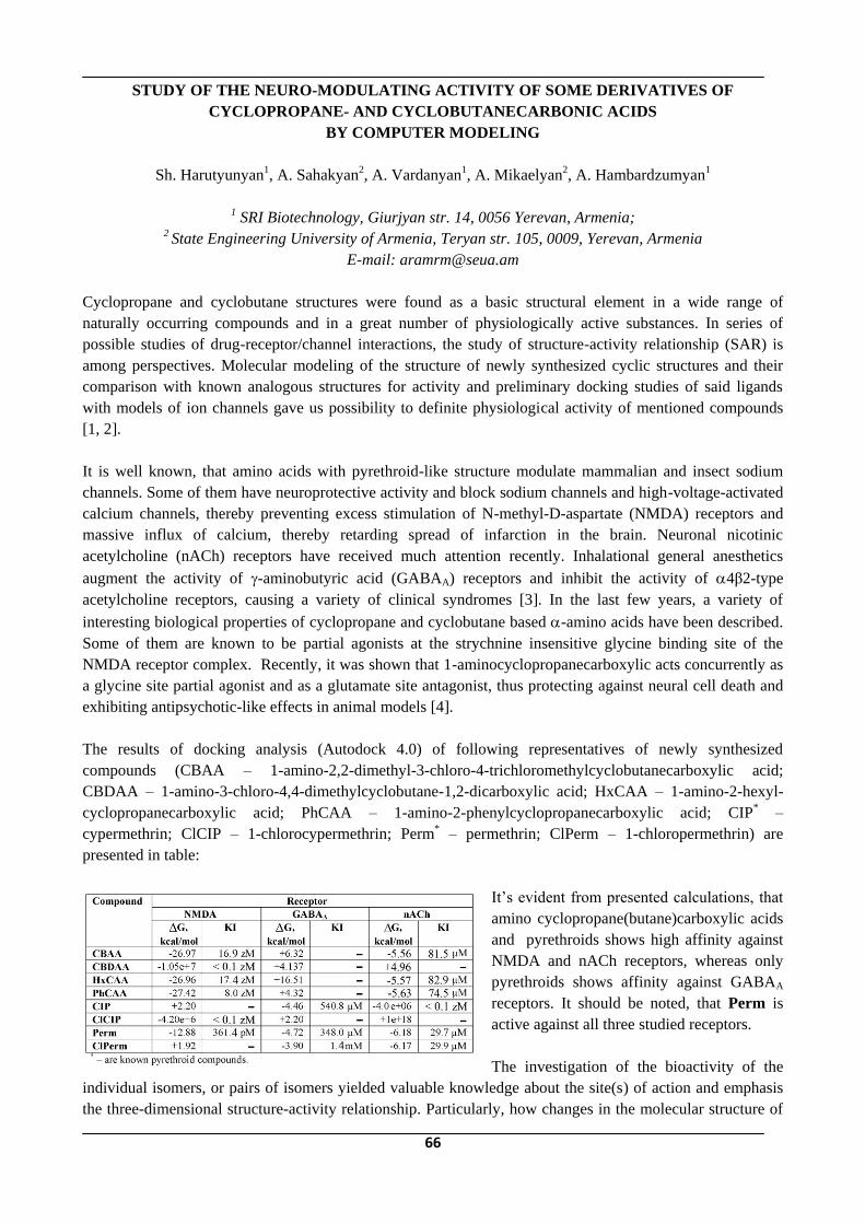
66
STUDY OF THE NEURO-MODULATING ACTIVITY OF SOME DERIVATIVES OF
CYCLOPROPANE- AND CYCLOBUTANECARBONIC ACIDS
BY COMPUTER MODELING
Sh. Harutyunyan1, A. Sahakyan2, A. Vardanyan1, A. Mikaelyan2, A. Hambardzumyan1
1 SRI Biotechnology, Giurjyan str. 14, 0056 Yerevan, Armenia;
2 State Engineering University of Armenia, Teryan str. 105, 0009, Yerevan, Armenia
E-mail: [email protected]
Cyclopropane and cyclobutane structures were found as a basic structural element in a wide range of
naturally occurring compounds and in a great number of physiologically active substances. In series of
possible studies of drug-receptor/channel interactions, the study of structure-activity relationship (SAR) is
among perspectives. Molecular modeling of the structure of newly synthesized cyclic structures and their
comparison with known analogous structures for activity and preliminary docking studies of said ligands
with models of ion channels gave us possibility to definite physiological activity of mentioned compounds
[1, 2].
It is well known, that amino acids with pyrethroid-like structure modulate mammalian and insect sodium
channels. Some of them have neuroprotective activity and block sodium channels and high-voltage-activated
calcium channels, thereby preventing excess stimulation of N-methyl-D-aspartate (NMDA) receptors and
massive influx of calcium, thereby retarding spread of infarction in the brain. Neuronal nicotinic
acetylcholine (nACh) receptors have received much attention recently. Inhalational general anesthetics
augment the activity of -aminobutyric acid (GABAA) receptors and inhibit the activity of 4β2-type
acetylcholine receptors, causing a variety of clinical syndromes [3]. In the last few years, a variety of
interesting biological properties of cyclopropane and cyclobutane based -amino acids have been described.
Some of them are known to be partial agonists at the strychnine insensitive glycine binding site of the
NMDA receptor complex. Recently, it was shown that 1-aminocyclopropanecarboxylic acts concurrently as
a glycine site partial agonist and as a glutamate site antagonist, thus protecting against neural cell death and
exhibiting antipsychotic-like effects in animal models [4].
The results of docking analysis (Autodock 4.0) of following representatives of newly synthesized
compounds (CBAA – 1-amino-2,2-dimethyl-3-chloro-4-trichloromethylcyclobutanecarboxylic acid;
CBDAA – 1-amino-3-chloro-4,4-dimethylcyclobutane-1,2-dicarboxylic acid; HxCAA – 1-amino-2-hexyl-
cyclopropanecarboxylic acid; PhCAA – 1-amino-2-phenylcyclopropanecarboxylic acid; CIP* –
cypermethrin; ClCIP – 1-chlorocypermethrin; Perm* – permethrin; ClPerm – 1-chloropermethrin) are
presented in table:
It‘s evident from presented calculations, that
amino cyclopropane(butane)carboxylic acids
and pyrethroids shows high affinity against
NMDA and nACh receptors, whereas only
pyrethroids shows affinity against GABAA
receptors. It should be noted, that Perm is
active against all three studied receptors.
The investigation of the bioactivity of the
individual isomers, or pairs of isomers yielded valuable knowledge about the site(s) of action and emphasis
the three-dimensional structure-activity relationship. Particularly, how changes in the molecular structure of

67
drugs and receptors/channels result in kinetic changes in the functions of receptors/channels. These results
afforded predictive capability for further synthetic endeavors.
1. Badanyan Sh.H., Stepanyan A.N., Mikaelyan A.R. et al., Zhurnal Organicheskoy Khimii (Rus.) 1997,
33(1):44-52.
2. Terzyan S.S., Mikaelyan A.R., Badanyan Sh.H. Zhurnal Strukturnoy Khimii (Rus.), 1998, 39(1):172-
175.
3. Narahashi T. The Journal of Pharmacology and Experimental Therapeutics, 2000, 294(1):1-26.
4. Truong M. et al. Tetrahedron: Asymmetry, 2003, 14:1063–1072.
68
PECULIARITIES OF PHOSPHOLIPIDS AND THEIR FATTY ACIDS COMPOSITION
ABNORMALITIES IN RAT BRAIN CHROMATIN STRUCTURE
AT UNILATERAL GANGLIOSYMPATHECTOMY
G.A. Hoveyan, M.A. Badalyan, A.G. Hoveyan*
H. Buniatian Institute of Biochemistry, NAS RA, 5/1 Paruir Sevak str., 0014 Yerevan, Armenia
*Yerevan State Medical University after M. Heratsi, Yerevan, Armenia
E-mail: [email protected]
Nowadays the revealing of the biomolecular mechanisms of wide scope of the sympathetic nervous
system functional activity becomes an issue of crucial importance. In this respect the role of the upper
cervical sympathetic ganglion as a modulator of organism's adaptational-trophical reactions; particularly
in its compensatory reactivity concerning to the hard-metabolizing compounds in the brain cells is
incomparably high. Its role is especially as a peripheral neuroendocrine center in regulation of metabolic
processes of phospholipids (PL) and their fatty acids (FA) in the intranuclear structures of the brain cells
that underlie many pathological states etiology and pathogenesis. The purpose of the present investigation
is to study the role of the right upper cervical sympathetic ganglion in processes of neuroendocrine
regulation and in regulation of the brain immunobiological activity by means of qualitative and
quantitative determining of changes in PL and their FA composition, as well as their physical and
chemical properties. The changes in contents and in ratio of the individual PL and their FA have been
studied at rats‘ unilateral gangliosympathectomy (removal of upper cervical right sympathetic ganglion).
The decrease of total PL contents testifies the depression of chromatin matrix activity as a result of
changes of hydrophobic interaction between DNA and histones as well as the conformational changes of
proteins with a new type of protein-protein interrelations inducing the decrease of RNA-polymerase
accessibility to the promotor region of gene. In the realization of those changes the decrease of
sphyngomyelins (SM) and increase of lysophosphatidylcholines (LPC) are of an important significance.
The decrease of SM also induces the decrease of RNA-polymerase I and II activity and changes in
quantitative correlation between bound and free form of enzyme. It is very important the variable changes
in the contents of Mono-, Di- and Triphosphoinositides. Those changes carry out expressed decreasing
character in PL of ganglioectomated site chromatin and also testify the destroying in the
phosphatidylcholinic and phosphoinositidic cycles functioning, which are well known in signal
transduction processes. In this case the increase of the level of saturated FA and the decrease of the level
of unsaturated, especially polyenic FA of chromatin PL in ganglioectomated hemisphere are of a
significant importance and are also the statement of RNA-polymerase activity inhibition. In chromatin of
the intact hemisphere the increase of the relative quantity of polyenic FA, particularly of arachidonic acid
takes place. An obvious increase of DPI, TPI and especially arachidonic acid, a slight increase of LPC
and relatively PS testify the possible compensator perturbations.
So, the results obtained indicate the significant role of sympathetic nervous system, especially its
peripheral part - UCSG, in regulation of minor components of chromatin - PL metabolism and their fatty
acids, testifying the adaptational-trophical action of sympathetic nervous system.
69
THE SEARCH OF RELATIONS BETWEEN AGGREGATIVE STATE OF BLOOD AND
INTERLEUKIN IL-1ß AT PATIENTS WITH ISCHEMIC STROKE
Hovhannisyan I.G., Hovhannisyan G.A., Tunyan Yu.S.
National Institute of Health after Academician S.Kh. Avdalbekyan, Ministry of Health of RA, Yerevan,
Armenia
Interdependence between containing of interleukin, aggregative state of blood cell‘s disorder and
neurological state was investigated at 114 patients with ischemic stroke (IS) during acute and sub acute
periods of the disease. 32 practically healthy persons were examined as a control group.
Carrying out examination allowed to determine that during acute period of IS, there are marked disorders of
aggregative peculiarities of blood cells which are characterized both by the raise of aggregative peculiarities
of erythrocytes and blood platelets, and by lowering of erythrocytes deformability. At the same time the
indices IL-1ß raise the level of norm almost fourfold. During sub acute period of the disease indicating
indices in comparison with period of diseases also expose to marked improved changes, however, they,
nevertheless, don‘t reach the level of norm, and the indices IL-1ß are considerably lowed and practically
don‘t distinguish from the norm. During both periods of IS between indices IL-1ß positive correlative
relations was revealed which is the evidence of accompaniment of aggregated platelet lyses with expression
of interleukin IL-1ßsynthesis.
It is noteworthy that during both periods of diseases between indices of blood platelet aggravation,
interleukin IL-1ß and neurological status positive, correlative relations is revealed which is more marked
during acute period at the same time the correlative relation was absent at died patients (42 men).
The results of our investigation let us suppose that previously supposed interrelation between two processes
– activation, aggravation and degranulation of platelets and expression of interleukin synthesis are proved
facts. However, it only testifies that the lysis of aggravated platelets is accompanied by the raise of
interleukin IL-1ß but it can‘t be the evidence of worsening of clinical state of health of the patients with IS,
owing to revealed relation. So, the present investigation ought to be considered as an attempt of search of
interrelations between considered factors. Conducting of wide-ranging, randomized investigations would
contribute for revealing of cause-effect relationship between the state of microcirculation and the system of
cytokine at different pathological states.
70
THE ROLE OF BIOLOGICAL ACTIVE SUBSTANCES IN THE ACTIVITY OF
SUCCINATDEHYDROGENASE IN SOME BRAIN STRUCTURES UNDER VIBRATION
Hovhannisyan K.R., Hovhannisyan H.H., Minasyan S.M.
Department of Human and Animal Physiology, Faculty of Biology, Yerevan State University,
1 A. Manoogian St., 0025 Yerevan, Armenia
E-mail: [email protected]
In present ecological conditions the influence of negative environmental factors on human organism is in
great interest. Vibration, due to reflector impact on CNS, causes disturbances in biological redox processes
of tissues and organs. It can lead to changes of the level of enzymatic activity of brain structures. The activity
of anaerobic metabolism enzyme such as succinatdehydrogenase is rather sensitive characteristic of cell
respiration under changed physiological state of organism in the conditions of vibration influence. The
natural preparations, mainly photogenes with adaptive activity, are used to increase immunity, resistance of
organism, prevention and complex treatment of many diseases. Biological active substances of plants are
close to natural metabolites of organism and well compatible with them, many of them are necessary for vital
functions. Licorice roots (Glicyrrhiza glabra L.) are widely used in medicine for prevention and treatment of
vide range of diseases. At present, number of effective preparations with immunopotentiating, adaptive,
antioxidant, antistress activities is produced from them. It is experimentally proved that such properties of
Licorice roots are caused by variety of biological and pharmacological active substances in them. In the
present work we investigated changes in the activity of succinatdehydrogenase in thalamus and
hypothalamus of rabbits under combined influence of vibration and Licorice roots.
Experiments were carrying out on 16 mature rabbits of breed the Chinchilla. Animals were provided by
Licorice roots (150 mg per 100 g weights of body). Animals were exposed to vibration influence on
vibroplatform EV-1 with frequency of 60 Hz, amplitude 0,4 mm for 30 days, 2 hours session daily. The
criterion of the enzyme activity was bleaching time of methylene blue in homogenate in vitro in anaerobic
conditions. The decolouration time, the above activity of enzyme is shorter. Three groups of animals were
studied. The analysis of the received data has shown that 30-day-long influence of vibration (I group) led to
fall of succinatdehydrogenase activity in studied brain areas. In the II group under combined influence of
vibration and Licorice roots there was marked fall of the enzyme activity, i.e. decolouration time of
methylene blue was extended in thalamus by 12,0 % (p<0,001), in hypothalamus - by 203,6 % (p<0,001) in
comparison with I group. In the III group exposed to combined influence of vibration and Licorice roots (30
days) after preliminary 30 day feedings in homogenate and solutions the hyperactivity of enzyme in studied
brain structures was observed, i.e. decolouration time methylene blue was shortened in thalamus by 35,7 %
(p<0,001), and in hypothalamus - by 32,7 % (p <0,001) in comparison with the vibrated animals.
In anaerobic metabolism conditions the requirement of cells in glucose is more and its natural intake usually
cannot satisfy to available requirement. It is supposed that mono- and disaccharides (to 20%) as well as
water-soluble polysaccharides that Glicyrrhiza glabra L. roots‘ contains have compensatory value for
maintenance of requirement of tissues in these substances, so they stimulated anaerobic metabolism, and
therefore, compensatory power supply of tissues. The above-mentioned fact allows concluding that
preliminary 30-day feeding of rabbits with Glicyrrhiza glabra L., further 30-day combined influence
Glicyrrhiza glabra L. and vibration substantially raises the activity of succinatdehydrogenase in thalamus
and hypothalamus. This testifies activation of compensatory possibilities of anaerobic metabolism under
vibration as a stress factor directed for maintenance of energetic homeostasis of the brain in the conditions
of long influence of vibration.

71
ULTRASTRUCTURAL PECULIARITIES OF WHITE RAT BRAIN MITOCHONDRIUMS IN
EXPERIMENTAL EPILEPTIC ATTACK BY KORAZOL AND IN CYTOPROTECTIVE ACTIONS
OF ANTIOXIDANTS
Hovnanyan K.O. 1, Simonyan A.A. 2, Simonyan L.A. 2, Margaryan A.S. 2, Batikyan I.G. 2,
Hovnanyan M.K. 1, Asatryan R.G. 1
1Institute of Molecular Biology, NAS RA, 7 Hasratyan St., 0014 Yerevan, Armenia;
2 H. Buniatian Institute of Biochemistry, NAS RA, 5/1 Paruir Sevak str., 0014 Yerevan, Armenia
E-mail: [email protected]
Now among various ways to cause experimental convulsive condition most adequate to epileptic human attacks
is the psychomotor form of epilepsy induced by corazol. Considering that mitohondria of eukaryotic cells is the
most sensitive indicator, defining functional activity and energetic power supply of the cells, essential interest
represents the analysis of ultrastructural changes of mitochondria in conditions of real processes proceeding in
vivo. The purpose of the present research consisted in carrying out special observation in directions of
ultrastructural changes of rats brain mitochondria after corazol influence , and on a background of application
by antioxidants actions a-tocopherol (TF) and thiosulfat sodium (TSN), having in view available information
on volatile reorganizations of mitochondria morphology.
White rats (180-200 grams, not purebred) were used in these experiments. Epileptic-like concussions were induced
by disposable intramuscular injection of corazol at the rate of 8 mg per 100 gram of animal weight. Control animals
received 1 ml of physiological solution. With the purpose of preliminary sensibilization factors endogenous
antioxidant activity, disposable intramuscular injections of 1 mg TSN and 0,4 mg of -TF oil solution per weight of
animal were performed. After euthanasia, brain slices from control and experimental animals were collected and
received mitochondrial fractions for transmission electronic microscopy were fixed by 2,5% solution of
glutaraldehyde in phosphate buffer with subsequent postfixing by 1% solution of osmium tetroxide in phosphate
buffer. After water draining in alcohol and steep in araldits, samples were concluded in a mix of araldits. Ultrathin
sections made on ultramicrotome Ultracut-Reichert, after contrasting with uranyl acetate and lead citric acid were
observed under Electron microscope Tesla BS-500 at an accelerating pressure of 80 kv.
Investigation of isolated mitochondria ultrastructure in suspension and in brain tissue after corazol injection
shown the role of corazol in their dynamic re-structuring and establish mitochondrial ultrastructure changes
polymorphism in the form of energetically defined conditions of mitochondrial ultrastructure, i.e. by
configuration changes. The structures of mitochondria well correlated with their functional condition. We
found that isolated mitochondria were affected by corazol, i.e. condensation of matrix, expansion of
intermembranous space, occurrence of vacuoles and separation of external membrane.
Also increased volume of intermembranous space can be noticed in such cases. Mitochondria in suspension
and in fabrics of brain after -TF and TSN influence in the greater part restore structural integrity. Revealed
by us various configuration conditions of mitochondria is not the nonspecific answer of mitochondrial
ultrastructure on developing pathological process. These changes of mitochondrial structure can be explained
on the basis of classic research data regarding interrelations of mitochondrial ultrastructure and their
functions. TF and TSN render cytoprotective effect and thus could be used for reduction of cytoprotective
effect.
Thus, results of electron microscope analysis make possible an estimation of functional condition of brain at
EFP according to ultrastructural changes of mitochondria, and also cytoprotective correcting effect of
antioxidants (TF, SST).

72
OXIDATIVE MODIFICATION OF PROTEINS IN WHITE RAT BLOOD SERUM UNDER NOISE
AND Α2–ADRENOBLOCKER INFLUENCE
L.S. Hunanyan,* G.A. Poghosyan,** O.P. Sotsky,** Shirinyan E. A.,***M.M. Melkonyan*
*Yerevan State Medical University after M. Heratsi , Chair of Medical Chemistry, Yerevan, Armenia;
**Laboratory of Biochemical and Biophysical Studies of YSMU Research Centre, Yerevan, Armenia;
***Institute of Fine Organic Chemistry, NAS RA, Yerevan, Armenia
E-mail: [email protected]
A great amount of real facts concerning the influence of oxygen active forms on the functional state of cells
and its role in the pathogenesis of many diseases has been accumulated. The recent studies showed that
products of oxygen metabolism first damage protein structures.
We represent here the results of investigations on the influence of noise (acute acoustic stress) and –
adrenoblocker on blood serum protein oxidative modification (POM). POM was evaluated basing on
dinitrophenylhydrasone (the interaction products of protein oxidized aminoacid residuals with 2,4-
dinitrophenylhydrasine) content shifts, which were registered using spectrophotometer SP-26 at wave
lengths 356, 370 and 430 nm.
Experimental animals (white male rats, 130-150 g) were subdivided into 6 groups. Rats of the first group
(intact animals) served as control group. Rats of the 2-nd, 5-th and 6-th groups underwent noise influence with
level of 91 dBA with maximum energy in the range of medium and high frequency waves during 2 hours.
The animals of the 3-rd - 6th groups were administered -adrenoblockers intraperitoneally in the dose of 2
mg per body mass.
Beditine (for the first time synthesized in the Institute of Fine Organic Chemistry, NAS RA) and isadoxan, as
a comparison standard, were used as -adrenoblockers. All experimental animals were decapitated in 2
hours; serum was collected for studies.
Marked increase in POM compared to the intact group animals was revealed in the result of acoustic
influence that was shown by elevated level of dinitrophenylhydrason in blood serum. It is worth mentioning
that the increase degree of the latter at various wave lengths is different, what is obviously explained by
POM different derivative product formation.
Thus, if POM product level registered at the wave length of 356 nm is almost twice higher under acoustic
influence, whereas at the wave length of 430 nm it is higher about 20 times. Intraperitoneal administration of
- adrenoblockers also increases POM intensity, though in a lesser degree compared to the acoustic
exposure, especially in case of beditine administration. The study of POM products registered at wave
lengths of 356 nm and 370 nm has revealed their higher growth under isadoxane influence than that of
beditine, whereas POM product amount at wave length of 430 nm was almost the same under the influence
of the studied -adrenoblockers.
Beditine preliminary administration, unlike idasoxane, not only fails to increase POM product amount, but
significantly prevents their increase. It is important to mention that dinitrophenylhydrasone level decrease
under beditine and noi
Thus, summing up the data obtained we can come to the following conclusions.
I. Noise influence causes expressed increase of dinitrophenylhydrasone content.
II. -adrenoblockers increase blood serum POM compared to the intact animals, in which
beditine does it in a lesser degree.
III. Beditine preliminary administration prevents the increase of blood serum POM products.

73
CALCINEURIN IS A KEY ENZYME UNDERLYING THE MOLECULAR MECHANISM OF
HEMORPHINS ACTION IN PATHOPHYSIOLOGY OF SARCOMA-45
Hunanyan O.1, Stepanyan H.2, Sarukhanyan F.1, Zakaryan H.1, and Barkhudaryan N.1
1 H. Buniatian Institute of Biochemistry, NAS RA, 5/1 Paruir Sevak str., 0014 Yerevan, Armenia 2 Institute of Fine Organic Chemistry, NAS RA, Yerevan, Armenia
E-mail: [email protected]
In an early in vitro study we showed that hemorphins modulate brain and lymphocytes
Ca2+/calmodulin(CaM)-dependent protein phosphatase calcineurin activity by binding with CaM, exhibiting
concentration-dependent biphasic response on enzyme activity. We have identified CaM as an essential
component in starting up the molecular mechanisms of hemorphins action and calcineurin as a key enzyme
underlying the molecular mechanisms of hemorphins action in the brain and immune system. It is well
known that calcineurin play an important role in the brain and immune system. This enzyme is involved in
the signal transduction cascade leading to T-cell activation by involvement in gene expression and
production of several cytokines (interleukin-2 (IL-2), IL-4, tumor necrosis factor-alpha (TNF) and etc.) via
dephosphorylation of nuclear factor of activated T-cells (NFAT) family members. In the brain calcineurin
regulates neurotransmitters and neuropeptides and neurohormones release. Calcineurin is involved in
pathophysiology of severe diseases, such as Alzheimer disease, diabetes, cancer and etc. In present work we
study the influence of hemorphins on plasma and brain calcineurin activity in pathophysiology of sarcoma-
45 (S-45). In the experiments were used 3 hemorphins: LVVYPW, LVV-hemorphin-7 (LVVYPWTQRF)
and hemorphin-7 (YPWTQRF).
For the experiments were used rats (Wistar line, 100-120 g). Rats were randomly divided into 5 groups
(n=10 in every group): control group injected by saline, and 4 experimental groups inoculated with sarcoma-
45. 3 of experimental groups after inoculation of S-45 received treatment with hemorphins. Each group
received the treatment with specific hemorphin during 8 days (single intraperitoneal injection of hemorphin
(1 mg/kg) daily).
Results obtained indicated down regulation of calcineurin activity in rats bearing S-45 in both plasma (2.7
fold) and brain (2.3 fold). All 3 groups of rat received treatment with different hemorphins exhibited increase
in calcineurin activity. More pronounced results in all 3 groups were received for plasma calcineurin activity,
when enzyme activity was almost reaching to the level of calcineurin activity in the control group. It is
necessary to emphasize that all 3 hemorphins demonstrated the inhibition of tumor growth. It is suggested
that hemorphins as a members of endogenous protective system of the organism come into play mainly in
response to pathophysiological conditions. In that case hemorphins, like other pleiotropic neuropeptides,
serve as one of the factors that switch on the compensatory systems in the organism. This is based on several
mechanisms and by implication of different signaling pathways in order to recover the homeostatic
disturbance. We think that in given case the hemorphines act as homeostatic agents via modulation of
calcineurin-NFAT pathway.

74
SOME CLINICAL LABORATORY PECULIARITIES IN PATIENTS WITH
NEUROENDOCRINE SYNDROM WITH NORM- AND HYPERPROLACTINEMIA
Karapetyan I.O., Beglaryan G.A.
Center of Family Planning and Sexual Health; Department of Obstetrics and Gynecology # 1,
Yerevan State medical University after M.Heratsi, Yerevan, Armenia
E-mail: ob&[email protected]
There were studied 107 female patients with neuroendocrine syndrome (NES) at the age of 23-35
(26.8 ±0.6). After clinical diagnostic verification the patients were divided into 2 clinical groups: the I group
involved the patients with normoprolactinemia (40 patients) and the II one - with hyperprolactinemia (67
patients). According to the case histories 77 females (71.9%) had the diagnosis Hypothalamic syndrome in
puberty period (HTSPP) and only half of them underwent treatment what indicates transformation of
NES from HTSPP. Hereditary aggravation (on endocrine pathology) was determined in 22.5% patients of the
group and 40.2%o - of the II one, and expressed premorbid state in patients with normoprolactinemia
was revealed in 10.0% and reliably (p<0.001) more (by 55.2%) in women with hyperprolactinemia.
The leading complaint in absolute majority of the patients was infertility (in the I group secondary infertility
was noticed twice often), dysfunction of menstrual cycle according to the secondary amenorrhea type
(10.0%; 37.3%), opsomenorrhea (72.5%; 62.7%). The maintained menstrual rhythm was noticed only in the
I clinical group (17.5%o). Galactorrhea was detected in 31 (46.3%) patients with hyperprolactinemia which
didn't depend on PRL level. Headache (31.3%), weakness (4.4%) paresthesia of various types (7.4%) were
nonspecific complaints observed only in the II group patients. Objective data revealed that 63 (58.9%)
patients were overweight, among them 41 women of the II group. Body mass index (BMI) was on average
29.8 ± 0.4. Microadenoma not more than 2 mm in size was revealed by CT and MRI methods only in 4
patients in the II group. Hormonal monitoring exactly revealed normoprolactinemia (PRL - 17.9 ± 1.24
ng/ml, in norm - 8.39 ±20.15 ng/ml) and hyperprolactinemia 29.3 ± 2.06 ng/ml), p<0.001. LH level was
lower than the average norm index ( 3.76 ± 0.07 mlU/ml; 2.81 ±0.16 mlU/ml) in both groups with reliable
difference (p< 0.002) unlike FSH which was almost the same. Hypoestrogenia was typical for both clinical
groups and that reflected both cycle dysfunction character (secondary amenorrhea, opsomenorrhea) and PRL
level. All patients were euthyroid. The changes in blood lipid spectrum were revealed in 44 (41.1%) patients.
The changes were characterized by general cholesterol (CS) increase and pCS - low density lipoproteins
(LDL) but the difference between the I and II groups was not reliable (p<0.05).
Ultrasonic study method of the small pelvis determined that 2 uteral sizes became less (posterioanterior
and width) and that was directly associated with the cycle dysfunction type (amenorrhea, opsomenorrhea)
and E2 level in blood.

75
SPECIFIC FEATURES OF PHOSPHOLIPID MOIETY OF PROTEOLIPIDS FROM PRIMARY
AND SECONDARY LYMPHOID ORGANS OF SOME MAMMALS
K.H.Manukian, K.L.Levonian, T.I.Kazarian, H.A.Stepanian, L.G.Kurakosian, H.A.Harutyunyan
H. Buniatian Institute of Biochemistry, NAS RA, 5/1 Paruir Sevak str., 0014 Yerevan, Armenia
In the course of investigations aimed to elicit the specific features of integral hydrophobic membrane
proteins-proteolipids (PLs) and lipids bound with them in cellular membranes of brain, especially rich in
these compounds, and other organs, it was important to study peculiarities of protein and lipid moieties of
PL, isolated from the primary (thymus) and secondary (spleen, lymphatic node) lymphoid organs of the
immune system of some mammals. Recently in definite cells of these three lymphoid organs expression of
major PL protein of myelin and its isoform-protein DM-20 was revealed. These proteins parallel with basic
protein of myelin are candidate antigens of autoimmune diseases including multiple sclerosis.
Investigations had shown that PLs are present in quite definite amounts, comparable with other organs, in all
lymphoid tissues studied. Concentration of proteolipid proteins (PLPs) was higher in lymphatic node (cattle)
– 1,73 mg/g of fresh tissue, somewhat lower in spleen (2,5- and 4- months old rats) – 1,38 mg/g and several
times lower in thymus of rats (2,5- and 4- months old) and rabbits (3- and 5-months old) – 0,45 and 0,35
mg/g of fresh tissue respectively.
The main task of present work was isolation of soluble preparations of PLs from lymphoid organs (thymus,
spleen – 3- and 4-months old rats; lymphatic node, armpit-cattle) and investigation of composition and
quantity of phospholipids, loosely bound and more tightly bound with PLPs. Simultaneously the
concentration of individual phospholipids in total lipid extracts of studied organs was determined. Isolation
of PLs and further analysis of their lipid (phospholipid) components were performed according to methods
described earlier [1].
Investigations had shown that total lipid extracts of all three lymphoid organs don`t significantly differ in
their phospholipid composition. They all were characterized by relatively high concentration of
phosphatidylserine, phosphatidylinositol and sphingomyelin. Together with similiarities quite definite
differences were observed particularly in the phospholipid composition of more tightly bound with PLPs
lipid fractions of lymphoid organs. Loosely bound with PLPs, lipid fractions of all three organs contained
mainly neutral phospholipids, viz., phosphatidylcholine, phosphatidylethanolamine, and sphingomyelin,
which in sum constituted 81-86% of all loosely bound phospholipids. These lipid fractions of lymphoid
organs were characterized by relatively high concentration of sphingomyelin (9,62-12,65% of all loosely
bound phospholipids). Relatively high content of loosely bound sphingomyelin was also specific feature of
PLs from brain and especially kidney. In lipid fractions more tightly bound wit PLPs the acidic
phospholipids prevailed, forming 75,4-80% of total of the tightly bound phospholipids. The PLs of three
lymphoid organs studied were characterized by the predominance of quite definite more tightly bound acidic
phospholipids. PLs of thymus and spleen were quite similar in phospholipid composition of tightly bound
with PLPs lipid fraction. It was characterized by high concentration of two acidic phospholipids:
phosphatidylserine and phosphatidylinositol, which constituted 32,18 and 28,22% of all phospholipids
tightly bound with PLPs in thymus and 30,53 and 29,65% in spleen respectively PLs of such type were
revealed by us earlier in kidney medulla and in liver microsomes. In contrast to these two lymphoid organs,
tightly bound with PLPs lipid fraction of lymphatic node PLs, was characterized by predominance of
phosphatidylserine, which made 39,83% of all tightly bound phospholipids, concentration of
phosphatidylinositol was two fold lower – 20,06%. Phospholipid composition of PLs of lymphatic node was

76
more similar with PLs of brain and its myelin, but in myelin the content of phosphatidylserine more tighyly
bound with PLP was considerably higher – 60-70% and that of phosphatidylinositol-lower – 14,6%.
Comparing presented data with ones obtained by us earlier on phospholipid composition of PLs from
different organs and their subcellular particles it may be concluded that in tissues of immune system, besides
of myelin PLs, other cellular membranes of PLs are also present. Only further investigation of cellular
elements of lymphoid organs may reveal localization of different PLs in these tissues.
Reference
1. Manukian, K.G., Kazarian T.I., Kirakosian L.G. et al. Neurokhimia, 2003, vol.20, pp.101-104.
77
MECHANISMS OF AGGREGATION OF GLOBULAR OLIGOMERIC PROTEINS AND
SUPPRESSION OF AGGREGATION BY CHAPERONES
Markossian K.A*, Kurganov B.I.
*Bach Institute of Biochemistry, Russian Academy of Sciences, Moscow, Russia
E-mail: [email protected]
Protein aggregation arises as a result of protein misfolding in response to mutations, posttranslational
modifications or changes of local conditions such as pH and temperature. Protein aggregation underlies
pathogenesis of a considerable part of human neurodegenerative diseases (such as Alzheimer‘s, Parkinson‘s
and Huntington‘s diseases, spinocerebellar ataxia, prion encephalopathies, amyotrophic lateral sclerosis,
systemic amyloidosis). Many misfolded proteins in cells expose hydrophobic areas on their surface, which
can interact leading to formation of disordered (amorphous) or ordered protein aggregates (amyloid fibrils).
The kinetics of aggregation of globular oligomeric proteins (bovine lens beta-crystallin, rabbit skeletal
muscle glyceraldehyde-3-phosphate dehydrogenase (GAPDH) and creatine kinase, pig heart mitochondrial
aspartate aminotransferase) has been studied by dynamic light scattering (DLS). The DLS data show that
the initial stage of thermal aggregation of proteins is formation of the start aggregates. Further sticking
of the start aggregates and aggregates of higher order resulting with formation of large-sized amorphous
aggregates proceeds in the regime of diffusion-limited cluster-cluster aggregation (DLCA). It is evident
that the start aggregates resulting in formation of amorphous aggregates are similar to protofibrils formed in
the course of assembly of fibrils.
A comparative study of the anti-aggregation effect of small heat-shock protein alpha-crystallin and
chaperonin GroEL has been carried out. Both chaperones suppress thermal aggregation of GAPDH. The
analysis of the initial parts of the dependences of the hydrodynamic radius of protein aggregates on time
shows that in the presence of alpha-crystallin or GroEL the kinetic regime of GAPDH aggregation is
changed from the DLCA regime, wherein the sticking probability for the colliding particles is equal to unity,
to the reaction-limited cluster-cluster aggregation regime, wherein the sticking probability for the colliding
particles becomes lower the unity. The interaction of alpha-crystallin with unfolded GAPDH blocks the
formation of the start aggregates. Suppression of aggregation by alpha-crystallin is due to the diminution of
the size of the start aggregates and increase in the duration of the latent period. The interaction of GroEL
with unfolded GAPDH does not affect the formation of the start aggregates. However GroEL incorporated in
the start aggregates reduces the sticking probability for the colliding start aggregates.
This study was funded by the Russian Foundation for Basic Research (grant 08-04-00666-a) and the Program
―Molecular and Cell Biology‖ of the Presidium of the Russian Academy of Sciences.

78
INPUT OF QUERCETIN IN PHOTOTOXITY OF H. PERFORATUM EXTRACTS
Martirosyan A.S., Vardapetyan H.R., Tiratsuyan S.G., Hovhannisyan A.A.
Russian-Armenian (Slavonic) University, H.Emin str.123, 0051 Yerevan, Armenia
Email: [email protected]
Apart from the ancient methods of use, modern techniques result in numerous innovations, where St John‘s
wort (Hypericum perforatum) can be used in the field of health care. H. perforatum is used mainly in
disturbances such as depression, nervous breakdown, stress, aggressiveness, etc. There are numerous
compounds within H. perforatum preparations, where the major classes are naphthodianthrones, e.g.
hypericin (HY) (1,3,4,6,8,13-hexahydroxy 10,11-dimethylphenanthroperylene-7, 14-dion) and flavonoids,
the representative of which is quercetin (2-(3, 4-dihydroxyphenyl)-3,5,7-trihydroxy-4H-1-benzopyran-4).
Nowadays these secondary metabolites are of great interest due to antioxidantive, anti-inflammatory,
immunomodulatory, gastro- and neuroprotective activities (quercetin), as well as because of their potential
application in photodynamic therapy of cancer, in blood sterilization and transfusion (HY) [1]. As drugs are
often injected to patients through blood system, study of the influence of H. perforatum components on
blood elements, in particular, erythrocytes, becomes an issue of current importance. In order to increase
therapeutic efficacy of H. perforatum preparations the overall mechanism, as well as the action of separate
components must be studied, the primary targets must be unrevealed. In the present work the influence of H.
perforatum herbal preparations and its separate components on model cells, such as erythrocytes was
studied.
The light-induced phototoxicity of H. perforatum extracts and HY were evaluated by calculating the
erythrocyte resistance. The ―thermal treated‖ extract (TE) and ―cold extract‖ (CE) were received from
flowers and leaves of H. perforatum. TE was received by Soxhlet extraction in methanol with further
lyophilization and dilution in ethanol, CE – by storing in 96% ethanol a week. Human erythrocytes were
received by centrifugation at 800g 15 min at 50C with further resuspension in buffer. After 10 min irradiation
by visible light (100W, 30mW/cm2 on the sample surface) photohemolysis degree was determined at 680 nm
on spectrophotometer Specord M400 (Carlzeiss, Germany).
Under the influence of visible light both extracts – CE and TE, as well as HY have expressed photodynamic
activity and reduce erythrocyte resistance. Irradiation of erythrocytes with CE and ascorbic acid has revealed
the protective effect of the latter despite its concentration on the contrary with pure HY and TE. It was
revealed that in CE extract there are some components, e.g. quercetin that suppresses photohemolysis
sensitized by HY. The latter was certified by conducting erythrocyte photosensitization in the presence of
quercetin. Suppression of erythrocyte photodestruction by quercetin is possibly connected with scavenging
of 1O2 and other radicals, generated during photosensitization reaction. The latter prevents apoptosis that is
considered as one of the causes of neurodegenerative disorders [2]. It could be concluded that TE and CE are
more powerful therapeutic tools and that agents existing in extracts, such as quercetin, can change outcomes
of HY action and make it more favorable for therapeutic application.
1. Miskovsky P. Current Drug Targets, V.3, P.55-84, 2002.
2. Middleton E., et al., Pharmacol. Rev., 52 (4), 673–751, 2000.
79
PRADER-WILLI SYNDROME AS HYPOTHALAMIC PATHOLOGY MANIFESTATION
Matevosyan A.A.1,3, Grigoryan E.G. 2, Simonyan K.G.1,3, Akopyan A.V. 1,3,
Melik-Pashayan O.E.3
1Yerevan State Medical University after M.Heratsi, Yerevan, Armenia; 2Scientific-Research Institute SPA Treatments and Physical Medicine, Ministry of Health of RA,
Yerevan, Armenia; 3“Neurohereditary Diseases” Charity Association
Prader-Willi syndrome (PWS) is genetic pathology which is manifested by wide spectrum of
neuroendocrinal, neurosomatic, neurovegetative and others disorders, in particular by obesity and
hyperphagia, growth deficiency, mental retardation, by disorders of behavior, breath, thermoregulation,
reproductive function, menstrual cycle, accompanied by respective changes in hormonal profile. Frequency
of diseases is 1:15 000 - 1:20 000 population. Clinical manifestations of the disease, neurological hormonal
researches (decrease of prolactin, estrol level in girls, gonadotropic hormone, etc.) testify of hypothalamic
character of the disorders.
It is established that PWS in caused by disorder in gene expression owing to defect of imprinting in the area
of 15 chromosomes (in region of a segment q11.2-q13). Critical region involve more than 12 genes leading
to dysfunction of hypothalamic area that causes neuroendocrinal disorders.
Interest to this pathology is caused by great attention to obesity problem. However, the disease is seldom
diagnosed in clinical practice, in spite of introduced descriptions of phenotype and clinical symptoms in
the literature, diagnostic opportunity of this pathology by modern cytogenetic and molecular genetic
researching methods (FISH and mutilation test) , worked out recently introduction of the new treatment
methods.
Dysfunction of hypothalamic area leads to fat metabolism disorder with over 10 times increase of fat
synthesis from acetate and rather decreased process of lipolysis. Hypogonadism on hypogonadal type can be
connected to hypothalamus dysfunction mainly of ventromedial and ventrolateral nucleus. Patients of both
sexes are usually sterile. Inguinal and umbilical hernia, joint hypertension, posture disorders, x-shaped
curvature of low extremities, etc., are apparently caused by muscular hypertension, hypertension joints, etc.
Increased risk of development of leukemia and other tumors in patients with Prader-Willi syndrome are
apparently caused by low reparation DNA ability, etc.
However there are no researches devoted to neuroimmunological changes in the given pathology in the
literature, though wide spectrum of hypothalamic neuroendocrinal and neurovegetative disorders should
assume an opportunity of neuroimmune regulation disorders in the pathogenesis of disease.
Carried out studies together with the International Association of Prader-Willi Syndrome (IPWSO) and,
Lency Found (USA) of PWS problem in Armenia, in particular, diseases prevalence, as well as clinical
peculiarities of syndrome, including neuromuscular disorders in PWS by using modern immunological
researches, allowed revealing some immunological alteration requiring further deep investigation.
Investigations allow to determine degree of neuroimmune system participation in pathogenesis of Prader-
Willi syndrome and to open new page in the study of this disease.

80
HYPOTHALAMIC PROLIN-RICH PEPTIDE AGAINST BRAIN DEGENERATION EVOKED BY
BETA-AMYLOID PEPTIDES
I.B. Meliksetyan1, J.S. Sarkissian1, Z.E. Avakyan1, E.A. Chavushyan1, K.B. Yenkoyan2, O.A. Nazaryan1, N.N.
Melkonyan1, A.A. Galoyan3
1L.Orbeli Institute of Physiology, NAS RA, 22 Orbeli Str., 0028 Yerevan, Armenia; 2 Yerevan State Medical University after M. Heratsi, Yerevan Armenia;
3 H. Buniatian Institute of Biochemistry, NAS RA, 5/1 Paruir Sevak str., 0014 Yerevan, Armenia
E-mail: [email protected]
By histochemical methods of revealing of Са2+-dependent acidic phosphate activity a comparative studies were
carried out of morpho-functional state of hippocampal and cerebral cortex cellular structures on Alzhimer‘s
diseases models. The model was induced by bilateral intracerebroventrical injection of β–amyloid peptides
Аβ 25–35, Аβ 1–40 and Аβ 1-42, combined with systemic administration of proline-rich peptide – 1 (PRP-1).
Morpho-histochemical study were carried out 18-22 weeks later after Аβ injection.
Morphological pictures of processes abundance of neurodegenerative changes in cerebral cortex under Аβ 25–35
is characterized by absence of areas continuity. The analysis of data concerning distribution of
neurodegenerative processes deep into and along cerebral cortex has shown that pyramidal layer is the most
frequently damaged area. The majority of neurons are subjected to central chromatolysis. However cells with
normal morphology were also detected. The analogous picture was observed in hippocampal slices. In the
CA3 and CA4 areas it is characterized by rounding and swelling of neuronal soma of big pyramidal cells of
dentate gyrus. Although these cells were more stable to amyloid, they also undergo to tendency the
cytoplasm clearing. The processes of neuronal splitting were accompanied by cancellous alveolar state of
neuronal cytoplasm.
At Аβ 1–40 injection the process of chromatolysis was also detected in cerebral cortex. However, here
greater in quantity and volume of ―gapped‖ was observed compared to Аβ 25–35. As a result, in cerebral
cortex occurs process of diffuse or limited evacuation of cellular layers, the spaces of which remains empty
owing to absence of reaction of glial cells nuclei. In hippocampal slices at Аβ 1–40 the swelling and
rounding of cellular soma with nucleus ectopy is also occurred. The central surface of cell appeared clear,
and in some neglected stages it was glassy.
At Аβ 1-42 injection in СА1 area of hippocampus slices we observed total disappearance of neurons reaction
in the form of semicircular pockets on equal distance from each other. As characteristic signs, we found big
amorphous phaeochrous neurons of dentate gyrus of hippocampus and ІІІ – ІV layers of cerebral cortex, with
disorderly located neurofibrils in cytoplasm. The mentioned data testify the processes of
hyperphosphoryllation that precedes late stages of Alzheimer‘s disease, and characterized by tau-related
neurofibrillar changes.
At Аβ 25-35 and 1-40 injections and PRP-1 administration, density of cells disposition in cerebral cortex and
in hippocampus increased. In cerebral cortex state of tensity of nerve cells markedly decreased; thicker
apical dendrites of pyramidal neurons started to act in response. The configurations became irregular,
however, further with development of regenerative processes, the dimensions decreased, the nucleus is
transformed into the center of the cell; the cytoplasm is filled up by precipitate granules. Different intensity
of neuronal staining was observed in slices, i.e. occured both dark-stained cells and neurons, in cytoplasm of
which on the light background a Homori–positive granules were visible. The similar picture we observed in
hippocampus. Everywhere the blood vessels were revealed, on external wall of which one can see a dark
pericytes with processes. At Аβ 1-42 injection and PRP-1 administration, there was sharp decrease of dark
cells number with neurofibrillar changes in cerebral cortex, suggecting that PRP-1 delayed tau-

81
phosphorylation of neurons. In СА1 area of hippocampus slices the devastated places were filled up by
neural cells with weak enzymatic activity.
Thus, results of these studies on PRP-1 use after different Аβ peptides injection allow suggesting that PRP-1
causes positive changes of structural properties of neurons, increase of metabolism that determines survival
of cells.
82
MECHANISM OF ENZYMATIC HYDROLYSIS OF DICHOLINE ESTER OF SEBACIC ACID BY
HUMAN SERUM BUTYRYLCHOLINESTERASE
M.R. Mnatsakanyan1, A.H. Papoyan1, V.A. Samokish2, G.P. Halebyan1
1Institute of Biotechnology, Gurjyan 14, Yerevan 0056, Armenia;
E-mail: [email protected]; 2 Institute of Analytical Instrument Engineering RAS, St. Petersburg, Russia
E-mail: [email protected]
Dicholine esters of aliphatic dicarboxylic acids (DChE) with the following general formula are bioactive
compounds of medical interest ascurarelike compound.
N
O
O
(CH2)n
O
O
N 2Hal-
The best known compound of this family succinyldicholine (DChE2, ditilin) is widely used in anesthesiology
as muscle relaxant of short action [F. Brucke, , Pharmacol. Rev. 1956, 8, 265-335. D. Bovet, Neuromuscular
blocking agents, in: J. Cheymol (Ed.), 1973. p.244]. The duration of their action is conditioned by their
hydrolysis by plasma butyrylcholinesterase (BChE). In the works of several authors it is shown that the
enzymatic hydrolysis of DChE by BuChE occurs by two sequential steps. In view of large difference
between values of Кm for DChE and product of its hydrolysis monocholine ester of dicarboxylic acid
(MChE), the latter is hydrolyzed forming diacid only after exhaustion of DChE in the reaction medium.
However during the study of enzymatic hydrolysis of DChE with n≥6 we discovered a deviation from
proposed mechanism whose essence consists in the formation of diacid in the first stage of the reaction
[Mnatsakanyan, M.R.; Hakobyan, L. M.; Sargsyan, J.V.; Samokish, V.A.; Halebyan, G.P. Biological Journal
of Armenia 2007, 59, 239-247]. In the present work in purpose to explain the observed phenomenon the
kinetics of enzymatic hydrolysis of DChE and MChE of sebacic acid (DChE8 and MChE8) by human serum
BChE was investigated as well as the values of the parameters of Michaelis-Menten equation were
determined for these substrates. It was shown that in contrast to succinyldicholine and succinylmonocholine,
for which Km(DChE)/Km(MChE) 10-2 and Vm(DChE) / Vm(MChE) 7, in case of DChE8 (Km(DChE) – 3.0 0.38 10-5
М, kcat(DChE) – 16.4 103 min-1) and MChE8 (Km(MChE) – 2.2 0.28 10-4 М , kcat(MChE) – 6.09 103 min-1), the
values of this parameters were closer: Km(DChE)/ Km(MChE) 0.15 Vm(DChE) / Vm(MChE) 2.7. The obtained
parameters for DChE8 and MChE8 were applied for plotting theoretical dependences of all reactants
concentration on time. As attested analysis of the obtained results, the individual parameters of Michaelis-
Menten equation for DChE8 and MChE8 do not explain observed accumulation of sebacic acid in the reaction
medium (which is nearly 1.7 10-4 М if substrate initial concentration is 5 10-4 М) in the first stage of
reaction if there is no deviation from pure competitive mechanism of hydrolysis. The proposed scheme of the
first stage of hydrolysis explaining the observed deviation from competitiveness is based on an assumption
that the part of MChE8 molecules (formed as a result of DChE8 hydrolysis) manages to be hydrolyzed due to
MChE8 slow diffusion from the region of active site of enzyme into reacting medium according to the
following scheme.
E + DChE [E -DChE]
E + MChE + choline
[E - MChE] E + diacid+choline

83
EFFECT OF CORONARY CONSTRICTORY NEUROPEPTIDE ON [35
S] TAURINE UPTAKE IN
RATS HEART AT THE BLOCKADE WITH STRYCHNINE – INHIBITOR OF TAURINE
RECEPTORS
Muradyan M. Sh., Sekhyan L.A.
H. Buniatian Institute of Biochemistry, NAS RA, 5/1 Paruir Sevak str., 0014 Yerevan, Armenia
In hypothalamus neurosecretory cells (NO and NPV) there are synthesized releasing hormones, as well as
cardiotrop neurohormones, which are specific regulators of cardiac blood circulation and cardio-vascular
system in a whole. In the adaptation mechanisms it was very important to find mediators, feed-back between
heart and hypothalamus. In 1967 A.A.Galoyan indicated that biosynthesis of peptide hormones and
peptidergic system takes place in atria ganglionary cells, which are signal systems for cardiotrop hormones
―К‖ and ―С‖ release into general blood flow. The neurohormones ―К‖ and ―С‖ are mediators like
acetylcholine and catecholamines. The sulfoamino acid taurine belongs to the system of mediators, which
changes the cardiac muscle reaction to adrenaline and acetylcholine influencing their receptors. A complex
regulatory system exists in heart, including Са, К, GAMP and taurine. To understand the conjunction of
those mediators in the regulation of cardiac blood circulation, we have studied the effect of coronary
constrictory neuropeptides (CCF) and strychnine (taurine blockader) on [35S] taurine uptake rate in different
parts of the heart at incubation for 10 min, 30 min, and 60 min. It is established that CCF in concentration of
3.10-8М during 10 min increases taurine uptake in the right and left atria for 53% and 84.2%, respectively,
and in left ventricle for 72%. At blockade of taurine receptors with strychnine in concentration 5.10-4М,
significant change of taurine uptake is not observed in all studied parts of heart. Combination of CCF and
strychnine induces increase of the uptake only in the left atria for 129.5%. The investigations fulfilled
indicate that at incubation of the slices from different parts of the heart during 30 min does not induce a
noticeable change in taurine uptake. At 60 min incubation an increase of taurine uptake is observed only in
the left atria for 129%, and in all other parts of heart an inhibition of the uptake is observed in the right and
left atria for 81.7% and 59%, respectively, and in cardiac ventricle for 67%. Strychnine removes the CCF
inhibiting effect and induces increase of uptake in the right ventricle, in the right and left atria for 101%,
125% and 120%, respectively. CCF in combination with strychnine an inhibition of taurine uptake takes
place in all studied parts of heart except the right atrium, where an increase of uptake is observed for 136%.
Thus, the obtained results indicate that in the basis of taurine physiological effect on the heart is the change
of cardiac muscle stimulation under CCF and taurine through the change of cell membrane permeability. The
effects of CCF and strychnine can be considered on taurine receptors level.

84
DNA METHYLATION AND ITS POSSIBLE CHANGES IN THE BRAIN
AT THE RETROGRADE AMNESIA
Nersesyan L., Danielyan I., Aharonyan A., Khudaverdyan N.*, Dzhagatspanyan I., Gharibyan J.
Institute of Fine Organic Chemistry of Scientific Technological Center of Organic and Pharmaceutical
Chemistry, NAS RA, Yerevan, Armenia;
*National Center of Oncology of the Ministry of Health of RA, Yerevan, Armenia
E-mail: [email protected]
The aim of present study was to investigate DNA methylation reaction in the cerebral cortex and cerebellum
of animals with amnesia before and after injection of the psychotropic preparations piracetam and
fenozepam. It is estimated the antiamnestic action during the formation of the conditioned passive avoidance
reflex with the subsequent use of electric shock as a factor of amnesia. It is established that during the
conditioned reflex formation the content of 5-methylcytosine in cerebral cortex is increased (80%) as
compared with the control (47%). Piracetam activated the function of neocortex and increased methylation
level of DNA in the cerebral cortex of rat brain. Fenozepam is not related to the conditioned reflex activity
and does not induce the changes of DNA methylation both in two studied regions of rat brain. Thus, DNA
methylation may correlate with the functional activity of brain cells in the process of the conditioned reflex
formation. Piracetam modulating the functional activity of brain cells can also change methylation level of
genome.

85
EXPERIMENTAL APPROACH TO STUDY THE SECRETED PROTEASES OF BRAIN
Onufriev M., Lyzhin A.*, Stepanichev M., Chaspekov L.*, Gulyaeva N.
Institute of Higher Nervous Activity and Neurophysiology RAS; Moscow, Russia;
*Institute of Neurology Rus. Acad. Med. Sci., Moscow, Russia
E-mail: [email protected]
The cysteine proteases caspase 3 and cathepsins are involved in both neuronal plasticity and neuropathology.
Caspase-3 and cathepsin B activities in extracellular medium (balanced salt solution, BSS) of primary rat
neuroglial cerebellar cultures were studied. At acidic pH, cathepsin B cleaved Ac-DEVD-AMC, a synthetic
caspase 3 substrate, this activity being significantly higher than that of caspase 3 at pH 7.4. This activity is
blocked by peptide inhibitors of both caspase 3 and cathepsin B. Substitution of culture medium for balanced
salt solution stimulated cathepsin B secretion in both types of cultures. Ischemia (oxygen–glucose
deprivation) significantly decreased secretion of cathepsin B activities into the culture medium.
This work was supported by the Russian Foundation for Basic Research (grant No.08-04-00870).

86
INFLUENCE OF NEW ADRENAL MEDULLARY CARDIOACTIVE PEPTIDES ON BRAIN AND
SOME ORGANS GLYCOGENPHOSPHORYLASE ACTIVITY
Paronian Z.Ch., Abelian J.G., Sahakian F.M., Srapionian R.M.
H. Buniatian Institute of Biochemistry, NAS RA, 5/1 Paruir Sevak str., 0014 Yerevan, Armenia
E –mail: zoyaparonyan@ rambler.ru
Development of new approaches and techniques to investigate specific CNS protein has led to quick progress
in studies of the adrenal medullary protein spectra. In adition to the catecholamines and nucleotides large
amounts of chromogranines and some identified and anidentified proteins and peptides: vasoactive intestinal
polypeptide, substance P, neuropeptide Y, somatostatin, neurotensine, opioid peptides, dopamine-ß-
hydroxylase, O-glycosylated polypeptides. Even this short list provides some perspectives onto the search for
new bioactive compounds in the adrenal medulla. Cardioactive compounds we discoverd earlier [1,2] in the
bovine adrenal medulla are interesting in this respect. Interest in these compounds as well as further studies
on their identification are associated with the involvement of adrenals in the realization of the
coronarodilating effect of hypothalamic neurohormones "K" and "C" [3].
Eight cardioactive compounds have been isolated from the bovine adrenal medulla. These compounds differ
from each other in terms of their pharmacological characteristics (the duration of action, the time taken to
increase and the magnitude of the peak effect). These differences appear to be associated with their structural
characteristics and particularly with the amino acid composition. The most interesting are two of these
compounds, lettered as M31 and M42 with Mr (4100-4200 D) and can be classified as the glycopeptides. M31
and M42 factors as a hypothalamic cardiotropic neurohormone "C" are resistent to various denaturing agents,
high temperature (110oC) and enzymatic treatment, to the effect of alkali (1N NaOH) and acids (6N HCl).
These factors demonstrate analogous to hypothalamic neurohormone "C" inhibition of cAMP PDE activity
and dependence of this effect from their coronary dilating potency as well.
In this study we attempted to investigate the possible participation of mentioned (M31 and M42)
compounds in the regulation of glycogenphosphorylase activity in brain and some organs of white rats.
Experimental data showed that M31 factor results in distinct changes of phosphorylase in the heart and
skeletal muscle. On the background of a decrease of a total phosphorylase activity balance there is some
prefential increase in the activity of Pa in the heart (48%) and skeletal muscle (62%), M42 does not effect on
a total phosphorylase activity, but markedly increase activity Pa in the brain and in the heart.
Glycogenphosphorylase is known to be controlled in several ways. In our case, probably there is covalent
modification on this enzyme which manifests itself in the alteration of the Pa/Pb coefficient in the heart and
skeletal muscle especially.
Results obthained provide indirect evidence about the intensification of glycogenolysis in these tissues. This
assumption has been confirmed alongside whit the data by observation on studies of altered glycogen level in
heart an skeletal muscle,where M31 factor decreased it (by 60%) and (by 42%) respectively. From these data
we can specifically see the coronary dilatory effect of adrenal medullary factors M31 and M42.
REFERENCES
1. Paronian Z.Ch., Srapionian R.M., Abramian S.S. et al., Neurokhimiya,1989,vol.8,
pp.365-370.
2. Srapionian R.M., Paronian Z.Ch., Sahakian S.A., Uspechi Physiol. Nauk,1998, vol 29,
pp.3-10
3. Galoyan А.А.,Srapionian R.M.,Sahakian S.А., ДАN ARM.SSR,1973, vol.56, pp.102-106.

87
RECOVERY PROCESSES IN RATS AFTER UNILATERAL PYRAMIDOTOMY, DESTRUCTION
OF RUBROSPINAL TRACT AND ACTION
OF BACTERIAL MELANIN
Petrosyan T.R.¹, Gevorkyan O.V.¹, Meliksetyan I.B.¹, Hovsepyan A.S.²,
Sagiyan A.S.², Manvelyan L.R.¹
¹ L.A. Orbeli Institute of Physiology, NAS RA, 22 Orbeli Str., 0028 Yerevan, Armenia;
² “Institute of Biotechnology” CJSC, Yerevan, Armenia
E-mail: [email protected]
Effects of bacterial melanin on the processes of posttraumatic recovery of instrumental reflex (IR) and limb
movements were studied after unilateral pyramidotomy on the bulbar level (n=12) and destruction of
rubrospinal tract on the level of C3-C4 segments in adult white mongrel rats (n=12), weighing 180 - 220 g
and initially trained to the IR. These operations caused unilateral limb paresis. On the next day after the
operation, one part of the animals was injected intramuscularly with the solution of bacterial melanin (BM),
obtained by biotechnological method, at a concentration of 6 mg/ml with an estimated optimally tolerable
dose of 170 mg/kg.
It is well known that corticospinal and rubrospinal tracts have almost the same function in the organization of
movements and this explains the process of functional compensation after lesion in one of these tracts.
Clinical signs for the isolated damage of each tract are also very similar and in both cases unilateral paresis is
manifested. Recovery of IR and balancing movements in pyramidotomized rats of control group was
completed within 19 days in average, and in rats with the destruction of rubrospinal tract - within 13.7±1.3
days in average. In rats injected with BM the complete recovery was over in average within 3 days in
pyramidotomized animals and within 2 days after the rubrospinal tract destruction.
Such a complete and rapid recovery was also confirmed by morpho-histochemical examination of brain
tissue of operated animals, shows that in these experiments applied bacterial melanin has a role of a
neuroprotector in the animal organism, accelerating the process of posttraumatic rehabilitation. Similarity of
responses registered from lumbar motoneurons of ―melanin‖ and intact animals by extracellular registration
in response to tetanic stimulation of pyramidal tract above the transection area, confirms as well the fact of
impulse conduction complete recovery in pyramidal tract after trauma.

88
PROLINE-RICH POLYPEPTIDE REGULATED THE ACTIVITY OF MEMBRANE
TRANSPORTING PROCESSES AT RADIATING DEFEAT
Sahakyan L. S., Ghazaryan P.A., Daghbashyan S.S., Galoyan A. A.*
Center of Haematology of the Ministry of Health of RA, Yerevan, Armenia;
*H. Buniatian Institute of Biochemistry, NAS RA, 5/1 Paruir Sevak str., 0014 Yerevan, Armenia
E-mail: [email protected]
The Aim of this work was the research of membrane transporting processes by radiation stress and the
possibilities of regulation of the broken ways by studying of Na/K-ATPase activity before and after
application of proline-rich peptide (PRP).
Materials and Methods Experiments were carried out on Wistar white male rats. Animals were exposed to
rays on the apparatus RUM-17. After 10 days of the exposure of 3Grej, PRP was introduced intraperitoneally
during 3 days at a rate of 5mg per 100g of animal weight.
Results According to the received data at a unitary total irradiation, the activity of Na/K - and Мg-ATФаз in
lymphocytes, thymocytes and spleen tissue was decreased, whereas in erythrocytes and brain it authentically
increased (р<0.001). After application of PRP in immunocompetent cells there was tendency to
normalization of studied enzymes activity; however in erythrocytes it remains to blood raised. The problem
of possible pathogenetic therapy of irradiation pathology by inclusion of PRP into the complex of therapeutic
means is discussed.

89
EFFECT OF PROLONGED VIBRATION CONJOINED WITH ACTION OF PRP-1
ON RESPONSES OF SUPERIOR VESTIBULAR NUCLEUS NEURONS,
EVOKED BY STIMULATION OF HYPOTHALAMIC
PARAVENTRICULAR AND SUPRAOPTIC NUCLEI
Sargsyan S.G.1, Minasyan S.M.1, Chavushyan V.A.3, Meliksetyan I.B.3, Sarkissian J.S.3, Galoyan A.A.2
1Department of Human and Animal Physiology, Faculty of Biology, Yerevan State Uuniversity, 1 A.
Manoogian St., 0025 Yerevan, Armenia
E-mail: [email protected]; 2 H. Buniatian Institute of Biochemistry, NAS RA, 5/1 Paruir Sevak str., 0014 Yerevan, Armenia
3L. Orbeli Institute of Physiology, NAS RA, 22 Orbeli Str., 0028 Yerevan, Armenia
E-mail: [email protected]
As known, the vibration action (VA), even by weak of their level, can be evoked the vibration disease,
striking the nervous, the cardiovascular and motor systems. Under prolonged VA observes the disturbance of
vestibular analyzer functions in type of subjective complaints on giddiness, violation of movement and other. In
present time the important place take in the main new approach, directed to the setting of CNS functions by
means use of endogenous neurohumoral factors of peptide nature. The special point in this respect take a new
hypothalamic prоline-rich peptide-1 (PRP-1) produced by paraventricular (PVN) and supraoptic (SON) nuclei
of hypothalamus.
The studies of plasticity were made, detected by changes of synaptic transmission and survival of superior
vestibular nucleus (SVN) neurons in conditions of prolonged VA and PRP-1 administration. On the Albino
rats were studied the hypothalamic-vestibular connections by means bilateral recording of evoked activity of
SVN single neurons at high frequency stimulation (HFS 100 Hz, 1sec) of PVN and SON. Experiments were
carried out at three series: 1 – intact animals; 2 – animals subjected by 15 days VA (frequency 60 Hz,
amplitude of vibrations 0,4 mm, 2 hours daily); 3 – animals, whom parallel with 15 days VA in first two
days over 2 hours before exposition were injected PRP-1 (0,05 mg/kg i/m). The registration and programmed
mathematical analysis of impulse activity were made in real time on-line.
In the first series analysis of pattern reaction of neurons of right-side SVN on HFS of ipsi SON and contra
PVN was revealed: the most intensity of tetanic potentiation (TP) – 65,8% and 60,5% respectively, combined
with posttetanic (TP+PTP) – 23,7% and 13,2% respectively. In the neurons of left side SVN at HFS of contra
SON TP is composed 51,1%, ipsi PVN – 62,2%, TP+PTP – 22,2%, and 20,0%, respectively. In second series
on HFS contra PVN in right side SVN has been revealed reactions of neurons with TP – in 41,2% cells, PTP
– 32,5%, TP+PTP – 11,7%, but under stimulation ipsi SON – TP in 26,5%, PTP – 32,4%, TP+PTP – 23,5%
cells. The activity of neurons of leftside SVN on HFS of ipsi PVN originated TP in 20,0% cells, PTP – 28,6%,
TP+PTP – 11,4%, TD+PTP in 14,3%, but at HFS of contra SON were revealed TP in 31,4%, PTP – 22,9%,
TP+PTP in 17,1% of neurons. In 3-th series of experiments in conditions of PRP-1 administration and
prolonged VA of HFS of hypothalamic nuclei on SVN nuclei was revealed of following poststimulus
manifestations of excitability: at HFS of ipsi SON in right SVN with TP in 72,7% cells, PTP – 15,1%,
TP+PTD and TP+PTP per 6,1%, by absent of areactive neurons; by stimulation of contra PVN took place TP
– 54,5%, PTP – 6,1%, presence areactive cells – 36,4%. Under HFS of ipsi PVN in neurons of neurons of left
side SVN 74,4% were composed the cells with TP and PTP, TP+PTP per 2,8%, but by stimulation of contra
SON 60% with TP and 14,3% with TP+PTP.
The disturbances of vestibular functions originating in postvibration period in useful degree has been
conditioned by changes in neuromediatory processes, connected with involvement of powerful adaptive

90
systems. As known, under action of vibration significantly changes the reception of corticosteroids, reflecting
the dependence of shifts of cholin-monoamine and GABA-ergic neuronal activity from level of glucocorticoids
and functional state of hypothalamo-hypophyseal-adrenocortical system.
Thus, the mechanisms of neuromediatory adaptation in conditions of VA, covering the GABA-mediated
inhibition in CNS, evidently activates under action of PRP-1. PRP-1 systemic use was prevented the
pathological disbalance of excitatory and inhibitory hypothalamic-vestibular poststimule manifestations of
activity and provided in conditions of prolonged VA the adequate adaptive redistribution of neuromediation.
91
PROLINE SPECIFIC DIPEPTIDYL PEPTIDASES
Sharoyan S., Antonyan A., Harutjunyan H., Mardanyan S.
H. Buniatian Institute of Biochemistry, NAS RA, 5/1 Paruir Sevak str., 0014 Yerevan, Armenia
E-mail: [email protected]
The increasing number of biological processes appears controlled by proline-specific peptide hormones.
Proline is a unique residue which forms special conformation of peptide chain protecting it against
degradation by usual peptidases even with broad specificity. Proteolytic processing of proline neuropeptides
and hormones from precursors and their degradation is realized by proteases specific for prоline bonds.
Dipeptidyl peptidases (DPPs) compose a family of serine proteases, which remove N-terminal dipeptides
from polypeptides and proteins with proline or alanine on the penultimate position. DPPIV (E.C. 3.4.14.5) is
a more widespread and intensive studied member of DPP family. Its substrates are: chemokines,
neuropeptides, hormones, growth factors, etc. In the hematopoesis system, DPPIV is expressed on the
surface of resting and activated T-cells as antigen CD26. DPPIV/CD26 plays an important role in immune
response, in protection of cells from cytotoxic derivatives of adenosine through binding adenosine deaminase
(ADA, E. C. 3.5.4.4). CD26/DPPIV participates in activation of T-cells, cell adhesion, modulation of
proline-containing peptides, can act as a tumor suppressor or activator. At multiple sclerosis CD26/DPPIV
level on T-cells was found to be 3- 4-fold higher than in control. Another member of DPP family, DPPII
(E.C.3.4.14.2) is discovered in lysosomes of fibroblasts, macrophages, must cells, neutrophil granulocytes,
and lymphocytes of different tissue. Its high specific activity is observed in human hemisphere, pituitary
gland and hypothalamus of rats. DPPII activity in brain changed at neurodegeneration, increased in the brain
parts with damaged neurons. It is suggested that DPPII may influence on the cicatrization processes after
lesion of brain.
We studied the influence of acute and chronic experimental aluminum toxicosis as a neuro-degeneration
model, on the activities of DPPIV, DPPII and ADA in brain and blood of rats. In brain, a significant decrease
of ADA activity and a weak increase of DPPII were observed at both types of toxicosis. In blood plasma,
DPPIV, DPPII and ADA activities increased at both types of toxicosis. We monitored the DPPIV/CD26 and
ADA activities, as well as IgG and IgM titers, after rat‘s immunization with human erythrocytes. The
similarities between immunoglobulins and enzymes development allowed us to suggest the participation of
ADA in Th2-, and DPPIV – in both of Th2- and Th1- immunity pathways. Comparison of the kinetic
parameters of ADA with those of DPPIV-ADA complex in the deamination reactions manifested more
effective neutralization of toxic nucleosides in the extracellular medium due to complex formation between
ADA and DPPIV. The interaction of DPPIV-bound and free ADA with several inhibitors demonstrate
stronger inhibiting ability of some of them towards DPPIV-bound ADA which could be important both for
selective therapeutic use of these inhibitors, and for obtaining new information concerning the interaction of
ADA with DPPIV.
For the first time, we have shown the ADA-binding ability of DPPII, similar to DPPIV. The dissociation
constant of DPPII-ADA complex was estimated using different techniques.
92
THE ELEVATION OF CYTOCHROME b558 ISOFORMES LEVELS IN RAT TISSUE CELL
MEMBRANES, MITOCHONDRIA AND NUCLEI AFTER ELECTRICAL STIMULATION OF
SUPRAOPTIC AND PARAVENTRICULAR
NUCLEUS OF HYPOTHALAMUS
Simonyan R.M., Simonyan G.M., Babayan M.A., Arakelyan L.N., Melkonyan L.H., Chaushyan E.P.,
Simonyan M.A., and Galoyan A.A.
H. Buniatian Institute of Biochemistry, NAS RA, 5/1 Paruir Sevak str., 0014 Yerevan, Armenia
E-mail: [email protected]
The electrical stimulation of SON and PVN increases the immunoreactivity of rat‘s hypothalamus. The
molecular-biochemical mechanisms of this effect associated with the changes of the level of cytochrome
(cyt) b558 in cell membranes, mitochondria and nucleus (brain, liver, spleen, heart, kidney) has not been
determined yet. The present study aims to determine those mechanisms.
The SON and PVN of hypothalamus undergo electrical stimulation with the rectangular electrical impulse
during 1 sec (three times by 5 sec intervals). A cyt b558 isoform of acidic character from cell membranes,
mitochondria and nucleus was isolated and purified by a licensed biotechnological method, using the ion-
exchange chromatography method on the celluloses KM-52 and DE-52. The amount of cyt b558 was
determined by optical-spectral method.
The electrical stimulation of hypothalamus causes a sharp increase of the endogenous level of cyt b558 of
acidic character in cell membranes, mitochondria and nucleus of these tissues, particularly, in the spleen.
It is known that the isoforms of cyt b558 localized in the immune cells components are key components of
NADPH oxidase. These cyt b558s also possess NADPH depending O2- -producing and methemoglobin-
reducing activities.
On the basis of these results it is supposed that the molecular-biochemical mechanisms of immune reactivity
increase of the hypothalamus SON and PVN are associated with the elevation of the endogenous level of cyt
b558 in the cell membranes, mitochondria and nucleus with the corresponding stimulation of immune system,
oxygen homeostasis, regulation of genetic code, as well as the energetic balance in the respiratory burst of
mitochondria.

93
BRAIN IMMUNITY AND ALZHEIMER’S DISEASE: LESSONS FROM ANIMAL MODELS AND
NEW APPROACHES TO THERAPEUTIC INTERVENTIONS
Stepanichev M.Yu., Lazareva N.A., Onufriev M.V., Kuleshova E.P., Moiseeva Yu.V.,
Davydova O.N., Salozhin S.V., and Gulyaeva N.V.
Institute of Higher Nervous Activity and Neurophysiology, Russian Academy of Sciences, Moscow, Russia
E-mail: [email protected]
Alzheimer's disease (AD) is a neurodegenerative disorder characterized clinically by a progressive cognitive
decline and dementia. AD brains are marked by amyloid plaques and neurofibrillary tangles, neuronal cell
loss, a prominent activation of glial cells, and innate immune responses. A growing number of studies in AD
have also reported alterations in systemic immune responses including changes in lymphocyte and
macrophage distribution and activation, the appearance of autoantibodies, an abnormal cytokine production.
Studies in transgenic or injection animal AD models support the notion that immune cells infiltrating the
brain may modulate the disease. For example, administration of aggregated fragment (25-35) of amyloid-β
peptide (Aβ (25-35)) into rat hippocampus resulted in a significant cell loss. The extension of neuronal
degeneration measured in the CA1 field was significantly longer in the Aβ(25-35)-treated group as compared
to the contralateral vehicle-treated hemisphere. Aβ(25-35) injection caused reactive gliosis in the ipsilateral
hemisphere as demonstrated by upregulation of glial fibrillar acidic protein (GFAP) expression and the
presence of hypertrophic astrocytes in the hippocampus. The damaged area of the hippocampal CA1 field
and the lateral band of the dentate gyrus displayed multiple darkly stained round isolectin B4-positive
phagocyte-like microglial cells.
Activated astrocytes and microglia are the main internal sources of brain pro-inflammatory and anti-
inflammatory cytokines. Both types of cytokines can modulate Aβ-related neurotoxicity. We studied effects
of co-administration of Aβ(25-35) and proinflammatory cytokine, tumor necrosis factor-α (TNF), on
neuronal damage in rat hippocampus. TNF augmented the Aβ(25-35)-induced damage significantly
increasing the extension of degenerating area. It also increased expression of GFAP-positive astrocytes as
compared to the Aβ(25-35)-treated rats. Moreover, injection of TNF into the cerebral ventricles prevented
Aβ(25-35)-induced increase in hippocampal caspase-3 activity.
Increased macrophage infiltration of the AD brain is accompanied by a decrease in the level of pro-
inflammatory and an increase in the level of anti-inflammatory cytokines that represents a form of brain
immune defense against AD-related pathological cascade. In our study, we used anti-inflammatory cytokine,
interleukin 4 (IL-4), to modulate the effects of Aβ(25-35). In the brain, IL-4 can be synthesized by astrocytes
and microglia, receptors for IL-4 being located in all types of neurons. Co-administration of IL-4 with
Aβ(25-35) resulted in a substantial decrease in the size of hippocampal damage possibly due to inhibition of
apoptosis-like neuronal death.
Cytokines are key participants in brain immune defense, which is activated in AD. Their roles are nuanced,
highly regulated and not completely understood. Sometimes, the roles of cytokines are quite opposite to their
function in other diseases, as we have demonstrated for TNF-α. Although cytokine-modulating therapies are
not currently used for the treatment of AD, cytokines can ameliorate pathology in certain experimental
models of AD, suggesting a potential for future therapeutic opportunities. Methods of targeted cytokine gene
delivery to the brain using viral vector-based technology offer a promising opportunity to develop new
therapeutics against AD.
This study was supported by RFBR grant #09-04-01157a and RFH grant #09-06-00490a.

94
APPLICATION POSSIBILITIES OF BILAYER LIPID MEMBRANES SUPPORTED ON METAL
OR THE SALT BRIDGE FOR RECONSTRUCTION OF IONIC CHANNELS
A. Vardanyan1, A. Sahakyan2, A. Hambardzumyan1, C. Martirosyan2, A. Mikaelyan2
1 SRI Biotechnology, Giurjyan str. 14, 0056 Yerevan, Armenia;
2State Engineering University of Armenia, Teryan str. 105, 0009 Yerevan, Armenia
E-mail: [email protected]
The selective conductance of sodium ions across the plasma membrane by the voltage-gated sodium channel
underlies the propagation of action potentials in neuronal cells of both vertebrates and invertebrates. The
critical role of the sodium channel in the functioning of the nervous system has made it the target of a diverse
array of toxins during evolution. Now the basic tool for studying native ionic channels is patch-clamp.
However this method adjusted certain restrictions on a choice of specific objects and techniques of
researches. Reconstruction of a functional ion translocation system from the minimal number of components
required to mimic a chemically activated membrane would certainly foster our understanding of membrane
excitability in general. One of the most useful approaches, in this respect, is the reconstruction of the simple
membrane system using self assembled lipid bilayers, such as planar bilayers lipid membranes (BLMs) and
liposomes. Conventional BLM is ideal system for basic research. However, the membrane of thickness about
5 nm separating two aqueous solutions is too fragile to be useful for practical applications. Besides,
conventional BLM is very sensitive to electrical and mechanical disturbances. In order, to overcome these
disadvantages, a novel, yet very simple method was found in the late 1980s for forming planar BLMs on a
solid support (s-BLM) [1], and later agar gels in the form of salt bridge support (sb-BLM) [2].
To study the molecular mechanism of interaction of
newly synthesized by as pyrethroid insecticides [3]
with Na channels of insects and mammalian species
we modified s-BLM and sb-BLM techniques,
developed by Tien and coworkers. Typical results
with application of s-BLM are presented in fig. 1.
From fig. 1 it‘s follows, that s-BLM received on the
basis of fraction of the general lipids of bovine
brain, extracted and purified by Folch's chloroform-
methanol method, is completely restored after
electric disruption (at ±0,8V) and shows a typical
curve of Tien's cyclic voltamperometry, with -
parametres - C=4.83x10⁻⁹ F and R=1.36x10⁹ Ohm.
At immersing received s-BLM in a solution of
membrane proteins the capacity increases in more than 20 times and resistance of BLM decreases almost in
as much time (C=8.38x10⁻⁸ F and R=7.30x10⁷ the Ohm). Similar measurements on sb-BLM have shown
peculiar to this system asymmetry of CVA and its sensitivity to presence in BLM of membrane proteins.
1. Ottova-Leitmannova A., Martynski T., Wardak A., Tien H.T. In: Birge X. editor, Molecular electronics
and bioelectronics, Advances in chemistry series, Vol.240, ACS, Washington, DC, 1994, pp.439-454.
2. Lu X., Ottova A., Tien H.T. Bioelectrochem. Bioenerg. 1996, 39:285-289.
3. Sh.H. Badanyan, A.N. Stepanyan, A.R. Mikaelyan, E.M. Hovivyan, H.A. Panosyan, Zhurnal
Organicheskoy Khimii (Rus.) 1997, 33(1):44-52.

95
ALTERATIONS OF THE HUMORAL IMMUNITY FACTORS UNDER THE CONDITIONS OF
EARLY HYPOKINESIA
Zalinyan S.Yu, Mkheyan L.D., Hovhannisyan M.S.
Department of Medical Microbiology, Yerevan State medical University M. Heratsi, Yerevan, Armenia
At present there are no doubts that hypokinesia (HK) becomes a reason of deep and irreversible changes in
different organs and systems of organism, including the immune system as well. The latter involves into
these reactions at early stages of the hypokinetic stress, what results in expressed negative effect onto the
immunological functions. That is why the study of the hypokinetic impairments‘ mechanisms and
development of the preventive methods and means against the negative consequences become not only
urgent, but of social significance as well.
A few literature data referring to the NK influence on the immune system are contradictory and
controversial, what could be predetermined by different conditions and duration of HK. The abovementioned
become a base for investigation of the peculiarities of the immune response formation and its course under
the conditions of immunization in different terms of HK. For this objective realization the primary immune
response dynamics to the sheep erythrocytes (SE) was studied under the HK conditions of 3-, 7- and 14-days
duration with the following immunization.
It was implemented by means of counting the antibody-forming cells (AFC) and the rosella-forming cells
(RFC) number in the spleen at the 5-th day of immunization and also by determination of the
haemagglutinogens‘ titer in the peripheral blood at the 5-th, 10-th and 15-th days of immunization.
Experiments were performed on 157 white mongrel male-rats weighing 150-170g. The animals were divided
into 4 groups. The I-st was the control one, the II-nd was exposed to HK of 3-days duration, the III-rd – to
HK of 7-days duration and the IV-th - to HK of 14 days duration. All the animals underwent to
immunization intraperitoneally per 0.5ml of 8% SE suspension immediately after the HK term.
Counting of the AFC number in the spleen under the HK conditions of 3-, 7- and 14-days duration on the 5-
th day of immunization showed the expressed inhibition of the index by 2.0; 1,7 and 2.6 times
correspondingly in comparison with the control group.The similar pattern was observed in the RFC number
counting (reductions was by 1.9; 1.8 and 1,5 times). All these changes definitely were reflected onto the
antibodies synthesis and accumulation processes in the dynamics of immune response. A significant decrease
in the antibodies titer logarithms was determined under the HK conditions of different durations, especially
on the 5-th and the 7-th days of immunization. In case of 7-days HK the spike of antibodies accumulation
shifted from the 7-th day onto the 10-th one, what was a result of the primary immune response development
in comparably slower way. In the other terms of HK in the experimental and the control groups as well, the
spike of antibodies accumulation was registered on the 7-th day of immunization.
The data obtained justify that HK as a multicomponent stress-factor evokes an inhibitory effect on the
antibodies formation intensiveness and its dynamics. On the one hand, it is probably connected with the
stress-realizing mechanisms, particularly with the hypothalamo-hypophysear-adrenocortical and the
sympathoadrenal systems‘ activation, which inhibit the process of antibody formation, and on the other hand,
with inhibition of the IL-1, 2, 6 TNF-α and -IF synthesis and secretion, what is testified by the data of recent
years investigations.

96
We extend a Special Thanks to Our Sponsors for Making International
Symposium
Immune System of the Brain:
Neurochemical and Neuroendocrine Aspects a Success!