international journal of surgery case reports · case report – open access international journal...
TRANSCRIPT

Mc
AFa
b
c
d
a
ARRAA
KMIGC
1
aptluiCgahnaw
d
C
h2c
CASE REPORT – OPEN ACCESSInternational Journal of Surgery Case Reports 26 (2016) 50–52
Contents lists available at ScienceDirect
International Journal of Surgery Case Reports
j ourna l h om epage: www.caserepor ts .com
esenteric Meckel’s diverticulum or intestinal duplication cyst: Aase report with review of literature
strit R. Hamzaa,b, Besnik X. Bicaja,b, Fisnik I. Kurshumliub,c, Valon A. Zejnullahua,atos E. Sadab,d, Avdyl S. Krasniqia,b,∗
Department of Abdominal Surgery, Clinic of Surgery, University Clinical Centre of Kosova (UCCK), 10000, Prishtina, KosovoFaculty of Medicine, University of Prishtina, 10000, Prishtina, KosovoInstitute of Anatomic Pathology, University Clinical Centre of Kosova (UCCK), 10000, Prishtina, KosovoDepartment of Anesthesiology and Reanimation, University Clinical Centre of Kosova (UCCK), 10000, Prishtina, Kosovo
r t i c l e i n f o
rticle history:eceived 24 March 2016eceived in revised form 27 June 2016ccepted 27 June 2016vailable online 6 July 2016
eywords:eckel’s diverticulum
ntestinal duplication cyst
a b s t r a c t
INTRODUCTION: A Meckel’s diverticulum (MD) is the most common congenital anomaly of the gastroin-testinal (GI) tract. They arise from the middle-to-distal ileum. Contrary to MD, intestinal duplication cyst(IDC) is uncommon congenital anomaly of GI, but can occur anywhere from the tongue to the anus.PRESENTATION OF CASE: Here we report an 18-year-old male who presented to the department of abdom-inal surgery with chronic abdominal pain, frequent vomiting and mild abdominal distension. Followingradiological investigation, a laparotomy was performed with the preoperative diagnosis of a mesentericcyst. Intraoperativelly it became apparent that the cystic mass was on the mesenteric aspect of the smallbowel without intestinal communication. Resection of the cyst was performed. Histological examina-
astric mucosaase report
tion of the specimen revealed the presence of gastric tissue, which resembles MD. Although, the exactdiagnosis of this cystic mass is ambiguous between MD and IDC, because of similar clinical signs, theircomplications and presence of gastric mucosa, however surgical treatment is gold standard of both.CONCLUSION: This case report underlines the necessity of how to differentiate between MD and IDC,although, surgical management is recommended for both.
© 2016 The Author(s). Published by Elsevier Ltd on behalf of IJS Publishing Group Ltd. This is an openhe CC
access article under t. Introduction
Meckel’s diverticulum (MD) is the most common congenitalnomaly of the gastrointestinal (GI) tract. It results from incom-lete obliteration of the vitelline duct leading to the formation of arue diverticulum of the small intestine [1]. MD is a true diverticu-um as it contains all layers of the small bowel wall. It can occur inp to 2% of the population and can manifest as obscure GI bleed-
ng, Meckel’s diverticulitis, obstruction, and intussusceptions [2].ontrary to MD, intestinal duplication cyst (IDC) is uncommon con-enital anomaly that can occur anywhere from the tongue to thenus [3]. Most of these cysts occur in the small bowel and aboutalf are in the mesenteric border of the ileum [3,4]. IDC commu-
icates only rarely with the intestinal lumen, although the cystsre attached to the intestine and may even share a common wallith the adjacent alimentary tract [4–6]. In general, MD arises fromAbbreviations: MD, Meckel’s diverticulum; GI, gastrointestinal; IDC, intestinaluplication cyst; CT, computed tomography.∗ Corresponding author at: Department of Abdominal Surgery, University Clinicalentre of Kosova, 10000, Prishtina, Kosovo.
E-mail address: [email protected] (A.S. Krasniqi).
ttp://dx.doi.org/10.1016/j.ijscr.2016.06.043210-2612/© 2016 The Author(s). Published by Elsevier Ltd on behalf of IJS Publishing
reativecommons.org/licenses/by-nc-nd/4.0/).
BY-NC-ND license (http://creativecommons.org/licenses/by-nc-nd/4.0/).
the antimesenteric border of the middle-to-distal ileum. However,there are very few published cases of mesenteric-sided MD andseveral authors have suggested that what some call a mesenteric-sided MD may in fact be an IDC [7–12]. We report a case of IDCwithout intestinal communication, but histopathological analysishas shown a gastric mucosa that resembles MD.
2. Presentation of case
A previously fit and healthy 18 year old man presented to thedepartment of gastroenterology, with a two year history of chronicabdominal pain, frequent vomiting and mild abdominal distension.On examination the abdomen was soft, non-tender, bowel soundswere present and on digital rectal examination there was only softstool. There was no history of trauma to the abdomen. All bloodresults were within normal range. Abdominal ultrasound showeda cystic mass measuring 9.0 × 8.0 cm in the right lower quadrant ofthe abdomen.
A contrast-enhanced abdominal computed tomography (CT)
scan revealed a well-circumscribed peritoneal hypodense cysticmass measuring 120 × 69 × 58 mm, located in the right lower quad-rant of the abdomen (Fig. 1). The patient was transferred to thedepartment of abdominal surgery for surgical treatment.Group Ltd. This is an open access article under the CC BY-NC-ND license (http://

CASE REPORT – OPEN ACCESSA.R. Hamza et al. / International Journal of Surgery Case Reports 26 (2016) 50–52 51
Fl
aitaetosll
Ft
ig. 1. Computed tomography (CT) revealed a well-circumscribed cystic massocated in the right lower quadrant of the abdomen (arrow).
Laparotomy was performed with the preoperative diagnosis of mesenteric cyst. At laparotomy, a large cystic mass was foundn the terminal ileal mesentery (Fig. 2). The mass was attachedo the intestine and shared a common wall with the adjacentlimentary tract. Incising into the cyst, clear fluid gushed out. How-ver the cyst had no distinct communicating path with intestinalract. The mass was excised free of surrounding soft tissue with-
ut iatrogenic injury to the surrounding intestinal and mesenterictructures. The mass had a dedicated vascular pedicle, which wasigated and removed (Fig. 3). The specimen was sent for histopatho-ogical examination. Biopsy of the cystic mass revealed ectopicig. 2. A cystic mass located on the mesentery of the terminal ileum attached tohe intestine and shared a common wall with the adjacent alimentary tract.
Fig. 3. Vascular pedicle of the cystic mass, which was simply ligated and removed.
gastric tissue, which resembles MD (Figs. 4 and 5). The postoper-ative course was uneventful and the patient was discharged fromhospital without any complications. This manuscript is written inaccordance with the CARE (CAse Report) guidelines [13].
3. Discussion
Classical feature of MD is its location on the anti-mesentericborder of the intestine, whereas mesenteric location is very rareand only few cases have been published [8–12]. There are sev-eral characteristics unique to MD that aid its identification whichinclude: location two feet proximal to the ileocecal valve, the pres-ence of an independent vessel supplying the structure involvingall the five layers of small intestine, and majority of specimenshave ectopic mucosa of either gastric, pancreatic or another ori-gin other than small intestine [7]. Regarding IDC, in general ilealduplication cysts makeup about 44% of GI tract duplications andthe wall of small bowel duplication cysts can contain two-mucosallayers sharing a common muscle layer [14,15]. Our case is an obvi-ous example of a cystic mass in the mesenteric side of the small
bowel that required cystic resection because of its clinical presenta-tion. However, the exact diagnosis of this mass is rather ambiguous.Histological analysis revealed gastric mucosa, which resembles MDFig. 4. Macroscopically view of cystic mass after fixation with 4% formaldehydesolution.
CASE REPORT – O52 A.R. Hamza et al. / International Journal of S
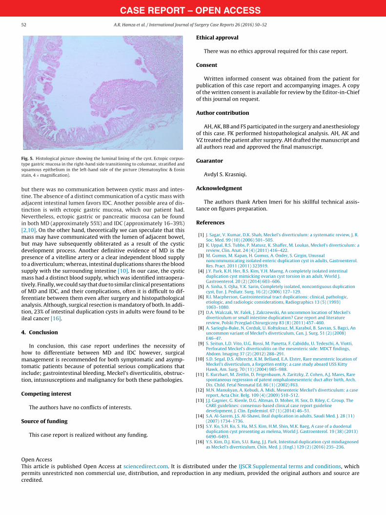
Fig. 5. Histological picture showing the luminal lining of the cyst. Ectopic corpus-tss
btatNi[mbdptsmtofati
4
hmtit
C
S
[
[
[
[
OTpc
ype gastric mucosa in the right-hand side transitioning to columnar, stratified andquamous epithelium in the left-hand side of the picture (Hematoxylinc & Eosintain, 4 × magnification).
ut there was no communication between cystic mass and intes-ine. The absence of a distinct communication of a cystic mass withdjacent intestinal lumen favors IDC. Another possible area of dis-inction is with ectopic gastric mucosa, which our patient had.evertheless, ectopic gastric or pancreatic mucosa can be found
n both MD (approximately 55%) and IDC (approximately 16–39%)2,10]. On the other hand, theoretically we can speculate that this
ass may have communicated with the lumen of adjacent bowel,ut may have subsequently obliterated as a result of the cysticevelopment process. Another definitive evidence of MD is theresence of a vitelline artery or a clear independent blood supplyo a diverticulum; whereas, intestinal duplications shares the bloodupply with the surrounding intestine [10]. In our case, the cysticass had a distinct blood supply, which was identified intraopera-
ively. Finally, we could say that due to similar clinical presentationsf MD and IDC, and their complications, often it is difficult to dif-erentiate between them even after surgery and histopathologicalnalysis. Although, surgical resection is mandatory of both. In addi-ion, 23% of intestinal duplication cysts in adults were found to beleal cancer [16].
. Conclusion
In conclusion, this case report underlines the necessity ofow to differentiate between MD and IDC however, surgicalanagement is recommended for both symptomatic and asymp-
omatic patients because of potential serious complications thatnclude; gastrointestinal bleeding, Meckel’s diverticulitis, obstruc-ion, intussusceptions and malignancy for both these pathologies.
ompeting interest
The authors have no conflicts of interests.
ource of funding
This case report is realized without any funding.
[
[
[
pen Accesshis article is published Open Access at sciencedirect.com. It is distribermits unrestricted non commercial use, distribution, and reproductredited.
PEN ACCESSurgery Case Reports 26 (2016) 50–52
Ethical approval
There was no ethics approval required for this case report.
Consent
Written informed consent was obtained from the patient forpublication of this case report and accompanying images. A copyof the written consent is available for review by the Editor-in-Chiefof this journal on request.
Author contribution
AH, AK, BB and FS participated in the surgery and anesthesiologyof this case. FK performed histopathological analysis. AH, AK andVZ treated the patient after surgery. AH drafted the manuscript andall authors read and approved the final manuscript.
Guarantor
Avdyl S. Krasniqi.
Acknowledgment
The authors thank Arben Imeri for his skillful technical assis-tance on figures preparation.
References
[1] J. Sagar, V. Kumar, D.K. Shah, Meckel’s diverticulum: a systematic review, J. R.Soc. Med. 99 (10) (2006) 501–505.
[2] K. Uppal, R.S. Tubbs, P. Matusz, K. Shaffer, M. Loukas, Meckel’s diverticulum: areview, Clin. Anat. 24 (4) (2011) 416–422.
[3] M. Gumus, M. Kapan, H. Gumus, A. Onder, S. Girgin, Unusualnoncommunicating isolated enteric duplication cyst in adults, Gastroenterol.Res. Pract. 2011 (2011) 323919.
[4] J.Y. Park, K.H. Her, B.S. Kim, Y.H. Maeng, A completely isolated intestinalduplication cyst mimicking ovarian cyst torsion in an adult, World J.Gastroenterol. 20 (2) (2014) 603–606.
[5] A. Sinha, S. Ojha, Y.K. Sarin, Completely isolated, noncontiguous duplicationcyst, Eur. J. Pediatr. Surg. 16 (2) (2006) 127–129.
[6] R.I. Macpherson, Gastrointestinal tract duplications: clinical, pathologic,etiologic, and radiologic considerations, Radiographics 13 (5) (1993)1063–1080.
[7] D.A. Walczak, W. Falek, J. Zakrzewski, An uncommon location of Meckel’sdiverticulum or small intestine duplication? Case report and literaturereview, Polski Przeglad Chirurgiczny 83 (8) (2011) 457–460.
[8] A. Sarioglu-Buke, N. Corduk, U. Koltuksuz, M. Karabul, B. Savran, S. Bagci, Anuncommon variant of Meckel’s diverticulum, Can. J. Surg. 51 (2) (2008)E46–47.
[9] S. Seitun, L.D. Vito, U.G. Rossi, M. Panetta, F. Cabiddu, U. Tedeschi, A. Viotti,Perforated Meckel’s diverticulitis on the mesenteric side: MDCT findings,Abdom. Imaging 37 (2) (2012) 288–291.
10] S.D. Segal, D.S. Albrecht, K.M. Belland, E.A. Elster, Rare mesenteric location ofMeckel’s diverticulum, a forgotten entity: a case study aboard USS KittyHawk, Am. Surg. 70 (11) (2004) 985–988.
11] E. Kurzbart, M. Zeitlin, D. Feigenbaum, A. Zaritzky, Z. Cohen, A.J. Mares, Rarespontaneous regression of patent omphalomesenteric duct after birth, Arch.Dis. Child. Fetal Neonatal Ed. 86 (1) (2002) F63.
12] M.N. Manukyan, A. Kebudi, A. Midi, Mesenteric Meckel’s diverticulum: a casereport, Acta Chir. Belg. 109 (4) (2009) 510–512.
13] J.J. Gagnier, G. Kienle, D.G. Altman, D. Moher, H. Sox, D. Riley, C. Group, TheCARE guidelines: consensus-based clinical case report guidelinedevelopment, J. Clin. Epidemiol. 67 (1) (2014) 46–51.
14] S.A. Al-Sarem, J.S. Al-Shawi, Ileal duplication in adults, Saudi Med. J. 28 (11)(2007) 1734–1736.
15] S.Y. Ko, S.H. Ko, S. Ha, M.S. Kim, H.M. Shin, M.K. Baeg, A case of a duodenalduplication cyst presenting as melena, World J. Gastroenterol. 19 (38) (2013)6490–6493.
16] Y.S. Kim, D.J. Kim, S.U. Bang, J.J. Park, Intestinal duplication cyst misdiagnosedas Meckel’s diverticulum, Chin. Med. J. (Engl.) 129 (2) (2016) 235–236.
uted under the IJSCR Supplemental terms and conditions, whichion in any medium, provided the original authors and source are