international journal of noni research · noni is widely used as a dietary supplement by human...
TRANSCRIPT

International Society for Noni Science12, Rajiv Gandhi Road, Chennai - 600 096.
Phone : 044-4901 1111 Fax : 044-4901 1149E-mail : [email protected] Visit : www.isns.in
InternationalJournal of
Noni ResearchVolume 10 Numbers 1 & 2 January & July 2015
Editor-In-Chief
Dr. Kirti Singh
Editor
Dr. P. Rethinam
Joint Editor
Dr. T. Marimuthu

CONTENTS
Pharmacological properties and clinicalapplications of Morinda citrifolia L.Srinivasan ThyagaRajan, P. Rethinam and Uday P. Pratap1
Effect of Noni (Morinda citrifolia L.) on plaqueinduced gingivitis - A microbiological studyDeepshika Saravanan, Saravanan Rethinam, T. Preethi Ganga andB. Sakthi Sowparanika
Etiology of dry and soft rot of Noni(Morinda citrifolia, L) fruitsS. Nakkeeran, Chandrasekar, P. Renukadevi and T. Marimuthu
Disease management in medicinal cropsB. Meena
Management of dry and soft rot of Noni(Morinda citrifolia L.) fruitsS. Nakkeeran, Chandrasekar, P. Renukadevi and T. Marimuthu
1
InternationalJournal ofNoni Research
Volume 10 Numbers 1 & 2 January & July 2015
19
28
43
58

Pharmacological properties and clinicalapplications of Morinda citrifolia L.
Authors’ affiliation :1. Integrative Medicine Laboratory,Department of Biotechnology,School of Bioengineering, SRMUniversity, Kattankulathur 603 203,Tamil Nadu, India;2. World Noni Research Foundation,Chennai, India1. Corresponding author: Email:[email protected]
SrinivasanThyagaRajan1
P. Rethinam2 andUday P. Pratap1
Abstract : Morinda citrifolia, also commonly referred to as Noni, has beenwidely used since ancient times in traditional medicine in South and SoutheastAsia with various parts of the plant; roots, stems, barks, leaves, and fruitsfinding a variety of use in treatment of human ailments. In recent years, it hasbeen increasingly used as a dietary supplement because its more than 200phytochemical constituents possess multiple biological activities and thus, exertcurative effects in a number of diseases such as inflammatory diseases, diabetes,cardiovascular diseases, infectious diseases, cancer, and female-specific problemssuch as menstrual difficulties, cognitive disorders, etc. Several in vitro and invivo studies involving cell cultures and rodents, and a few human clinicalstudies have demonstrated that Noni’s potential health benefits are a result ofits antioxidant, immunostimulatory, anti-inflammatory, anti-tumorigenic andpro-apoptotic properties, and the direct modulation of intracellular signallingpathways by its phytochemicals. This review provides an update of findings onthe pharmacological functions and clinical benefits of the phytochemicals atthe cellular level in various diseases to emphasize the need for furtherexperimental and clinical studies to explore the mechanism(s) of action(s) ofalready and yet to be determined phytochemicals of M. citrifolia. AlthoughNoni is considered safe for consumption, it is essential to develop properrecommendations so that it may be safely used by various age groups of humanbeings as there is increased utilization of Noni as a dietary supplement.
Introduction
Morinda citrifolia (Noni) is one among many herbal medicines used widely inIndian Ayurveda and Siddha medicine practice, and Polynesian folk-lore medicinesfor the past 2000 years (Singh et al., 2011; Ramaswamy et al., 2012; Whistler1985). It is also called commonly referred to as Indian Mulberry, Ba Ji Tian, Nonoor Nonu, cheese fruit, and Nhau in various medicinal practices that use it for treatingvarious diseases because of its wide-ranging health benefits including therapy forcancer, arthritis, diabetes, hypertension, pain, infectious diseases, etc. (Whistler.,1991). Morinda citrifolia, belonging to the species Rubiaceae (coffee family), is
Correspondence to :Srinivasan ThyagaRajanIntegrative Medicine Laboratory,Department of Biotechnology,School of Bioengineering, SRMUniversity, Kattankulathur 603 203,Tamil Nadu, India;Corresponding author: Email:[email protected]
Keywords : Noni, antioxidant, diabetes, inflammation, heart, cognition,phytochemicals
Intl. J. Noni Res., 2015, 10 (1 & 2) 1

a small evergreen tree found in coastal regions characterized by a straight trunk,bright green and elliptical leaves, white tubular flowers, and ovoid greenish-yellowfruit. The mature Noni fruit has an unpalatable taste and odor but processing of thefruit has enabled it to be marketed as a nutraceutical for the treatment of variousdiseases (Wang et al., 2002; Potterate et al., 2007). Although all parts of the planthave been used in treatment of diseases, it is the leaves that are traditionally andwidely used, followed by roots of the plant; but now the fruit juice is widely consumedas a supplement. Many secondary metabolites such as iridoid glycosides andtriterpenoids (ursolic acid) in the fruit, and anthraquninones in roots havecontributed to the beneficial therapeutic effects of Noni. Traditional use of Nonivaries widely in Southeast Asia, South Pacific where it originated and Africa, HawaiianIslands, and Caribbean where it has been brought for cultivation. Its root, stem,bark, leaves, and fruit is used externally as a poultice or applied directly to the body,or consumed orally as an infusion without or with fermentation. Topical applicationis meant for the treatment of sores, burns, cuts, and inflammation besides as a reliefagent from headaches. Oral consumption of Noni has been used to treat diversehealth problems that include improving immunity, regulation of female-specificproblems such as menstruation, and cancer, helminthic, diabetes, mental diseasessuch as depression and drug addiction, heart diseases, obesity, and a wide varietyof other ailments.
Phytochemicals in Morinda citrifolia
Noni is widely used as a dietary supplement by human beings all over the worldbecause of increased publicity and marketing as a general cure for several acuteand chronic diseases (Fairchild., 2004), as well as several scientific studiesdemonstrating its usefulness in the treatment of such diseases. Although the presenceof xeronine and its precursor, proxeronine, have been reported to be in Noni(Heinicke., 1985), no peer-reviewed publications exist to support this data.
Analysis for phytochemical constituents of M. citrifolia by NMR spectroscopy andmass spectrometry (MS), and gas chromatography MS revealed the presence ofmore than 200 compounds (Potterate., 2007; Singh., 2012). These primarily consistof a number of anthraquinones (damnacanthal) and anthraquinone glycosides, fattyacids and their derivatives, iridoids and iridoid glycosides, lignans, neolignans,flavonol glycosides, phenylpropanoids, saccharides, triterpenoids and fatty acids.Several new compounds and those belonging to the glycoside and polysaccharidefamily have been identified that may have potential health benefits (Lv et al., 2011;Beh et al., 2012; Hu et al., 2012). The presence of carotenoids in the leaves, bark,and fruits, and especially its increased content in the leaves confers the ability tocorrect vitamin A deficiency (Aalbersberg et al., 1993). Noni fruits from Australiahave been found to contain enough vitamin C and potassium to meet the dailyrequirements for humans (Peerzada et al., 1990). Vitamin E production was
Intl. J. Noni Res., 2015, 10 (1 & 2) 2
Srinivasan ThyagaRajan et al. Pharmacological properties and clinical applications of Morinda citrifolia
enhanced in the callus of M. elliptica by modifying the medium conditions in vitroindicating that this vitamin may also contribute to the health benefits of Noni (Chonget al., 2004). The polysaccharide, mostly pectic polysaccharide content of the fruitsfrom Vietnam was found to contain several monosaccharides and their productssuch as arabinose, galactose, galacturonic acid, and rhamnose (Bui et al., 2006).
Ten anatomical parts (bark, branches, flowers, leaves, pulp, immature and maturefruit, stem, heartwood and root) of Morinda citrifolia L. was taken for analysis ofbioactive compounds using Reverse Phase – High Performance LiquidChromatography (RP-HPLC). It revealed that polydatin and physcion were themajor anthraquinones in plant parts, rhein and resveratrol in the roots,âcryptoxanthin, zeaxanthin, rutin, quercetin and myrecitin were the major carotenoidsin noni parts, and gallic acid was the major phenolic in all the nine parts of noniexcept the immature fruit. Mature noni fruits had the maximum number of bioactivecompounds suggesting that it can be used for various therapeutic purposes (Singhet al., 2012).
Pharmacological basis for the biological activities of M. citrifolia
Several studies have demonstrated antioxidant, analgesic and anti-inflammatory, anti-cancer, anti-microbial, anti-diabetic, cardioprotective, cognitive protective functions,etc. in both crude and pure constituents of Noni through in vitro and in vivostudies.
Antioxidant Activity of M. citrifolia has been one of the key activities responsiblefor several of Noni’s therapeutic effects in humans (Wang et al., 2002). Recentstudies have attempted to provide the phytochemical basis for the antioxidant propertiesof Noni. Oral administration of deacetylasperulosidic acid isolated from the Noni fruithas been demonstrated to reduce lipid peroxidation and enhance superoxidedismutase and catalase activity (Ma et al., 2013). The presence of vitamins C andE, and scopoletin and damnacanthal in Noni fruit have the capability to bind to ³-D-crystallin, a lens protein with equal affinity, which may be useful in the preventionof cataract formation (Rentala et al., 2013). The antioxidant property of Noni fruitjuice examined through HPLC-DAD (Diode Array Detector) and Electro SprayIonization Mass Spectrometric detection (HPLC-ESI-MS) may be associated withphenolic compounds, iridoids, and ascorbic acid (Dussossoy et al., 2011).Spectroscopic analysis of Noni fruit revealed several compounds out of which aneolignan, americanin A, has been found to have potent antioxidant activity (Su etal., 2005).
Anti-inflammatory Activity of M. citrifolia is another important property ofNoni that is critical to its therapeutic properties. The iridoid glycoside, monotropein,in M .officinalis root significantly promoted antinociceptive and anti-inflammatoryaction in mice with carrageenan-induced edema and inflammation that may be
Intl. J. Noni Res., 2015, 10 (1 & 2) 3
Srinivasan ThyagaRajan et al. Pharmacological properties and clinical applications of Morinda citrifolia
facilitated through the inhibition of pro-inflammatory cytokines, inducible nitricoxide synthase (iNOS), cyclooxygenase-2 (COX-2), and nuclear factor-ºB (NF-ºB)activity (Choi et al., 2005; Shin et al., 2013). In addition, four saccharide fatty acidesters isolated from M. citrifolia fruit exhibited strong anti-inflammatory activity inmice (Akihisa et al., 2007). It is probable that these phytochemicals may beresponsible for the inhibition of matrix metalloprotease-9 release from immunecells, thus causing analgesic and anti-inflammatory effects in mice and providingrelief from debilitating pain and joint destruction found in arthritis (Basar et al.,2010). These effects of Noni may be also mediated through the direct inhibition ofthe key players in the inflammation process such as COX-1 and COX-2 activities andthe production of nitric oxide (NO) and prostaglandins E2 (Dussossoy et al., 2011).Another compound that had potent anti-inflammatory activity was damnacanthalisolated from the Noni root and it mediated its action through its ability to bindweakly to H1 receptor (Okusada et al., 2011). Besides its effects at the receptorlevel, damnacanthal’s mechanism of action include suppression of the NF-ºB activityleading to the inhibition of the expression of cytokines, COX-2, and iNOS and thus,can be potentially used for the treatment of diseases involving inflammation (Nualsanitet al., 2011). The leaves of Noni also exerted anti-inflammatory activity probablythrough selective inhibition of yet to be determined subsets of leukocytes (Serafiniet al., 2011).
Anti-cancer Property of M. citrifolia may be mediated through its ability toenhance antioxidant properties and anti-tumor immunity. Damnacanthal treatmentof K-rasts-NRK cells reversed the abnormal morphology and cytoskeletal structurewithout changing the amount and localization of Ras while its addition to epidermoid,breast, hepatocellular, cervical cancer cell lines suppressed their proliferation (Thaiet al., 2010, Hiramatsu et al., 1993). Such anti-cancerous property of damnacanthalmay be due to induction of caspase activity to promote apoptosis through theproapoptotic protein nonsteroidal anti-inflammatory activated gene-1 (NAG-1) thatmay enhance transcription factor CCAAT/enhancer binding protein ² (C/EBP²)(Nualsanit et al., 2012). Inhibition of HIF-1± protein and interfering with theintracellular signaling pathways involving PKB, ERK-1/2, JNK-1 and S6 in cancer cellsor repressing IL-1², an inducer of HIF-1± by Noni may explain its anti-cancerfunctions besides its anti-angiogenic activity (Beh et al., 2012; Jang 2012; Hornicket al., 2003). In vivo administration of MMTV-neu transgenic mice with TahitianNoni fruit juice resulted in significant reduction in tumor weight and volume and inlonger tumor doubling times in mice (Clafshenkel et al., 2012). This was accompaniedby significant inhibition of the growth of aggressive forms of cancer and also inducedadditional beneficial effects such as significant changes in mammary secondaryductule branching and lobuloalveolar development, serum progesterone levels, andestrous cycling. Phytochemical-rich Noni is able to influence the development andgrowth of cancer at the cellular and molecular level that can be exploited as atherapeutic option or as an adjunct therapy in the treatment of cancer.
Intl. J. Noni Res., 2015, 10 (1 & 2) 4
Srinivasan ThyagaRajan et al. Pharmacological properties and clinical applications of Morinda citrifolia
Another crucial mechanism through which Noni can exert anti-tumor effects is bythe augmentation of immunity that may improve immunosurveillance, enhancecytostatic and cytocidal activity of immune cells through immune molecules such ascytokines, or modulation of tumor antigen recognition. Polysaccharide-richconstituents in Noni were capable of stimulating the release of cytokines useful inthe elimination of tumor cells such as tumor necrosis factor-alpha (TNF-±),interleukin-1² (IL-1²), IL-10, IL-12 p70, interferon-gamma (IFN-³) and nitric oxide(NO) while it had no effect on IL-2 and suppressed IL-4 release in mice with Lewislung LLC peritoneal carcinoma (Hirazumi et al., 1999). More importantly, it alsoimproved survival time and combinatorial therapy with chemotherapeutic agentspromoted the curative effects indicating a probable use of Noni as an adjuncttherapy in cancer. Such anti-cancer immunity was shown to be mediated throughenhancement of T helper (Th)-1 cytokines and suppression of Th2 cytokinesinvolving macrophages, T cells, and natural killer (NK) cells (Furusawa et al.,2003). In a significant finding, fermented Noni exudate was demonstrated to quicklyimprove innate immune system functions through the upregulation of NK cell activityfollowed by adaptive immune system and thereby, facilitating elimination of tumorcells in mice (Li et al., 2008).
In addition, Noni was able to prevent carcinogen-induced DNA adduct formation invarious organs and increase the expression of p53 and pro-apoptotic Bax whiledecreasing anti-apoptotic Bcl-2, Bcl-XL, and survivin that resulted in an increase inthe activities of caspases-9 and -3 suggesting that Noni is capable of altering thecellular functional capacity to induce apoptosis (Wang et al., 2001; Gupta et al.,2013). Some of the glycosides in the Noni fruit juice suppressed 12-O-tetradecanoylphorbol-13-acetate (TPA)- and epidermal growth factor (EGF)-inducedAP-1 activity that blocked the phosphorylation of c-Jun in mouse epidermal cell lineindicating that such effects may also be possible in cancer (Liu et al., 2001).
Out of a total of 44 peer-reviewed publications on the use of Noni in cancer, 19directly dealt with cancer involving in vitro and in vitro studies in cell lines andanimals, and human cancer (Brown., 2012). The studies in cell lines and animalsdemonstrate that Noni (1) stimulates immune system as outlined above, (2) exertsin vitro cytocidal (0-36%) effect depending on the type of cancer cells, and (3)moderately augments (25-45%) the survival rate in rodents. Further studies arewarranted to focus on the isolation of active components and their mechanism(s)of action(s) in anti-cancer activity.
Anti-microbial Activity of M. citrifolia against bacteria, fungi, viruses, andparasites is one of the key functions that have been widely exploited as a traditionalremedy in a number of countries. Several components of Noni exhibit pronouncedanti-tubercular activity (Saludes et al., 2002). Extracts of Noni leaves inhibited thegrowth of M. tuberculosis by 89% while the anti-TB drug, Rifampicin, had a growth-
Intl. J. Noni Res., 2015, 10 (1 & 2) 5
Srinivasan ThyagaRajan et al. Pharmacological properties and clinical applications of Morinda citrifolia

inhibitory rate of 97%. Methanolic extract of M. citrifolia had potential antibacterialactivities to both gram-positive S. aureus and Methicillin Resistant S. aureus (MRSA)indicating that the phytochemical constituents of Noni exert potent anti-bacterialactivity against latest superbugs such as MRSA (Zaidan et al., 2005). Anthaquinonesfrom M. augustifolia showed antimicrobial activity against Bacillus subtilis,Escherichia coli, Micrococcus luteus, Sarcinalutea, Candida albicans andSaccharomyces sake (Xiang et al., 2008). Helicobacter pylorus (H. pylori) in thestomach is a common cause for stomach cancer, especially when the chronicinflammation caused by it is left untreated. The ethanol and ethyl acetate extractspromoted anti-adhesion of H. pylori to human gastric carcinoma AGS cells resultingin the down-regulation of CagA, IL-8, COX-2 and iNOS expression indicating Noni’santi-inflammatory properties in stomach (Huang et al., 2014). Presence of water-soluble components in M. citrifolia interfered with the morphological conversionof C. albicans and the germination of Aspergillus nidulans that provides an optionfor Noni as an anti-fungal therapy for candidiasis and aspergillosis (Banerjee et al.,2006; Jainkittivong et al., 2009).
Phytochemicals in M. citrifolia have been shown to exert strong antiviral activityagainst mild to severe pathogenic viruses. Powdered Noni fruit was able to provideprotection against the cytopathic effect of HIV-1(IIIB) in lymphocyte cell lines andinhibited the hepatitis C virus (HCV) replication that may be due to damnacanthaland pheophorbide-a, a major catabolite of chlorophyll a (Ali et al., 2000; Selvamet al., 2009; Ratnoglik et al., 2014).
Most studies have focused on the use of M. citrifolia in inhibiting malarial parasiteswhich is the major cause of mortality in the tropical countries. Although all the partsof M. lucida had significant effects on Plasmodium berghei, the stem bark extractsof the plant markedly inhibited the parasitaemia in mice because of the presenceof digitolutein, rubiadin 1-methyl ether, and damnacanthal (Koumaglo et al., 1992;Obih et al., 1985). Extracts of M. lucida induced a dose-dependent inhibition ofegg-hatching and larval migration of the parasitic nematode, Trichostrongyluscolubriformis (Hounzangbe-Adote et al., 2005). The aqueous and ethanolic extractsof M. citrifolia fruits had a significant anti-helminthic activity against Ascaridiagalliin chickens suggesting its potential use in preventing infestation with roundwormswhich is a major cause of health problems in countries situated in tropical regions(Brito et al., 2009). In agreement with this notion, alcoholic extract of the leavesof M. citrifolia showed significant in vitro anti-helmintic activity against humanAscaris lumbricoides (Raj., 1975).
Anti-diabetic Activity of M. citrifolia is another important function that can beused to treat increasing incidence of diabetes and post-diabetic complications suchas heart diseases and neuro- and retinopathy in India and abroad. Several ofMorinda sp. alcoholic extracts decreased the fasting glucose levels, improved hepatic
Intl. J. Noni Res., 2015, 10 (1 & 2) 6
Srinivasan ThyagaRajan et al. Pharmacological properties and clinical applications of Morinda citrifolia

and renal functions, and enhanced antioxidant activities in streptozotocin (STZ)-induced diabetic rodents possibly mediated through the bioactive molecules suchas damnacanthol-3-O-beta-D-primeveroside and lucidin 3-O-beta-D-primeveroside(Soon et al., 2002; Kamiya et al., 2008). In addition, aqueous extractsof M. lucida significantly inhibited ± -amylase and ± –glucosidase activities furtherpotentiating the anti-diabetic effects of Noni (Kazeem et al., 2013). Similarly, the anti-diabetic effects of fermented M. citrifolia was demonstrated in STZ-induced diabeticrats where it reduced glycosylated hemoglobin (HbA1c) levels, enhanced insulinsensitivity, and significantly decreased serum triglycerides and low-density lipoprotein(LDL) cholesterol and such effects may have been due to the activation of peroxisomeproliferator-activated receptor-(PPAR-) ³ and stimulated glucose uptake via stimulationof AMP-activated protein kinase (Lee et al., 2012). Such hypoglycemic effects maybeen due to altering the intracellular signaling by the Noni molecules with theultimate inhibition of hepatic FoxO1 mRNA expression and a concomitant increasein FoxO1 phosphorylation and nuclear expulsion of the proteins (Nerurkar et al.,2012). It is important to elucidate whether the phytochemicals in Noni have insulinmodulatory properties because fruit juice of M. citrifolia exerts synergistic functionswith insulin in diabetic rodents that may have been due to certain compounds inNoni that can inhibit protein tyrosine phosphatase 1B resulting in insulinomimeticeffects (Horsfall et al., 2008; Nguyen et al., 2013). Another complication in diabeticpatients is poor healing of wounds because of high glucose levels and Noni hastenedthe process of wound healing in rats with STZ-induced diabetes via its ligand-bindingto the Platelet-derived growth factor and A(2A) receptors (Palu et al., 2010; Nayaket al., 2007).
Cardioprotective Functions of M. citrifolia have been crucial to the treatmentof cardiovascular problems that may or may not be related to diabetic complications.Noni caused vasodilatation by relaxation of vascular smooth muscles throughendothelium-dependent involving nitric oxide-cGMP pathway and –independentpathways (Ettarh et al., 2004). Such vasodilatory effects and spasmolytic effects ofM. citrifolia may be mediated through the effects of phytochemicals that are capableof blocking voltage-gated calcium channels leading to the release of intracellularcalcium and thus, exerting ameliorating effects in hypertension (Gilani et al., 2010).Extracts of Noni have been shown to exert hypotensive effects, lower blood pressurein anesthetized dogs, and promote diuretic effects (Wang et al., 2002). Dyslipidemiaare disorders of lipoprotein metabolism that results in coronary heart disease andother cardiovascular problems directly or as an outcome of diabetes. It ischaracterized by a high plasma triglyceride concentration, low HDL cholesterolconcentration and increased concentration of small dense LDL-cholesterol particles.The aqueous-ethanolic extracts of fruits, leaves, and roots of M. citrifolia hadantidyslipidemic effect in high fat diet-fed rats through the inhibition of biosynthesis,absorption and secretion of lipids providing additional proof for its health benefit
Intl. J. Noni Res., 2015, 10 (1 & 2) 7
Srinivasan ThyagaRajan et al. Pharmacological properties and clinical applications of Morinda citrifolia

in the treatment of heart problems (Mandukhail et al., 2010). Few of theanthraquinones from the roots of M. officinalis enhanced adipocyte differentiationthat may be a mechanism for weight loss therapy and prove to be beneficial in thetreatment of diabetes and cardiovascular diseases (Liu et al., 2012).
Neuroprotective effects of M. citrifolia is another important property that maybe critical to the treatment of neuro-associated disorders such as memory impairment,cognitive dysfunctions, neurodegenerative disorders, etc in the elderly especially,when the population of elderly is on the rise in several countries of the world. Inulin-type oligosaccharides extracted from M. officinalis was shown to possessneuroprotective effects by suppressing the intracellular calcium overloading andsimultaneously enhance nerve growth factor mRNA expression in corticosterone-lesioned PC12 cells (Li et al., 2004). These cellular effects may explain the preventionof neuronal damage in rodents with cerebral ischemia by M. citrifolia fruit juice andthe reversal of neuronal deficits (Harda et al., 2009). It may also facilitate protectionfrom stress-induced impairment of cognitive functions by improving the blood vesseldensity in the hippocampal dentate gyrus and attenuating the development of glucoseintolerance (Muto et al., 2010; Harda et al., 2010).
Various extracts of Noni fruit juice markedly enhanced memory and cerebral bloodflow and reduced oxidative stress and acetylcholinesterase activity in a scopolamine-induced amnesia mouse model suggesting that Noni not only improves cellularfunctions in the brain, but also reverses the behavioral deficits (Pachauri et al.,2012). Further proof for this beneficial effect of Noni on improving memory deficitswas observed in mice where Noni extract inhibited the STZ-induced memorydysfunction that was demonstrated to be due to increased levels of Brain-derivedneurotrophic factor (BDNF), a key growth factor for the maintenance of neurons,acetylcholine, and ATP levels leading to a better cholinergic neurotransmission andantioxidative functions (Pachauri et al., 2013). Similarly, administration ofoligosaccharides of M. officinalis in rats with beta-amyloid-induced dementiaenhanced the activities of antioxidant enzymes, acetylcholine, and Na+ /K+ -ATPasesupporting the earlier studies where Noni improved brain energy metabolism andcholinergic neuronal activity (Chen et al., 2013). The improvement in short- andlong-term memory and exploratory behavior by Noni in dementia mouse model notonly enhanced cholinergic neurotransmission but also inhibited monoamine oxidase-A activity that may have been responsible for the increase in the levels of serotoninand dopamine in the brain (Muralidharan et al., 2010). These limited evidence forthe improvement of neurobehavioral functions by Noni may be helpful in its use asa dietary supplement in the elderly population, people with cognitive dysfunctions,and patients with neurodegenerative disorders.
Clinical Applications of Morinda citrifolia
There are very limited studies pertaining to Noni’s health benefits in human beings.
Intl. J. Noni Res., 2015, 10 (1 & 2) 8
Srinivasan ThyagaRajan et al. Pharmacological properties and clinical applications of Morinda citrifolia

In one of the studies, it was used in women to determine whether it had anti-inflammatory and pain-reducing effects as reported in animals. Administration ofNoni capsules to women with primary dysmenorrhea did not provide any healthbenefits such as attenuating bleeding or menstrual pain that may have been relatedto dosage, failure to monitor the compliance of Noni capsule consumption, samplesize, etc. (Fletcher et al., 2013).
Drinking of Noni fruit juice by adult heavy smokers daily for a month significantlyreduced cholesterol levels, triglycerides, and hs-CRP in association with a decreasein LDL and homocysteine, as well increases in HDL suggesting the beneficial of effectsof Noni in preventing oxidative stress, dyslipidemia, and systemic inflammation(Wang et al., 2012). These beneficial biochemical effects may reduce the cancerrisk in heavy cigarette smokers where drinking 1 to 4 oz of Noni juice daily for amonth may block carcinogen-DNA binding or excising DNA adducts from genomicDNA (Wang et al., 2009). A gel containing the ethanol extract and juice pressedfrom the leaves was safe for topical use and may be useful in preventing UVB-induced skin injury, and therefore may be potentially considered for prophylacticand therapeutic use in skin cancer (West et al., 2009).
Controlled human trials documenting anti-diabetic effects of noni supplementationis not available but four population-based surveys have provided a link between noniconsumption and the resulting increased glucose uptake (Nerurkar et al., 2015).In another study, noni fruit juice consumption by Indian diabetic patients for aperiod of 6 months reduced fasting plasma glucose and LDL cholesterol levels whileaugmenting the HDL cholesterol level (Sathishkumar, 2007). Similar beneficial anti-diabetic effects of noni fruit juice was provided by another study where a month-long oral consumption of noni fruit juice resulted in substantial decrease in HbA1c,fasting blood sugar, total cholesterol, LDL, triglyceride aspartate aminotransferaseand alanine aminotransferase levels (Ramesh et al., 2013). These effects may havebeen facilitated by bioavailability of scopoletin, iridoid glycosides, monotropein, anddeacetylasperulosidic acid, or flavonoid quercetin (3,30,40,5,7-pentahydroxyflavone),and its glycoside, rutin (quercetin-3-O-b-rutinoside). However, proper randomizedhuman trials examining the pharmacokinetics and bioavailability of these bioactivecompounds have to be performed to establish the anti-diabetic activity of Noni inhumans.
The following studies demonstrated some therapeutic effects in human beings:
(1) Aqueous extract of M. citrifolia enhanced the rate and the extent of ranitidineabsorption by improving the gastrokinetic activity that may be due to its bioactivecomponent, scopoletin, stimulating the 5-HT(4) receptor (Nima et al., 2012).
(2) Extracts of M. citrifolia stems and the morindicone and morinthone, bioactivemolecules of Noni had potent anti-leishmanial activity against L. major that cause
Intl. J. Noni Res., 2015, 10 (1 & 2) 9
Srinivasan ThyagaRajan et al. Pharmacological properties and clinical applications of Morinda citrifolia
cutaneous leishmaniasis in humans (Sattar et al., 2012).
(3) Noni capsules containing dried fruit at a dose of 600 mg Noni extract significantlyreduced the postoperative nausea and vomiting during the first 6 hours (Prapaitrakoolet al., 2010).
(4) Oral consumption of three or four capsules four times daily (6-8 g) in 51advanced cancer patients was better in controlling fatigue, pain, and maintainingphysical function and scopoletin may be one of the many bioactive moleculesexerting such curative effects (Issell et al., 2009).
Although the above studies may not strictly conform to the standard human clinicaltrials, the results from these disparate studies point to possible beneficial therapeuticeffects in human beings and emphasize the need for a thorough systematic approachin establishing the usefulness of M. citrifolia in the treatment of diseases and cancer.
Conclusions and Future Directions
For several centuries, various parts of M. citrifolia have been used as a medicinalplant and it is currently used as a dietary supplement in a number of countriesbecause of its anti-oxidant, anti-inflammatory, analgesic, anti-cancer, anti-diabetic,and anti-hypertensive properties. These findings are based on studies involving invitro and in vivo cell cultures and animal models. The biological activity of Nonimay be related to more than 200 phytochemicals that include anthraquinones,flavonoids, glycosides, iridoids, lignans, and triterpenoids but their individualcontribution to the biological activity of Noni is yet to be fully elucidated. It is criticalto develop techniques to identify and characterize yet to be isolated phytochemicalsand also, examine the complex interactions between these phytochemicals as it iswell known that biological effects are not mediated by a single component butthrough synergistic interactions among the various bioactive compounds.
Despite the wide-ranging benefits of Noni reported in literature, it is critical that someof the physiological and therapeutic effects of it are explored in detail in animalstudies so that the mechanism(s) of action(s) are understood. It is important thatpharmacokinetics of the various bioactive compounds are examined in vivo so thatthe contributions of their metabolites to the beneficial effects of noni can be fullyascertained. Such in-depth analysis will pave the way for development of targeteddrugs to various ailments afflicting humans.
Although anecdotal and traditional knowledge about M. citrifolia is driving its usein humans as a dietary supplement, it is imperative that proper clinical studies areessential to examine the cellular, biochemical, and molecular mechanism of itsaction. It involves designing randomized double-blinded clinical studies withappropriate placebo controls in humans that will provide information about
Intl. J. Noni Res., 2015, 10 (1 & 2) 10
Srinivasan ThyagaRajan et al. Pharmacological properties and clinical applications of Morinda citrifolia
bioavailability, safety, and benefits of long-term usage of noni and its bioactivecomponents. This will aid in the development of Noni products with appropriateingredients with reproducible quality useful not only for clinical trials but alsomarketing.
References
Aalbersberg, W. G. L., Hussein, S., Sotheeswaran, S., Parkinson, S. 1993. Carotenoidsin the leaves of Morinda citrifolia. J. Herbs Spices Med. Plants 2: 51–54
Akihisa T, Matsumoto K, Tokuda H, Yasukawa K, Seino K, Nakamoto K, Kuninaga H,Suzuki T, Kimura Y.2007. Anti-inflammatory and potential cancer chemopreventiveconstituents of the fruits of Morinda citrifolia (Noni). J Nat Prod.70:754-7.
Ali AM, Ismail NH, Mackeen MM, Yazan LS, Mohamed SM, Ho AS, Lajis NH. 2000.Antiviral, cyototoxic and antimicrobial activities of anthraquinones isolated from theroots of. Pharm Biol. 38:298-301.
Banerjee S, Johnson AD, Csiszar K, Wansley DL, McGeady P. 2006. An extract ofMorinda citrifolia interferes with the serum-induced formation of filamentousstructures in Candida albicans and inhibits germination of Aspergillus nidulans.Am J Chin Med. 34: 503-9:
Basar S, Uhlenhut K, Högger P, Schöne F, Westendorf J. 2010. Analgesic and anti-inflammatory activity of Morinda citrifolia L. (Noni) fruit. Phytother Res.24:38-42.
Beh HK, Seow LJ, Asmawi MZ, Abdul Majid AM, Murugaiyah V, Ismail N, Ismail Z.2012. Anti-angiogenic activity of Morinda citrifolia extracts and its chemicalconstituents. Nat Prod Res. 26:1492-7.
Brito DR, Fernandes RM, Fernandes MZ, Ferreira MD, Rolim FR, da Silva Filho ML.2009. Anthelmintic activity of aqueous and ethanolic extracts of Morinda citrifoliafruit on Ascaridia galli. Rev Bras Parasitol Vet. 18:32-6.
Brown AC.2012. Anticancer activity of Morinda citrifolia (Noni) fruit: a review.Phytother Res. 26:1427-40.
Bui, A. K. T., Bacic, A., Pettolino, F. 2006. Polysaccharide composition of the fruitjuice of Morinda citrifolia (noni). Phytochemistry. 67: 1271–1275
Chen DL, Zhang P, Lin L, Zhang HM, Liu SH. 2013. Protective effect of oligosaccharidesfrom Morinda officinalis on beta-amyloid-induced dementia rats. Zhongguo ZhongYao ZaZhi. 38:1306-9.
Choi J, Lee KT, Choi MY, Nam JH, Jung HJ, Park SK, Park HJ.2005 Antinociceptiveanti-inflammatory effect of Monotropein isolated from the root of Morinda officinalis.
Intl. J. Noni Res., 2015, 10 (1 & 2) 11
Srinivasan ThyagaRajan et al. Pharmacological properties and clinical applications of Morinda citrifolia
Biol Pharm Bull. 28:1915-8.
Chong TM, Abdullah MA, Fadzillah NM, Lai OM, Lajis NH. 2004. Anthraquinonesproduction, hydrogen peroxide level and antioxidant vitamins in Morinda ellipticacell suspension cultures from intermediary and production medium strategies. PlantCell Rep. 22:951-8.
Clafshenkel WP, King TL, Kotlarczyk MP, Cline JM, Foster WG, Davis VL,Witt-EnderbyPA. 2012. Morinda citrifolia (Noni) Juice Augments Mammary Gland Differentiationand Reduces Mammary Tumor Growth in Mice Expressing the Unactivated c-erbB2Transgene. Evid Based Complement Alternat Med. 487423.
Dussossoy E, Brat P, Bony E, Boudard F, Poucheret P, Mertz C, Giaimis J, Michel A.2011. Characterization, anti-oxidative and anti-inflammatory effects of Costa-Ricannoni juice (Morinda citrifolia L.). J Ethnopharmacol. 133:108-15.
Ettarh RR, Emeka P. 2004. Morinda lucida extract induces endothelium-dependentand-independent relaxation of rat aorta. Fitoterapia.75:332-6.
Fairchild, D. 2004. Noni: aspirin of the ancients. Flyana.com, Anahola, HI
Fletcher HM, Dawkins J, Rattray C, Wharfe G, Reid M, Gordon-Strachan G. 2013.Morinda citrifolia (Noni) as an Anti-Inflammatory Treatment in Women with PrimaryDysmenorrhoea: A Randomised Double-Blind Placebo-Controlled Trial. ObstetGynecol Int. 2013:195454.
Furusawa E, Hirazumi A, Story S, Jensen J. 2003. Antitumour potential ofapolysaccharide-rich substance from the fruit juice of Morinda citrifolia (Noni) onsarcoma 180 ascites tumour in mice. Phytother Res. 17:1158-64.
Gilani AH, Mandukhail SU, Iqbal J, Yasinzai M, Aziz N, Khan A, Najeeb-ur-Rehman.2010. Antispasmodic and vasodilator activities of Morinda citrifolia root extract aremediated through blockade of voltage dependent calcium channels.BMC ComplementAltern Med. 13: 10:2.
Gupta RK, Banerjee A, Pathak S, Sharma C, Singh N. 2013. Induction of mitochondrial-mediated apoptosis by Morinda citrifolia (Noni) in human cervical cancer cells.Asian Pac J Cancer Prev.14:237-42.
Harada S, Fujita-Hamabe W, Kamiya K, Mizushina Y, Satake T, Tokuyama S. 2010.Morinda citrifolia fruit juice prevents ischemic neuronal damage through suppressionof the development of post-ischemic glucose intolerance. J Nat Med. 64:468-73.
Harada S, Hamabe W, Kamiya K, Satake T, Yamamoto J, Tokuyama S. 2009. Preventiveeffect of Morinda citrifolia fruit juice on neuronal damage induced by focal ischemia.Biol Pharm Bull. 32:405-9.
Intl. J. Noni Res., 2015, 10 (1 & 2) 12
Srinivasan ThyagaRajan et al. Pharmacological properties and clinical applications of Morinda citrifolia

Heinicke, R. M. 1985. The pharmacologically active ingredient of noni. Pac. Trop.Bot. Gard. Bull. 15: 10–14
Hiramatsu T, Imoto M, Koyano T, Umezawa K. 1993. Induction of normal phenotypesin Ras-transformed cells by damnacanthal from Morinda citrifolia. CancerLett.73:161-6.
Hirazumi A, Furusawa E.1999. An immunomodulatory polysaccharide-rich substancefrom the fruit juice of Morinda citrifolia (noni) with antitumour activity. PhytotherRes. 13:380-7.
Hornick CA, Myers A, Sadowska-Krowicka H, Anthony CT, Woltering EA. 2003.Inhibition of angiogenic initiation and disruption of newly established human vascularnetworks by juice from Morinda citrifolia (noni). Angiogenesis. 6:143-9.
Horsfall AU, Olabiyi O, Aiyegbusi A, Noronha CC, Okanlawon AO. 2008. Morindacitrifolia fruit juice augments insulin action in Sprague-Dawley rats with experimentallyinduced diabetes. Nig Q J Hosp Med. 18:162-5.
Hounzangbe-Adote S, Fouraste I, Moutairou K, Hoste H. 2005. In vitro effects of fourtropical plants on the activity and development of the parasitic nematode,Trichostrongylus colubriformis. J Helminthol. 79:29-33.
Hu MX, Zhang HC, Wang Y, Liu SM, Liu L. 2012. Two new glycosides from the fruitsof Morinda citrifolia L. Molecules. 17:12651-6.
Huang HL, Ko CH, Yan YY, Wang CK. 2014. The Anti-adhesion and Anti-inflammationof Noni (Morinda citrifolia) Fruit extracts on AGS cells During Helicobacter pyloriInfection. J Agric Food Chem. 62:2374-83.
Issell BF, Gotay CC, Pagano I, Franke AA. 2009. Using quality of life measures in aPhase I clinical trial of noni in patients with advanced cancer to select a Phase IIdose. J Diet Suppl. 6:347-59.
Jainkittivong A, Butsarakamruha T, Langlais RP. 2009. Antifungal activity of Morindacitrifolia fruit extract against Candida albicans. Oral Surg Oral Med Oral PatholOral Radiol Endod. 108:394-8.
Jang BC. 2012. The fruit juice of Morinda citrifolia (noni) downregulates HIF-1±protein expression through inhibition of PKB, ERK-1/2, JNK-1 and S6 inmanganese-stimulated A549 human lung cancer cells. Int J Mol Med. 29:499-504.
Kamiya K, Hamabe W, Harada S, Murakami R, Tokuyama S, Satake T. 2008. Chemicalconstituents of Morinda citrifolia roots exhibit hypoglycemic effects instreptozotocin-induced diabetic mice. Biol Pharm Bull. 31:935-8.
Intl. J. Noni Res., 2015, 10 (1 & 2) 13
Srinivasan ThyagaRajan et al. Pharmacological properties and clinical applications of Morinda citrifolia

Kazeem MI, Adamson JO, Ogunwande IA. 2013. Modes of inhibition of ± -amylaseand ±-glucosidase by aqueous extract of Morinda lucida Benth leaf. Biomed ResInt.2013:527570.
Koumaglo K, Gbeassor M, Nikabu O, de Souza C, Werner W. 1992. Effects of threecompounds extracted from Morinda lucida on Plasmodium falciparum. PlantaMed. 58:533-4.
Lee SY, Park SL, Hwang JT, Yi SH, Nam YD, Lim SI. 2012. Antidiabetic Effect ofMorinda citrifolia (Noni) Fermented by Cheonggukjang in KK-A(y) Diabetic Mice.Evid Based Complement Alternat Med. 2012:163280.
Li J, Stickel SL, Bouton-Verville H, Burgin KE, Yu X, Wong DK, Wagner TE, Wei Y.2008. Fermented Noni exudate (fNE): a mediator between immune system and anti-tumor activity. Oncol Rep. 20:1505-9.
Li YF, Liu YQ, Yang M, Wang HL, Huang WC, Zhao YM, Luo ZP. 2004. The cytoprotectiveeffect of inulin-type hexasaccharide extracted from Morinda officinalis on PC12cellsagainst the lesion induced by corticosterone. Life Sci. 75:1531-8.
Liu G, Bode A, Ma WY, Sang S, Ho CT, Dong Z. 2001. Two novel glycosides fromthe fruits of Morinda citrifolia (noni) inhibit AP-1 transactivation and celltransformation in the mouse epidermal JB6 cell line. Cancer Res. 61:5749-56.
Liu Q, Kim SB, Ahn JH, Hwang BY, Kim SY, Lee MK. 2012. Anthraquinones fromMorinda officinalis roots enhance adipocyte differentiation in 3T3-L1 cells. NatProd Res. 26:1750-4.
Lv L, Chen H, Ho CT, Sang S. 2011. Chemical components of the roots of Noni(Morinda citrifolia) and their cytotoxic effects. Fitoterapia. 82:704-8.
Ma DL, Chen M, Su CX, West BJ. 2013. In vivo antioxidant activity ofdeacetylasperulosidic Acid in noni. J Anal Methods Chem.2013:804504.
Mandukhail SU, Aziz N, Gilani AH. 2010. Studies on antidyslipidemic effects ofMorinda citrifolia (Noni) fruit, leaves and root extracts. Lipids Health Dis. 20:9:88.
Muralidharan P, Kumar VR, Balamurugan G. 2010. Protective effect of Morindacitrifolia fruits on beta-amyloid (25-35) induced cognitive dysfunction in mice: anexperimental and biochemical study. Phytother Res. 24:252-8.
Muto J, Hosung L, Uwaya A, Isami F, Ohno M, Mikami T. 2010. Morinda citrifoliafruit reduces stress-induced impairment of cognitive function accompanied byvasculature improvement in mice. Physiol Behav. 101:211-7.
Intl. J. Noni Res., 2015, 10 (1 & 2) 14
Srinivasan ThyagaRajan et al. Pharmacological properties and clinical applications of Morinda citrifolia

Nayak BS, Isitor GN, Maxwell A, Bhogadi V, Ramdath DD. 2007. Wound-healingactivity of Morinda citrifolia fruit juice on diabetes-induced rats. J Wound Care.16:83-6.
Nerurkar PV, Hwang PW, Saksa E. 2015. Anti-Diabetic Potential of Noni: The Yin andthe Yang. Molecules. 20:17684-17719.
Nerurkar PV, Nishioka A, Eck PO, Johns LM, Volper E, Nerurkar VR. 2012. Regulationof glucose metabolism via hepatic forkhead transcription factor 1 (FoxO1) byMorinda citrifolia (Noni) in high-fat diet-induced obese mice. Br J Nutr. 108:218-28.
Nguyen PH, Yang JL, Uddin MN, Park SL, Lim SI, Jung DW, Williams DR, Oh WK.2013. Protein tyrosine phosphatase 1B (PTP1B) inhibitors from Morinda citrifolia(Noni) and their insulin mimetic activity. J Nat Prod. 76:2080-7.
Nima S, Kasiwong S, Ridtitid W, Thaenmanee N, Mahattanadul S. 2012. Gastrokineticactivity of Morinda citrifolia aqueous fruit extract and its possible mechanism ofaction in human and rat models. J Ethnopharmacol. 142:354-61.
Nualsanit T, Rojanapanthu P, Gritsanapan W, Kwankitpraniti T, Min KW, Baek SJ.2011. Damnacanthal-induced anti-inflammation is associated with inhibition of NF-ºB activity. Inflamm Allergy Drug Targets. 10:455-63.
Nualsanit T, Rojanapanthu P, Gritsanapan W, Lee SH, Lawson D, Baek SJ. 2012.Damnacanthal, a noni component, exhibits antitumorigenic activity in humancolorectal cancer cells. J Nutr Biochem. 23:915-23.
Obih PO, Makinde M, Laoye OJ. 1985. Investigations of various extracts of Morindalucida for antimalarial actions on Plasmodium bergheiberghei in mice. Afr J MedMed Sci.14:45-9.
Okusada K, Nakamoto K, Nishida M, Fujita-Hamabe W, Kamiya K, Mizushina Y, SatakeT, Tokuyama S.2011. The antinociceptive and anti-inflammatory action of the CHCl3-soluble phase and its main active component, damnacanthal, isolated from the rootof Morinda citrifolia. Biol Pharm Bull. 34:103-7.
Pachauri SD, Tota S, Khandelwal K, Verma PR, Nath C, Hanif K, Shukla R, SaxenaJK, Dwivedi AK. 2012. Protective effect of fruits of Morinda citrifolia L. on scopolamineinduced memory impairment in mice: a behavioral, biochemical and cerebral bloodflow study. J Ethnopharmacol. 139:34-41.
Pachauri SD, Verma PR, Dwivedi AK, Tota S, Khandelwal K, Saxena JK, Nath C. 2013.Ameliorative effect of Noni fruit extract on streptozotocin-induced memory impairmentin mice. Behav Pharmacol. 24:307-19.
Intl. J. Noni Res., 2015, 10 (1 & 2) 15
Srinivasan ThyagaRajan et al. Pharmacological properties and clinical applications of Morinda citrifolia
Palu A, Su C, Zhou BN, West B, Jensen J. 2010. Wound healing effects of noni(Morinda citrifolia L.) leaves: a mechanism involving its PDGF/A2A receptor ligandbinding and promotion of wound closure. Phytother Res. 24:1437-41.
Peerzada, N., Renaud, S., Ryan, P. 1990. Vitamin C and elemental composition ofsome bush fruits. J. Plant Nutr. 13: 787–793
Potterat O, Hamburger M. 2007. Morinda citrifolia (Noni) fruit—phytochemistry,pharmacology, safety. Planta Med. 73:191-9.
Prapaitrakool S, Itharat A. 2010. Morinda citrifolia Linn. for prevention ofpostoperative nausea and vomiting. J Med Assoc Thai. 93:S204-9.
Raj RK. 1975. Screening of indigenous plants for anthelmintic action against humanAscaris lumbricoides: Part-II. Indian J Physiol Pharmacol.19: 47-9.
Ramaswamy RS, Prathyusha N, Saranya R, Sumathy H, Mohanavalli KT, Priya RJ,Venkhatesh JR, Babu CS, Manickavasakam K, Thanikachalam S. 2012. Acute toxicityand the 28-day repeated dose study of a Siddha medicine Nuna Kadugu in rats. BMCComplement Altern Med. 12:190.
Ramesh S, Muthubalaji R, Elangomathavan R and Patharajan S. 2013. Therapeuticvalue of Divine Noni Gold in type 2 diabetic patients: Effect on insulin resistance, lipidprofile and antioxidant status. Intl. J. Noni Res., 8:58-67.
Ratnoglik SL, Aoki C, Sudarmono P, Komoto M, Deng L, Shoji I, Fuchino H, KawaharaN, Hotta H. 2014. Antiviral activity of extracts from Morinda citrifolia leaves andchlorophyll catabolites, pheophorbidea and pyropheophorbidea, against hepatitis Cvirus. Microbiol Immunol.doi:10.1111/1348-0421.12133.
Rentala S, Konada S, Chintala R, Mangamoori LN, Upadhyayula SM, Dhurjeti SM.2013. Docking studies of Vitamin C, Vitamin E, Damnacanthal and Scopoletin withhumanlens gamma D-crystalline. Bioinformation.9:721-4.
Saludes JP, Garson MJ, Franzblau SG, Aguinaldo AM. 2002. Antitubercular constituentsfrom the hexane fraction of Morinda citrifolia Linn. (Rubiaceae). Phytother Res.16:683-5.
Sathish Kumar G. 2007. The effect of Noni (Morinda citrifolia L.) in Type 2diabetes mellitus in inadequately controlled patients. Noni Cli. Res. J., 1:20-24
Sattar FA, Ahmed F, Ahmed N, Sattar SA, Malghani MA, Choudhary MI. 2012. Adouble-blind, randomized, clinical trial on the antileishmanial activity of a Morindacitrifolia (Noni) stem extract and its major constituents. Nat Prod Commun.7:195-6.
Intl. J. Noni Res., 2015, 10 (1 & 2) 16
Srinivasan ThyagaRajan et al. Pharmacological properties and clinical applications of Morinda citrifolia

Selvam P, Murugesh N, Witvrouw M, Keyaerts E, Neyts J. 2009. Studies of AntiviralActivity and Cytotoxicity of Wrightia tinctoria and Morinda citrifolia. Indian JPharm Sci.71:670-2.
Serafini MR, Santos RC, Guimarães AG, Dos Santos JP, da Conceicão Santos AD, AlvesIA, Gelain DP, de Lima Nogueira PC, Quintans-Júnior LJ, Bonjardim LR, deSouzaAraújo AA. 2011. Morinda citrifolia Linn leaf extract possesses antioxidant activitiesand reduces nociceptive behavior and leukocyte migration. J Med Food. 14:1159-66.
Shin JS, Yun KJ, Chung KS, Seo KH, Park HJ, Cho YW, Baek NI, Jang D, Lee KT. 2013.Monotropein isolated from the roots of Morinda officinalis amelioratesproinflammatory mediators in RAW 264.7 macrophages and dextran sulfate sodium(DSS)-induced colitis via NF-ºB inactivation. Food ChemToxicol.53:263-71.
Singh AK, Singh K, Peter PI. 2011. Revisiting the origin of the domestication of noni(Morinda citrifolia L.). Plant Genetic Resources. 9: 549-556.
Singh DR.2012. Morinda citrifolia L. (Noni): A review of the scientific validation forits nutritional and therapeutic properties. J Diabetes Endocrinol. 3:77-91.
Singh DR, Singh S, Shajeeda Banu V. 2012. High Performance Liquid Chromatography(HPLC) analysis of different parts of Morinda citrifolia L. Intl. J. Noni Res.7:55-61.
Soon YY, Tan BK. 2002. Evaluation of the hypoglycemic and anti-oxidant activitiesof Morinda officinalis in streptozotocin-induced diabetic rats. Singapore Med J.43:077-85.
Su BN, Pawlus AD, Jung HA, Keller WJ, McLaughlin JL, Kinghorn AD. 2005. Chemicalconstituents of the fruits of Morinda citrifolia (Noni) and their antioxidant activity.J Nat Prod.68:592-5.
Thani W, Vallisuta O, Siripong P, Ruangwises N. 2010. Anti-proliferative and antioxidativeactivities of Thai noni/Yor (Morinda citrifolia Linn.) leaf extract. Southeast AsianJ Trop Med Public Health.41:482-9.
Wang MY, Peng L, Lutfiyya MN, Henley E, Weidenbacher-Hoper V, Anderson G. 2009.Morinda citrifolia (noni) reduces cancer risk in current smokers by decreasingaromatic DNA adducts. Nutr Cancer. 61:634-9.
Wang MY, Peng L, Weidenbacher-Hoper V, Deng S, Anderson G, West BJ. 2012. Nonijuice improves serum lipid profiles and other risk markers in cigarette smokers.Scientific World Journal. 2012:594657.
Wang MY, Su C. 2001. Cancer preventive effect of Morinda citrifolia (Noni). AnnN Y Acad Sci. 952:161-8.
Intl. J. Noni Res., 2015, 10 (1 & 2) 17
Srinivasan ThyagaRajan et al. Pharmacological properties and clinical applications of Morinda citrifolia

Wang MY, West BJ, Jensen CJ, Nowicki D, Su C, Palu AK, Anderson G. 2002 .Morinda citrifolia (Noni): a literature review and recent advances in Noni research.Acta Pharmacol Sin. 23:1127-41.
West BJ, Deng S, Palu AK, Jensen CJ. 2009. Morinda citrifolia Linn. (Rubiaceae)leaf extracts mitigate UVB-induced erythema. J Nat Med. 63:351-4.
Whistler WA. Herbal medicine in the Kingdom of Tonga. 1991. J Ethnopharmacol.31:339-72.
Whistler WA. Traditional and herbal medicine in the Cook Islands. 1985. JEthnopharmacol. 13: 239-80.
Xiang W, Song QS, Zhang HJ, Guo SP. 2008. Antimicrobial anthraquinones fromMorinda angustifolia. Fitoterapia.79:501-4.
Zaidan MR, Noor Rain A, Badrul AR, Adlin A, Norazah A, Zakiah I. 2005. In vitroscreening of five local medicinal plants for antibacterial activity using discdiffusionmethod. Trop Biomed. 22:165-70.
Intl. J. Noni Res., 2015, 10 (1 & 2) 18
Srinivasan ThyagaRajan et al. Pharmacological properties and clinical applications of Morinda citrifolia

Effect of Noni (Morinda citrifolia L.) onplaque induced gingivitis - Amicrobiological study
Authors’ affiliation :*Associate Professor,Department of Periodontics# Professor, Department of Oraland Maxillofacial Surgery.1, 2 Intern R.V.S. Dental Collegeand Hospital, Kumaran KottamCampus, Kannampalayam,Coimbatore, Tamil NaduCorresponding Author:E-mail:[email protected]
Deepshika Saravanan*,Saravanan Rethinam#,T. Preethi Ganga1
andB. SakthiSowparanika2
Abstract : Morinda citrifolia L. (Noni) is a common traditional medicinalplant of the native people of the south pacific, which is used for the treatmentof a broad variety of disease including gingivitis This scientific study wasmade to evaluate the effect of noni juice on plaque, gingivitis and colonyforming units. Twenty samples with mild to moderate plaque and gingivitiswere select of which. 10 used Noni fruit juice as mouth rinse for 28 days andthe rest 10 continued with their normal oral hygiene measures. Their plaqueand gingival scores and CFU counts were assessed before and after using Nonifruit juice. The results were positive as the noni fruit juice as mouth rinseresulted in decrease in plaque, gingival scores and number of bacteria inmouth. This paper explains about the dental benefits of Noni fruit juice onplaque, gingivitis and mouth residing bacteria.
Introduction
Gingivitis and periodontitis are wide spread disease and are responsible for earlyloss of teeth. Gingivitis is usually caused by inadequate oral hygiene which has takenplace over a certain period of time. Gingivitis is triggered by a plaque accumulationof the bacteria. Antibacterial mouth rinses like chlorhexidine are used for thetreatment of gingivitis but they are accompanied with side effects like staining, allergyand lingering after taste. All these suggest for a natural remedy and nowadays usageof natural products have been increasingly popular. A daily treatment with Noni fruitjuice is not much heard of and its effect on dental aspect lacks sufficient scientificresearch. The objective of the study was to find out the effectiveness of Noni fruitjuice on plaque induced gingivitis. This is a type of study deals with clinical findingssupported by microbiological evidence.
Morinda citrifolia L. (Noni) has been identified as edible plant by Merill (1943)in his technical manual-emerging food plants and poisonous plants of the Island ofPacific. It is a common traditional medicinal plant of the native people of the southpacific, which is used for the treatment of a broad variety of disease includinggingivitis (Johnas et al., 2013; Solomon, 1999). .During the last 15 years, numerous
Correspondence to :Deepshika SaravananAssociate Professor,Department of PeriodonticsCorresponding Author:E-mail:[email protected]
Keywords : Noni, Plaque index, gingival index.
Intl. J. Noni Res., 2015, 10 (1 & 2) 19

in vitro and in vivo investigations have been published about the biological activities,chemical constituents and preliminary phytochemicals of Noni (Lavand and Larson,1979; Moorthy and Reddy, 1970; Simonsen ,1920). Sang et al. (2001) identifiediridoid glycosides from the leaves of Morinda citrifolia L..
The research studies on Noni in dentistry are very few hence the future looks brightfor this plant in curing dental diseases. Among many positive effects on health,current research results claim that Noni fruit juice is beneficial for fungal infections,bacterial infections, inflammations, immune enhancements and analgesic effects(Younos et al., 1990). In the present study, we studied the influence of a dailytreatment of teeth and gingiva with a fruit juice prepared from Morinda citrifoliaL. (Noni). The reason for the effectiveness of noni juice against gingivitis could beeither a reduction of the bacterial count and/or the reduction of inflammationassociated with gingivitis/periodontitis. Noni juice contains scopoletinanthroquinonesand terpenes which also decreases inflammation. Heinicke R. (1985) had reportedthe pharmacological activities of Noni. The study can promote large scale researcheson noni and promote awareness among the people to use Noni fruit juice as aremedy for various health problems.
Materials and Methods
Twenty subjects are selected and divided into Group A (10 - control) and GroupB (10 experimental) based on inclusion and exclusion criteria by random sampling.Group B participants were instructed to maintain proper oral hygiene measuresalong with rinsing the mouth using Noni fruit juice for duration of 4 weeks. Thesubjects were students of RVS dental college and hospital, Kannampalayam. And thestudy was conducted in department of periodontology.
The following materials were used for the study:
1. 0.85 % Na Cl
2. Sterile containers for collecting saliva sample
3. Mouth mirror
4. Periodontal probe
5. Explorer
6. Noni fruit juice
7. Containers and measuring jar
Statistical analysis used
Students paired ‘t’ test and Wilcoxon signed rank test are used to compare plaquescores, gingival scores and CFU count before and after using noni juice.
Intl. J. Noni Res., 2015, 10 (1 & 2) 20
Deepshika Saravanan et al. Effect of Noni (Morinda citrifolia L.)on plaque induced gingivitis - A microbiological study

The following criteria for inclusion and exclusion were used:
Inclusion criteria for groups (A & B)
1. Subjects in the age group of 18-21years
2. Subjects who have not used any mouth wash/rinse for past 6 months
3. Subjects having at least 20 natural teeth in the permanent dentition.
4. Subjects with mild to moderate plaque and gingivitis.
5. Subjects who were willing to use noni juice as instructed for 4weeks (group Bonly).
Exclusion criteria for groups A & B
1. History of antibiotic use in the past 3-4 weeks.
2. Subjects having allergy to noni juice.
3. Smokers – past and present.
4. Pregnant & lactating women.
Informed consent procedures
Informed consent was obtained from the subjects before starting the study.
Base line index scoring
The plaque index, gingival index and papilla bleeding index of all participants arerecorded on day 0 and day 28 (after 4 weeks).
Plaque index
The plaque index by Silness.P & Loe .H (1967) is used to assess the thickness ofplaque at the gingival area of the tooth on the index teeth.
Gingival index
Gingival index by Loe. H & Silness. P (1963) is used to assess the severity of gingivitison the index teeth.
Sampling- Baseline CFU
On day 0, all the subjects are instructed to wash their mouth with the physiologicalsaline (0.85% Nacl). This saline is collected in a sterile container and is seriallydiluted and plated in nutrient agar plates. The plates are incubated aerobically at37degree for 24hours. After this incubation period, the number of colonies presentin 1ml of saline is calculated.
Colony count is calculated by the formula:
Number of bacteria/ml = number of colonies / dilution × amount plated
Intl. J. Noni Res., 2015, 10 (1 & 2) 21
Deepshika Saravanan et al. Effect of Noni (Morinda citrifolia L.)on plaque induced gingivitis - A microbiological study

Group A
Participants were instructed to continue their routine oral hygiene practice.
Group B
Procedure to practice noni mouth rinse
Patients in the noni group also instructed in methods to achieve proper oral hygiene,additionally they have to rinse the mouth with 30ml of noni juice plus 30ml waterfor 2min with subsequent swallowing once a day for a total duration of four weeks.
Subjects are instructed to take noni juice after brushing and in empty stomach.
They are also advised to take food half an hour after rinsing with noni juice.
Indices at day 28
- On day 28, the plaque index, gingival index of all the subjects are recorded asmentioned before.
CFU score at day 28
- After 28 days, the same procedure is followed for sample collection in both thegroups and results are tabulated.
Results
The results of this study is presented and discussed below in tables 1 to 6 anddiscussed.
Table 1 : Comparison of plaque score before and after Noni treatmentusing paired t-test
Group Mean SSD SEM t-value p-value
Group A Before 0.8900 .27366 0.0865 1.3528 0.0106After 1.050 .25495 0.0806
Group B Before 0.9800 .27809 0.088 2.851 0.1929After 0.6200 .22998 0.081
The mean plaque score of group B declined from 0.98to 0.62 after 28 days.However there is no significant reduction in group A.
Intl. J. Noni Res., 2015, 10 (1 & 2) 22
Deepshika Saravanan et al. Effect of Noni (Morinda citrifolia L.)on plaque induced gingivitis - A microbiological study

Table 2 : Comparison of gingival score before and after noni treatmentusing paired t-test
Group Mean SD SEM t-value p-value
Group A Before 0.8750 .31114 0.0984 2.512 0.0218After 1.1900 .24585 0.0777
Group B Before 1.0350 .34323 0.1122 0.7554 0.4598After 0.8150 .33170 0.1216
The mean gingival score of group B declined from 1.03 to 0.81 after 28 days.However there is no significant reduction in group A.
Table 3: Comparison of CFU score before and after noni treatmentusing paired t-test
Group Mean SD SEM t-value p-value
Group A Before 43.00 4.667 1.476 1.602After 43.4 13.335 4.217
Group B Before 52.50 12.039 3.807 5.028After 39.40 6.753 2.135
The mean no of colonies in the case of group B declined from 52.5*103 to 39.4*103
after 28 days. However there is no significant reduction in group A.
Table 4 : Comparison of each parameters before and after Nonitreatment using Wilcoxon signed rank test.
Group Group A Group B
Z Asymp.sig Z Asymp.sig(2-tailed) (2-tailed)
Plaque index beforewith plaque index after 2.376 0.180 2.810 0.005
Gingival index beforewith gingival index after 2.809 0.016 2.318 0.005
No. of colonies beforewith no. of colonies after 1.602 0.182 1.387 0.127
Intl. J. Noni Res., 2015, 10 (1 & 2) 23
Deepshika Saravanan et al. Effect of Noni (Morinda citrifolia L.)on plaque induced gingivitis - A microbiological study
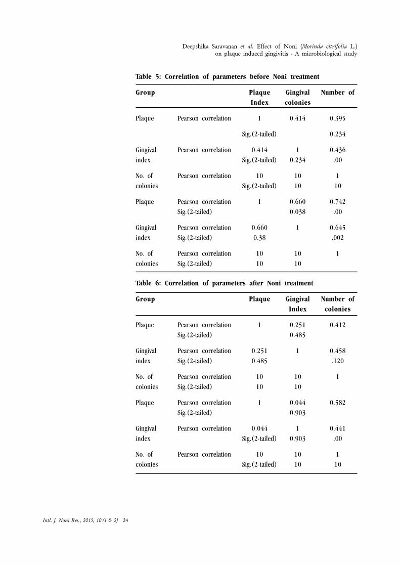
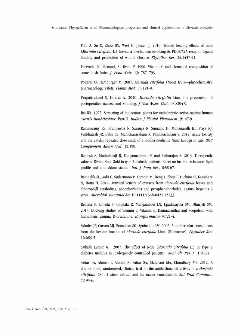
Table 5: Correlation of parameters before Noni treatment
Group Plaque Gingival Number ofIndex colonies
Plaque Pearson correlation 1 0.414 0.395
Sig.(2-tailed) 0.234
Gingival Pearson correlation 0.414 1 0.436index Sig.(2-tailed) 0.234 .00
No. of Pearson correlation 10 10 1colonies Sig.(2-tailed) 10 10
Plaque Pearson correlation 1 0.660 0.742Sig.(2-tailed) 0.038 .00
Gingival Pearson correlation 0.660 1 0.645index Sig.(2-tailed) 0.38 .002
No. of Pearson correlation 10 10 1colonies Sig.(2-tailed) 10 10
Table 6: Correlation of parameters after Noni treatment
Group Plaque Gingival Number ofIndex colonies
Plaque Pearson correlation 1 0.251 0.412Sig.(2-tailed) 0.485
Gingival Pearson correlation 0.251 1 0.458index Sig.(2-tailed) 0.485 .120
No. of Pearson correlation 10 10 1colonies Sig.(2-tailed) 10 10
Plaque Pearson correlation 1 0.044 0.582Sig.(2-tailed) 0.903
Gingival Pearson correlation 0.044 1 0.441index Sig.(2-tailed) 0.903 .00
No. of Pearson correlation 10 10 1colonies Sig.(2-tailed) 10 10
Intl. J. Noni Res., 2015, 10 (1 & 2) 24
Deepshika Saravanan et al. Effect of Noni (Morinda citrifolia L.)on plaque induced gingivitis - A microbiological study

Discussion
Plants have always been among the coon sources of medicines, either processed astraditional preparation or used to extract active principles ( Maiden, 1889) Morindacitrifolia L. has been used for more than 2000 years as traditional folk medicinefor various diseases like headache, fever, arthritis, Gingivitis, diabetes, etc. (Mathivananet al., 2005; Wang et al., 1999; Brett, 2012). It is known to have dental benefits butsufficient scientific research has not been carried out on this field. Noni has anti-bacterial action and anti-inflammatory action. So noni extract has positive effects onplaque induced gingivitis (John Glang, 2013).
Plaque and gingival index were assessed for 20 subjects as clinical evidence. Thiswas done to find the effect of Noni fruit juice on plaque induced gingivitis.
The plaque index scores at day 0 and day 28 for group A is taken. Based on resultsand statistical analysis, there is no significant reduction in plaque scores was seenin the case of group A. Mean scores indicate that there is an increase in plaquescores after 28 days in case of group A. This indicates that with time without anypreventive measures there is increased accumulation of plaque. The mean plaquescore of group B declined from 0.9800 to 0.6200 after 28 days indicating that thereis significant reduction in plaque scores due to gargling of Noni fruit extract whichsupports our study.
The mean gingival score of group B declined from 1.035 to 0.8150 after 28 daysindicating that there is significant reduction in gingival scores due to gargling of noniextract which supports our study. However no significant reduction was seen in caseof group A. Mean scores indicate increase in gingival index after 28 days in caseof group A. Thus gingivitis when left untreated can progress to periodontitis.
CFU count is taken to find out anti-bacterial effect of noni juice which provides themicrobiological evidence. The mean number of colonies in case of group Bdeclined from 52.50 to 39.40 after 28 days indicating that there is significantreduction in number of colonies due to gargling of noni extract. This indicates thatNoni juice has anti-bacterial effect. However no significant reduction was seen incase of group A. Mean scores indicate increase in number of colonies after 28 daysin case of group A.
Conclusion
As the results are positive it can be said that Noni fruit juice goggling causesreduction in plaque as well as gingivitis even it is practised for such a short time of28 days. If practised daily it can develop as a healthy habit. From CFU count resultsit is evident that Noni has anti-bacterial effect as results show reduction in numberof bacteria per ml after 28 days of n Noni fruit juice gargling. While the results on
Intl. J. Noni Res., 2015, 10 (1 & 2) 25
Deepshika Saravanan et al. Effect of Noni (Morinda citrifolia L.)on plaque induced gingivitis - A microbiological study
control group is significant as it shows increase in plaque and gingival index scores.Also CFU count increases when sufficient oral hygiene measures are not taken intime. Both the plaque and gingival scores tally with CFU count which indicates thatnumber of bacteria present in mouth is related to plaque and gingivitis. All theseresults indicate that Noni extract has significant effect on plaque and gingivitis. HenceNoni fruit juice has a array of therapeutic activities and has a promising note as aplaque control agent. From the results obtained it can be concluded that Noni fruitjuice has significant effect on plaque and gingivitis.
Acknowledgement
The principal investigator is also very much thankful to Chairman, Noni BioTech,Chennai for providing Noni fruit juice as well as the fund for conducting the study.
The principal investigator is also grateful to Dr. P. Rethinam who has been a pillarof support to conduct the study. Help rendered by Co-Principal Investigator andassociates in successfully conducting the project is gratefully acknowledged.
The principal investigator is very grateful to RVS Dental College and hospital forhaving given me the opportunity to work on this project.
References
Brett, J. West, Stephen K. Palmer, Shixin Deng and Afa K. Palu. 2012. Antimicrobialactivity of an Iridoid rich extract from Morinda citrifolia ,L.fruit. Research andDevelopment. Tahikian Noni International, USA.,4(1)52-54
Heinicke, R. 1985. The pharmacologically active ingredient of Noni. Bulletin of theNational Tropical Botanical Garden
Jonas Glang, Wolfgang Falk and Johannes Westends. 2013. Effect of Morinda citrifoliaL. fruit juice on gingivitis / Periodontitis. ............2:21-27.
Levand, O and Larson, H. O. 1979. Some chemical constituents of Morinda citrifoliaL. Planta Med., 36: 186-187.
Mathivanan, N., Surendiran, G., Surendiran, K., Sagadevan, E and Malarvizhi, K.2005. Review on the current scenario of Noni research; Taxonomy, distribution,chemistry, medicinal and therapeutic values of Morinda citrifolia L. Intl. J. NoniRes., 1 (1):1-16.
Merrill, E.D. 1943. Noni (Morinda citrifolia L.) as an edible plant. In: Technicalmanual: emergency food plants and poisonous plants of the islands of the pacific.Washington DC: US Government Printing Office; 75-110.
Moorthy, N.K. and Reddy, G. S. 1970. Preliminary phytochemical and pharmacologicalstudy of Morinda citrifolia Linn. Antiseptic, 67: 167-171.
Intl. J. Noni Res., 2015, 10 (1 & 2) 26
Deepshika Saravanan et al. Effect of Noni (Morinda citrifolia L.)on plaque induced gingivitis - A microbiological study

Sang, S., Cheng, X., Zhu, N., Wang, M., Jhoo, J.W. and Stark, R.E. 2001 Iridoidglycosides from the leaves of Morinda citrifolia L. J Nat Prod., 64: 799-800.
Simonsen, J. L. 1920. Note on the constituents of Morinda citrifolia L. J. Chem. Soc.,117: 561-564.
Solomon, N. 1999 The tropical fruit with 101 medicinal uses, NONI juice. 2nd ed.Woodland Publishing.
Wang, M., Kikuzaki, H., Csiszar, K., Boyd, C. D., Maunakea, A. and Fong, S. F. 1999.Novel trisaccharide fatty acid ester identified from the fruits of Morinda citrifolia(Noni). Agric Food Chem., 47: 4880-4882.
Younos, C., Rolland, A., Fleurentin, J., Lanhers, M. C., Misslin, R. and Mortier, F.1990. Analgesic and behavioural effects of Morinda citrifolia L. Planta Med., 56:430-434.
Youngken, H. W., Jenkins, H. J. and Butler, C.L. 1960. Studies on Morinda citrifoliaL. II. J Am Pharm Assoc., 49: 271-273.
Deepshika Saravanan et al. Effect of Noni (Morinda citrifolia L.)on plaque induced gingivitis - A microbiological study
Intl. J. Noni Res., 2015, 10 (1 & 2) 27
Etiology of dry and soft rot of Noni(Morinda citrifolia, L) fruits
Authors’ affiliation :1. Department of PlantPathologyTamil Nadu AgriculturalUniversity,Coimbatore- 641 0032. Additional Director,World Noni ResearchFoundation, Chennai – 600 096Corresponding author-E-mail:
S. Nakkeeran1,Chandrasekar1,P. Renukadevi1 andT. Marimuthu2
Abstract : Survey of Noni orchards at Tamil Nadu and Kerala revealed thepresence of both dry and wet fruit rot. The pathogen associated with dry fruitrot of Noni was Colletotrichum gloeosporioides and Alternaria alternata. Thewet or soft rot of Noni was caused by Pantoea agglomerans. The pathogenassociated with Noni wet rot was confirmed for its identity through fatty acidmethyl ester analysis , 16s rDNA analysis and through biochemicalcharacterization. The primary spread ofNnoni dry fruit rot caused by C.gloeosporioides was due to the presence of dormant mycelium in the stems andbarks and due to the fruiting body namely the acervuli. The primary spread byA. alternata is through infected crop leaf residues. The secondary spread ofboth the pathogen was through air borne conidia. Flowers on the fruits werealso found to be infected by both C. gloeosporioides and A. Alternata.Aerodynamic studies revealed the association of conidia of both the dry rotpathogens in air. Presence of continuous inoculums in the Noni orchardsfavour the disease spread. Presence of scars on the fruit surface coupled withdew and increased humidity is highly favourable for the fruit rot epidemic.However, the wet fruit rot is severe only during the monsoon seasons.
Introduction
Noni (Morinda citrifolia L.) is known for its therapeutic use and grown throughoutthe tropical countries. The extracts ofNoni fruits stimulate the immune system andcure most of the diseases. Extensive cultivation of Noni has resulted in outbreak ofdiseases such as leaf blight, anthracnose, black flag, fruit rot, stem blight, sootymould, stem canker and algal leaf spot. Fruit rot diseases leads to considerable yieldreduction in terms of quantity and quality of fruits. Recent survey in the usual Nonigrowing areas of Tamil Nadu and Kerala revealed the outbreak of fruit rot diseases.However, perusal of literature revealed that the etiology behind the fruit rot diseasewas obscure. Hence, the present investigation was carried out to understand the hostparasite relationship of fruit rot pathogens.
Correspondence to :S. NakkeeranDepartment of PlantPathologyTamil Nadu AgriculturalUniversity,Coimbatore- 641 003Corresponding author-E-mail:
Keywords :
Intl. J. Noni Res., 2015, 10 (1 & 2) 28
Materials and Methods
Isolation of pathogens associated with dry and soft fruit rot
Pathogens associated with fruit rot were isolated from lesions of Noni flowers, fruits,and infected twigs through tissue segment method on potato dextrose agar (PDA)and nutrient agar for fungal and bacterial pathogens respectively. Plates were incubatedat room temperature (28-300 C) and observed periodically. The growing edges offungal hyphae developing from the infected tissues were then transferred asepticallyto PDA slants and pure culture was stored at 40 C.
Identification of pathogens associated with dry fruit rot.
The pathogen was identified up to species level based on cultural and morphologicalcharacters. A loopful of fungal culture grown on PDA plates were taken on a glassslide and observed under image analyzer (CETI, with Capture pro software MedlineScientific Ltd, UK) under 40X magnifications for the presence of conidia andconidiophore. After confirming the spores, the culture was purified by single sporeisolation technique and stored at 4 ï C on PDA slants.
Identification of bacterial pathogen associated with soft /wet fruit rot.
Bacterial pathogen associated with soft rot of noni was isolated on NA medium,based on the cultural, morphological and biochemical characterization the pathogenwas identified.
The identification was further confirmed through, FAME analysis and 16s DNA.
Morphological characteristics of bacterial pathogen
Morphological and cultural characters of bacterial pathogen was observed throughmicroscope with the help of capture pro software.
Growth of bacterial pathogen in different media
The growth of bacterial pathogen was assessed in nutrient agar (NA) Luria bertanimedium (LB) and Crystal Violet Pectate medium (CVP). An 18 hours old bacterialculture in a growing slant was transferred to 1 ml of sterile water and thoroughlyshaked to get uniform suspension of bacterium. The concentration of bacterial cellswas adjusted to 106 cells ml-1 using spectrophotometer. Twenty ml of the sterile agarmedium in a test tube was inoculated with 100 µ1 of inoculum prepared as indicatedabove. Later it was shaked well and plated on to in nutrient agar medium Luriabertani and CVP medium in sterile Petri plates .The plates were incubated at (28± 2oC). The bacterial colonies were observed after 20 hours after inoculation.
S. Nakkeeran et al. Etiology of dry and soft rot of Noni (Morinda citrifolia, L) fruits
Intl. J. Noni Res., 2015, 10 (1 & 2) 29
Biochemical properties of the bacterial pathogen
Gram staining
Gram staining performed using 24 h old bacterial culture as described here under.The uniform suspension of the isolate prepared in sterilized distilled water wassmeared on the cleaned glass slide and air dried. The smear was gently exposedto flame for two min and covered with crystal violet solution for 30 sec. Then theslide was gently washed with distilled water for a few min and covered with Lugolsiodine solution for 30 seconds. The iodine solution was washed by using 95 percent ethyl alcohol until no more color flows from the smear. The slide was againwashed with distilled water, drained and safranin (counter-stain) was applied on theslide for 30 sec. The slide was then washed with distilled water, blotted using acountry filter paper and air dried. The slide was then examined in the microscopeunder oil immersion (Aneja, 1993).
Biochemical characterization of bacterial pathogen was performed with gram negativebacteria identification kit supplied from Tulip diagnostics (P) Ltd, India. Theidentification kit comprises test such as 1.Lysine utilization test, 2.Ornithindecarboxylation test, 3.Phenyalanine deamination, 4.Urease detection test, 5. Nitratereduction test, 6. H
2S production test, 7. Citrate utilization test 8. Glucose utilization
test, 9. Adonitol utilization test, 10. Arabinose utilization test, 11. Lactose utilizationtest, 12. Sorbitol utilization test, 13. Oxidase detection test.
Kit containing 13 vials with differential medium were inoculated with 100 µl of 18-24 h old culture and incubated at 35-38o c for 24 h. Experiment was observed toexamine color change after incubation so as to identify the pathogen. Pathogen wasidentified as Pantoea agglomerans. In addition, reaction of bacterial pathogen alsotested for KOH, Simmon citrate utilization and catalase test.
Fatty acid methyl ester of P. agglomerans
The bacterial fatty acid methyl derivatives were analyzed with Midi Sherlock®MicrobialIdentification System (Sherlock TSBA Library version 3.80; Microbial ID). Liquidculture (24h old) of bacteria was used to assess fatty acid. The fatty acids areextracted by saponification in dilute sodium hydroxide/methanol solution followedby derivatization with dilute hydrochloric acid/methanol solution to give the respectivemethyl esters (FAMEs). The FAMEs were then extracted from the aqueous phase bythe organic solvent and the resulting extract was analyzed by Gas Chromatography(GC) the bacterial cells were killed by saponification. FAMEs are more volatile thantheir respective fatty acids and therefore more suitable to gas chromatographyanalysis. The Sherlock software automates all analytical operations and uses asophisticated pattern recognition algorithm to match the unknown FAME profile tothe stored library entries for identification.
S. Nakkeeran et al. Etiology of dry and soft rot of Noni (Morinda citrifolia, L) fruits
Intl. J. Noni Res., 2015, 10 (1 & 2) 30

Molecular characterization of P. agglomerans through amplification of16s ribosomal RNA of P. agglomerans
Genomic DNA was isolated from the bacterial culture by using the bacterial GenomicDNA Isolation Kit (RKT09).The bacterial DNA were amplified with forward primer16s forward primer: (5’AGAGTRTGATCMTYGCTWAC3) and16s reverse primer:(5’CGYTAMCTTWTTACGRCT-3')’ .The 20 ìl mixture approximately contain 50 ng oftotal DNA, 5 mM each dNTPs, 20 pmol of each forward primer and reverse primerand 0.5 U of Taq DNA polymerase (Bangalore Genei, India). PCR amplification wasperformed in a thermocycler (Thermal Cylcer ABI2720, Chromous Biotech Pvt. Ltd,Bangalore, India).
Sequencing of bacterial 16s ribosomal RNA and sequence analysis ofPantoea agglomerans
Sequencing of amplicons were performed with ABI 3130 Genetic Analyzer (Chemistry:Big Dye Terminator version 3.1"Cycle sequencing kit. (Chromous Biotech Pvt. Ltd,Bangalore, India.). The biosynthetic gene homology searches were performed usingthe BLAST program though the internet server at the National Centre for BiotechnologyInformation, National Institutes of Health, and Bethesda, USA. Sequences andaccession numbers for compared isolates were retrieved from the Gene Bankdatabase. Sequence pair distances among related and different Pantoea spp isolateswere scored with the CLUSTAL X (1.81) program and phylogenetic tree analysis wasperformed with Phylogenetic Tree Builder. Newly obtained sequences were submittedin the Gen Bank database, New York, USA.
Pathogenicity of dry fruit rot
The unripe maturedNoni fruits were used for pathogenicity.
The C. gloeosporioides and A. alternata were used for artificial inoculation on tounriped fruits at the rate of 5 x 10⁵ conidia ml-1. Conidia of C. gloeosporioides andA. alternata were harvested using 14 days old PDA culture by grinding it withphosphate buffer pH 7.0 in a sterile pestle and mortar. The fruits were washed withrunning tap water and surface sterilized with 0.1 per cent mercuric chloride andwashed with sterile distilled water. The washed fruits were inoculated with conidialsuspension after pin prick using sterile entomological pins.
The fruits inoculated with sterile distilled water served as control. The inoculatedfruits were incubated in sterile poly propylene (PP) bags and sprayed with sterilewater. To maintain humidity sterile cotton wools were moistened with sterile waterand placed inside the PP bags. The bags were incubated at room temperature (28± 2ºC) for 72 hours. The infection was recorded periodically and the per centinfection of fruits was calculated. Three replications were maintained each replicationconsist of five fruits.
S. Nakkeeran et al. Etiology of dry and soft rot of Noni (Morinda citrifolia, L) fruits
Intl. J. Noni Res., 2015, 10 (1 & 2) 31
Pathogenicity of C. gloeosporioides on twigs
The healthy green twigs were collected from noni orchard. Twigs were sliced in tosmall bits (4-6 cm) and washed with running tap water, surface sterilized with 0.1per cent mercuric chloride and washed with sterile distilled water. Later the surfacesterilized twigs were inoculated with spore suspension of C. gloeosporioides at therate of 5 x 10⁵ conidia ml-1. The twigs inoculated with sterile distilled water servedas control. After inoculation the twigs were incubated in sterile poly propylene (PP)bags and sprayed with sterile water. To maintain humidity sterile cotton wools weremoistened with sterile water and placed inside the PP bags. The bags were incubatedat room temperature (28 ± 2ºC) for 72 hours.
Pathogenecity of C. gloeosporioides on flowers
Apparently healthy flowers of Noni were collected from noni orchard.
The collected noni flowers were surface sterilized with 0.1 per cent mercuricchloride for one minute and later serially washed thrice in sterile distilled water.Then the flowers were blot dried in sterile what man. No.1 filter paper. Ten sterilizedflowers were placed at equal distance on the plates The conidia of C. gloeosporioideswas harvested from 14 days old culture grown on PDA and suspended in phosphatebuffer pH 7.0. The conidial suspension was adjusted to a load of 5 x 10⁵ conidiaml-1. The conidial suspension of C. gloeosporioides was sprayed to the flowers onplain agar in sterile Petri plate. Plates were incubated for three days at 28 ± 2 ºC.The flowers after the incubation were assessed for the colonization by C.gloeosporioides (Sergeeva et al., 2008).
Pathogenicity test in vitro for wet fruit rot
Moist chamber method
Pathogenecity was performed with healthy matured unriped fruit. Ten sterile tissueculture bottles (12.5X 6.00) were taken; inner side of bottle was lined with sterile moistfilter paper. The surface sterilized fruits were kept inside the bottles placed with sterilemoist cotton laid upto a height of 3 cm at the bottom. The healthy unripe fruits wereinoculated with l00 µ1 of 18 h old bacterial pathogen after making four pinpricksrandomly at the apical end of the fruit at the concentration of 106 cells ml-1. Inoculatedfruits were incubated at room temperature (28 ± 2 ºC) for 24 to 48hours. Controlswere maintained by keeping sterile moist cotton at the apical end of fruit. The infectionwas recorded periodically and the per cent infection of fruits was calculated. Threereplications were maintained each replication consist of 5 fruits.
Pathogenicity test in field
The bacterial pathogen was grown to log phase (48 h) in NA broth, and the cellswere harvested by centrifugation at 4000 rpm for 10 min, washed in Phosphate
S. Nakkeeran et al. Etiology of dry and soft rot of Noni (Morinda citrifolia, L) fruits
Intl. J. Noni Res., 2015, 10 (1 & 2) 32

Buffer Saline (PBS) and resuspended in the same volume of PBS. The optical densitywas adjusted using a spectrophotometer to contain 106 cells ml-1. Healthy unripedfruits were sprayed with bacterial pathogen at the rate of 1 ml by suspending it in100 mg of carborundam powder to create mechanical injury to fruits using anaspirator. The inoculated fruits were then covered with polythene bags with 100gauge thickness. The symptoms of the disease were observed at periodical intervals.A total of ten fruits were assessed for pathogenicity.
Histopathological studies of infected twigs and fruits
Histopatholgical studies were made by microtome sectioning of infected and uninfectedtwigs and fruits of noni followed by staining and microscopic observations. Infectedfruit and twigs were examined for the presence of propagules of fungal pathogenwith the help of image analyzer. At 10X x 40X magnification with CETI, capture prosoftware supplied by Medline Scientific Ltd, UK.
Mode of Spread
Source of infection
Twigs and fruits exhibiting brown lesion with pin head shaped acervuli were collectedfrom noni orchard. Twigs and fruits were sliced in to small bits (4-6 cm) and washedwith running tap water, surface sterilized with 0.1 per cent fmercuric chloride andwashed with sterile distilled water. Later twigs and fruits were incubated at 5ïC for48 hours. After incubation the bits were examined under stereo zoom microscopefor the presence of fungal growth.
Aerodynamic studies
Clean microscopic slides were evenly smeared with commercially available wax onone side of slide. Later the slides were tied with Noni tree in East, West, North andsouth directions at 30 cm, 60 cm, 90 cm, 120 cm, 150 cm, 180 cm and 210 cmheight intervals from the ground level. After 24 hours the slides were examinedunder microscope for the presence of air borne conidia.
Results and Discussion
Survey for the incidence of fruit rot diseases
Survey was conducted in major Noni growing areas of Tamil Nadu and Kerala duringSeptember, 2010 .Survey revealed that the incidence of dry and wet fruit rot werefound in surveyed area . Among five districts surveyed across two states, the highincidence of dry fruit rot was recorded in Coimbatore about 71.40 per cent followedby Aliyar Nagar of Coimabtore accounted 63.20 per cent. The lowest incidence ofdry fruit rot (Colletotrichum) was observed in kodunallur, Ernakulum district ofKerala it ranged 24.80 per cent disease incidence. Among surveyed area Thrissurdistrict was found with more incidence of Alternarial dry fruit rot about 45.00 per
S. Nakkeeran et al. Etiology of dry and soft rot of Noni (Morinda citrifolia, L) fruits
Intl. J. Noni Res., 2015, 10 (1 & 2) 33
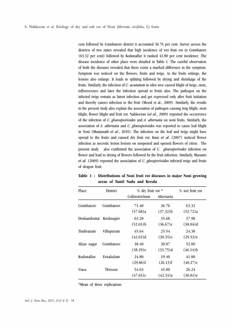
cent followed by Coimbatore district it accounted 36.76 per cent .Survey across thedistricts of two states revealed that high incidence of wet fruit rot in Coimbatore(63.32 per cent) followed by Kodunallur it ranked 41.80 per cent incidence .Thedisease incidence of other place were detailed in Table 1. The careful observationof both the diseases revealed that there exists a marked difference in the symptom.Symptom was noticed on the flowers, fruits and twigs. As the fruits enlarge, thelesions also enlarge. It leads to splitting followed by drying and shrinkage of thefruits. Similarly, the infection of C. acutatum in olive tree caused blight of twigs, stem,inflorescence and later the infection spread to fruits also. The pathogen on theinfected twigs remain as latent infection and get expressed only after fruit initiationand thereby causes infection to the fruit (Moral et al., 2009). Similarly, the resultsin the present study also explain the association of pathogen causing twig blight, stemblight, flower blight and fruit rot. Nakkeeran (et al., 2009) reported the occurrenceof the infection of C. gloeosporioides and A. alternata on noni fruits. Similarly, theassociation of A. alternata and C. gloeospoioides was reported to cause leaf blightin Noni (Manjunath et al., 2010). The infection on the leaf and twigs might havespread to the fruits and caused dry fruit rot. Kaur et al. (2007) noticed flowerinfection as necrotic lesion lesions on unopened and opened flowers of citrus. Thepresent study also confirmed the association of C. gloeosporiodes infection onflower and lead to drying of flowers followed by the fruit infection. Similarly, Masantoet al. (2009) reported the association of C. gleosporioides infected twigs and fruitsof dragon fruit.
Table 1 : Distributions of Noni fruit rot diseases in major Noni growingareas of Tamil Nadu and Kerala
Place District % dry fruit rot * % wet fruit rotColletotrichum Alternaria
Coimbatore Coimbatore 71.40 36.76 63.32(57.68)a (37.32)b (52.72)a
Denkanikottai Krishnagiri 63.20 35.68 37.98(52.65)b (36.67)c (38.04)d
Tindivanam Villupuram 45.64 25.54 24.30(42.65)d (30.35)e (29.53)e
Aliyar nagar Coimbatore 38.40 30.87 52.00(38.29)e (33.75)d (46.14)b
Kodunallur Ernakulam 24.80 19.40 41.80(29.86)f (26.13)f (40.27)c
Viasa Thrissur 54.63 45.00 26.24(47.65)c (42.33)a (30.81)e
*Mean of three replications
S. Nakkeeran et al. Etiology of dry and soft rot of Noni (Morinda citrifolia, L) fruits
Intl. J. Noni Res., 2015, 10 (1 & 2) 34

In a column, means followed by a common letter are not significantly different at5% level by DMRT; Values in parentheses are arcsine transformed values
Symptoms of dry fruit rot caused by Colletotrichum gloeosporioides
Colletotrichum gloeosporioides infect all parts of noni plant irrespective of thestages of crop growth. The symptoms were observed on twigs, flowers and fruits.Symptoms of the infection caused by C. gloeosporioides on the flowers appear asdull brown lesions. The infected flowers dried within 48 h after infection. Examinationof the flowers under stereo zoom microscope revealed the presence of brownnecrotic spots on the corolla tube adhering on the noni fruit. The twigs infected byC. gloeosporioides were characterized by the presence of necrotic brown lesionswith yellow halo. Later the lesions enlarge in size which was characterized with greycentre. On the grey centre number of minute, pin head shaped fructifications of theacervuli was observed .The necrotic lesions spread gradually towards fruits throughpeduncle and leads to the development of infection on flowers and fruits.
Symptoms of dry fruit rot occurring on the fruits incited by C. gloeosporioides arecharacterized by the presence of small circular reddish brown, slightly sunkennecrotic spot on fruits. It later turns, into dark brown lesions, expands both upwardand downward, coalesces, dry and shrinks. As fruits enlarge, the lesion also expands,leading to splitting and drying of fruits. Subsequently, small pinhead like acervuliappears on the infected tissues. Subsequently, the infected fruits shrinks, dries offand get mummified. After mummification the infected fruits were colonized bysaprophytic moulds like Fusarium spp, Aspergillus, and Penicillium.
Symptoms of dry fruit rot caused by A. alternata
The dry fruit rot caused by A. alternata is characterized by the presence of blacknecrotic sunken spot of 2-3mm dia on the green unripe fruits. During favorableenvironment spot turns to dark black lesion and coalesced. Centre of the lesionsare black in color with alternate concentric zonations. As the fruit expands it leadsto splitting and drying of the fruits coupled with the saprophytic infection of othermoulds.
Symptoms of soft rot caused by Pantoea agglomerans on fruits
The bacterial infection was associated with matured un-riped fruits. Symptoms werecharacterized by the presence of brown colored water soaked lesion on externalsurface of the fruit. Later with in 24 to 48 h after infection, the lesion spread to theentire fruit and the infected fruits emit a bad odour. The affected tissue becomessoftened and rots subsequently. Later, the infection extends up to the peduncle andthe fruits fall down after 24-48 h after infection.
Identification of dry rot pathogens
S. Nakkeeran et al. Etiology of dry and soft rot of Noni (Morinda citrifolia, L) fruits
Intl. J. Noni Res., 2015, 10 (1 & 2) 35
Colletotrichum gloeosporioides
The colony of the pathogen was white to pale grey on PDA medium. The pathogenproduced hyaline septate, conidiophores bearing ovoid to cylindrical shape conidia,one celled, with one or two oil globules. The length and width of the conidia was11.25 µm and 6.5 µm respectively.
Alternaria alternata
The colony colour on the PDA medium was dark brown. The colony was characterizedby flat growth on PDA medium. It produced abundant, branched, septate, brownmycelia. Conidiophores were simple, olive-brown with septations. The conidia weresmooth, brown to dark brown in colour, obclavate measuring 30.55 to 42.35µmx 11.73 to 17.30 mm, muriform or irregular with 0-5 cross septa.
Isolation and identification of pathogen associated with Soft / Wet fruitrot.
The pathogen associated with bacterial fruit rot was isolated on nutrient agar medium.Based on the morphological and biochemical characterization the pathogen wasidentified as Pantoea agglomerans. The identification was further confirmed through,FAME analysis and 16s rDNA.
Cultural and morphological characterization
The bacterial colony was yellow in colour. Colony morphology was mucoid withsmooth margin on NA medium. . Bacterial cells were rod shaped. The bacteriashowed negative reaction to Gram staining, indicating that it is a Gram negativebacterium.
Biochemical properties of bacterial pathogen
Series of biochemical test were performed with micro express kit for confirmationof bacterial pathogen. According to kit specification, the pathogenic bacteria posses90% positive reaction for Citrus utilization, Nitrate reduction, Glucose utilization andArabinose utilization, 75-89% for Phenylalanine Deamination, 26- 75% for Ornithineutilization and 25% for lactose utilization . Pathogenic bacteria exhibited negativereaction to lysine utilization, Urease detection, H
2S production, Adonital utilization,
Sorbitol utilization and Oxidase detection (Table 3) Based on the micro express kitanalysis, the pathogen was confirmed as P. agglomerans. In addition thebacterium also expressed positive reaction for KoH, Simmon citrate utilization andcatalase test.
S. Nakkeeran et al. Etiology of dry and soft rot of Noni (Morinda citrifolia, L) fruits
Intl. J. Noni Res., 2015, 10 (1 & 2) 36

Table 3 : Biochemical characterization of P.agglomerans
S. No Biochemical assay Pantoea .spp
1 Lysine utilisation test -
2 Ornithine decarboxylation d
3 Phenylalanine deamination (+)
4 Urease detection -
5 Nitrate reductase test +
6 H2S production -
7 Citrate utilisation +
8 Gucose utilisation +
9 Adonitol utilisation -
10 Arabinose utilisation +
11 Lactose utilisation (-)
12 Sorbitol utilisation -
13 Oxidase detection -
+ = positive (more than 90%); (+) = 76-89% positive; (-) = 11-25% positive; d=26-75% Positive; - = negative (more than 90%)
Confirmation of P. agglomerans by FAME analysis
Identification of P. agglomerans was confirmed by Fatty acid methyl ester (FAME)analysis. Microbial identification system identified pathogenic bacteria as P.agglomerans with the similarity index (SI) of 0.536 with 0.3 separation of betweenthe first and second choice. FAME analysis confirmed that the pathogen associatedwith soft rot of noni was P. agglomerans. This was in same line with the finding ofGermida and Siciliano (2001) who identified wheat root colonizing bacteriumEnterobacter agglomerans (Pantoea agglomerans) with the similarity index of0.535. Coutinho et al. (2002) confirmed Eucalyptus leaf blight pathogen P. ananatisby fatty acid profiling.
Molecular confirmation of P. agglomerans through 16S rDNA analysis
The identity of soft rot bacterial pathogen was confirmed through PCR. The bacterial16 rDNA was amplified with universal bacterial forward and reverse primers.Amplification of 16 rDNA revealed the presence of 1500bp amplicon. Afteramplification, the amplicon of 1500bp was purified from PCR product and sequenced.Partial sequences of P. agglomerans was submitted to NCBI, Gen bank, New York,USA, bearing accession no. JN036646. The sequence of the 1500bp of the ampliconis as follows.
S. Nakkeeran et al. Etiology of dry and soft rot of Noni (Morinda citrifolia, L) fruits
Intl. J. Noni Res., 2015, 10 (1 & 2) 37

16 S rDNA sequences analysis of pathogen
The 16S rDNA homology searches were performed using the BLAST programthough the internet server at the National Center for Biotechnology Information, USA.The comparison of sequences of pathogens revealed 99 per cent homology withP.agglomerans (Table 4). The unweighted pair group method with arithmetic means(UPGMA) tree resulting rDNA sequences of pathogen had 99 percent similarity withsequences of P. agglomerans deposited in gen Bank. Genetic distance among thesequences from gen bank and sequence of 16S rDNA regions of pathogen is shownin Table 4. Based on the results, the pathogen was confirmed as P. agglomerans.The results are in agreement with the findings of Medrano and Bell (2007) wherein they reported the amplification of 16 S r DNA region of P. agglomerans with16s-F and 16s -R specific primers that yielded 1.5 kb amplicon. Similarly Coutinho etal. (2002) differentiated P. ananatis from other species using 16 S rRNA primers.Krawczyk et al. (2010) identified P. ananatis associated with leaf spot disease ofmaize by using 16 s rDNA specific primers it yielded 1.5 kb amplicon.
Table 4 : Genetic similarity score between P. agglomerans and GeneBank sequences
S. No. Accession No. Similarity score Organism
1 HM130693 99.00 Pantoea agglomerans strain BJCP2
2 EU931561 99.00 Pantoea agglomerans strain ZFJ-6
3 JN036646* 99.00 Pantoea agglomerans
4 HQ443233 99.00 Pantoea agglomerans strain T2
5 HQ647265 99.00 Pantoea agglomerans
6 EF522820 98.00 Pantoea sp. 092305
7 GQ200831 99.00 Pantoea dispersa strain ND4
8 FJ560472 99.00 Pantoea sp. M3S5
9 JF958137 96.00 Erwinia sp. JSC-N3-112-2
10 HM130694 99.00 Pantoea agglomerans strain BJCP3
Pathogenicity
Pathogenicity of dry fruit rot by C. gloeosporioides
Koch postulates were proved for flower infection with C. gloeosporioides isolatedfrom the infected flowers. The apparently healthy flowers sprayed after sterilization,with the conidial suspension of C. gloeosporioides (5 x 10⁶ spore’s ml-1) resultedin the expression of brown necrotic spots on the corolla tube. It was followed bythe colonization of C. gloeosporioides observed after 48h of incubation. Similarly,artificial inoculation of spore suspension of C. gloeosporioides (5 x 10⁶ spores ml-
S. Nakkeeran et al. Etiology of dry and soft rot of Noni (Morinda citrifolia, L) fruits
Intl. J. Noni Res., 2015, 10 (1 & 2) 38
1) on twigs resulted with reproduction of brown lesions characterized with greycentre. The association of C. gloeosporioides with twig infection was confirmed byreisolation Inoculation of C. gloeosporioides on the fruits at the rate of 5 x 10⁶
spores ml-1 after making pin prick injuries with sterile needles on to the greenimmature fruits developed necrotic brown, sunken spots after four days of inoculation.The spots later expand and coalesced. It resulted in the shrinkage and rotting offruit. The pathogen was reisolated from the fruits and was confirmed as C.gloeosporioides based on their morphological and conidial characters.
Pathogenicity of dry fruit rot by A. alternata
Inoculation of A. alternata (5 x 10⁶ spores’ ml-1) on to the healthy green immaturefruits expressed small black necrotic spots after four days of inoculation. The spotslater enlarged and were characterized by the presence of black concentric rings.Re isolation of pathogen from the infected tissue confirmed the association of A.alternata.
Pathogenicity of wet rot in vitro
Apparently healthy fruits were inoculated with the suspension of bacterial pathogen(106 cell m-1) after pinprick. Later inoculated fruits were incubated in sterile bottlesmoistened with sterile absorbent cotton. Infected fruits expressed water soakedlesions at the point of inoculation after 24h of inoculation. The brown lesionsextended both upward and downward leading to rotting of fruits coupled with theemission of a foul smell. The pathogen was reisolated and Koch’s postulate wascompleted to confirm the pathogenicity.
Pathogenicity of wet rot in field
Soft-rot symptoms were observed on noni fruit after 24 - 48 h of inoculation ofpathogen under field condition. The fruit without wound did not show any fruit rotsymptoms. The bacteria caused soft rot disease and were reisolated from Noni fruitexpressing the soft-rot symptoms. The Koch’s postulates were also confirmed. However,the symptoms were not observed on uninoculated control.
Mode of Spread
Primary and secondary source of infection
Incubation of infected twigs and fruit bits in humid chamber at 5ïC leads to germinationof fructification. Microscopic observation of incubated twigs and fruit bits understereo zoom microscope revealed the presence of dormant whitish mycelial growthand acervuli. Thin sections from incubated twigs revealed the presence of acervuliwith black setae and capsule shaped conidia of C. gloeosporioides. Percentassociation of C. gloeosporioides on flowers, pericarp, twigs and fallen leaves were
S. Nakkeeran et al. Etiology of dry and soft rot of Noni (Morinda citrifolia, L) fruits
Intl. J. Noni Res., 2015, 10 (1 & 2) 39

32.5%, 28.0%, 62.0% and 23.0% respectively. Similarly, the per cent association ofA. alternata on fallen leaves and fruit pericarp was 19.5% and 16.0% respectively(Table 2).
Table 2 : Per cent association of dry fruit rot pathogen of Noni
Source material Per cent association of pathogens
C. gloeosporioides A. alternata
Flowers 32.5 0.0
Pericarp 28.0 16.0
Twigs 62.0 0.0
Leaves fallen on to soil 23.0 19.5
*Mean of three replications
In a column, means followed by a common letter are not significantly different at5% level by DMRT; Values in parentheses are arcsine transformed values
Microscopic observation of slides after 24 h revealed the presence of the conidiaof C. gloeosporioides and A. alternata. The number of conidia per microscopicfield varied as per the height where the slides were tied on to the noni trees. Boththe conidia were observed at a distance of 120 – 150 cm. The number ofColletotrichum conidia was 50 to 57 at 120 and 150 cm respectively. Similarly themaximum numbers of 52 to 54 conidia of Alternaria were observed at the heightof 120-150 cm (Table 5). The deep seated dormant mycelium in the twigs and stemmight colonize during the conducive period and lead to the production of acervuliduring the death of the tissues. Hence, the dormant mycelium may serve as theprimary source of inoculum to cause fruit rot of noni. Subsequently the conidiareleased from the fructifications may serve as the secondary source of inoculum forthe further spread of the disease. Similar observation was noticed in olive fruitsinfected by anthracnose disease (Moral et al., 2009). Moreover, the aerodynamicstudies on the dry fruit rot pathogens, indicated that the pathogens were air bornein nature.. This study suggested that the fungal spores of both pathogens were onmiddle part of the tree.
S. Nakkeeran et al. Etiology of dry and soft rot of Noni (Morinda citrifolia, L) fruits
Intl. J. Noni Res., 2015, 10 (1 & 2) 40
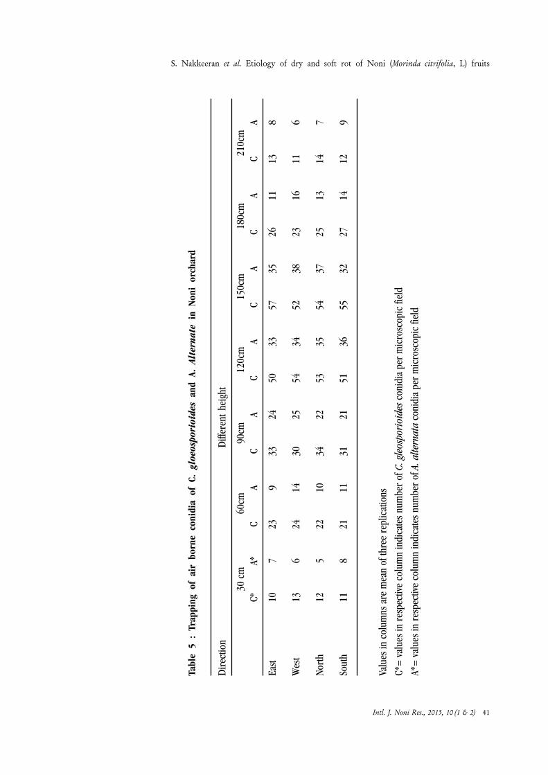
Tabl
e 5
: Tr
appi
ng o
f ai
r bo
rne
coni
dia
of C
. gl
oeos
pori
oide
s an
d A.
Alt
erna
te i
n N
oni
orch
ard
Dir
ectio
nD
iffer
ent
heig
ht
30 c
m60
cm90
cm12
0cm
150c
m18
0cm
210c
mC*
A*C
AC
AC
AC
AC
AC
A
East
107
239
3324
5033
5735
2611
138
Wes
t13
624
1430
2554
3452
3823
1611
6
Nort
h12
522
1034
2253
3554
3725
1314
7
Sout
h11
821
1131
2151
3655
3227
1412
9
Valu
es in
col
umns
are
mea
n of
thre
e re
plic
atio
ns
C*=
val
ues
in r
espe
ctiv
e co
lum
n in
dica
tes
num
ber
of C
. gle
ospo
rioi
des
coni
dia
per
mic
rosc
opic
fiel
d
A*=
val
ues
in r
espe
ctiv
e co
lum
n in
dica
tes
num
ber
of A
. alte
rnat
a co
nidi
a pe
r m
icro
scop
ic fi
eld
S. Nakkeeran et al. Etiology of dry and soft rot of Noni (Morinda citrifolia, L) fruits
Intl. J. Noni Res., 2015, 10 (1 & 2) 41

References
Coutinho, T.A., Preisig, O., Mergaert, J., Cnockaert, M.C., Riedel, K.H., Swings, J. andWingfield, M.J. 2002. Bacterial blight and dieback of Eucalyptus species, hybrids,and clones in South Africa. Plant Dis., 86: 20-25.
Kaur, R., Rewal, H.S. and Sethi, A. 2007. Pre-Harvest Stem-End Rot in Citrus Cultivarsdue to Colletotrichum gloeosporioides. Europ. J. Hort. Sci., 72 (1): 20–25.
Krawczyk, K., Kamasa, J., Zwolinska, A. and Pospieszny, H. 2010. First report ofPantoea ananatis associated with leaf spot disease of maize in Poland. Journal ofPlant Pathology ,92 (3): 807-811.
Manjunath, H., Nakkeeran, S., Raguchander, T., Anand, T. and Renukadevi, P. 2010.Physiological characterization of Colletotrichum gloeosporioides, the incitant ofanthracnose disease of noni in India. Archives of Phytopathology and PlantProtection, 44(11): 1105–1114.
Masanto, M., Kamaruzaman, S, Awang, Y., Ghazali, M and Satar, M. 2009. The FirstReport of the Occurrence of Anthracnose Disease Caused by Colletotrichumgloeosporioides (Penz.) Penz. & Sacc. on DragonFruit (Hylocereus spp.) inPeninsular Malaysia. American Journal of Applied Sciences, 6 (5): 902-912.
Medrano, E.G. and Bell. A.A. 2005. Role of Pantoea agglomerans in opportunisticbacterial seed and boll rot of cotton (Gossypium hirsutum) grown in the field.Journal of Applied Microbiology, ISSN 1364-5072
Morales.V., Silva-Rojas, H.V. and Ochoa-Martínez. D. 2007. First Report of
Pantoea agglomerans Causing Leaf Blight and Vascular Wilt in Maize and Sorghumin Mexico. J. Pdis., 91: 10-13.
Nakkeeran. S., Manjunath, H., Vijay, R., Chandrasekar, G., Renugadevi, P. andRaguchander, T. 2009. Explotation of antibiotic producing plant growth promotingrhizobacteria and fungal antagonist for the management of foliar diseases of Noni.International Journal of Noni Research, 4: 1-2.
S. Nakkeeran et al. Etiology of dry and soft rot of Noni (Morinda citrifolia, L) fruits
Intl. J. Noni Res., 2015, 10 (1 & 2) 42

Disease management in medicinal crops
Authors’ affiliation :Department of Medicinal andAromatic cropsTamil Nadu Agricultural University,Coimbatore - 641 003, Tamil Nadu,IndiaEmail : [email protected]
B. Meena
Abstract : India is one of the twelve mega biodiversity hot spot regions ofthe world and one fifth of all the plants found in India are used formedicinal purpose. Medicinal plants are important for pharmacologicalresearch and drug development, not only when constituents are used directlyas therapeutic agents, but also as starting materials for the synthesis ofdrug or as models for pharmacologically active compounds. The worldmarket for plant derived chemicals viz., pharmaceuticals, fragrances, flavoursand colour ingredients exceed several billion dollars per year. Diseases cancause substantial losses in medicinal plants. Yield loss due to diseases isone of the important factors that limit successful production of allhorticultural crops. Coleus (Coleus forskohlii) rose into prominence byvirtue of its alkaloid, forskolin, a labdane diterpene present in the swollenprimary roots (tubers). The productivity of Coleus has been hampered byits susceptibility to nematode, root rot and wilt diseases. The occurrenceof major diseases of important medicinal plants and their management arediscussed.
Introduction
World Health Organization (2003) estimates that 80% of the world’s populationdepends on traditional medicine for their health needs. In many developed countries,traditional herbal remedies are making a comeback as alternatives to modernmedicine. Exports from India have increased from Rs. 460 crores in 1995 to 1200crores in 2000 (Ghosh, 2000). The world of naturals is storming the globe withscientific rationale and trends that are fast emerging to support better health and lifethrough plant and plant products. The demand for the products from the medicinalplants such as phytochemical, steroidal, biologically active compounds, alkaloidsetc. is increasing in the national and international market.
According to the Hindu Survey of Indian Agriculture report, there was about US $60 billion sales of herbal medicines in the world and projected to increase at anaverage annual growth rate of 6.4 per cent reaching US $ trillion by the end of 2050(Anon., 2007). India being the second largest exporter, next to China account for
Correspondence to :B. MeenaDepartment of Medicinal andAromatic cropsTamil Nadu Agricultural University,Coimbatore - 641 003, Tamil Nadu,IndiaEmail : [email protected]
Keywords : Medicinal plants, diseases, fungicides, biological control
Intl. J. Noni Res., 2015, 10 (1 & 2) 43

about 13 per cent of global imports. To meet the demand, medicinal plants are beingcollected indiscriminately from forests resulting in dwindling supplies and endangeringthe survival of the species themselves.
Crop protection (pest and disease control) is a new area of research for medicinalplants. There is very little information available on the types of diseases on medicinalplants, and the effects of them on the plants. Yield loss due to diseases is one of theimportant factors that limit successful production of all horticultural crops. Thesuccess of controlling the diseases lies in correct identification of the pathogen andknowledge about their life cycles on a particular crop.
Coleus (Coleus forskohlii)
Coleus (Coleus forskohlii Briq.) syn. Coleus barbatus [(Andr.) Benth.] is animportant medicinal plant belonging to the family Lamiaceae (Mint family). Firstfound in India, it is now grown all over the world. C. forskohlii is a hardy perennial,having thrived for 3,000 years already. Mint family plants are also usually verystrongly fragrant because they contain vast amounts of essential oils. The plant isfleshy and has a scent very similar to camphor. The plant has very high medicinalvalue. This compound is used for the treatment of glaucoma, congestivecardiovascular diseases, asthma, allergies, psoriasis, cancer metastases, obesity andweight loss (Shah et al., 1980). This alkaloid has the unique property of activatingall hormone-sensitive adenylate cyclase enzymes in biological systems (De souza &Shah, 1988). Forskolin content has been found to vary from 0.07 to 0.59% of drytubers and just 1 g of forskolin costs $85, showing the importance of this crop(Gowda, 2000). In addition, forskolin is reported to have been used in the preparationof medicines preventing grey hair and restoring grey hair to its normal colour.Recently, with increase in the demand of forskolin, its cultivation has picked up insouthern states with annual production of about 100 tonnes from 700 ha in India(Anon., 2004). Root juice is given to children suffering from constipation. Its foliageis employed in treating intestinal disorders and used as condiment since timesimmemorial.On account of increased exports, cultivation of Coleus is expanding inTamil Nadu, Karnataka, Andhra Pradesh and the present area is about 5,000 acres.In Tamil Nadu, it is cultivated in Salem (Attur), Erode (Sathyamanagalam), Dindugaldistricts in about 2,000 acres under contract farming. Recently farmers have startedto grow it as a crop because of its economic potential (Vishwakarma et al., 1988).
Diseases
The crop is subjected to attack by many diseases viz., stem blight caused byPhytophthora nicotianae var. nicotianae, wilt caused by Sclerotium rolfsii, leafspot caused by Botryodiplodia theobromae, bacterial root rot caused byPseudomonas aeruginosa, dry root rot caused by Macrophomina phaseolina,aerial blight caused by Rhizoctonia solani, collar rot complex caused by Rhizoctonia
B. Meena. Disease management in medicinal crops
Intl. J. Noni Res., 2015, 10 (1 & 2) 44
bataticola, Fusarium chlamydosporum, Sclerotium rolfsii and root-knot nematode(Meloidogyne species). Among these, root-knot and wilt disease complex is occurringin severe form and cause heavy losses. Due to this disease complex the yield losswas ranged up to 50 to 60 per cent. Therefore, it is necessary to take effectivecontrol measures to save the crop from complete devastation.
Leaf spot
The lesions are initially brown and punctiform, becoming elliptic, subcircular toirregular and pale brown in colour. They are well delimited with a dark brown rim(up to 5 mm in diameter), distributed on the lamina, sometimes coalescing andleading to extensive necrosis and yellowing. A dematiaceous fungus (Corynesporacassiicola) was consistently found sporulating in the centre of the lesions.
Blight
It is common during monsoons or during period of high humidity. Symptomsinclude water soaked leaf spots that increased rapidly in size becoming light tan tobrown and later necrotic. Severe infection results in defoliation and death of plants.Rhizoctonia solani has been reported to cause the leaf blight of C. forskohlii.
Root-rot/wilt
It is the major disease of C. forskohlii causing heavy losses (>50%) in south India.Root rot is a disease caused by a variety of fungi/bacterium species that love standingwater. Disease shows various symptoms like yellowing and wilting of leaves, brownto black roots, oozing, putrefaction and decaying of roots and unhealthy plants.Fusarium chlamydosporum was found to be the causal agent of the disease. Thesymptoms include gradual yellowing, marginal necrosis and withering of leavesfollowed by loss in vigour and premature death. Such plants show discoloration ofroots and complete decaying of tap and lateral root system. Such affected plants arefinally killed due to severe root and collar rots. The infected tubers rot and emit badodour.
Ralstonia solanacearum was reported to be causing vascular wilt of C. barbatus.The symptoms include initially brown later on becomes black roots due to decaying,oozing and putrefaction of roots. Boby and Bagyaraj (2003) reported that thefungus Fusarium chlamydosporum causes root rot of C. forskohlii. Kamalakannanet al. (2005) reported root rot disease (Macrophomina phaseolina) on C.forskohlii. The symptoms observed were yellowing and drooping of the leaves,blackening of the stem, rotting of the root, basal stem and bark peeling. Thepresence of black sclerotia was observed on the rotted portion. The mycelium wasinitially hyaline and later became grey in colour. Sclerotia were minute, black, roundto oblong or irregular in shape with mycelial attachment.
B. Meena. Disease management in medicinal crops
Intl. J. Noni Res., 2015, 10 (1 & 2) 45

Plant parasitic nematode fungal interactions
Senthamarai et al. (2006) observed nematode fungal disease complex involvingMeloidogyne incognita and M. phaseolina on C. forskohlii. Patil et al. (2003)reported that the prevalence and severity of different diseases and nematode problemsin north eastern Karnataka, on medicinal crops such as Coleus, Long pepper,Ashwagandha etc. and estimated the loss to the tune of 5-20 per cent due to thepresence of root-knot, damping off, rhizome rot, seed rot, seedling blight anddieback diseases. Krishna Rao and Krishnappa (1994) observed that inoculationof nematode (M. incognita) along with fungus (Fusarium oxysporum f. sp.ciceri) at lower levels resulted in 19.88 per cent wilt incidence when compared to6.66 per cent with fungus alone. However, maximum wilt incidence (66.70%) wasobserved when inoculated with 2 juveniles per g of soil and 25 g inoculum per 500g soil of nematode and fungus respectively. Presence of root-knot nematode alongwith fungus adversely affected the root-knot disease.
Senthamarai et al. (2006) carried a glasshouse experiment to study the interactionof M. incognita and M. pahseolina on C. forskohlii. The nematode multiplicationwas adversely affected when fungus was inoculated prior to nematode. Simultaneousinoculation of nematode and fungus as well as nematode followed by fungus 15 dayslater, caused 100 per cent root-knot disease and significant reduction in plantgrowth compared to the inoculation of fungus alone or fungus inoculation prior tonematode. Significant reduction in number of branches, shoot length, root length,fresh shoot weight, dry shoot weight, dry root weight and tuber yield per plant wasobserved in simultaneous inoculation of M. incognita and F. chlamydosporumfollowed by M. incognita seven days prior to inoculation of F. chlamydosporumover untreated control.
Screening of popular Coleus genotypes for their reaction to Meloidogyneincognita and Fusarium chlamydosporum
In view of the long range programmes in management of complex diseases basedon the use of resistant cultivars, the screening study helps in identifying promisingcultivars having some degree of resistance/ tolerance to the nematode and/or fungusdisease complex. Eleven genotypes were tested for their reaction to M. incognitaand F. chlamydosporum together under glasshouse condition. Among 11 genotypes,yellow tubers showed maximum shoot and root length, whereas, minimum shootand root length was noticed in Sunadolli local. Yellow tubers which showed leastnumber of galls, root-knot index, nematode population, as compared to othergenotypes. With respect to root rot index minimum root rot index was observed ingenotype like Rabakavi local and yellow tubers (Kumar, 2008). Among 11 genotypes,yellow tuber and Rabakavi local genotypes showed resistant reaction, Orange tuberand Nimbanur local genotypes showed moderately resistant reaction. The study
B. Meena. Disease management in medicinal crops
Intl. J. Noni Res., 2015, 10 (1 & 2) 46
indicated that the ability of nematode to induce wilt susceptibility was not uniformlyconsistent (Kumar, 2008).
Management of root-knot and wilt disease complex by organicamendments, plant products and bioagents
Amendments
Among the several methods of managing the plant diseases, soil amendments is oneof the effective method. Amendments in the form of plant debris, green manures,farmyard manures, compost, oil cakes and fertilizers are known to improve cropproductivity by improving nutrient status and soil tilth. Addition of amendments tosoils might have increase microbial activities in soil to suppress diseases. Sivakumarand Marimuthu (1986) reported a significant reduction of 44.40 per cent overcontrol in nematode population with neem cake applied at 100 kg per ha. Groundnutcake and neem cake along with carbofuran were used to manage Meloidogyneincognita (Reddy and Khan, 1991).
Goswami et al. (1993) indicated that organic amendments showed fluctuations inthe mortality of M. incognita and among the organic amendments tested, mustardwas the most nematoxic. Amendments of soil with decomposable organic matter isrecognized as the most efficient method of changing soil and rhizosphere environment,thereby adversely affecting the life cycle of pathogens and enabling the plant to resistthe attack of pathogens through better vigour or altered physiology. It was alsoreported that chemicals like ammonia and fatty acids liberated during thedecomposition of neem cake could be one of the factors involved in nematodecontrol. Apart from this, the neem cake itself contains formaldehyde (0.25%), whichis another factor responsible for nematode control. Similarly, in the soil amendedwith saw dust, phenols are liberated which inhibit the growth and development ofroot-knot nematode (Sitaramaiah and Singh, 1978).
Effect of plant products on fungal wilt pathogens
Management of disease through fungicides alone leads to cause soil residual problemand health hazards, besides involving higher input cost. One of the recent approachesfor plant disease management is exploitation of plant products. Mansoor (2006)reported neem leaf, seed powder, oil cake and two nematicides viz., carbofuran andphorate alone and in combination reduced the root-knot development caused byM. incognita. Highest reductions in the nematode infections and correspondingimprovement in plant growth was noted in pots treated with cake combined withcarbofuran as well as increasing shoot length, shoot dry weight, root fresh weight,number of leaves of Japanese mint. Neem cake at two per cent reduced number ofroot galls and population densities of M. incognita.
B. Meena. Disease management in medicinal crops
Intl. J. Noni Res., 2015, 10 (1 & 2) 47
Sahayarani (2003) reported wintergreen oil at a concentration of 0.05 per centeffectively inhibited the spore germination of powdery mildew pathogen of Phyllanthusniruri. In recent days, a special attention is being shown to the formulation ofbotanical fungicides since the formulated products are more effective than crudeplant oils for its easy applicability and required in smaller quantity. Vanitha (2010)reported that 30 and 40 EC formulations of wintergreen oil, lemongrass oil and theircombination significantly inhibited the mycelial growth and spore germination ofAlternaria chlamydospora which causes leaf blight disease of Solanum nigrum.The compatibility of the formulations with biocontrol agents viz., Trichodermaviride, Pseudomonas fluorescens and Bacillus subtilis showed that these threeformulations were not inhibit the growth of biocontrol agents. The EC formulationstested for its storability at room temperature for different periods showed that theyretained their antifungal effect up to 60 days.
The aqueous and 50% ethanol extract of Annona squamosa, Azadircta indica,Eucalyptus spp., Ocimum sanctum, Lawsonia inermis, Allium schoenoprasum,Cinnamomum verum, Zingiber officinale, Piper nigrum, Calendula officinalisspecies were found to have antifungal activity against Fusarium chlamydosporumcausing root rot of Coleus amboinicus and C. forskohlii (Chathuri et al., 2011).
Biocontrol agents
Chemical or physical methods against soil-borne diseases have limited efficacy or aretoo expensive. Biological control of plant pathogens is considered as a potentialcontrol strategy in recent years, because chemical control results in accumulationof harmful chemical residues which may lead to serious ecological problems. Theliterature on biological control of soil borne pathogens of medicinal plants is verylimited. Fungi belonging to the genus Trichoderma are the most promising biocontrolagents against a range of plant pathogens under a variety of environmental conditions(Chet, 1987). Another important group of biocontrol agents is the pseudomonads.Even though Pseudomonas fluorescens is mainly considered as a PGPR, it cansuppress wide range of pathogens including Fusarium, Rhizoctonia and Pythium(Nautiyal, 1997). Vesicular arbuscular mycorrhizal (VAM) fungi suppressing theactivity of root pathogens is also a well documented phenomenon (Mohan andVerma, 1996).
A field study was undertaken to study the possibility of controlling the root rotdisease of C. forskohlii using three biocontrol agents viz., Glomus mosseae,Pseudomonas fluorescens, Trichoderma viride, singly and in combination. Plantingof coleus cutting was done in wilt sick soil. Inoculation with T. viride + G. mosseaegave the best result in controlling the disease. The same treatment also resulted inmaximum growth, yield and root forskolin concentration of Coleus. Plants treatedwith T. viride + G. mosseae showed a disease severity index of 33.28% compared
B. Meena. Disease management in medicinal crops
Intl. J. Noni Res., 2015, 10 (1 & 2) 48

to uninoculated control plants, which had a maximum disease severity index of85.5%. The fungicide Emisan (0.2%) was not as effective as the biocontrol agentsin controlling the pathogen (Boby and Bagyaraj, 2003).
Combination of NSKE 5 g per kg + P. lilacinus 10 g per kg + T. viride 10 g perkg + P. fluorescens 10 g per kg as soil application recorded lowest number of galls,root-knot index, root rot index, number of egg masses and nematode population.Tripathi and Singh (2006) reported combination of fungal biocontrol agents alongwith oil cakes is an ideal for controlling nematode population and increasing plantgrowth. Ramaprasad Shresti (2005) also reported application of T. viride + nematofor controlling wilt incidence, nematode population of root-knot nematode andnumber of galls on C. forskohlii under field conditions.
Effect of arbuscular mycorrhizal fungi and Pseudomonas fluorescenson root rot and wilt, growth and yield of Coleus forskohlii
Biological control involving arbuscular mycorrhizal (AM) fungi (Mohan and Verma,1996) and P. fluorescens (Nautiyal, 1997) is considered as a preferred diseasemanagement strategy because chemical control results in accumulation of harmfulchemical residues leading to serious ecological and health problems. Managementof root rot disease under low and high inoculum levels was assessed with fourarbuscular mycorrhizal (AM) fungi and a strain of P. fluorescens. The AM fungusGlomus fasciculatum and P. fluorescens were the most effective that reduced theseverity of root-rot and wilt of C. forskohlii by 56-65% and 61-66%, respectively,under lower and higher levels of pathogen F. chlamydosporum, G. fasciculatum.In plants treated with P. fluorescens, an increase of 97-223% and 97-172% in dryshoot and root weight, respectively, was observed. Increase in yields with both thebiocontrol agents was accompanied by increase in P uptake (230-303%) and inK uptake (270-335%). The forskolin content of the roots was significantly increased(14-21%) by G. fasciculatum, P. fluorescens or G. mosseae under lower inoculumlevel of pathogen (Rakshapal Singh et al., 2009).
AM fungi are known to increase the resistance of plants to soil-borne pathogens bymodification of cell wall, production of antimicrobial compound and alteredrhizosphere microflora (Sampangi and Bagyaraj, 1989). The disease suppressiveeffects of P. fluorescens are also well established and may result from productionof antibiotics, siderophores, hydrocyanic acid, salicylic acid and competition fornutrients (Sharma, 2006).
Downy mildew disease
A new downy mildew disease that affects Coleus has recently been discovered andis causing alarm for both Coleus growers and researchers. The microscopicappearance of the coleus pathogen looks like Peronospora lamii, a familiar downy
B. Meena. Disease management in medicinal crops
Intl. J. Noni Res., 2015, 10 (1 & 2) 49
mildew that affects some members of the mint family, such as Salvia. Downy mildewsare not fungi; instead they are relatives of Pythium and Phytophthora and are moreclosely related to algae. They are favored by very moist, humid environments, suchas those prevalent during plant propagation in the spring (Margery, 2006).
Symptoms
The symptoms include irregular, brown leaf spots, leaf twist and leaf drop. The leafspotting can take the form of squamish patches bounded by larger veins. The fungusproduces branched conidiophores that protrude through the stomata on theundersurface of leaves; ovoid conidia are produced on the pointed tips of thebranches. At first, the spores are transparent and then turn grayish-brown as theymature, making the leaf undersurface appear somewhat ‘dirty’ to the naked eye. Thesporulation can be sparse, but sometimes a thick, downy coating develops even onleaves that look perfectly normal. The disease can be spread when infected plantsare moved or it gets on worker’s hands or clothing. It also can be splashed or blownfrom one plant to another. The effective fungicides include Stature DM(dimethomorph), Chipco Aliette (fosetyl-Al), Alude (phosphorus acid), Compass(trifloxystrobin) and Heritage (azoxystrobin). For protection against downy mildews,mancozebs are effective and coppers are also helpful. Keeping stock plants after anoutbreak of downy mildew will be a very risky proposition. Keeping seed-growncoleus separate from cuttings-grown coleus will be especially desirable. Theimportation of unrooted coleus cuttings from other countries is another possibleavenue for introducing downy mildew, so offshore production sites also need toestablish downy mildew management programs.
Noni (Morinda citrifolia) Noni is susceptible to attack by a wide rangeof pests and disease-causing pathogens. However, the damage depends upon thepest or pathogen and upon the environment.Diseases In damp, high-rainfallor flooded areas, noni is prone to certain plant diseases caused by fungi, leaf spot(Colletotrichum sp.) and stem, leaf and fruit blights (Phytophthora sp. andSclerotium rolfsii). They can be minimized by sanitation (picking up or removingseverely diseased leaves) or by periodic application of approved fungicides. Somefoliar diseases caused by fungi (fungal leaf spots or black flag disease caused by thefungus-like Phytophthora) may significantly inhibit leaf growth and fruitdevelopment.Black flag disease Noni black flag was first discovered in 2000in the Puna district near Opihikao on the island of Hawaii. The disease is causedby a Phytophthora sp. Foliar symptoms of noni black flag include: black leaf spotsand leaf blight; brown to black stem blight; brown to black soft rot of fruits; fruitmummification; severe defoliation (hanging, diseased leaves are referred to “blackflags”); blackened leaf veins; death of stems; plant death (Nelson, 2004). Roots andwoody portion of the plant are not normally infected. Noni black flag is a majorthreat to noni farms in areas where the disease it occurs. Large yield losses and even
B. Meena. Disease management in medicinal crops
Intl. J. Noni Res., 2015, 10 (1 & 2) 50

plant death are possible. Noni black flag is favored by frequent rains, high winds,warm weather and high relative humidity. Spores are dispersed by splashing rain andwind.
Control:
Inspect noni plants regularly during and after periods of extended rainfall.Promptly prune, remove and destroy symptomatic foliage and fruits to reducepathogen inoculum levels.
Remove fallen or pruned branches, stems, leaves and fruits.
Promote air circulation within the noni canopy to ensure rapid drying of leavesand fruits.
Ensure good soil drainage.
Control weeds around the noni plants.
The most common and severe problem for noni is root-knot disease caused by root-knot nematodes (Meloidogyne spp.). These soil-dwelling, root-parasitic roundwormsare very destructive to noni and must be kept out of the nursery. The disease cancause farm failure. To keep nematodes out of nurseries, use soil-less media or onlyheat-treated soil for seedlings. Once established in a field, root-knot nematodes arevirtually impossible to eradicate and can eventually result in plant death. Root-knotnematodes are best controlled by avoiding them i.e. by starting with nematode-freeseedlings and by planting the seedlings in a nematode-free location. Avoid plantingnoni where it does not grow naturally and avoid fields where other crops have beenplanted. Rocky soils are best for noni cultivation. Proper use of irrigation, fertilizer,and composts can help in minimizing the damage caused by root-knot nematodes.
Glory Lily (Gloriosa superba)
Gloriosa superba L. (Liliaceae) is a striking tuberous climbing plant with brilliantwavy-edged yellow and red flowers. The name Gloriosa comes from the wordgloriosus, which means handsome and superb from the word superba clearlyalluding to the beautiful flowers which appear from November to March. Flame liliesare grown commercially for a chemical compound. They are also used by certainpeople to treat intestinal worms, bruises, infertility, skin problem and impotence. Itis also said that sap from the leaf tip is used for pimples and skin eruptions.
Leaf blight – Alternaria alternata
Leaf blight disease was found on Gloriosa in West Bengal, India in 2004. Smallbrownish spots on leaves developed into concentric rings, which eventually darkenedand coalesced to blight the entire leaf. The causal fungus was morphologicallyidentified as Alternaria alternata (Fr.) Keissler. Symptoms appear in all stages.Initially, small, pale to brown, irregular or round spots appear on the leaves. Each
B. Meena. Disease management in medicinal crops
Intl. J. Noni Res., 2015, 10 (1 & 2) 51
spot has a central necrotic lesion with concentric rings. In advanced stages, severalspots coalesce together to from large blighted areas.
Management strategies
Remove the infected plant debris from the field.
Spray mancozeb (0.2%) or propiconazole (0.1%) or tebuconazole (0.1%) atthe time of infection. Four to five sprays may be given at 15 days interval to checkthe leaf blight severity.
Root rot – Macrophomina phaseolina
Yellowing of leaves, discolouration and rotting of roots and dark brown lesions onthe stem are the prominent symptoms of root rot disease. Rotting of tubers alsooccur. The presence of sclerotial bodies as small, black dot like structures, are seenon the stem portions. The pathogen survives in the soil for several years. The yieldloss ranged up to 50 to 60 per cent due to this disease. The pathogen producespycnidia which are black, flask shaped structures with an ostiole. Numerouspycnidiospores are released from the pycnidium.
Management
Application of Trichoderma viride or Pseudomonas fluorescens at 2.5 kg/hahelps to prevent further multiplication of the pathogen.
Soil drenching Bacillus subtilis (0.2%) can control the disease.
Tuber rot - Sclerotium spp.
It affects the underground tuber causing death of the plant. In the initial stages,infected tubers start becoming soft and the foliage exhibits yellow appearance. Inadvanced stages, the whole tuber gets infested giving an appearance of discolouredmass and the plant dies off.
Management
Soil drenching with carbendazim (0.2%) can control the disease.
Application of Trichoderma viride or Pseudomonas fluorescens at 2.5 kg/hahelps to prevent further multiplication of the pathogen.
Provision of drainage facility, decreasing the frequency of irrigation can help incontrol of the disease.
Periwinkle (Catharanthus roseus)
Periwinkles are an extremely popular bedding flower because of their wide rangeof arresting colors and their heat- and drought-tolerance. However, these beautifulflowers are subject to a number of diseases that can turn gorgeous flower garden
B. Meena. Disease management in medicinal crops
Intl. J. Noni Res., 2015, 10 (1 & 2) 52

into a wasteland if we do not properly prevent and control the issues. Carefulplanning and observation of periwinkle plants should enable us to keep them safefrom these problematic infections.
Botrytis blight
Botrytis is a fungal infection that tends to manifest itself in small, black dots thatcluster on the edges of the leaves of the periwinkle, then extend inward. The sporesof the fungus splash onto the leaves during wet weather, then grow if the environmentcontinues to be moist. This can be controlled by planting periwinkles in full sun andby removing any infected leaves as soon as we spot them.
Foliar blight
The disease was caused by Phytophthora parasitica and Phytophthora nicotianae.This infection creates dark brown streaks on the stem, then extends to leaves and,in some cases, flowers as well. Diseased leaves are gray-olive green to brownish. Therots progress from leaves to the stem and branches of the plant are killed. Yellowleaves and black leaf rots also occur near the stem. Movement of the fungus intothe primary stem causes wilting of the entire plant.
Phytophthora nicotianae produces microscopic, hyaline (clear) spores calledsporangia. These asexual spores are papillate (have a pointed tip), nearly spherical(round), and release swimming spores when water is available. They are attractedto host tissue or organic matter. If the environment is not wet enough, sporangiagerminate by producing fungal threads or germ tubes. The fungus also producesa spherical spore with thickened walls. These are chlamydospores and do notrelease zoospores. They germinate by producing germ tubes. These spores are ableto survive in soil for many months in the absence of the host. The fungus is favoredby warmer temperatures (25 to 28°C) and moisture (Nagata and Aragaki, 1989).
Management
Use clean seeds and monitor seedlings for any sign of disease. Remove any diseasedseedling immediately. Removal of diseased plants and plant parts reduces the amountof spores that spread disease. Fungicides are available that reduce disease levels.However, no chemical will eliminate this pathogen and the pathogen will always bepresent once it is introduced among susceptible plants. Mancozeb is a good contactfungicide and can reduce the number of leaf spots. Metalaxyl is an effective systemicfungicide that will protect leaves and new shoots.
Root and Stem Rot
This occurs when the roots and stems of periwinkles develop any of a variety offungal infections. The stems turning black and the leaves are wilted and brown asif the plant is not getting any water. If there are plants with root and stem rot, remove
B. Meena. Disease management in medicinal crops
Intl. J. Noni Res., 2015, 10 (1 & 2) 53

them from the bed immediately and treat the remaining plants with a fungicide toprevent the spread of the disease.
Leaf spot
Fungal leaf spot disease thrives on moist leaf surfaces and cause transparent, brownor black spots that disfigure periwinkle leaves. Flecks or black dots, the spore-bearing fruiting bodies surround some fungal spots. Often spots come together toform larger patches of dead tissue. Dig up and discard seriously infected plantstogether with the soil surrounding their roots. Spray at 10 day intervals with aflowable sulfur spray.
Canker and Dieback
Shoots blacken and die back to ground because of dieback disease. The tips ofperiwinkle vines turn dark brown or black, wilt and die back to the soil. The diseaseis most troublesome during rainy weather. Remove and destroy infected plants orplant parts. A spray of Bordeaux mixture may give some control, possibly reducingthe spread of the disease to adjacent plants.
Conclusion
Protection of crop plants from disease causing agents has been the focal point ofthe scientists in dealing with plant pathogens. Various control strategies are adoptedfor the improvement of the productivity and quality of medicinal plants. But, beforeadopting any pest control strategies, assessment of population as well as estimationof the damage, are to be given due importance. In depth analysis and realizationabout the starting features of microbial ecology have impelled workers to replacethe term ‘control’ with ‘management’. No single method of management has givena lasting solution. Prohibitive costs of chemicals and their adverse ecological impactsof major compulsion invariably ask for diversions of research priorities from chemicalmethod to other alternatives. The successive shifting of priorities from chemical tocultural and currently to integrated strategies demonstrates the elasticity of thescientific ideas. Integrated management systems constitute best management strategiesand tactics for the disease complex at hand. There is hardly any comprehensiveeffort of highlight the significance of multi-pathogenic scenario vis-a-vis their integratedmanagement.
References
Anonymous, 2004. Horticulture Crop Statistics of Karnataka State at a Glance,Government of Karnataka, Directorate of Horticulture, Lalbaugh, Bangalore, Karnataka,India, 28p.
Anonymous, 2007. Need for strategic approach. Hindu Survey of IndianAgriculture, pp. 186-189.
B. Meena. Disease management in medicinal crops
Intl. J. Noni Res., 2015, 10 (1 & 2) 54
Boby, V. U., Bagyaraj, D. J. 2003. Biological control of root-rot of Coleus forskohliiBriq. using microbial inoculants. World Journal of Microbiology & Biotechnology,19: 175–180.
Chathuri, P.M., Jyothi, N.S., Uma, G.C., Girisha, S.T., 2011. Biocontrol of root-rotdisease of Coleus forskohlii and Coleus amboinicus by using plant extracts asantifungal agents. Archives of Phytopathology and Plant Protection, 44: 888-893.
Chet, I., 1987. Trichoderma application, mode of action and potential as biocontrolagent of soil borne plant pathogenic fungi. In : Innovative Approaches to PlantDisease Control, Ed. I. Chet. John Wiley and Sons publishers, New York, pp. 137–160.
DeSouza, N.J., Shah, V., 1988. Forskolin – An adenylate cyclase activating drug fromIndian herb. In : Economic and Medicinal Plant Research, Vol. 2, Academic PressLtd., New York.
Ghosh, S.P., 2000. Medicinal and aromatic plants : present status and futureperspective. Indian Journal of Arecanut, Spices and Medicinal Plants, 2: 10-11.
Goswami, B.K., Rao, U., Minton, M.K., 1993. Efficacy of decomposed products ofsome oil cakes collected at different time intervals on hatching and mortality ofMeloidogyne incognita. Curr. Nematol., 4: 215-217.
Gowda, S., 2000. ‘Coleus forskohlii’. Agro India, 8: 27–28.
Kamalakannan, A., Mohan, L., Valluvaparidasan, V., Mareeswari, P., Karuppiah, R.,2005. First report of Macrophomina root rot (Macrophomina phaseolina) onmedicinal coleus (Coeus forskohlii) in India. New Disease Reports, 11: 48.
Krishna Rao, V., Krishnappa, K., 1994. Interaction between Meloidogyne incognitaand Fusarium oxysporum f. sp. ciceri at different inoculum levels on chickpea.Indian J. Nematol., 24: 112-115.
Kumar, B., 2008. Studies on root-knot and wilt complex in Coleus forskohlii(Wild.) Briq. caused by Meloidogyne incognita (Kofoid and White) Chitwoodand Fusarium chlamydosporum (Frag. and Cif.) Booth. M.Sc. (Agri.) Thesis,University of Agricultural Sciences, Dharwad, pp. 76.
Mansoor, A.S., 2006. Management of plant parasitic nematodes on tomato usingneem products and nematicides. J. Ecofriendly Agric., 1: 73-74.
Margery, D., 2006. New disease attacking Coleus. Greenhouse Product News, 16:1-3.
Mohan, V., Verma, N., 1996. Interaction of VA Mycorrhizal fungi and rhizospheremicroflora of forest tree species in arid and semiarid regions. In : An impact of
B. Meena. Disease management in medicinal crops
Intl. J. Noni Res., 2015, 10 (1 & 2) 55
diseases and pests in tropical forests, Proceedings of IUFRO Symposium, pp. 222-231.
Nagata, N.M., Aragaki, M., 1989. Etiology and control of Phytophthora leaf blightof golden-fruited palm. Plant Disease, 73: 661-663.
Nautiyal, C.S., 1997. Selection of chickpea rhizosphere competent Pseudomonasfluorescens NBR11303 antagonistic to Fusarium oxysporum f.sp. ciceris,Rhizoctonia bataticola, Pythium spp. Current Microbiology, 35: 52-58.
Nelson, S.C., 2004. Black flag of noni (Morinda citrifolia) caused by a Phytophthoraspecies. Plant Disease, 1-4.
Patil, M.B., Kattimani, K.N., Nargund, V.B., Sataraddi, A.R., Naik, M.K., 2003. Diseasesscenario in medicinal and aromatic plants in north-eastern Karnataka. Symp. onRecent Dev. in the Diagnosis and Management of Plant Diseases, 19-20, December2003, 94p.
Rakshapal Singh, Parameswaran, T.N., Prakasa Rao, E.V.S., Puttanna, K., Alok Kalra,Srinivas, K.V.N.S., Bagyaraj, D.J., Divya, S., 2009. Effect of arbuscular mycorrhizalfungi and Pseudomonas fluorescens on root-rot and wilt, growth and yield ofColeus forskohlii. Biocontrol Science and Technology, 19: 835-841.
Ramaprasad Shresti, A.Y., 2005. Studies on collar rot complex of Coleus forskohliiBriq. M.Sc. (Agri.) Thesis, Univ. Agric. Sci., Dharwad, Karnataka, India, pp. 100.
Reddy, P.P., Khan, R.M., 1991. Integrated management of root-knot nematodesinfecting okra. Curr. Nematol., 2: 115-116.
Sahayarani, S., 2003. Management of powdery mildew (Oidium phyllanthi) diseasein Phyllanthus niruri Linn. M.Sc. (Ag.) Thesis, Tamil Nadu Agricultural Univeristy,Coimbatore, India, 98p.
Sampangi, R.K., Bagyaraj, D.J., 1989. Root Diseases and Mycorrhizae. Journal ofPhytopathological Research, 2: 1-6.
Senthamarai, M., Poornima, N., Subramanian, S., 2006. Nematode-fungal diseasecomplex involving Melodogyne incognita and Macrophomina phaseolina on Coleusforskohlii Briq. Indian J. Nematol., 36: 181-184.
Shah, V., Bhat, S.V., Bajwa, B.S., Domacur, H., DeSouza, N.J., 1980. The occurrenceof forskolin in Labitae. Planta Medica, 39: 183-185.
Sharma, Y.R., 2006. Recent Trends in the Use of Antagonistic Organism for DiseaseManagement in Spice Crops. In : Current Status of Biological Control of PlantDiseases Using Antagonistic Organism in India. Eds. B. Ramanujam and R.J.Rabindra, PDBC publications, Bangalore, India, pp: 49-73.
B. Meena. Disease management in medicinal crops
Intl. J. Noni Res., 2015, 10 (1 & 2) 56
Sitaramaiah, K., Singh, R.S., 1978. Role of fatty acids in nematode control throughamendment of soil with margosa cake. Indian J. Agric. Sci., 48: 266-270.
Sivakumar, M., Marimuthu, T., 1986. Efficacy of different organic amendments againstPhytonematodes associated with betelvine. Indian J. Nematol., 16: 277-278.
Tripati, P.K., Singh, C.S., 2006. Effect of some compatible biocontrol agents alongwith mustard cake and furadon on Meloidogyne incognita infesting tomato plant.Indian J. Nematol., 36: 309-310.
Vanitha, S., 2010. Developing new botanical formulation using plant oils and testingtheir physical stability and antifungal activity against Alternaria chlamydosporacausing leaf blight in Solanum nigrum. Research Journal of Agricultural Sciences,1: 385-390.
Vishwakarma, R.A., Tyagi, B.R., Ahmed, B., Hussain, A., 1988. Variation in forskolincontent in the roots of Coleus forskohlii. Planta Medica, 54: 471-472.
B. Meena. Disease management in medicinal crops
Intl. J. Noni Res., 2015, 10 (1 & 2) 57
Management of dry and soft rot of Noni(Morinda citrifolia L.) fruits
Authors’ affiliation :Department of Plant PathologyTamil Nadu Agricultural University,Coimbatore - 641 003*Corresponding author's E-mail:[email protected]
S. Nakkeeran*ChandrasekarP. Renukadevi andT. Marimuthu
Abstract : Eighteen bacterial endophytes of Bacillus subtilis were screenedfor the management of Noni fruit rot under in vitro and in vivo. The studiesreflected that, the B. subtilis isolates BS2 and BS8 having fengycin, surfactin,bacillomycin and iturin biosynthetic genes were effective against Noni fruit rotboth under in vitro and in vivo. Studies on the extracellular metabolites ofthe isolates BS1, BS2 and BS8 through GSMS analysis confirmed the presenceof as L-glutamic acid dimethy ester, Dodecanoic acid, Petadecanoic acid, 14methyl ester and 1,2-benzenedicarboxylic acid, disooctyl ester, undecanoicacid,10-methyl ester, Hexadecanoic acid, Diglycerol, Tetradecanoic acid-12methyester, Teradecanoic acid, 12-methyl-, methyl ester and 2-hexadeconalwith antimicrobial properties.
Introduction
Extensive cultivation of Noni (Morinda citrifolia L.) has resulted in outbreak of dryand wet fruit rot. Fruit rot diseases leads to considerable yield diminish in terms ofquantity and quality of fruits. Recent survey in the usual Noni growing areas of TamilNadu and Kerala revealed the outbreak of fruit rot diseases during monsoonseasons. Hence, the present investigation was carried out to understand the hostparasite relationship of fruit rot pathogens and management of fruit rot with biocontrolagents.
Materials and Methods
Isolation and characterization of Bacillus spp. against fruit rot pathogenApparently healthy matured Fruits and were cut into sections 2-3 cm long. The tissuewas put in beaker, soaked in distilled water and drained. Fruit tissue was rinsed in70% ethanol for 30 seconds and then sterilized with 0.1% HgCl2 for 3 minutes. Thetissue was then washed ten times with sterile water (Gagne et al., 1987). Surface-disinfected tissue was aseptically macerated with homogenizers. Macerated tissuewas diluted into 10-1 dilution by adding 9 volumes of sterile distilled water. Serial
Correspondence to :S. NakkeeranDepartment of Plant PathologyTamil Nadu Agricultural University,Coimbatore - 641 003*Corresponding author's E-mail:[email protected]
Keywords :
Intl. J. Noni Res., 2015, 10 (1 & 2) 58
dilution was made up to 10-6 dilution. 100µl from appropriate dilutions were spreadplated on NA medium in sterile Petri plates. The bacillus species were tested for thepresence of fengycin, surfactin, bacillomycin and iturin genes using the specificprimers of the respective genes.
In vitro screening of antagonists
In vitro efficacy of bacterial antagonists against C. gloeosporioides, A.alternata
The antifungal activity of Bacillus isolates were tested by dual culture technique(Dennis and Webster, 1971) using PDA medium. The radial growths of mycelia(mm) of antagonist and pathogen were measured and percent inhibition (PI) wascalculated.
In vitro efficacy of B. subtilis against P. agglomerans
Antibacterial activity of the isolates of B. subtilis was assessed by pouring the nutrientagar medium seeded with 1 ml of P. agglomerans (108 cells ml-1) in to sterile Petriplates. .Eighteen hours old B. subtilis cultures spotted at the rate of 5µl, one cmaway from the periphery of Petri plates and incubated at room temperature at 28± 2 ÚC for 72 h. The area of inhibition zone was measured.
Bioassay of crude antibiotics against Pantoea agglomerans by agardiffusion method
NA medium was poured in sterile Petri plate and allowed to solidify. A 5mm diametersterilized cork borer was placed inside the medium and wells were made in centerof the Petri plate. The crude antibiotics extracted was poured at the rate of 100 m1.After drying of crude antibiotics 18 hrs old P. agglomerans was evenly streaked overNA medium. The inhibition zone was recorded after 24 hrs of incubation (28 ± 2ÚC). Crude antibiotics of B. subtilis isolates which showed inhibition against bothfungal and bacterial pathogens of noni were taken for GC-MS analysis to identifyantimicrobial compounds responsible to arrest the growth of pathogens.
GC-MS analysis of crude antibiotics
Identification of the antibacterial metabolites was done by GC-MS analysis. The m/z peaks representing mass to charge ratio characteristic of the antimicrobial fractionswere compared with those in the mass spectrum library of the correspondingOrganic compounds.
Effects of B.subtilis against Noni fruit rot under field condition
Dry and wet fruit rot
S. Nakkeeran et al. Management of dry and soft rot of Noni (Morinda citrifolia L.) fruits
Intl. J. Noni Res., 2015, 10 (1 & 2) 59

Field experiment was conducted during 2010-2011 in Noni fields located at medicinalgarden established in botanical garden at TNAU, Coimbatore. Field trail was aimedto assess the efficacy of liquid culture (48 h old) of B.subtilis in controlling thefungal and bacterial fruit rot of Noni. The trials were carried out in a randomizedblock design (RBD) with three replications. Treatment details of field spray aredescribed below.
S. No Treatments Treatments Details*
1 T1 Foliar spray BS2 liquid formulation @0.1%
2 T2 Foliar spray BS2 liquid formulation @0.5%
3 T3 Foliar spray BS2 liquid formulation @1%
4 T4 Foliar spray BS8 liquid formulation @0.1%
5 T5 Foliar spray BS8 liquid formulation @0.5%
6 T6 Foliar spray BS8 liquid formulation @1.0%
7 T7 Foliar Spray with Difenaconazole@ 0.5%
8 T8 Untreated Control
As the disease outbreak was severe during monsoon season, pre monsoon spraywas given during June 2010, followed by monthly spraying of antagonistic strains upto the month of October 210. Similarly untreated control was also maintained toassess the efficacy of B. subtilis under field conditions against fruit rot. *But 500ppm of streptocycline was used as chemical check for the management of wet fruitrot.
Results and Discussion
Isolation of B. subtilis
Eighteen isolates of endophytic bacteria were isolated from various crops.
Detection of antibiotic biosynthetic genes of B. subtilis isolates
Surfactin
Biosynthetic gene specific primers SUR1F and SUR1R amplified a 441 bp ofsurfactin gene, for all 18 strains (BS1 to BS18) (Table 1). However, thesequence analysis confirmed the presence of surf genes in the isolates BS2,BS6 and BS15. The BLAST searches for amplified region had 97-100% homologywith surf genes deposited in gen bank sequences like AF233756, AF534916,and AY040867. The bankit numbers for the strains BS2, BS6 and BS15 are1445962, 1445855, 1451152 respectively.
S. Nakkeeran et al. Management of dry and soft rot of Noni (Morinda citrifolia L.) fruits
Intl. J. Noni Res., 2015, 10 (1 & 2) 60

Iturin
Iturin specific primers amplified with the fragment size 647bp of iturin gene. Amongall strains of B.subtilis BS1, BS2, BS3, BS4, BS6, BS7, BS8, BS10, BS11, BS13, BS14and BS18 strains were amplified for iturin gene (Table 1). Partial sequences ofBS1,BS2, BS6, BS 8 iturin genes was sequenced and submitted to NCBI, Gen bank,New York, USA, bearing accession no. JF926689, JF926691, JF926690, andJF926692 respectively.
Table 1 : Molecular detection of antibiotics genes from the isolates ofBacillus spp.
S.No. Isolates Surfactin Iturin Bacillomycin Fengycin
1 BS1 + + - +
2 BS2 + + + +
3 BS3 + + - -
4 BS4 + + - +
5 BS5 + - - -
6 BS6 + + + +
7 BS7 + + - +
8 BS8 + + + +
9 BS9 + - - -
10 BS10 + + - -
11 BS11 + + - +
12 BS12 + - - -
13 BS13 + + - -
14 BS14 + + - +
15 BS15 + - - -
16 BS16 + - - +
17 BS17 + - - -
18 BS18 + + - +
+ = Presence of biosynthetic gene; - = Absence of biosynthetic gene
Bacillomycin
Strains of B.subtilis BS2, BS6, and BS8 expressed bacillomycin gene amplified at875 bp (Table 1).. Blast searches revealed that these sequences had 98-100 percent similarity with gene bank sequences HM210890, HM234098, AY137375.Sequences of bacillomycin gene were submitted with NCBI bearing the bankit no1455644(BS2) and14553(BS7) respectively.
S. Nakkeeran et al. Management of dry and soft rot of Noni (Morinda citrifolia L.) fruits
Intl. J. Noni Res., 2015, 10 (1 & 2) 61
The genomic DNA of the strains BS1, BS2, BS4, BS6, BS7 BS8,BS11, BS14,BS16andBS18 were amplified at 964 bp .BLAST searches of this sequences showed 98 percent similarity with AJ011849 (Table 1). Sequences of fengycin gene submitted withNCBI bear the bankit no.1455621(BS2) , 1441653 (BS3). Ongena (2008) reportedcyclic lipopeptides of the surfactin, iturin and fengycin families from Bacillus, impartsuccessful biocontrol activity by direct suppression of phytopathogens and reinforcingof the potential host plant through stimulating induced systemic resistancephenomenon. Among Bacillus isolates used in this studies BS2, BS6, and BS8yielded positive for all antibiotic genes. However, the amplification of gene pertainingto zwittermycin with specific primers did not yield any amplicon, confirming absenceof the gene in all the isolates used under study.
In vitro screening of antagonists against dry fruit rot pathogens
C. gloeosporioides
All 18 Bacillus isolates were screened for their antagonistic activity against C.gloeosporioides. Among various isolates of Bacillus spp screened; the isolate BS2inhibited C. gloeosporioides to an extent of 56.67 percent over control. It wasfollowed by BS8, BS1 and BS6 with percent inhibition of 54.44, 52.22, and50.22 respectively. Isolates BS3, BS7 and BS17 were not found to differ significantly(Table 2). Similarly the PGPR was found effective against C. gloeosporioides bothunder in vitro and in vivo causing noni leaf blight (Nakkeeran et al., 2009;Manjunath et al., 2010).
A. alteranata
In vitro screening of Bacillus isolates against A. alternata revealed that the isolateBS2 inhibited A. alternata up to 61.11 per cent (Plate 19) .It was followed bythe isolates BS8, BS1, and BS6 which inhibited A. alternata up to 59.11, 58.44and 56.11 per cent respectively (Table 2). Similarly the PGPR was found effectiveagainst A. alternata both under in vitro and in vivo causing noni leaf blight(Nakkeeran et al., 2009; Manjunath et al., 2010).
S. Nakkeeran et al. Management of dry and soft rot of Noni (Morinda citrifolia L.) fruits
Intl. J. Noni Res., 2015, 10 (1 & 2) 62

Table 2 : In vitro antagonism of B. subtilis isolates againstC. gloeosporioides and A. alternata
*Mean of three replications
In a column, means followed by a common letter are not significantly different at5% level by DMRT; Values in parentheses are arcsine transformed values
In vitro efficacy of Bacillus spp. against P. agglomerans
Antagonistic activity of Bacillus isolates against P. agglomerans was assessed throughdual plate technique. Among the isolates of Bacillus spp. screened the isolate BS2was found to record inhibition zone of 28mm. It was followed by the isolate BS8,which inhibited P. agglomerans to an extent of 25mm (Table 3).
S. Nakkeeran et al. Management of dry and soft rot of Noni (Morinda citrifolia L.) fruits
Intl. J. Noni Res., 2015, 10 (1 & 2) 63
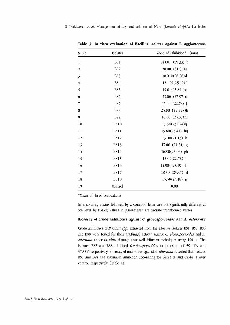
Table 3: In vitro evaluation of Bacillus isolates against P. agglomerans
S. No Isolates Zone of inhibition* (mm)
1 BS1 24.00 (29.33) b
2 BS2 28.00 (31.94)a
3 BS3 20.0 0(26.56)d
4 BS4 18 .00(25.10)f
5 BS5 19.0 (25.84 )e
6 BS6 22.00 (27.97 c
7 BS7 15.00 (22.78) j
8 BS8 25.00 (29.998)b
9 BS9 16.00 (23.57)hi
10 BS10 15.30(23.024)ij
11 BS11 15.80(23.41) hij
12 BS12 13.00(21.13) k
13 BS13 17.00 (24.34) g
14 BS14 16.50(23.96) gh
15 BS15 15.00(22.78) j
16 BS16 15.90( 23.49) hij
17 BS17 18.50 (25.47) ef
18 BS18 15.50(23.18) ij
19 Control 0.00
*Mean of three replications
In a column, means followed by a common letter are not significantly different at5% level by DMRT; Values in parentheses are arcsine transformed values
Bioassay of crude antibiotics against C. gloeosporioides and A. alternata
Crude antibiotics of Bacillus spp. extracted from the effective isolates BS1, BS2, BS6and BS8 were tested for their antifungal activity against C. gloeosporioides and A.alternata under in vitro through agar well diffusion techniques using 100 µl. Theisolates BS2 and BS8 inhibited C.goleosporioides to an extent of 59.11% and57.55% respectively. Bioassay of antibiotics against A. alternata revealed that isolatesBS2 and BS8 had maximum inhibition accounting for 64.22 % and 62.44 % overcontrol respectively (Table 4).
S. Nakkeeran et al. Management of dry and soft rot of Noni (Morinda citrifolia L.) fruits
Intl. J. Noni Res., 2015, 10 (1 & 2) 64

Table 4 : In vitro evaluation of antifungal and antibacterial activityof crude antibiotics from Bacillus spp.
*Mean of three replications
In a column, means followed by a common letter are not significantly different atthe 5% level by DMRT; Values in parentheses are arcsine transformed values
Bioassay of crude antibiotics against Pantoea agglomerans by agardiffusion method
Antibacterial activity of crude antibiotics was confirmed with agar diffusion method.Observation on control plates did not show any inhibition zone, where as platesamended with crude antibiotics 100 µl suppressed the growth of P. agglomerans.The crude antibiotics of the isolate BS2 recorded the maximum inhibition zone(29.00mm) followed by the isolate BS8 (Table 4). Several studies evidently explainedthat the antimicrobial compound synthesized from B. subtilis posses inhibitoryactivity against several phytopathogenic bacteria. These studies evidently revealedthat inhibitory action of Bacillus isolates used in this study might be because ofantimicrobial compound production.
Gas chromatography and mass spectrometry (GCMS)
Crude antibiotics were analyzed through GCMS to detect antimicrobial compoundproduced by different isolates of Bacillus spp. Result from three isolates BS1, BS2,and BS8 revealed the presence of antimicrobial compound belonging to fatty acid,lipopeptide, peptide, and aldehydes. NIST library search of the extracellular metaboliteproduced from Bacillus isolate BS2 revealed the presence of antimicrobial compoundsuch as L-glutamic acid dimethy ester, Dodecanoic acid, Petadecanoic acid, 14methyl ester and 1,2-benzenedicarboxylic acid, disooctyl ester. Similarly GCMS analysisfor the presence of metabolites in the effective isolate BS8 revealed the presence ofUndecanoic acid,10-methyl ester, Hexadecanoic acid, Diglycerol, and Tetradecanoicacid-12 methyester. Besides, compounds like Dodeconaic acid, Teradecanoic acid,12-methyl-, methyl ester and 2-hexadeconal were detected with isolate BS1 (Table5, 6, 7) . Shaligram and Singhal (2010) reported fatty ester group, tetradecanoic
S. Nakkeeran et al. Management of dry and soft rot of Noni (Morinda citrifolia L.) fruits
Intl. J. Noni Res., 2015, 10 (1 & 2) 65
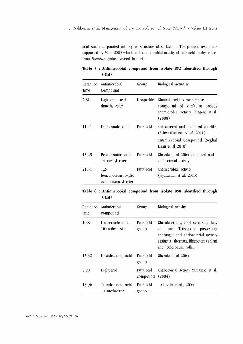
acid was incorporated with cyclic structure of surfactin . The present result wassupported by Melo 2009 who found antimicrobial activity of fatty acid methyl estersfrom Bacillus against several bacteria.
Table 5 : Antimicrobial compound from isolate BS2 identified throughGCMS
Retention Antimicrobial Group Biological ActivitiesTime Compound
7.81 L-glutamic acid Lipopetide Glutamic acid is main polardimethy ester compound of surfactin posses
antimicrobial activity (Ongena et al.(2008)
11.41 Dodecanoic acid Fatty acid Antibacterial and antifungal activities(Ashwanikumar et al. 2011)
Antimicrobial Compound (SeghalKiran et al 2010)
15.29 Petadecanoic acid, Fatty acid Ghazala et al 2004 antifungal and14 methyl ester antibacterial activity
21.51 1,2- Fatty acid Antimicrobial activitybenzenedicarboxylic (jayaraman et al. 2010)acid, disooctyl ester
Table 6 : Antimicrobial compound from isolate BS8 identified throughGCMS
Retention Antimicrobial Group Biological activitytime compound
10.8 Undecanoic acid, Fatty acid Ghazala et al ., 2004 sauturated fatty10-methyl ester group acid from Tetraspora possessing
antifungal and antibacterial activityagainst A. alternata, Rhizoctonis solaniand Sclerotium rolfsii
15.32 Hexadecanoic acid Fatty acid Ghazala et al 2004group
5.20 Diglycerol Fatty acid Antibacterial activity Yamazaki et al.compound (2004)
13.96 Tetradecanoic acid- Fatty acid Ghazala et al., 200412 methyester group
S. Nakkeeran et al. Management of dry and soft rot of Noni (Morinda citrifolia L.) fruits
Intl. J. Noni Res., 2015, 10 (1 & 2) 66

Table 7 : Antimicrobial compound from isolate BS1 identified throughGCMS
Retention Antimicrobial Group Biological activitytime compound
11.59 Dodeconaic acid Fatty acid Antimicrobial compound(Seghal Kiran et al ., 2010)
Antifunagal and Antibacterial activity(Robbel et al., 2010)
13.95 Tetradecanoic acid, Fatty acid Ghazala et al., 2004 reported12-methyl-,methyl ester antifungal and antibacterial activity of
tetradecanoic acid against fungalpathogens.
15.08 2-hexadeconal Volatile Antimicrobial compound fromcompound Naringi crenulata (Sarada et al.,
2011)
Effect of Bacillus spp., on the incidence of fungal and bacterial fruit rotdiseases under field conditions.
Dry fruit rot
Field experiment was laid out in the noni field located at Coimbatore. Differenttreatments were carried out with standard chemical check for the management ofboth fungal and bacterial fruit rot disease. Among the different treatments foliarapplication of liquid culture with 1% of BS2 was most effective under field condition.Minimum disease incidence of about 16.23 percent was recorded in plots sprayedat 1% of the Bacillus isolate (BS2). It was followed by the foliar application of BS8which recorded 18.25 per cent fruit rot. But the disease incidence was maximum(43.80 per cent) in control plots. Noni trees sprayed with BS2 and BS8 recordedthe fruit yield of 51.80 t/ha and 50.67 t/ha with cost benefit ration of 1:2.59 and1: 2.53 respectively. Comparison of percent incidence recorded in chemical checkresulted that the bio agents are quiet inferior in controlling fruit diseases (Table.8).Similarly the PGPR was found effective against C. gloeosporioides and A. alternataunder in vivo causing noni leaf blight (Nakkeeran et al., 2009; Manjunath et al.,2010).
S. Nakkeeran et al. Management of dry and soft rot of Noni (Morinda citrifolia L.) fruits
Intl. J. Noni Res., 2015, 10 (1 & 2) 67

Table 8 : Effects of B.subtilis against noni dry fruit rot (C.gloeosporioides and A. alternata) under field condition
*Mean of three replications
In a column, means followed by a common letter are not significantly different at5% level by DMRT
Values in parentheses are arcsine transformed values.
Wet / soft rot
Foliar application of bio formulation had good impact in controlling bacterial fruitrot disease of Noni .Among treatments, foliar application with isolate BS2 at 1 % hadcontrolled bacterial fruit rot to an extent of 65.53 percent over control with the fruityield of 48.33 t/ha . It was followed by the foliar application of BS8, which reducedthe incidence of fruit rot to an extent of 61.34% over control with fruit yield of 46.22t/ha. However, among all the treatments, foliar application of antibiotic namelystreptomycin sulphate @ 0.5% was effective than biocontrol agents (Table.9).
S. Nakkeeran et al. Management of dry and soft rot of Noni (Morinda citrifolia L.) fruits
Intl. J. Noni Res., 2015, 10 (1 & 2) 68
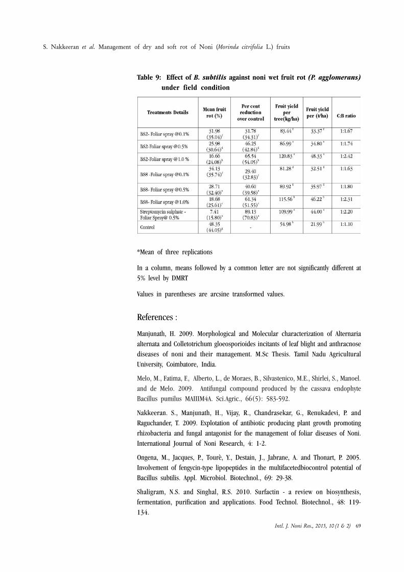
Table 9: Effect of B. subtilis against noni wet fruit rot (P. agglomerans)under field condition
*Mean of three replications
In a column, means followed by a common letter are not significantly different at5% level by DMRT
Values in parentheses are arcsine transformed values.
References :
Manjunath, H. 2009. Morphological and Molecular characterization of Alternariaalternata and Colletotrichum gloeosporioides incitants of leaf blight and anthracnosediseases of noni and their management. M.Sc Thesis. Tamil Nadu AgriculturalUniversity, Coimbatore, India.
Melo, M., Fatima, F., Alberto, L., de Moraes, B., Silvastenico, M.E., Shirlei, S., Manoel.and de Melo. 2009. Antifungal compound produced by the cassava endophyteBacillus pumilus MAIIIM4A. Sci.Agric., 66(5): 583-592.
Nakkeeran. S., Manjunath, H., Vijay, R., Chandrasekar, G., Renukadevi, P. andRaguchander, T. 2009. Explotation of antibiotic producing plant growth promotingrhizobacteria and fungal antagonist for the management of foliar diseases of Noni.International Journal of Noni Research, 4: 1-2.
Ongena, M., Jacques, P., Tourè, Y., Destain, J., Jabrane, A. and Thonart, P. 2005.Involvement of fengycin-type lipopeptides in the multifacetedbiocontrol potential ofBacillus subtilis. Appl. Microbiol. Biotechnol., 69: 29-38.
Shaligram, N.S. and Singhal, R.S. 2010. Surfactin - a review on biosynthesis,fermentation, purification and applications. Food Technol. Biotechnol., 48: 119-134.
S. Nakkeeran et al. Management of dry and soft rot of Noni (Morinda citrifolia L.) fruits
Intl. J. Noni Res., 2015, 10 (1 & 2) 69

Guidelines to AuthorsAims and ScopeInternational Journal of Noni Research(IJNR) publishes original research andreview articles on all aspects on Noni(Morinda citrifolia L.) and other speciesof Morinda. All submissions will bereviewed by the editorial board or by externalreferees. The journal covers diversity,cultivation, phytochemistry and clinicalresearch, etc. related to Noni.Three categories of paper will be consideredfor publication in IJNR.1) Reviews2) Full-length papers3) Short communications.Submission of Manuscript (Hard andSoft copy)Authors are advised to submit theirmanuscripts along with a covering letter tothe Editor, International Journal ofNoni Research, # 12, Rajiv Gandhi Road,Perungudi, Chennai - 600 096, IndiaE-Mail SubmissionAuthors are encouraged to submit theirmanuscripts via e-mail as attachment fileto the E-mail ID : [email protected]/ [email protected] (A coveringletter to be sent with the manuscript).Preparation of ManuscriptTwo typed copies of manuscripts using MSWord should be submitted. They should betyped on one side of the paper only, double-spaced with 1.2” margins in all the sides.All pages should be numbered.All the accepted articles will be subjectedto editorial revision. A singed statement fromall the authors should be accompanied withthe manuscript saying that1. the content of the article has not been
published in whole or in part elsewhere2. the article is not currently being
considered for publication elsewhere3. all necessary ethical safeguards have
been met regarding patient and animalexperimentation, etc.
Full-length PapersThere is no page restriction on overall lengthof article. It should be divided into1) Abstract (100- 200 words) followed by
keywords upto six words2) Introduction3) Materials and Methods4) Results5) Discussion6) Acknowledgements7) ReferencesResults and discussion may be combined.
Title page should be prepared in a separatesheet, which should provide the title ofpaper, name(s) of the author(s), name(s)and address of the institution(s) where thework has been carried out and details ofthe corresponding author with e-mail ID.Short CommunicationsIt should not exceed 2 pages with ashort summary of 50 words. Shortcommunications should not be divided intodifferent headings. However, headings canbe used for acknowledgements andreferences. Figures and Tables should berestricted to a maximum of 2 each. All otherstyle should be as for Full-length papers.Figures and TablesFigures, legends and tables should be typedon a separate sheet and numberedconsecutively in Arabic numerals.Photographs should be sharp with glossyprints. Micrographs etc should include a barmarker to provide an internal measure ofscale or magnification should be mentionedin each microscopic photograph.ReferencesReferences in the text should be cited asfollowsSingle author: Surendiran (2004) or(Surendiran, 2004)Two authors: Surendiran and Mathivanan(2005) or (Surendiran and Mathivanan,2005)Three or more authors: Mathivananet al. (2006) or (Mathivanan et al., 2006)Where two or more than two references arequoted consecutively in the text,chronological order should be followed. Ifthe references are within a year, alphabeticalorder must be followed. Where referencesare made to papers by the same author(s)in the same year, it should be followed by a,b, c, etc.References must be listed alphabetically atthe end of the manuscript. The followingstyle must be followed to cite the references:Journal articlesAbbott, I.A. 1985. The geographic originof the plants most commonly used formedicine by Hawaiians. Journal ofEthnopharmacology, 14: 213–22.Surendiran, G. and Mathivanan, N. 2006.Antifungal activity of Morinda citrifolia andMorinda pubescens. InternationalJournal of Noni Research, 2: 18-23.Mathivanan, N., Surendiran, G., Srinivasan,K., Sagadevan, E. and Malarvizhi, K. 2005.Review on the current scenario of Noni
research: Taxonomy, distribution, chemistry,and medicinal and therapeutic values ofMorinda citrifolia L. InternationalJournal of Noni Research, 1:1 – 9.Books or monographsLalithakumari, D. 2000. Fungal Protoplast:A Biotechnological Tool. Oxford & IBHPublishing Co., Pvt., Ltd., New Delhi. p184.Single author volumesMathivanan, N. 2004. Current scenario ofthe biocontrol potential of Trichodermafor the management of plant diseases. In:(Eds. Bagyanarayana, G., Bhadraiah, B. andKunwar, I.K.), Emerging Trends inMycology, Plant Pathology and MicrobialBiotechnology, BS Publications,Hyderabad, India. pp. 364-382.Multi-authors volumesMathivanan, N., Bharati N. Bhat, Prabavathy,V.R., Srinivasan, K. and Chelliah, S. 2003.Trichiderma viride: Lab to land for themanagement of root diseases in differentcrops. In: (Eds). Mathivanan, N., Prabavathy,V.R. and Gomathinayagam, S.), InnovativeMethods and Techniques for IntegratedPest and Disease Management Centre forAdvanced Studies in Botany, University ofMadras, Chennai, India. pp : 52-58.Thesis / DissertationSurendiran, G. 2004. Antimicrobial andWound Healing Activity of Morindatinctoria. M. Sc. Thesis, University ofMadras, Chennai, India. p.150.ProofsProof will be sent to the correspondingauthor for checking and making essentialcorrections. At this stage, no extensivegeneral revision or alteration will beallowed. Proof should be corrected andreturned to the editor within 5 days ofreceipt of MS.OffprintsCorresponding author will receive the PDFformat of the article by e-mail free of charge.A copy of the journal will be sent by post tothe corresponding author after publication.Page ChargesNo page charge for publishing the paper inthe International Journal of Noni Research.CopyrightInternational Society for Noni Science isholding the copyright of all the papers thatare published in the International Journalof Noni Research. Authors may use thematerial elsewhere after publication andauthors themselves are responsible forobtaining permission to reproducecopyright material from other sources.